2009 biochem 201 oxidphos handout - school of medicine€¦ · the electron transport chain...
TRANSCRIPT
9/21/09
1
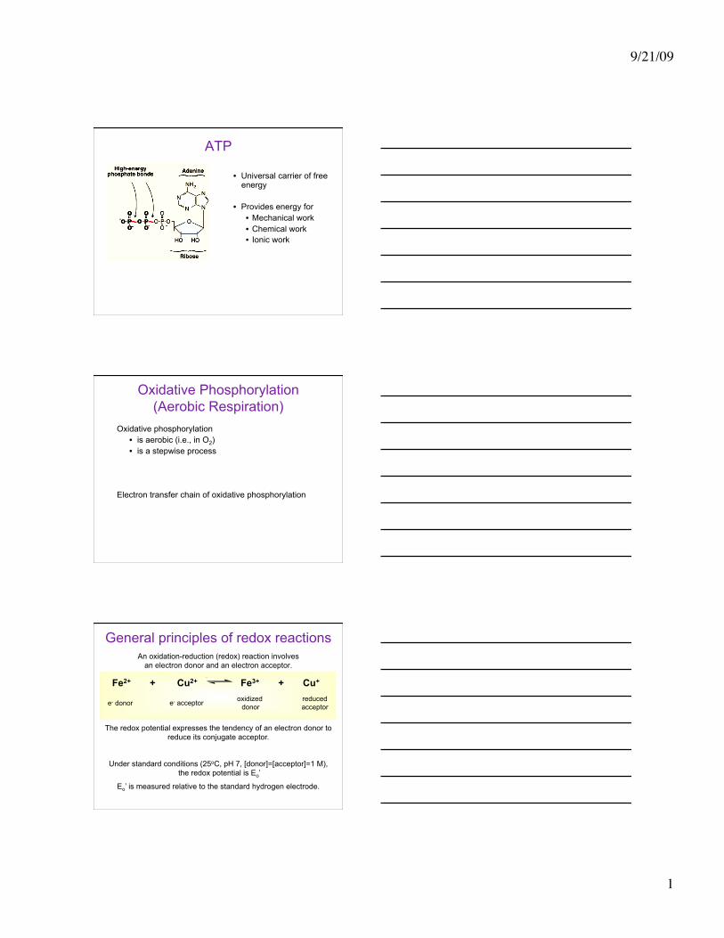
ATP
• Universal carrier of free energy
• Provides energy for • Mechanical work • Chemical work • Ionic work
Oxidative Phosphorylation (Aerobic Respiration)
Oxidative phosphorylation • is aerobic (i.e., in O2) • is a stepwise process
Electron transfer chain of oxidative phosphorylation
General principles of redox reactions An oxidation-reduction (redox) reaction involves
an electron donor and an electron acceptor.
The redox potential expresses the tendency of an electron donor to reduce its conjugate acceptor.
Under standard conditions (25oC, pH 7, [donor]=[acceptor]=1 M), the redox potential is Eo’
Eo’ is measured relative to the standard hydrogen electrode.
Fe2+ + Cu2+ Fe3+ + Cu+
e- donor e- acceptor oxidized donor
reduced acceptor
9/21/09
2
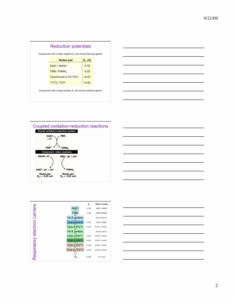
Reduction potentials Compounds with a large negative Eo are strong reducing agents.
Compounds with a large positive Eo are strong oxidizing agents.
Redox pair Eo’ (V)
NAD+ / NADH
FMN / FMNH2
Cytochrome b Fe3+/Fe2+
1/2 O2 / H2O
-0.32
-0.22
+0.07
+0.82
Coupled oxidation-reduction reactions
Res
pira
tory
ele
ctro
n ca
rrie
rs
NAD+
FMN
Fe-S centers
Coenzyme Q
Cyto b (Fe3+)
Fe-S centers
Cyto c (Fe3+)
Cyto a (Fe3+)
Cyto a3 (Fe3+)
O2
Cyto c (Fe3+)
- 0.32
- 0.30
+ 0.04
+ 0.07
+ 0.23
+ 0.29
+ 0.55
+ 0.82
+ 0.25
Eo’
NAD+ / NADH
FMN / FMNH2
Fe3+S / Fe2+S
Fe3+S / Fe2+S
H-Fe3+ / H-Fe2+
H-Fe3+ / H-Fe2+
H-Fe3+ / H-Fe2+
H-Fe3+ / H-Fe2+
CoQ / CoQH2
O2 / H2O
H-Fe3+ / H-Fe2+
Redox couples
9/21/09
3
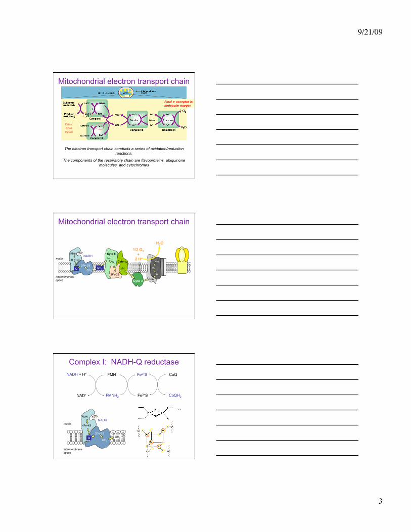
Mitochondrial electron transport chain
The electron transport chain conducts a series of oxidation/reduction reactions.
The components of the respiratory chain are flavoproteins, ubiquinone molecules, and cytochromes
Final e- acceptor is molecular oxygen
2 e¯
Citric acid cycle
Mitochondrial electron transport chain
bH bL
2Fe-2S
c1
Cyto b
Cyto c1
Cyto c
Q 2Fe-2S
QH2
FMN
4Fe-4S NADH
QH2
matrix
intermembrane space CuA
a
a3
CuB
1/2 O2 +
2 H+
H2O
Complex I: NADH-Q reductase NADH + H+
NAD+
FMN
FMNH2
Fe2+S
Fe3+S
CoQ
CoQH2
Q 2Fe-2S
QH2
FMN
4Fe-4S
NADH
QH2
matrix
intermembrane space

9/21/09
4
Redox states of coenzyme Q
Reduced form of coenzyme Q
(QH2, ubiquinol)
H3CO
H3CO
OH
OH
CH3
CH2
CH
CH2C
CH3H
10
H3CO
H3CO
O
OH
CH3
R
H3CO
H3CO
O
O
CH3
R
e- + H+ e- + H+
Semiquinone intermediate
(QH•)
Oxidized form of coenzyme Q
(Q, ubiquinone)
Complex III: cytochrome bc1
QH2
QH• Cyt 2+ b Fe2+S
Fe3+S Cyt 3+ b
Q
QH•
Cyt 3+ c1
Cyt 2+ c1
Cyt 2+ c
Cyt 3+ c
N
N N
N
OH OHO O
Fe
matrix
intermembrane space
bH
bL
2Fe-2S c1
Cyto b
Cyto c1
Cyto c
QH2
Complex IV: cytochrome oxidase
Cyto c
matrix
intermembrane space CuA
a
a3
CuB
1/2 O2 +
2 H+
H2O
Cyt 2+ a3
Cyt 3+ a3
Cyt 3+ a
Cyt 2+ a
Cyt 2+ c
Cyt 3+ c H2O
1/2 O2
9/21/09
5
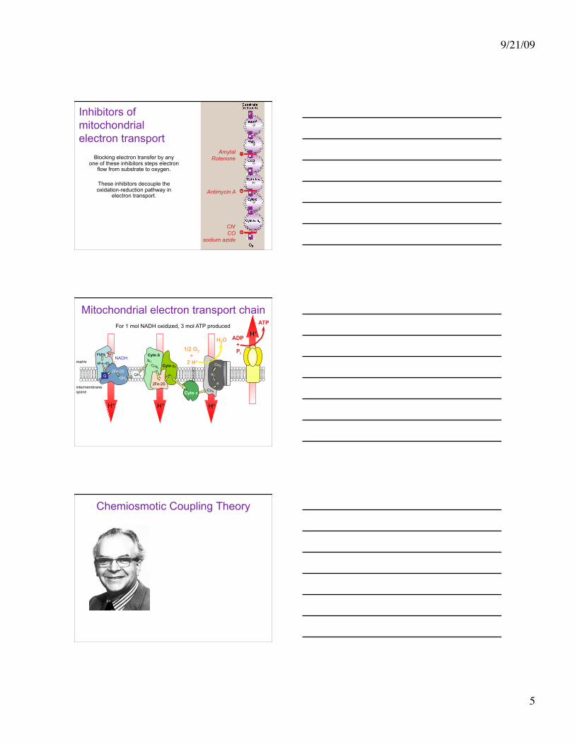
Inhibitors of mitochondrial electron transport
Blocking electron transfer by any one of these inhibitors steps electron
flow from substrate to oxygen.
These inhibitors decouple the oxidation-reduction pathway in
electron transport.
Amytal Rotenone
Antimycin A
CN- CO
sodium azide
H+
H
H+ H+ H+
Mitochondrial electron transport chain
bH bL
2Fe-2S
c1
Cyto b
Cyto c1
Cyto c
Q 2Fe-2S
QH2
FMN
4Fe-4S NADH
QH2
matrix
intermembrane space CuA
a
a3
CuB
1/2 O2 +
2 H+
H2O
ATP
ADP + Pi
For 1 mol NADH oxidized, 3 mol ATP produced
Chemiosmotic Coupling Theory
9/21/09
6
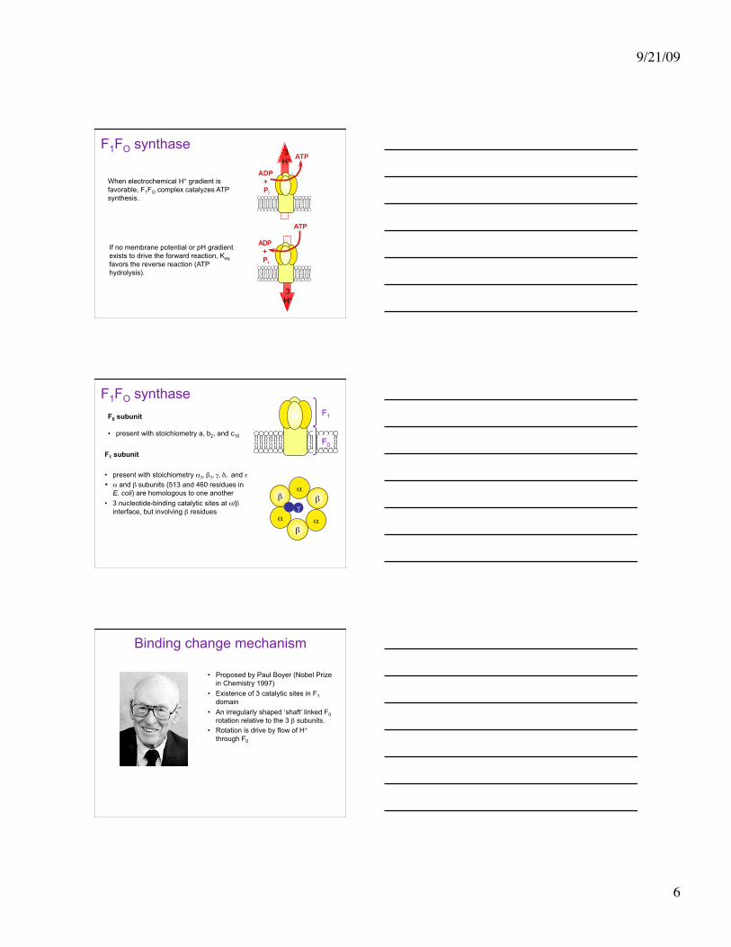
F1FO synthase 3 H+ ATP
ADP + Pi
ATP
ADP + Pi
When electrochemical H+ gradient is favorable, F1FO complex catalyzes ATP synthesis.
If no membrane potential or pH gradient exists to drive the forward reaction, Keq favors the reverse reaction (ATP hydrolysis).
3 H+
F1FO synthase
F1 subunit
• present with stoichiometry α3, β3, γ, δ, and ε • α and β subunits (513 and 460 residues in
E. coli) are homologous to one another • 3 nucleotide-binding catalytic sites at α/β
interface, but involving β residues
F0 subunit
• present with stoichiometry a, b2, and c10
F1
F0
α β
α α β
β γ
Binding change mechanism
• Proposed by Paul Boyer (Nobel Prize in Chemistry 1997)
• Existence of 3 catalytic sites in F1 domain
• An irregularly shaped ‘shaft’ linked F0 rotation relative to the 3 β subunits.
• Rotation is drive by flow of H+ through F0
9/21/09
7
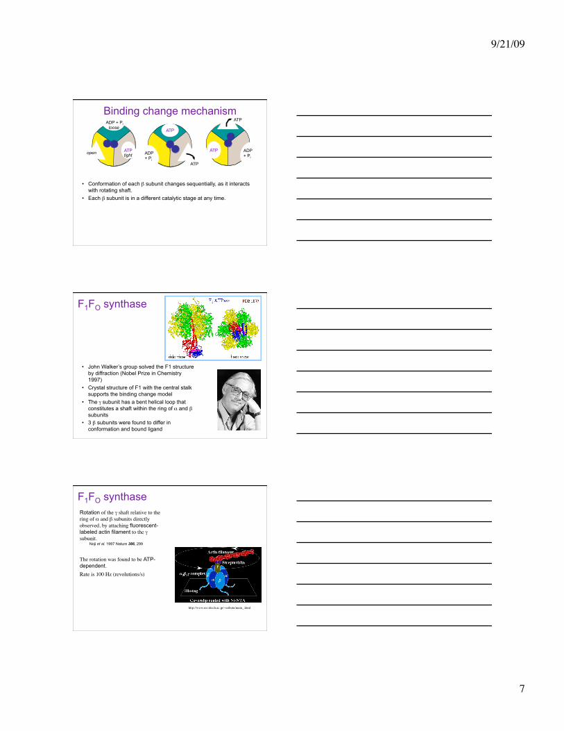
Binding change mechanism
• Conformation of each β subunit changes sequentially, as it interacts with rotating shaft.
• Each β subunit is in a different catalytic stage at any time.
ATP
ATP ATP open
ATP
ATP
ADP + Pi
ADP + Pi
ADP + Pi tight
loose
F1FO synthase
• John Walker’s group solved the F1 structure by diffraction (Nobel Prize in Chemistry 1997)
• Crystal structure of F1 with the central stalk supports the binding change model
• The γ subunit has a bent helical loop that constitutes a shaft within the ring of α and β subunits
• 3 β subunits were found to differ in conformation and bound ligand
Rotation of the γ shaft relative to the ring of α and β subunits directly observed, by attaching fluorescent-labeled actin filament to the γ subunit.
Noji et al. 1997 Nature 386, 299
The rotation was found to be ATP-dependent. Rate is 100 Hz (revolutions/s)
F1FO synthase
http://www.res.titech.ac.jp/~seibutu/main_.html