© 2009 benjamin lee predmore - university of...
TRANSCRIPT
1
INVESTIGATION INTO THE MECHANISMS OF HYDROGEN SULFIDE SIGNALING IN THE CARDIOVASCULAR SYSTEM AND THE EFFECTS OF AGE AND CALORIC
RESTRICTION
By
BENJAMIN LEE PREDMORE
A DISSERTATION PRESENTED TO THE GRADUATE SCHOOLOF THE UNIVERSITY OF FLORIDA IN PARTIAL FULFILLMENT
OF THE REQUIREMENTS FOR THE DEGREE OFDOCTOR OF PHILOSOPHY
UNIVERSITY OF FLORIDA
2009

2
© 2009 Benjamin Lee Predmore

3
To my Grandparents, Roy and Dorothy Predmore and my Parents, Roy and Donna Predmore

4
ACKNOWLEDGMENTS
I would like to thank my committee for their valuable input throughout my work on this
dissertation: David Julian, Dave Evans, Lou Guillette, Christiaan Leeuwenburgh, and Charles
Wood. I thank my parents for their support of my academic pursuits. I would also like to thank
the members of the various labs I have worked in who have helped me to complete my work.
From the Julian lab, I thank Joanna Joyner-Matos, Jennessa Andrzejewski, Maikel Alendy, and
Khadija Ahmed. From the Leeuwenburgh lab, I thank Stephanie Wohlgemuth, Brian Bouverat,
Emanuele Marzetti, and Jinze Xu, and Hazel Lees. I thank Arturo Cardounel and from his lab Pat
Kearns, Kanchana Karuppiah, Scott Forbes, and Arthur Pope. I would also like to thank Christy
Carter, Drake Morgan, and Tom Foster for their donation of aorta tissue used in some of the
experiments. This work was supported in part by a Multidisciplinary Training Program in
Hypertension (NIH T32 HL083810) through the UF Hypertension Center.

5
TABLE OF CONTENTS
Page
ACKNOWLEDGMENTS ...............................................................................................................4
LIST OF FIGURES .........................................................................................................................8
ABSTRACT...................................................................................................................................10
CHAPTER
1 HYDROGEN SULFIDE: PAST, PRESENT AND FUTURE ...............................................12
Introduction.............................................................................................................................12Hydrogen Sulfide is an Environmental and Industrial Toxin.................................................12Hydrogen Sulfide is an Energy Source for Chemosynthetic Communities ...........................14Hydrogen Sulfide is a Gasotransmitter and Physiological Modulator ...................................15Enzymatic Production of Hydrogen Sulfide...........................................................................16Regulation of Hydrogen Sulfide Production ..........................................................................16Physiological Actions of Hydrogen Sulfide ...........................................................................17Interactions between the Gasotransmitters .............................................................................21Hydrogen Sulfide Chemistry and Technical Considerations..................................................23
2 NITRIC OXIDE, ADENOSINE TRIPHOSPHAE (ATP)-SENSITIVE POTASSIUM CHANNELS, AND ARACHIDONIC ACID METABOLITES MODULATE THE TRIPHASIC RESPONSE TO HYPOXIA AND HYDROGEN SULFIDE IN RAT AORTA...................................................................................................................................26
Abstract...................................................................................................................................26Introduction.............................................................................................................................26Materials and Methods ...........................................................................................................28Results.....................................................................................................................................31Discussion...............................................................................................................................33
Initial Contraction Phase .................................................................................................34Relaxation Phase .............................................................................................................35Second Contraction Phase ...............................................................................................36Conclusion.......................................................................................................................37
3 HYDROGEN SULFIDE INCREASES NITRIC OXIDE PRODUCTION FROM ENDOTHELIAL CELLS BY A PROTEIN KINASE B (AKT)-DEPENDENT MECHANISM........................................................................................................................44
Abstract...................................................................................................................................44Introduction.............................................................................................................................44Materials and Methods ...........................................................................................................45
Chemicals ........................................................................................................................45Bovine Arterial Endothelial Cell Culture ........................................................................45

6
Hydrogen Sulfide Exposure ............................................................................................46Akt Blockade ...................................................................................................................46Electron Paramagnetic Resonance Detection of Nitric Oxide ........................................46Western Blotting..............................................................................................................47Statistics...........................................................................................................................48
Results.....................................................................................................................................48Discussion...............................................................................................................................49
4 THE HYDROGEN SULFIDE SIGNALING SYSTEM: CHANGES DURING AGING AND THE BENEFITS OF CALORIC RESTRICTION........................................................55
Abstract...................................................................................................................................55Introduction.............................................................................................................................56Materials and Methods ...........................................................................................................58
Chemicals ........................................................................................................................58Animals............................................................................................................................58Western Blotting..............................................................................................................58RNA Extraction ...............................................................................................................59Real-Time PCR ...............................................................................................................59Hydrogen Sulfide Production ..........................................................................................60Myography ......................................................................................................................62Statistics...........................................................................................................................63
Results.....................................................................................................................................63Cystathionine Gamma-Lyase and Cystathionine Beta-Synthase Protein Expression.....63Cystathionine Gamma-Lyase and Cystathionine Beta-Synthase Messenger
Ribonucleic Acid (mRNA) Expression .......................................................................64Hydrogen Sulfide Production ..........................................................................................64Contractile Response to Hydrogen Sulfide in Aorta .......................................................65
Discussion...............................................................................................................................66Cystathionine Gamma-Lyase and Cystathionine Beta-Synthase Protein Expression.....66Cystathionine Gamma-Lyase and Cystathionine Beta-Synthase mRNA Expression.....68Hydrogen Sulfide Production ..........................................................................................69Contractile Response to Hydrogen Sulfide in Aorta .......................................................70Conclusions .....................................................................................................................71
5 SYNTHESIS: UNDERSTANDING HYDROGEN SULFIDE SIGNALING IN VASCULAR SMOOTH MUSCLE AND THE TRIPHASIC RESPONSE...........................81
Introduction.............................................................................................................................81What is Modulating Phase 1 of the Triphasic Response?.......................................................82What is Modulating Phase 2 of the Triphasic Response?.......................................................83What is Modulating Phase 3 of the Triphasic Response?.......................................................85A Proposed Time-course and Mechanism of the Triphasic Response ...................................85Conclusions and Future Directions.........................................................................................86
LIST OF REFERENCES...............................................................................................................89

7
BIOGRAPHICAL SKETCH .......................................................................................................104
8
LIST OF FIGURES
Figure Page
1-1 Enzymatic production of hydrogen sulfide by cystathionine beta-synthase and cystathionine gamma-lyase................................................................................................25
2-1 Representative tracings of the effect of hypoxia or hydrogen sulfide ...............................38
2-2 Duration of the triphasic response to hypoxia or hydrogen sulfide ...................................39
2-3 Magnitude of the first phase of the triphasic response to hypoxia or hydrogen sulfide with inhibitors of nitric oxide, adenosine triphosphate (ATP)-sensitive potasium channels, and arachadonic acid metabolites ......................................................................40
2-4 Magnitude of the second phase of the triphasic response to hypoxia or hydrogen sulfide with inhibitors of nitric oxide, ATP-sensitive potasium channels, and arachadonic acid metabolites .............................................................................................41
2-5 Magnitude of the third phase of the triphasic response to hypoxia or hydrogen sulfide with inhibitors of nitric oxide, ATP-sensitive potasium channels, and arachadonic acid metabolites .................................................................................................................42
2-6 Magnitude of the triphasic response to hypoxia or hydrogen sulfide with or without an intact endothelium.........................................................................................................43
3-1 The effect of hydrogen sulfide on nitric oxide production by bovine arterial endothelial cells .................................................................................................................51
3-2 The effect of hydrogen sulfide on endothelial nitric oxide phosphorylation.....................52
3-3 The effect of Akt inhibition on hydrogen sulfide-stimulated nitric oxide production.......54
4-1 Effect of age and diet on cystathionine gamma-lyase and cystathionine beta-synthase protein expression ..............................................................................................................73
4-2 Effect of diet on cystathionine gamma-lyase and cystathionine beta-synthase mRNA expression ..........................................................................................................................74
4-3 Effect of diet on hydrogen sulfide production. ..................................................................75
4-4 Representative tension tracing of a rat aorta ring to hydrogen sulfide ..............................76
4-5 Effect of age on potassium chloride- and norepinephrine-induced contractions and the first phase contraction to hydrogen sulfide ..................................................................77
4-6 Effect of diet on potassium chloride-and norepinephrine-induced contractions. ..............78

9
4-7 Effect of diet on hydrogen sulfide-induced contractions...................................................79

10
Abstract of Dissertation Presented to the Graduate Schoolof the University of Florida in Partial Fulfillment of theRequirements for the Degree of Doctor of Philosophy
INVESTIGATION INTO THE MECHANISMS OF HYDROGEN SULFIDE SIGNALING IN THE CARDIOVASCULAR SYSTEM AND THE EFFECTS OF AGE AND CALORIC
RESTRICTION
By
Benjamin Lee Predmore
August 2009
Chair: David JulianMajor: Zoology
The gasotransmitter hydrogen sulfide (H2S) modulates vascular tone in vertebrates.
Hydrogen sulfide and hypoxia elicit similar contractile responses in vertebrate smooth muscle,
while both H2S and nitric oxide (NO) elicit synergistic vasodilatory responses. Aging has
negative impacts on the cardiovascular system, which can be attenuated by caloric restriction
(CR). The mechanisms behind H2S and hypoxia signaling and the synergy with NO are
unknown, as well as whether aging and CR affect hydrogen sulfide signaling. I investigated the
mechanisms through which aorta rings respond to hydrogen sulfide and hypoxia, how hydrogen
sulfide regulates endothelial NO production, and how aging and CR affect the H2S signaling
system. I used bovine arterial endothelial cells and Fisher 344 x Brown Norway rats, 6-38
months of age, maintained on an ad libitum (AL) or CR diet. To investigate aging and CR, I
measured protein and mRNA expression of the hydrogen sulfide producing enzymes
cystathioneine –lyase (CSE) and cystathionine –synthase (CBS) and the rate of hydrogen
sulfide production from aorta and liver tissues, in addition to functional assessment using aorta
rings. In the first study, I found that hypoxia and hydrogen sulfide elicit a triphasic, contraction-
relaxation-contraction response. However, the mechanisms are not the same between hypoxia

11
and hydrogen sulfide. In the second study, I found that hydrogen sulfide stimulated a two-fold
increase in NO production from endothelial nitric oxide synthase (eNOS). Phosphorylation of
eNOS at Ser 1177 was also significantly increased, and inhibition of Akt attenuated this. In the
third study, I found that the first phase contraction increased in sensitivity to hydrogen sulfide
with age, while CR increased the magnitude of all phases. AL aorta CSE and CBS protein
expression increased with age, but remained unchanged with CR. Liver CSE and CBS protein
expression remained constant with age. Aorta CSE and CBS mRNA expression was higher with
CR. Hydrogen sulfide production was also higher with CR in aorta and liver. I conclude that in
rat aorta the triphasic responses to hypoxia and H2S are mediated by different mechanisms,
hydrogen sulfide up-regulates eNOS/NO production through an Akt-dependent mechanism, and
the increased sensitivity to hydrogen sulfide and increased protein expression of CSE and CBS
with age in aorta point to a drop in hydrogen sulfide bioavailability, while CR maintains
CSE/CBS function. These studies reveal novel, age-sensitive mechanisms of hydrogen sulfide
action to regulate vascular tone. They also illustrate the benefit of CR on the hydrogen sulfide
signaling system. This is quite timely, given the emerging roles of hydrogen sulfide in
cardiovascular pathologies.

12
CHAPTER 1HYDROGEN SULFIDE: PAST, PRESENT AND FUTURE
Introduction
Hydrogen sulfide, a name often used to refer to sum of the chemical species H2S, HS- and
S2-, has had a long and interesting development in the scientific literature. The “face” of
hydrogen sulfide has changed within the last 40 years from an environmental and industrial
toxin, to an energy source for chemosynthetic communities, and most recently as a gaseous
physiological modulator, or gasotransmitter (Wang 2002), and a potential therapeutic agent in
medicine. To familiarize the reader with the many aspects of hydrogen sulfide, this chapter will
briefly examine the chemistry and toxicology of hydrogen sulfide as well as the chemosynthetic
communities that it fuels, and then focus on its recent induction into the gasotransmitter family
and its transition into biomedical research and therapeutic applications.
Hydrogen Sulfide is an Environmental and Industrial Toxin
There are many toxic and potentially lethal actions of hydrogen sulfide, the most
significant of which is the reversible inhibition of the electron transport chain via cytochrome c
oxidase (Nicholls 1975; Nicholls et al. 1982; Khan et al. 1990). This reversible inhibition can
occur at 1-40 μM sulfide in isolated mitochondria (Nicholls et al. 1982; Bagarinao 1992), and is
mechanistically very similar to cyanide poisoning (Nicholls et al. 1982; Shepherd et al. 2008).
Other mechanisms of sulfide toxicity include opening of the mitochondrial permeability
transition pore (Eghbal et al. 2004; Julian et al. 2005a); increasing production of reactive oxygen
species (including superoxide) (Chen et al. 1972; Tapley et al. 1999; Eghbal et al. 2004; Julian et
al. 2005a); out-competing oxygen for hemoglobin binding, causing formation of sulfhemoglobin
(Bagarinao et al. 1992; Kraus et al. 1996; Völkel et al. 2000); and inhibition of about 20 other
enzymes (Bagarinao 1992).
13
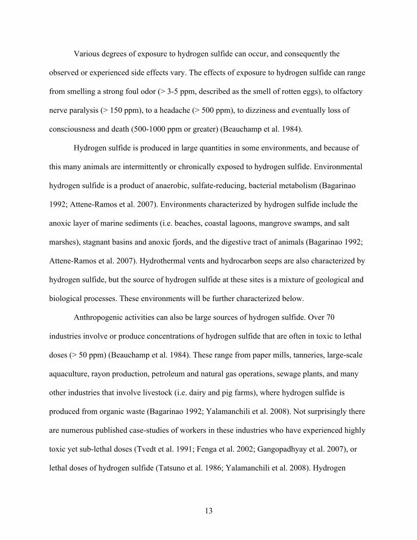
Various degrees of exposure to hydrogen sulfide can occur, and consequently the
observed or experienced side effects vary. The effects of exposure to hydrogen sulfide can range
from smelling a strong foul odor (> 3-5 ppm, described as the smell of rotten eggs), to olfactory
nerve paralysis (> 150 ppm), to a headache (> 500 ppm), to dizziness and eventually loss of
consciousness and death (500-1000 ppm or greater) (Beauchamp et al. 1984).
Hydrogen sulfide is produced in large quantities in some environments, and because of
this many animals are intermittently or chronically exposed to hydrogen sulfide. Environmental
hydrogen sulfide is a product of anaerobic, sulfate-reducing, bacterial metabolism (Bagarinao
1992; Attene-Ramos et al. 2007). Environments characterized by hydrogen sulfide include the
anoxic layer of marine sediments (i.e. beaches, coastal lagoons, mangrove swamps, and salt
marshes), stagnant basins and anoxic fjords, and the digestive tract of animals (Bagarinao 1992;
Attene-Ramos et al. 2007). Hydrothermal vents and hydrocarbon seeps are also characterized by
hydrogen sulfide, but the source of hydrogen sulfide at these sites is a mixture of geological and
biological processes. These environments will be further characterized below.
Anthropogenic activities can also be large sources of hydrogen sulfide. Over 70
industries involve or produce concentrations of hydrogen sulfide that are often in toxic to lethal
doses (> 50 ppm) (Beauchamp et al. 1984). These range from paper mills, tanneries, large-scale
aquaculture, rayon production, petroleum and natural gas operations, sewage plants, and many
other industries that involve livestock (i.e. dairy and pig farms), where hydrogen sulfide is
produced from organic waste (Bagarinao 1992; Yalamanchili et al. 2008). Not surprisingly there
are numerous published case-studies of workers in these industries who have experienced highly
toxic yet sub-lethal doses (Tvedt et al. 1991; Fenga et al. 2002; Gangopadhyay et al. 2007), or
lethal doses of hydrogen sulfide (Tatsuno et al. 1986; Yalamanchili et al. 2008). Hydrogen
14
sulfide poisoning still remains an everyday risk for those working in the petroleum, sewer,
maritime, and mining industries (Yalamanchili et al. 2008).
Animals that are adapted to environments with hydrogen sulfide have avoidance
behaviors in addition to antioxidant defenses and detoxification mechanisms to protect
themselves from acute and/or chronic exposure to hydrogen sulfide (Bagarinao 1992). Most of
these animals have a very similar set of detoxification mechanisms, including specialized
hemoglobin that can bind both oxygen and hydrogen sulfide (Martineu et al. 1997; Zal et al.
1997; Zal et al. 1998; Hourdez et al. 2000), conversion of sulfide to thiosulfate (Levitt et al.
1999; Doeller et al. 2001), and storage of sulfide as both taurine and thiotaurine (Joyner et al.
2003). However, in some invertebrates, hydrogen sulfide can also be used as the terminal
electron acceptor in aerobic respiration (Doeller et al. 2001; Kraus et al. 2004).
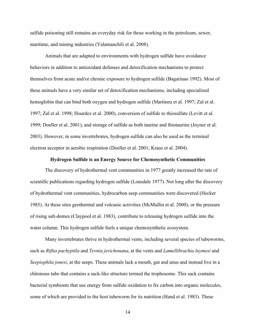
Hydrogen Sulfide is an Energy Source for Chemosynthetic Communities
The discovery of hydrothermal vent communities in 1977 greatly increased the rate of
scientific publications regarding hydrogen sulfide (Lonsdale 1977). Not long after the discovery
of hydrothermal vent communities, hydrocarbon seep communities were discovered (Hecker
1985). At these sites geothermal and volcanic activities (McMullin et al. 2000), or the pressure
of rising salt-domes (Claypool et al. 1983), contribute to releasing hydrogen sulfide into the
water column. This hydrogen sulfide fuels a unique chemosynthetic ecosystem.
Many invertebrates thrive in hydrothermal vents, including several species of tubeworms,
such as Riftia pachyptila and Tevnia jerichonana, at the vents and Lamellibrachia luymesi and
Seepiophila jonesi, at the seeps. These animals lack a mouth, gut and anus and instead live in a
chitonous tube that contains a sack-like structure termed the trophosome. This sack contains
bacterial symbionts that use energy from sulfide oxidation to fix carbon into organic molecules,
some of which are provided to the host tubeworm for its nutrition (Hand et al. 1983). These

15
tubeworms have specialized hemoglobin to transport oxygen and hydrogen sulfide from the
respiratory surface to the bacteria without the animal becoming poisoned itself (Martineu et al.
1997; Zal et al. 1997; Zal et al. 1998; Hourdez et al. 2000). In addition to specialized
hemoglobin, hydrocarbon seep tubeworms also have posterior extensions, termed “roots”, to
obtain hydrogen sulfide from below the sediment-water interface (Julian et al. 1999).
Hydrogen Sulfide is a Gasotransmitter and Physiological Modulator
Since hydrogen sulfide has had a long history as a toxin, it was very surprising to find
that hydrogen sulfide can be endogenously produced in a variety of animal tissues and that it has
both neuromodulatory (Abe et al. 1996) and cardiovascular regulatory effects in mammals
(Hosoki et al. 1997). Since its discovery of its neuromodulatory abilities (Abe et al. 1996),
hydrogen sulfide has been added to the family of gasotransmitters (Wang 2002), which also
includes nitric oxide (NO) and carbon monoxide (CO).
The discovery of gasotransmitters began with the work on the actions of NO by Murad,
Furchgott and Ignarro from 1977-1986 (Furchgott 1999), who in 1988 shared the Nobel Prize in
Physiology or Medicine for their work. The discovery of the gasotransmitter function of CO by
Verma and colleagues followed shortly thereafter in 1993 (Verma et al. 1993). Finally,
investigations of hydrogen sulfide as a physiological modulator began in 1996-1997 with the
work of Abe and Kimura (Abe et al. 1996) and Hosoki, Matsuki and Kimura (Hosoki et al.
1997).
All three gasotransmitters, CO, NO and H2S, share several similarities. They are
endogenously produced, small gas molecules that are capable of physiological action (Wang
2002). They can easily diffuse across cell membranes to exert their function (Wang 2002). They
do not require a mechanism of degradation or reuptake because they are all very reactive, and
they use heme as a common sink (Wang 2002).

16
Enzymatic Production of Hydrogen Sulfide
All of the gasotransmitters are endogenously produced by enzymatic reactions. Nitric
oxide is produce from L-arginine by nitric oxide synthase (NOS). Carbon monoxide is produced
from heme by heme oxygenize (HO). In mammalian tissues, hydrogen sulfide is primarily
produced from L-cysteine by two PLP (pyridoxal-5’-phosphate)-dependent, cysteine metabolic
enzymes: cystathionine gamma-lyase (CSE) and cystathionine beta-synthase (CBS) (Julian et al.
2002; Zhao et al. 2003). However, several other enzymatic pathways exist for the production of
hydrogen sulfide, including via cysteine amino transferase or cysteine lyase (Julian et al. 2002).
CSE is expressed in endothelial cells and vascular smooth muscle cells (Hosoki et al. 1997;
Wang 2002; Wang 2003; Yang et al. 2008), and is the predominant enzyme for hydrogen sulfide
production in the cardiovascular system. In the CSE reaction (Figure 1-1), L-cysteine must first
dimerize to form cystine, which is then transformed into pyruvate, thiocystine and NH3 by CSE.
CSE can then catalyze the reaction of thiocystine with other thiol compounds to from H2S and
CysSR (Julian et al. 2002). Alternatively, thiocystine may form H2S and cystine non-
enzymatically (Cavallini et al. 1962). CBS is the predominant enzyme for hydrogen sulfide
production in the nervous system (Abe et al. 1996). In the CBS reaction (Figure 1-1), L-cysteine
is hydrolyzed to yield equimolar amounts of H2S and L-serine (Cavallini et al. 1962).
Julian and colleagues showed that hydrogen sulfide is produced in invertebrate tissues
from CBS and CSE (Julian et al. 2002), revealing that gasotransmitters have a deeper phylogenic
history than just vertebrates. Hydrogen sulfide has been shown to act as a signaling molecule in a
variety of invertebrates (Julian et al. 1998; Julian et al. 2002; Gainey et al. 2005; Julian et al.
2005b), as has nitric oxide (Gainey et al. 2003; Palumbo 2005).
Regulation of Hydrogen Sulfide Production
Hydrogen sulfide production by CBS and CSE can be physiologically regulated, although

17
the mechanisms are poorly understood. Sex hormones appear to regulate brain hydrogen sulfide
production by CBS, with males having higher hydrogen sulfide concentration in the brain than
females (Eto et al. 2002b). This can be reversed either by castration of males or by testosterone
injection in females (Eto et al. 2002b). CBS activity in vitro can also be regulated by S-adenosyl-
methionine (SAM), an allosteric activator of CBS (Eto et al. 2002b). CSE also appears to be
calcium-calmodulin dependent, and can be stimulated through muscarinic receptor activation of
intracellular calcium (Yang et al. 2008).
Hydrogen sulfide production may also be regulated by the other gasotransmitters. NO and
CO may bind to and inactivate CBS, with CO being the more potent inactivator than NO (Taoka
et al. 2001; Puranik et al. 2006). In contrast, NO seems to stimulate hydrogen sulfide production
in the cardiovascular system (Lowicka et al. 2007): NO donors stimulate CSE-dependent
hydrogen sulfide production in aorta tissue homogenates in a cGMP-mediated manner (Zhao et
al. 2003), and incubation of vascular smooth muscle cells with NO donors increases CSE protein
and mRNA expression (Zhao et al. 2001). Sodium nitroprusside (SNP), an NO donor, increases
the activity of brain CBS in vitro, but this effect results from chemical modification of the
cysteine groups of CBS, rather than a direct action of NO itself (Eto et al. 2002a).
Physiological Actions of Hydrogen Sulfide
Abe and Kimura were the first to show a physiological role for hydrogen sulfide (Abe et
al. 1996). They demonstrated that hydrogen sulfide was not only produced in the brain by CBS,
but that it increased N-methyl-D-aspartic acid (NMDA) receptor-mediated responses and
facilitated hippocampal long-term potentiation (Abe et al. 1996). Since then many actions have
been reported. These include the ability to upregulate -aminobutyric acid B (GABAB) receptors
in the brain (Han et al. 2005), regulate cerebrovascular circulation (Leffler et al. 2006) and play a

18
role in cerebral ischemic damage after a stroke (Qu et al. 2006), mediate learning and memory
formation (Partlo et al. 2001), stimulate L-type calcium channels in neurons (Garcia-Bereguiain
et al. 2008), negatively regulate the hypothalamo-pituitary-adrenal axis (Dello Russo et al. 2000),
and protect neurons from oxidative stress (Kimura et al. 2004; Kimura et al. 2006).
A multitude of functions for hydrogen sulfide have been reported in the cardiovascular
system, particularly in the vascular smooth muscle. One of the most important sites of action of
hydrogen sulfide is the ATP-sensitive K+ channel (KATP) in smooth muscle cells, which causes
hyperpolarization and relaxation (Zhao et al. 2001). However, hydrogen sulfide has also been
shown to cause contraction, relaxation or multiphasic responses in aorta (Hosoki et al. 1997;
Zhao et al. 2001; Dombkowski et al. 2005), mesenteric artery (Cheng et al. 2004; Tang et al.
2005), cerebral artery (Leffler et al. 2006), gastric artery (Kubo et al. 2007c), mammary artery
(Webb et al. 2008), and pulmonary arteries (Dombkowski et al. 2005; Wang et al. 2008). These
responses, however, all depend on the concentration of hydrogen sulfide and O2, the specific
vessel examined, and the animal model used (Dombkowski et al. 2004; Dombkowski et al.
2005). Hydrogen sulfide has also been associated with the pathology of a number of
cardiovascular diseases, including hypertension and COPD (chronic obstructive pulmonary
disease), in addition to hydrogen sulfides involvement in septic shock (Chunyu et al. 2003; Du et
al. 2003; Hui et al. 2003; Yan et al. 2004; Chen et al. 2005). Additionally, hydrogen sulfide has
recently been implicated in vasodilation of the corpus cavernosum (Srilatha et al. 2007;
d'Emmanuele di Villa Bianca et al. 2009; Shukla et al. 2009) and the dysregulation of hydrogen
sulfide has been linked to erectile dysfunction (Srilatha et al. 2006).
Hydrogen sulfide also affects the heart itself. Hydrogen sulfide preconditioning of rat
cardiomyocytes induces a cardioprotection against ischemia and reperfusion injury (Hu et al.

19
2007). The proposed pathways in this protection include eNOS, KATP channels, protein kinase C,
extracellular signal regulated kinase (ERK 1/2), and phosphatidylinositol 3-kinase/proteine
kinase B (Akt) (Hu et al. 2007; Yong et al. 2008a). Other cardiovascular targets of hydrogen
sulfide are -adrenergic receptors (Yong et al. 2008b), carotid sinus baroreceptor (Xiao et al.
2007), NADPH oxidase-1, and Rac(1) (Ras-related C3 botulinum toxin substrate 1) (Muzaffar et
al. 2008) and cyclooxygenase, potentially altering arachadonic acid metabolite levels as well
(Koenitzer et al. 2007).
Recent evidence also suggests that hydrogen sulfide acts as an oxygen sensor in
vertebrates and may be involved in vascular responses to hypoxia (Dombkowski et al. 2006;
Olson et al. 2006; Olson 2008). The rationale is that the low oxygen levels during hypoxia allow
accumulation of hydrogen sulfide in vascular tissue, which then modulates vascular tone.
A role for hydrogen sulfide in the gastrointestinal system has also been emerging. As in
other tissues, hydrogen sulfide is produced in gastric and intestinal tissues from CBS and CSE
(Fiorucci et al. 2006), and has had several demonstrated functions. Hydrogen sulfide has been
reported to protect gastric mucosal epithelium from oxidative stress (Yonezawa et al. 2007) and
enhance ulcer healing in rats (Wallace et al. 2007). Hydrogen sulfide also affects gut motility and
secretion (Kubo et al. 2007c), relaxation of ileum (Hosoki et al. 1997), and can also inhibit motor
patterns in human, rat and mouse colon (Gallego et al. 2008). In the liver, hydrogen sulfide
regulates perfusion and biliary bicarbonate secretion (Fiorucci et al. 2005b; Fujii et al. 2005).
In addition to actions of hydrogen sulfide in the nervous, cardiovascular, and
gastrointestinal systems, there is evidence that hydrogen sulfide is involved in insulin secretion,
also working through KATP (Yang et al. 2005), as well as leukocyte adhesion and trafficking, via
KATP (Zhang et al. 2007), and leukocyte-mediated inflammation (Zanardo et al. 2006).

20
Hydrogen sulfide has a protective role or antioxidant capacity in many systems.
Hydrogen sulfide will readily scavenge hydrogen peroxide, and increase intracellular levels of
reduced glutathione (GSH) (Kimura 2002; Pryor et al. 2006) and the hydrogen sulfide signaling
system (including CSE and CBS) has both anti-oxidant (Kimura et al. 2004; Whiteman et al.
2004a; Kimura et al. 2006; Yan et al. 2006; Jha et al. 2008) and anti-inflammatory (Fiorucci et
al. 2005a; Zanardo et al. 2006; Wallace 2007b) actions. Not surprisingly, there is large interest in
its potential role as an NSAID and therapeutic agent for a variety of disorders, including
hypertension (Fiorucci et al. 2007; Lowicka et al. 2007; Szabo 2007; Wallace 2007a; Wallace
2007b; Li et al. 2009). However, hydrogen sulfide is also reported to increase
lipopolysaccharide-induced inflammation (Li et al. 2005).
One of the more dramatic actions of hydrogen sulfide is greatly reducing metabolism in
mice, resulting in a suspended animation-like state (Blackstone et al. 2005). While the
possibility of inducing a suspended animation has tremendous potential, this has not yet been
duplicated in animals larger than the mouse (Haouzi et al. 2008). Nonetheless, this discovery has
led the creation of the Ikaria Corporation, a gas-drug company investigating both therapeutic
potential of NO and H2S. Ikaria has produced a more stable form of Na2S called IK-1001, which
is purportedly suitable for injection, and INOmax® which is nitric oxide for inhalation. IK-1001
has been used in several research applications and finished Phase I clinical trials in 2008 (Elrod
et al. 2007; Szabo 2007; Jha et al. 2008; Kiss et al. 2008). The metabolic effects of hydrogen
sulfide are also seen in the nematode Caenorhabditis elegans, in which hydrogen sulfide
increases thermotolerance and lifespan in (Miller et al. 2007). While still in research and
development, it is clear that hydrogen sulfide has a very high therapeutic potential, much like
NO and NO-donors.

21
Interactions between the Gasotransmitters
Hydrogen sulfide and the other gasotransmitters may interact with each other, but the
interactions have not been firmly established. The early work on the interactions of hydrogen
sulfide and NO indicated that the relationship was synergistic. In 1997 Hosoki et al showed that
hydrogen sulfide increased the effects of the NO donor SNP by up to 13-fold (Hosoki et al.
1997). Later, Julian et al. showed that SNP potentiates hydrogen sulfide-induced contractions in
the body wall of the echiuran worm Urechis caupo (Julian et al. 2005b)..
Not surprisingly, there have also been reports of negative interactions between the
gasotransmitters. At a direct chemical level hydrogen sulfide can react with NO to form a
nitrosothiol (Whiteman et al. 2006), and hydrogen sulfide can increase CO production from HO-
1, which can then inhibit NO production from iNOS (Oh et al. 2006). In studies of hypertension
and pulmonary vascular structural remodeling (PVSR), exogenous hydrogen sulfide inhibits the
NO/NOS pathway and upregulates the CO/HO-1 pathway (Qingyou et al. 2004; Li et al. 2006).
Moreover, during hypertension the expression and activity of CSE decreases with a concomitant
decrease in plasma hydrogen sulfide concentration (Qi et al. 2004; Xiaohui et al. 2005), while
plasma NO levels and eNOS expression levels increase (Zhong et al. 2003). Accordingly,
application of hydrogen sulfide (as NaHS) rescues rats from hypertension (Du et al. 2003;
Qingyou et al. 2004; Yan et al. 2004) and application of hydrogen sulfide lessens aorta structural
remodeling, decreases NO levels, and increases CO levels (Qingyou et al. 2004; Yan et al. 2004;
Li et al. 2006). Similarly, increasing expression of HO-1 prevents development of hypertension
and inhibits PVSR (Zhao et al. 2001).
However, data have been published that contradict many of the interactions reported
between the gasotransmitters and their respective enzymes. These discrepancies are likely the
result of using different experimental systems, as well as differences in methodologies and

22
techniques while working with hydrogen sulfide, which could alter or skew the results (see
Technical Considerations below). For example, while several studies have demonstrated the
positive effect of hydrogen sulfide on NO signaling and NO production (Chapter 3, Hosoki et al.
1997; Yong et al. 2008a), several other studies show negative effects of hydrogen sulfide on NO
production (Geng et al. 2007; Kubo et al. 2007b; Kubo et al. 2007c). This discrepancy is likely
because the latter investigators waited for hours to look for an effect of hydrogen sulfide, when
the positive effect on NO production can be observed in minutes (Chapter 3). The absence of a
sustained effect is likely because H2S is so volatile and can rapidly oxidize in solution (see
below).
It is also unclear whether the action of hydrogen sulfide on KATP is universal. Rui Wang
and colleagues show that hydrogen sulfide causes a direct activation of KATP and that
glibenclamide (an KATP inhibitor) inhibits the action of hydrogen sulfide (Zhao et al. 2001; Wang
2002; Zhao et al. 2002; Tang et al. 2005; Yang et al. 2005). However, the precise mechanism for
this activation still remains unknown, and others, including myself (Chapter 2), have not seen the
same effect of glibenclamide. In these cases glibenclamide either does not work at all (Kiss et al.
2008), or only partially inhibits the relaxation to hydrogen sulfide (Chapter 2, Kubo et al. 2007a;
Kubo et al. 2007b; Kubo et al. 2007c; Webb et al. 2008).
An additional receptor of hydrogen sulfide, the Cl-/HCO3- exchanger, has recently been
revealed (Kiss et al. 2008). This receptor, when inhibited using the anion exchanger inhibitor
4,4'-Diisothiocyanatostilbene-2,2'-disulfonate (DIDS), completely blocks the hydrogen sulfide
relaxation (Kiss et al. 2008). From this it is evident that hydrogen sulfide is likely working
through both KATP and the Cl-/HCO3- exchanger. However, these receptors only apply to the

23
relaxation observed to hydrogen sulfide. The mechanism(s) behind the contractile, or multiphasic
responses to hydrogen sulfide have yet to be identified.
Hydrogen Sulfide Chemistry and Technical Considerations
Hydrogen sulfide exists as a gas (H2S, dihydrogen sulfide) and is a weak acid in solution,
dissociating into HS- and S2-. There are three commonly used methods to create hydrogen sulfide
solutions in the laboratory. One way is to bubble distilled water or a physiological buffer directly
with hydrogen sulfide gas. However, this method is not as accurate as using a known amount of
a chemical donor. Two chemical donors, sodium (Na2S) and sodium hydrosulfide (NaHS), are
widely used instead of hydrogen sulfide gas. Na2S crystals are clear, and once the oxidized
portions are rinsed from the crystals with distilled water, they can be measured out as any other
chemical. NaHS is in the form of yellow flakes, and the oxidized portions cannot be easily rinsed
before weighing since the flakes immediately dissolve upon contact with water. Na2S is argued
to be a better hydrogen sulfide donor than NaHS for making solutions because of its higher
purity. It is thought that oxidation products formed from the impurities in NaHS solutions may
interfere with physiological experiments (Koenitzer et al. 2007). However, Na2S is highly basic
and may cause confounding effects by altering pH of buffers if used in high concentrations.
Despite this, both donors have been commonly used in published studies. Working in a fume
hood is recommended when dealing with hydrogen sulfide because higher levels of hydrogen
sulfide can prevent its olfactory detection and these symptoms can rapidly progress during a
high, acute exposure. Therefore, it is important at first notice of hydrogen sulfide gas to move to
a well-ventilated area to avoid increased exposure and more severe symptoms.
In solution, hydrogen sulfide exists as three species: H2S, HS- and S2- (see Equation 1).
H2S HS- + H+ S2- + H+ (1)

24
Because the pKa for the first dissociation is 7.02-7.04 (Chen 1972; Beauchamp et al. 1984) and
the estimated pKa of the second dissociation is 12-15 (Chen 1972; Beauchamp et al. 1984;
Bagarinao 1992), at a physiological pH of 7.4 hydrogen sulfide exists as approximately 1/3 H2S
and 2/3 HS- with very little S2- (Beauchamp et al. 1984). This approximate relationship holds
true in fresh- and salt-water environments, although variation in pH will shift the H2S/HS-
equilibrium. Therefore when working with and discussing the effects of hydrogen sulfide, it is
important to take into account not only H2S gas, but the HS- anion. Throughout this dissertation
the term “hydrogen sulfide” will refer to the sum of the species H2S, HS- and S2-, unless
otherwise specified.
Hydrogen sulfide gas is very volatile (Cline 1969; Julian et al. 1998; Dorman et al. 2002)
and will readily come out of solution, causing a net loss of hydrogen sulfide. This loss is
exacerbated by the fact that hydrogen sulfide will readily oxidize in the presence of divalent
metals and oxygen (Tapley et al. 1999), a condition that is prevalent in most physiological
buffers, blood, sea water, and extracellular fluid. Therefore, an additional complication that faces
the experimenter when working with hydrogen sulfide is its ephemeral nature. This not only
makes the detection of hydrogen sulfide in low quantities a challenge, but rigorous
deoxygenation measures must be taken to make accurate stock solutions of hydrogen sulfide for
experimentation. All solutions should also be made immediately prior to experimentation to
assure the hydrogen sulfide concentration has not changed significantly due to oxidation and
volatilization.

25
Figure 1-1. Enzymatic production of hydrogen sulfide by cystathionine β-synthase (CBS) and cystathionine γ-lyase (CSE). Figure adapted from Julian et al. 2002. In the CBS reaction (1), L-cysteine is hydrolyzed to yield equimolar amounts of H2S and L-serine. In the CSE reaction (2), L-cysteine must first dimerize to form cystine, which is then transformed into pyruvate, thiocystine and NH3 by CSE. CSE can then catalyze the reaction of thiocystine with other thiol compounds to from H2S and CysSR. Alternatively, thiocystine may form H2S and cystine non-enzymatically.

26
CHAPTER 2NITRIC OXIDE, ADENOSINE TRIPHOPHATE (ATP)-SENSITIVE POTASSIUM
CHANNELS, AND ARACHIDONIC ACID METABOLITES MODULATE THE TRIPHASIC RESPONSE TO HYPOXIA AND HYDROGEN SULFIDE IN RAT AORTA
Abstract
Hypoxia and hydrogen sulfide elicit similar contractile responses in every vertebrate
smooth muscle thus far tested, but the mechanism of each is poorly understood. In aorta
preparations, the responses to hypoxia and hydrogen sulfide can be mediated by blockade of
nitric oxide (NO), ATP-sensitive potassium channels (KATP), and arachidonic acid metabolites,
but no study has determined whether the effects of blockade are similar in the same tissue
preparation. We tested this with aortic rings from Fisher 344 and Fisher 344 x Brown Norway
rats using standard vascular myography techniques. We found that both hypoxia and hydrogen
sulfide elicit a triphasic, contraction-relaxation-contraction response. The NO synthase inhibitor
L-NAME and the KATP inhibitor glibenclamide significantly reduced hypoxia-induced
contraction phases, while the hypoxia-induced relaxation phase was reduced only by
glibenclamide. An arachidonic acid metabolism inhibitor cocktail (AAM inhibitor cocktail) of
esculetin (to inhibit lipoxygenase), clotrimazole (to inhibit Cytochrome P-450) and indomethacin
(to inhibit cyclooxygenase) did not have an effect on either hypoxia-induced contraction or
relaxation. In contrast, the hydrogen sulfide response was affected only by the AAM inhibitor
cocktail, which reduced the second contraction phase. We conclude that in rat aortic smooth
muscle the triphasic responses to hypoxia and hydrogen sulfide are mediated by different
signaling mechanisms.
Introduction
Hydrogen sulfide (H2S) is the newest member of the gasotransmitter family, joining nitric
oxide (NO) and carbon monoxide (CO) (Wang 2002; Wang 2003). Hydrogen sulfide is produced

27
from L-cysteine by cystathionine -lyase in vascular smooth muscle and endothelial cells (Wang
2002; Wang 2003; Yang et al. 2008) and it elicits a variety of effects in the vasculature when
applied exogenously (Dombkowski et al. 2005). Furthermore, hydrogen sulfide may be intrinsic
to or interact with hypoxia signaling, since accumulation of endogenous hydrogen sulfide would
be favored by hypoxia and exogenous hydrogen sulfide mimics the effects of hypoxia in smooth
muscle (Olson et al. 2006). Consistent with this, the vascular response to hypoxia is decreased by
inhibitors of hydrogen sulfide production and enhanced by addition of L-cysteine (Olson et al.
2006; Olson et al. 2008).
The mechanisms underlying hypoxia-induced vascular responses, and whether they are
identical to hydrogen sulfide-induced responses, are poorly understood. Here we investigate the
mechanisms of the vascular responses to hypoxia and hydrogen sulfide in rat thoracic aorta by
blocking three potential downstream effectors: NO, which causes cyclic GMP-mediated
relaxation; ATP-sensitive potassium channels (KATP), which cause relaxation by hyperpolarizing
smooth muscle cells (Wang 2002; Wang 2003); and arachidonic acid metabolites (AAM), which
can be vasoconstrictors or vasodilators (Kompanowska-Jezierska et al. 2008). We hypothesized
that both hypoxia and hydrogen sulfide would cause a similar multiphasic response, potentially
mediated by NO, KATP, and AAM.
While the contractile responses to hypoxia and hydrogen sulfide may both result from a
reduction of bioavailable NO, the mechanisms may differ. Since NO synthase (NOS) is O2
dependent, hypoxia may reduce NO bioavailability (Besse et al. 2002) thereby increasing vessel
tension. Moreover, hypoxia may allow accumulation of endogenous hydrogen sulfide by
decreasing its spontaneous oxidation (Olson et al. 2006). Hydrogen sulfide may itself reduce NO
bioavailability by chemically reacting with NO to produce a nitrosothiol (Whiteman et al. 2006)

28
and reduce NO production by inhibiting NOS (Geng et al. 2007). Therefore, blockade of NO
production by inhibiting NOS should eliminate or reduce hypoxia- and hydrogen sulfide-induced
contractions if they are the result of inhibition of NOS and/or reduced NO bioavailability by
hypoxia and hydrogen sulfide. Furthermore, KATP channels hyperpolarize smooth muscle cells
and are activated by hydrogen sulfide (Wang 2002; Tang et al. 2005), so their blockade should
reduce relaxation. Finally, hydrogen sulfide may directly interact with the hemes of
cyclooxygenase and cytochrome P-450 (Koenitzer et al. 2007), potentially altering AAM
production and consequently vascular tone. Therefore blockade of AAM may affect hypoxia-
and hydrogen sulfide-induced responses.
Materials and Methods
Adult, male Fisher 344 x Brown Norway hybrid (N = 33) of ages 6-27 months of age were
housed individually under standard conditions and provided with food and water. Older animals
(19-27 mo) were used in the hypoxia and hydrogen sulfide inhibitor experiments, while younger
animals were used to investigate the role of the endothelium in the hydrogen sulfide response (6-
10 mo). There was no significant variability in the responses within these two age groups. To
reduce potential anesthesia artifacts on responses to hydrogen sulfide (Dombkowski et al. 2005),
euthanasia was performed by guillotine according to IACUC-approved methods. After
dissection, the thoracic aorta was carefully cleaned of fat and sectioned into 5 mm long rings.
Four rings were then randomly selected, with each receiving one of four treatments: control; 1
mmol/L L-NAME to block NOS; 1 µmol/L glibenclamide (a sulfonylurea derivative) to block
KATP; or an AAM inhibitor cocktail consisting of 10 µmol/L each of esculetin, clotrimazole, and
indomethacin to block AAM (Dombkowski et al. 2004). Vessel rings were attached with
stainless steel wire to a force transducer and mounted in a tissue bath system (Radnoti Glass
Technology, Monrovia, CA) containing 37°C Krebs bicarbonate buffer, pH 7.4, aerated with
29
95% air/ 5% CO2. A resting tension of 1.5 g was applied and maintained as vessels were allowed
to equilibrate for at least 1 h. This tension was found to give the best response based on a tension
–response curve was generated for the Fisher 344 x Brown Norway hybrid rat aorta.
At the start of each experiment, vessels were maximally contracted with 80 mmol/L KCl,
and subsequently washed twice with Krebs buffer. This was repeated after 30 min, at which
point the vessels had returned to resting tension of 1.5 g. This procedure ensures optimum in
vitro vessel activity (Dombkowski et al. 2005). The second KCl contraction was also used to
normalize the responses to the other agonists. To check for an intact endothelium, acetylcholine
(Ach, 10 µmol/L) was then added to cause relaxation. Vessels that failed to contract to KCl or
relax to ACh were discarded. After two additional washes, the vessels were incubated with the
respective inhibitors for 30 min and the tension on all rings was continuously adjusted to 1.5 g.
To determine the response to hypoxia, vessels were precontracted with 1 µmol/L phenylephrine
(PE) and the buffer aeration was switched from 95% air / 5% CO2 to 95% N2 / 5% CO2 after the
PE contraction had stabilized (approximately 5 min). To determine the response to hydrogen
sulfide, vessels were precontracted with PE, as above, but aeration with air was continued and
300 µmol/L total hydrogen sulfide (referring to the sum of the chemical species: H2S, HS- and
S2-) was added to each bath as either sodium hydrosulfide (NaHS, N = 18) or sodium sulfide
(Na2S, N = 6). After the response completed and vessel tension had stabilized, baths were
washed twice and vessels returned to resting tension (30 min). Some experiments were
performed with the endothelium removed. To achieve this, the rings were gently rubbed on the
inside with a stainless steel wire (Chung et al. 2007). Rings were then checked to make sure
endothelium was removed by addition of 10 µmol/L ACh. In some experiments the components
of the AAM inhibitor cocktail were added separately to investigate the individual contributions
30
of COX (indomethacin, N = 6), LOX (esculetin, N = 7), and Cyt P-450 (clotrimazole, N = 7). Of
the 33 animals tested, the final number that responded to both KCl and ACh, and therefore that
were used in subsequent statistical analysis were as follows: control treatment, N = 17 for
hypoxia and N = 20 for hydrogen sulfide; L-NAME treatment, N = 16 for hypoxia and N = 14
for hydrogen sulfide; glibenclamide treatment, N = 18 for hypoxia and N = 15 for hydrogen
sulfide; AAM inhibitor cocktail treatment, N = 14 for hypoxia and N = 11 for hydrogen sulfide;
removal of endothelium, N = 12 for hydrogen sulfide (hypoxia not tested).
The magnitude of the hypoxia- and hydrogen sulfide-induced triphasic responses for each
vessel were quantified in the following manner: the initial contraction was measured from the PE
pre-contracted tension to the peak of the first contraction, the relaxation was measured from the
peak of the first contraction to the base of the relaxation, and the second contraction was
measured from the base of the relaxation to the peak of the second contraction. The magnitude of
contractions and relaxations were subsequently normalized to the magnitude of the second KCl
contraction for that vessel (i.e., KCl contraction force = 100%). Significant differences from
control values were determined by one-way ANOVA with a Tukey post-hoc test. A two-tailed
Student’s t-test was used to compare the hypoxia-induced and hydrogen sulfide-induced control
phases. For each combination of treatment and inhibitor, at least 13 rings were used for analysis.
The statistical software used was JMP 7.0.1 (SAS Institute, Cary, NC USA), with p < 0.05
accepted as significant.
Total hydrogen sulfide concentrations were measured by a methylene blue assay (Gilboa-
Garber 1971) and were approximately 75% of calculated values (with the difference presumably
being due to volatilization and oxidation of sulfide). In physiological saline at pH 7.4, hydrogen
sulfide dissociation is approximately 1/3 H2S and 2/3 HS- (Beauchamp et al. 1984). Therefore the

31
true hydrogen sulfide gas (H2S) concentrations were typically ca. 75 µmol/L. Chemicals were
purchased from Thermo Fisher Scientific (Waltham, MA) or Sigma-Aldrich (St. Louis, MO).
Results
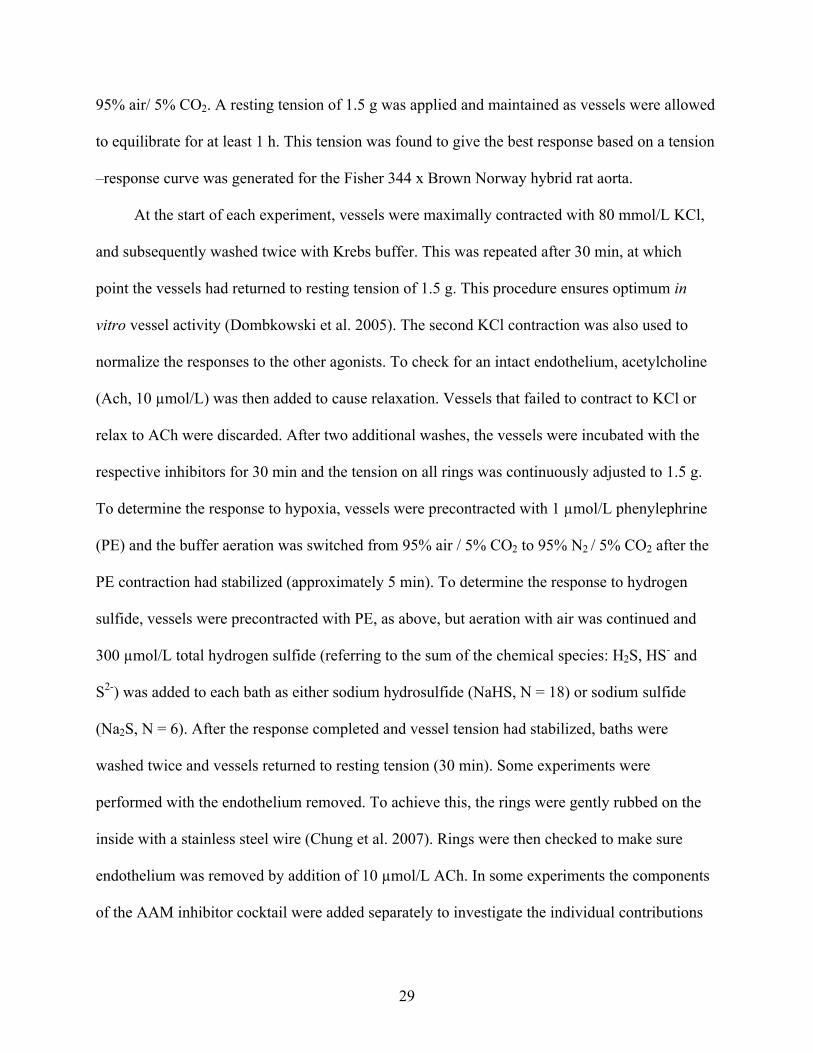
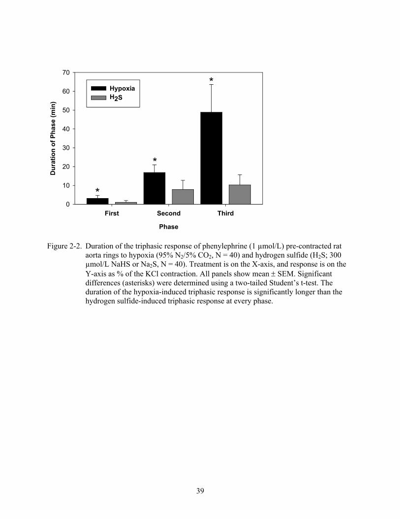
Isolated rat aorta rings exposed to hypoxia alone showed a triphasic contraction-relaxation-
contraction response that was complete in ca. 90 min (Figure 2-1a). Isolated rat aorta rings
exposed to hydrogen sulfide alone showed a similar triphasic contraction-relaxation-contraction
response (Figure 2-1b), although it was complete in ca. 30 min. The duration of all three phases
of the hypoxia-induced triphasic response was consistently, and significantly longer in duration
than the phases of the hydrogen sulfide -induced triphasic response (Figure 2-2; two-tail t-test, p
< 0.0001 for all). The triphasic response was the same when elicited by NaHS or Na2S as the
hydrogen sulfide donor. When the hypoxia-induced response was compared to the hydrogen
sulfide-induced response, the initial contraction and relaxation were significantly larger in
magnitude during hypoxia (Figure 2-3; two-tail t-test, p = 0.003 and Figure 2-4; two-tail t-test; p
< 0.001).
To test whether similar mechanisms are responsible for modulating the triphasic
responses to hypoxia and hydrogen sulfide, we inhibited NO production with L-NAME, KATP
channel opening with glibenclamide, and blocked AAM with an inhibitor cocktail of
indomethacin, esculetin, and clotrimazole, which inhibit cyclooxygenase, cytochrome P-450, and
lipoxygenase, respectively. When applied prior to hypoxia exposure, L-NAME significantly
reduced the initial contraction phase, but this phase was not significantly affected by
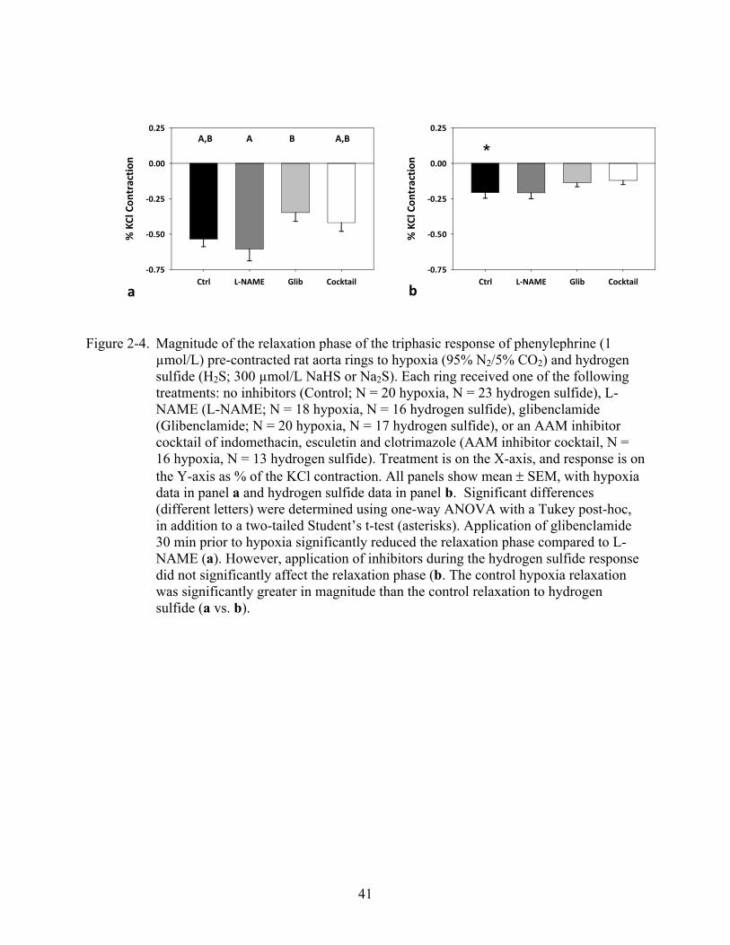
glibenclamide or the AAM inhibitor cocktail (Figure 2-3a, ANOVA, p = 0.0397). The relaxation
phase in response to hypoxia was not significantly affected by any of the inhibitors compared to
control, but glibenclamide significantly reduced the relaxation phase compared to L-NAME
(Figure 2-4a, ANOVA, p = 0.0340). The second contraction phase in response to hypoxia was

32
significantly reduced by L-NAME and glibenclamide, but not by the AAM inhibitor cocktail
(Figure 2-5a, ANOVA, p = 0.0215). In contrast, when applied prior to hydrogen sulfide
exposure, none of the inhibitors significantly affected the initial contraction phase (Figure 2-3b,
ANOVA, p = 0.272) or the relaxation phase (Figure 2-4b, ANOVA, p = 0.254). The second
contraction phase in response to hydrogen sulfide was not significantly affected by L-NAME or
glibenclamide, but was significantly reduced by the AAM inhibitor cocktail (Figure 2-5b,
ANOVA, p = 0.0176).
To further investigate the role of endothelium-derived products on the hydrogen sulfide-
induced response, we removed the endothelium from the aorta rings before experimentation.
There were significant differences in all three phases of the hydrogen sulfide-induced triphasic
response when comparing rings with no endothelium to control rings with an intact endothelium.
Compared to control rings, removal of the endothelium significantly reduced the magnitude of
the initial contraction phase (Figure 2-6, two-tail t-test, p = 0.0027), significantly increased the
magnitude of the relaxation phase (Figure 2-6, two-tail t-test, p = 0.0009), and significantly
reduced the magnitude of the second contraction phase (Figure 2-6, two-tail t-test, p < 0.0001).
To determine the effect of L-NAME without manipulating the tension before addition of
hydrogen sulfide as was done in the majority of the experiments, L-NAME was administered
after precontraction with PE and the ring was allowed to further constrict until stable. This
differed from the previous application of L-NAME which was 30 minutes before PE
preconstruction, and where the tension was adjusted back down to 1.5 g manually after L-NAME
was added. The initial contraction phase was significantly reduced while the second contraction
phase remained (N = 4, data not shown). Finally, when the AAM inhibitor cocktail components

33
were added separately, indomethacin, esculetin, and clotrimazole did not have a significant effect
on the second phase contraction (data not shown, N = 6-7, ANOVA, p = 0.157).
Discussion
This report demonstrates that hypoxia and hydrogen sulfide each elicit a characteristic,
triphasic contraction-relaxation-contraction response in rat aorta. While others investigators have
reported mono- or biphasic responses to hydrogen sulfide (Hosoki et al. 1997; Dombkowski et
al. 2005; Koenitzer et al. 2007), this is the first report of a triphasic response to hydrogen sulfide
in aorta. However, a triphasic response has been reported in rat pulmonary artery (Dombkowski
et al. 2005) and in response to acute hypoxia in rat aorta (Besse et al. 2002). While aorta has
been used in this study, and is commonly used to elicit the vascular actions of hydrogen sulfide
(Hosoki et al. 1997; Zhao et al. 2001; Dombkowski et al. 2005; Olson et al. 2006; Koenitzer et
al. 2007; Yang et al. 2008), it should be noted that this is a conduit vessel, not a resistance vessel.
Changing the tone of the aorta will effectively alter blood pressure and flow to the entire vascular
system not specific vascular beds, and would therefore not be an effective means of regulating
blood flow, as it would in a resistance vessel. In preliminary experiments with pulmonary artery
and mesenteric artery, I observed the same triphasic response as in aorta rings (Predmore,
unpublished data). Therefore, the aorta is used here as an experimental proxy for the resistance
vessels.
The concentration of hydrogen sulfide that accumulates in the aorta rings during hypoxia is
unknown, and for that matter so is the hydrogen sulfide concentration during hypoxia in vivo.
Therefore the incubation bath hydrogen sulfide concentrations reported are approximations of
what may actually accumulate during hypoxia (300 µmol/L total hydrogen sulfide or ca. 75
µmol/L H2S). This is within the range of previously reported plasma hydrogen sulfide
concentrations, which range from 10 to 300 µmol/L (Whitfield et al. 2008), although note that it
34
has recently been argued that free hydrogen sulfide is actually significantly lower (<100 nmol/L)
(Whitfield et al. 2008).
While the triphasic responses to hypoxia and hydrogen sulfide are broadly similar, they
differ temporally, in magnitude, and in their interaction with NO, KATP, and AAM. The hypoxia-
induced initial contraction and relaxation phases were significantly larger than the hydrogen
sulfide-induced counterparts, whereas the second contraction phase was similar. Interestingly,
the triphasic response to hydrogen sulfide is dose dependent: at concentrations of 100 and 600
µmol/L total hydrogen sulfide, the magnitude of the triphasic responses were similar, but at 900
µmol/L total hydrogen sulfide the magnitude of the phases increased slightly (data not shown).
Although all phases of the hypoxia-induced triphasic response could be reduced in magnitude by
one or more of the inhibitors, only the second contraction phase of the hydrogen sulfide-induced
triphasic response was significantly affected by inhibitors, suggesting that the triphasic responses
to hypoxia and hydrogen sulfide are differentially mediated.
Initial Contraction Phase
Our data suggest that the initial contraction phase in response to hypoxia is partially
mediated by NO bioavailability but not by KATP or AAM, and that the initial contraction phase in
response to hydrogen sulfide is not primarily mediated by NO bioavailability, KATP or AAM.
However, there must be some endothelium-derived factor mediating this response to hydrogen
sulfide, since removal of the endothelium severely reduced the initial contraction phase to
hydrogen sulfide.
In the initial experiments with L-NAME the baseline tension was maintained constant
throughout the incubation with L-NAME. Therefore any increase in tension after L-NAME
addition was reduced back to baseline before the addition of PE, hypoxia, or hydrogen sulfide.
The rationale behind this was to prevent the rings from maximally constricting after the PE was

35
added. If they were maximally constricted before addition of hypoxia or hydrogen sulfide, it
would be impossible to tell if it was an effect of NOS blockade, or simply the mechanical limits
of the rings to respond. In the experiments following when L-NAME was added after PE pre-
contraction, care was taken to ensure that the maximal tension, as determined from the KCl
contraction, was not reached before addition of hydrogen sulfide.
While the blockade of NO synthesis by L-NAME did not significantly alter the initial
contraction phase to hydrogen sulfide in the initial experiments, there was a non-significant
decrease in the hydrogen sulfide-induced initial contraction with L-NAME, similar to what was
observed with hypoxia (Fig. 2-3a vs. 2-3b). However, when L-NAME was added after the PE
precontraction, and the tension was not adjusted after L-NAME was added (which was the case
in the previous experiments), there was a significant reduction of the initial contraction phase to
hydrogen sulfide. Recent evidence also shows that hydrogen sulfide may cause a reduction of
cyclic AMP (cAMP) (Lim et al. 2008). This may occur in addition to the reduction of free NO
during hydrogen sulfide-induced contractions in vascular smooth muscle to mediate the observed
contractions.
Relaxation Phase
Our data suggest that the relaxation phase in response to hypoxia is partially mediated by
KATP, but not by NO or AAM, and that the relaxation phase in response to hydrogen sulfide is
not mediated by NO, KATP or AAM. Glibenclamide significantly reduced the relaxation phase
during hypoxia when compared to the L-NAME treatment (Fig. 2-4a), but it did not affect the
response to hydrogen sulfide (Fig. 2-4b). This was surprising given that hydrogen sulfide
activates KATP (Wang 2002; Tang et al. 2005). However, the glibenclamide concentration we
used (1 mol/L) may have been higher than that required for specific blockade of KATP, and may

36
have also blocked Cl- channels (Sheppard et al. 1997). A blockade of Cl- efflux could prevent
depolarization of smooth muscle cells (Kitamura et al. 2001), thereby reducing the effects of
KATP blockade. However, 10-100 µM glibenclamide is required for noticeable Cl- inhibition
(Sheppard et al. 1997; Robert et al. 2005). Recent evidence suggests that hydrogen sulfide-
induced vasorelaxation may be due to a reduction in ATP concentration by metabolic inhibition
followed by acidosis and activation of Cl-/HCO3- exchangers (Kiss et al. 2008), and therefore
may not primarily rely on KATP channels. It remains to be tested whether this occurs during the
triphasic response for both hypoxia and hydrogen sulfide, since only a monophasic relaxation
was observed in the aforementioned study. It is also interesting to note that the magnitude of the
relaxation phase during hydrogen sulfide-induced responses increased when the endothelium was
removed. This may be the result of removing the source of AAM that are vasoconstrictive (i.e.
thromboxanes, and some prostaglandins) and that may interfere with the relaxation pathway(s)
initiated by hydrogen sulfide (see below).
Second Contraction Phase
Our data suggest that the second contraction phase in response to hypoxia is partially
mediated by NO bioavailability and potentially KATP, but not mediated by AAM, and that the
second contraction phase in response to hydrogen sulfide is mediated by AAM but not by NO
bioavailability or KATP. This could indicate that hypoxia continues to inhibit the NO/NOS
system, whereas hydrogen sulfide induces production of one or more AAM. Both of these
possibilities would lead to contraction. This is similar to the response of the initial contraction, in
which the hypoxia-induced contraction may result from a decrease in NO bioavailability,
whereas the hydrogen sulfide-induced contraction may result from an alternative pathway such
as vasoconstrictive AAM or by decreasing cAMP (Lim et al. 2008). This is supported by the
observation that removal of the endothelium significantly reduced the second contraction phase,

37
indicating that the response is derived from endothelium. While the involvement of specific
enzymes and their products (i.e. cyclooxygenase, lipoxygenase or Cytochrome P-450) could not
be determined by adding the components of the AAM inhibitor cocktail alone, there may be an
additive or combined effect of multiple AAM causing the second phase contraction to hydrogen
sulfide. Our data provide no support for a role for of KATP channels in hydrogen sulfide-induced
contractions, which is also supported by Lim and colleagues (Lim et al. 2008), but our data do
support a role for KATP channels in hypoxia-induced contractions. However, if glibenclamide
non-specifically blocked chloride channels, and therefore Cl- efflux, in addition to KATP
(Sheppard et al. 1997; Kitamura et al. 2001), this could have artificially reduced the magnitude
of the hypoxia-induced second contractions.
Conclusion
Our data show that hypoxia and hydrogen sulfide produce similar responses in rat aorta
but that these responses result from different mechanisms. The data suggest that NO and KATP
contribute to the triphasic response to hypoxia, while AAM play a role in the hydrogen sulfide
response. However, no component of the triphasic response to any blockade was completely
eliminated, indicating that more signaling pathways are involved, potentially including, but
certainly not limited to, cAMP and metabolic inhibition. Additionally, the effect of the inhibitors
on higher or lower hydrogen sulfide concentrations remains to be determined, since we tested
only one concentration of hydrogen sulfide. Therefore, further investigations are warranted to
illuminate how these two signaling events are mediated and interconnected, and whether these
trends persist over a range of hydrogen sulfide concentrations.
38
Figure 2-1. Representative tracings of the effect of (a) hypoxia (95% N2/5% CO2) or (b) hydrogen sulfide (H2S; 300 µmol/L NaHS or Na2S) on phenylephrine (PE) (1 µmol/L) pre-contracted rat aorta rings. Both stimuli produced a triphasic contraction-relaxation-contraction. Horizontal scale bar indicates time (min) and vertical scale bar indicates aorta ring tension (g). To reduce noise, data were smoothed in Sigma Plot using a negative exponential 1st degree polynomial function with a 0.02 (hypoxia) or 0.05 (hydrogen sulfide) sampling proportion and nearest neighbors bandwidth method.
39
Phase
First Second Third
Du
rati
on
of
Ph
ase
(min
)
0
10
20
30
40
50
60
70
HypoxiaH2S
*
*
*
Figure 2-2. Duration of the triphasic response of phenylephrine (1 µmol/L) pre-contracted rat aorta rings to hypoxia (95% N2/5% CO2, N = 40) and hydrogen sulfide (H2S; 300 µmol/L NaHS or Na2S, N = 40). Treatment is on the X-axis, and response is on the Y-axis as % of the KCl contraction. All panels show mean SEM. Significant differences (asterisks) were determined using a two-tailed Student’s t-test. The duration of the hypoxia-induced triphasic response is significantly longer than the hydrogen sulfide-induced triphasic response at every phase.

40
Ctrl L-NAME Glib Cocktail
% K
Cl C
ontr
actio
n
0.00
0.25
0.50
*
Ctrl L-NAME Glib Cocktail
% K
Cl C
ontr
actio
n
0.00
0.25
0.50
ba
*
A A,BB A,B
Figure 2-3. Magnitude of the initial contraction phase of the triphasic response of phenylephrine (1 µmol/L) pre-contracted rat aorta rings to hypoxia (95% N2/5% CO2) and hydrogen sulfide (H2S; 300 µmol/L NaHS or Na2S). Each ring received one of the following treatments: no inhibitors (Control; N = 20 hypoxia, N = 23 hydrogen sulfide), L-NAME (L-NAME; N = 18 hypoxia, N = 16 hydrogen sulfide), glibenclamide (Glibenclamide; N = 20 hypoxia, N = 17 hydrogen sulfide), or an AAM inhibitor cocktail of indomethacin, esculetin and clotrimazole (AAM inhibitor cocktail, N = 16 hypoxia, N = 13 hydrogen sulfide). Treatment is on the X-axis, and response is on the Y-axis as % of the KCl contraction. All panels show mean SEM, with hypoxia data in panel a and hydrogen sulfide data in panel b. Significant differences (different letters) were determined using one-way ANOVA with a Tukey post-hoc, in addition to a two-tailed Student’s t-test (asterisks). Application of L-NAME 30 min prior to hypoxia significantly reduced the initial contraction phase (a). However, application of inhibitors during the hydrogen sulfide response did not significantly affect the initial contraction phase (b). The control hypoxia initial contraction was significantly greater in magnitude than the control initial contraction to hydrogen sulfide (a vs. b, asterisk).
41
Ctrl L-NAME Glib Cocktail
% K
Cl C
ontr
actio
n
-0.75
-0.50
-0.25
0.00
0.25
bCtrl L-NAME Glib Cocktail
% K
Cl C
ontr
actio
n
-0.75
-0.50
-0.25
0.00
0.25
a
*A,B BA A,B
Figure 2-4. Magnitude of the relaxation phase of the triphasic response of phenylephrine (1 µmol/L) pre-contracted rat aorta rings to hypoxia (95% N2/5% CO2) and hydrogen sulfide (H2S; 300 µmol/L NaHS or Na2S). Each ring received one of the followingtreatments: no inhibitors (Control; N = 20 hypoxia, N = 23 hydrogen sulfide), L-NAME (L-NAME; N = 18 hypoxia, N = 16 hydrogen sulfide), glibenclamide (Glibenclamide; N = 20 hypoxia, N = 17 hydrogen sulfide), or an AAM inhibitor cocktail of indomethacin, esculetin and clotrimazole (AAM inhibitor cocktail, N = 16 hypoxia, N = 13 hydrogen sulfide). Treatment is on the X-axis, and response is on the Y-axis as % of the KCl contraction. All panels show mean SEM, with hypoxia data in panel a and hydrogen sulfide data in panel b. Significant differences (different letters) were determined using one-way ANOVA with a Tukey post-hoc, in addition to a two-tailed Student’s t-test (asterisks). Application of glibenclamide 30 min prior to hypoxia significantly reduced the relaxation phase compared to L-NAME (a). However, application of inhibitors during the hydrogen sulfide response did not significantly affect the relaxation phase (b. The control hypoxia relaxation was significantly greater in magnitude than the control relaxation to hydrogen sulfide (a vs. b).

42
Ctrl L-NAME Glib Cocktail
% K
Cl C
ontr
actio
n
0.00
0.25
0.50
0.75
bCtrl L-NAME Glib Cocktail
% K
Cl C
ontr
actio
n
0.00
0.25
0.50
0.75
a
A A,BA,B BA BB A,B
Figure 2-5. Magnitude of the second contraction phase of the triphasic response of phenylephrine (1 µmol/L) pre-contracted rat aorta rings to hypoxia (95% N2/5% CO2) and hydrogen sulfide (H2S; 300 µmol/L NaHS or Na2S). Each ring received one of the following treatments: no inhibitors (Control; N = 20 hypoxia, N = 23 hydrogen sulfide), L-NAME (L-NAME; N = 18 hypoxia, N = 16 hydrogen sulfide), glibenclamide (Glibenclamide; N = 20 hypoxia, N = 17 hydrogen sulfide), or an AAM inhibitor cocktail of indomethacin, esculetin and clotrimazole (AAM inhibitor cocktail, N = 16 hypoxia, N = 13 hydrogen sulfide). Treatment is on the X-axis, and response is on the Y-axis as % of the KCl contraction. All panels show mean SEM, with hypoxia data in panel a and hydrogen sulfide data in panel b. Significant differences (different letters) were determined using one-way ANOVA with a Tukey post-hoc. Application of L-NAME and glibenclamide 30 min prior to hypoxia significantly reduced the second phase contraction (a). Application of the AAM inhibitor cocktail 30 min prior to hydrogen sulfide significantly reduced the second contraction phase (b).

43
Phase
First Second Third
% K
Cl C
on
trac
tio
n
-2.0
-1.5
-1.0
-0.5
0.0
0.5
1.0
+ Endo- Endo
*
*
*
Figure 2-6. Magnitude of the triphasic response of phenylephrine (1 µmol/L) pre-contracted rat aorta rings to hydrogen sulfide (H2S; 300 µmol/L NaHS or Na2S) with (N = 19) and without (N = 12) an intact endothelium. Endothelium was removed by rubbing the inside of the ring with stainless steel wire. Treatment is on the X-axis, and response is on the Y-axis as % of the KCl contraction. All panels show mean SEM. Significant differences (asterisks) were determined using a two-tailed Student’s t-test. Removal of the endothelium results in a significant reduction of the initial and second contraction phases to hydrogen sulfide, while it significantly increases the relaxation phase to hydrogen sulfide.

44
CHAPTER 3HYDROGEN SULFIDE INCREASES NITRIC OXIDE PRODUCTION FROM
ENDOTHELIAL CELLS BY A PROTEIN KINASE B (AKT)-DEPENDENT MECHANISM
Abstract
Hydrogen sulfide (H2S) and nitric oxide (NO) are both gasotransmitters that can elicit
synergistic vasodilatory responses in the in the cardiovascular system, but the mechanisms
behind this synergy are unclear. In the current study we investigated the molecular mechanisms
through which hydrogen sulfide regulates endothelial NO production. Initial studies were
performed to establish the temporal and dose-dependent effects of hydrogen sulfide on NO
generation using EPR spin trapping techniques. H2S stimulated a two-fold increase in NO
production from endothelial nitric oxide synthase (eNOS), which was maximal 30 min after
exposure to 25-150 µmol/L hydrogen sulfide. Following 30 min hydrogen sulfide exposure,
eNOS phosphorylation at Ser 1177 was significantly increased compared to control, consistent
with eNOS activation. Pharmacological inhibition of Akt, the kinase responsible for Ser 1177
phosphorylation, attenuated the stimulatory effect of hydrogen sulfide on NO production. Taken
together, these data demonstrate that hydrogen sulfide up-regulates NO production from eNOS
through an Akt-dependent mechanism. These results implicate hydrogen sulfide in the regulation
of NO in endothelial cells, and suggest that deficiencies in hydrogen sulfide signaling can
directly impact processes regulated by NO.
Introduction
Hydrogen sulfide (H2S) and nitric oxide (NO) are both gasotransmitters that function in
the cardiovascular system (Hosoki et al. 1997; Wang 2003). Recent reports indicate that the NO
and hydrogen sulfide signaling pathways interact on a variety of levels (Geng et al. 2007; Kubo
et al. 2007b; Kubo et al. 2007d; Yong et al. 2008a). Hydrogen sulfide and NO interact
synergistically in vitro, with hydrogen sulfide enhancing NO-mediated relaxation up to 13 fold in

45
isolated rat aorta (Hosoki et al. 1997), and treatment of Langendorff-perfused Sprague-Dawley
rat hearts with hydrogen sulfide immediately following ischemia confers cardioprotection
through NOS activation (Yong et al. 2008a). In contrast, other in vitro studies indicate that
incubation with hydrogen sulfide decreases NO production in aortic rings (Geng et al. 2007;
Kubo et al. 2007b), cell culture (Geng et al. 2007), and recombinant eNOS (Kubo et al. 2007d).
However, in these studies hydrogen sulfide incubation occurred 1-6 h before measurement of NO
production. Consequently, since hydrogen sulfide is volatile and oxidizes rapidly in the presence
of oxygen and free divalent metals (Tapley et al. 1999), the key effects of hydrogen sulfide in
those studies may have occurred before NO measurement began. In the present study we
investigated the ability of hydrogen sulfide to acutely modulate NO production in the
endothelium, with a specific focus on the mechanism of action.
Materials and Methods
Chemicals
Endothelial cell growth supplement was purchased from Upstate (Temecula, CA USA).
All other chemicals were purchased from Sigma-Aldrich (St. Louis, MO, USA) and were of the
highest quality available, unless otherwise noted.
Bovine Arterial Endothelial Cell Culture
Bovine Arterial Endothelial Cells (BAECs) were cultured in DMEM (1.0 g/L glucose)
supplemented with 10% FBS, 1% penicillin/streptomycin and endothelial cell growth
supplement (5 mg/L). Culture flasks were maintained in a 37 C incubator at 5.0% CO2.
Adherent endothelial cells were grown in 6-well plates for EPR studies and in 100 mm dishes for
western blotting studies.

46
Hydrogen Sulfide Exposure
Sodium sulfide (Na2S), a hydrogen sulfide donor, was prepared as a saturated stock
solution in distilled water and maintained at 4 C. At this temperature, the concentration of a
saturated solution of Na2S is 1.72 mol/L. From this stock, hydrogen sulfide dilutions were made
in Krebs buffer, of which 1.0 mL was added per well of a six-well plate, and 3.0 mL was added
per 100 mm Petri dish. The concentration of total hydrogen sulfide (H2S, HS- and S2-) in the
dilutions was determined using a methylene blue assay, with a standard curve generated with
deoxygenated, distilled water and Na2S as the reference (Gilboa-Garber 1971). After 30 min in a
37° C incubator, a 150 µmol/L hydrogen sulfide solution prepared in Krebs buffer showed no
detectable hydrogen sulfide, indicating virtually complete loss of hydrogen sulfide, presumably
by oxidation and volatilization (data not shown).
Akt Blockade
The Akt inhibitor Triciribine was used to prevent the phosphorylation of eNOS (Dieterle
et al. 2009). Triciribine (5.0 µmol/L) was added in Krebs buffer 30 min before experiments.
Cells were washed with PBS before and after addition of Triciribine.
Electron Paramagnetic Resonance Detection of Nitric Oxide
Spin-trapping measurements of NO were performed using a BrukerE-scan spectrometer
(BrukerBioSpin Corporation, Billerica, MA USA) with the iron spin trapping complex N-
methyl-D-glucaminedithiocarbamate (Fe-MGD) (Cardounel et al. 2002; Cardounel et al. 2007).
For measurements of NO produced by BAECs, cells were cultured as described above and spin
trapping was performed on cells grown in 6-well plates (1 x 10⁶ cells/ well). In these studies,
cells attached to the substratum were utilized since scraping or enzymatic removal leads to injury
and membrane damage with impaired NO generation. The medium from each well was removed
and the cells were washed with PBS (phosphate-buffered saline, without CaCl2 or MgCl2). Next,

47
0.15 ml of Krebs buffer containing the NO spin trap FE-MGD (0.5 mmol/L Fe2+, 5.0 mmol/L
MGD), and calcium ionophore (A23187, 1 mol/L) was added to each well and the plates were
incubated at 37 C under a humidified environment containing 5% CO2 /95% O2 for 20 min
(Cardounel et al. 2002; Cardounel et al. 2007). Following incubation, the medium from two
wells was removed and pooled as one 0.3 ml sample, frozen in liquid nitrogen and stored at -80
C. This yielded three samples per plate. The frozen NO spin-trap samples are stable, and were
later individually thawed, and trapped NO in the supernatants was quantified using the electron
paramagnetic resonance technique. Spectra recorded were obtained using the following
parameters: microwave power; 20 mW, modulation amplitude 3.16 G and modulation frequency;
100 kHz.
Western Blotting
BAECs from a 100 mm dish were scraped and suspended in 300 l
radioimmunoprecipitation assay (RIPA) buffer with Halt protease inhibitor cocktail (Thermo
Fisher Scientific, Rockford, IL USA), placed on ice, and sonicated to lyse the cells and suspend
the protein. The suspension was centrifuged at 12000 × g for 20 min at 4 C and the supernatant
removed, frozen in liquid nitrogen, and stored at -80 C. Western blotting was performed using
commercially-available polyclonal antibodies for eNOS and Ser 1177 eNOS (BD Biosciences,
San Jose, CA USA), monoclonal -actin (Cell Signaling Technology, Danvers, MA USA) and
secondary antibody conjugated to alkaline phosphatase (Sigma-Aldrich St. Louis, MO USA).
Protein was separated using SDS-PAGE and transferred onto PVDF membrane (Immobilon P,
Millipore, Billerica, MA USA) using a semidry blotter (BioRad, Hercules, CA USA). Using the
Snap-ID system (Millipore, Billerica, MA USA), membranes were blocked in 0.05% non-fat
milk in PBST (PBS with 0.05% Tween-20), washed in PBST and incubated 10 min with

48
appropriate primary antibodies (diluted 1:333 in PBST). Membranes were then washed in PBST
four times and subsequently incubated 10 min with secondary antibody diluted 1:3,333 in
blocking solution. Membranes were then washed two times in PBST, once in PBS, and once in
Tris-HCl (100 mmol/L, pH 9.5), after which chemiluminescence substrate was added (DuoLux
substrate, Vector Laboratories, Burlingame, CA USA). Images were captured with a digital
imager (GeneSnap, Syngene,Frederick, MD USA) and were analyzed using commercial software
(Quantity One,BioRad, Hercules, CA USA) to determine band intensity using local background
subtraction.
Statistics
All data were analyzed using one-way ANOVA with Dunnett’s post-hoc test for
significant differences from a control, with alpha 0.05 considered significant (JMP 7.0, SAS
Institute, Cary, NC USA).
Results
Initial experiments were conducted to establish the time course of hydrogen sulfide effects
on endothelial NO production. BAECs were exposed to the hydrogen sulfide donor Na2S (100
µmol/L) for 15, 30, 60, 120 and 240 min (Figure 3-1A). At each time point, endothelial-derived
NO production was measured using EPR. Peak NO production was observed at 30 min post
hydrogen sulfide treatment (ANOVA, p < 0.001); at which time mean NO production was
increased by 87%. This effect was not observed at later time points, suggesting a transient
activation of eNOS. To determine the dose-response to hydrogen sulfide, NO production was
measured 30 min after the addition of 5-600 µmol/L Na2S (Figure 3-1B). Mean NO production
increased by 39-62% at Na2S concentrations between 40-150 µmol/L (ANOVA, p = 0.001).
The transient nature of the hydrogen sulfide effects on endothelial NO production
suggested a change in eNOS phosphorylation status. Therefore, western blotting was used to
49
determine the phosphorylation state of eNOS after addition of hydrogen sulfide at 15 and 30 min.
Total eNOS expression was unchanged for all treatments (Figure 3-2A, ANOVA, p = 0.6727),
but after 30 min of incubation in the presence of 150 mol/L Na2S, eNOS phosphorylation at Ser
1177 increased by 88% compared to control (Figure 3-2B, ANOVA, p = 0.0031). Furthermore,
the ratio of phosphorylated Ser 1177 eNOS to total eNOS increased by 148% after 30 min
compared to control (Figure 3-2C, ANOVA, p = 0.0033). To determine whether increased Ser
1177 phosphorylation was responsible for the augmented NO production, BAECs were
pretreated for 30 min with the Akt inhibitor Triciribine (5 µmol/L), after which the cells were
exposed to 150 mol/L Na2S. Triciribine prevented the augmentation of endothelial NO
production (Figure 3-3, ANOVA, p = 0.0038). These results clearly indicate that hydrogen
sulfide increases endothelial NO production through Akt activation and subsequent increased
phosphorylation of eNOS at Ser 1177.
Discussion
Although an early study showed a synergistic effect of hydrogen sulfide on NO-induced
relaxation of blood vessel rings (Hosoki et al. 1997), later studies showed that hydrogen sulfide
inhibited eNOS-dependent NO production in aortic rings and cell culture, and from recombinant
proteins (Geng et al. 2007; Kubo et al. 2007b; Kubo et al. 2007d). However, these later studies
measured NO production 1-6 h after hydrogen sulfide addition. Since hydrogen sulfide is volatile
and rapidly oxidizes in the presence of oxygen and free divalent metals (Tapley et al. 1999), we
hypothesized that hydrogen sulfide acts within minutes of its application, and therefore that an
hour or longer delay between hydrogen sulfide application and measurement of NO production
could lead to a failure to detect an hydrogen sulfide effect. Here we demonstrate that hydrogen
sulfide up-regulates NO production from eNOS within 30 min. While we assume hydrogen

50
sulfide is causing this action, hydrogen sulfide in solution exists as H2S, HS-, and S2-. With the
extracellular ratio of H2S/HS- being between 1:3 and 1:5 and the intracellular ratio being
approximately equal (Olson et al. 2009), it is equally likely that HS- is causing the up-regulation.
To our knowledge, no one has yet definitively demonstrated that only H2S is causing the
observed effects of hydrogen sulfide, as opposed to HS-.
The comparatively rapid action led us to suspect that hydrogen sulfide was stimulating
phosphorylation of eNOS at Ser 1177 (Mount et al. 2007). We investigated this using western
blotting to detect total eNOS and phosphorylated eNOS. While total eNOS remained constant
for all treatment groups, 30 min exposure to 150 mol/L hydrogen sulfide resulted in a
significant increase in both phosphorylated eNOS and the ratio of phosphorylated eNOS to total
eNOS. To confirm that hydrogen sulfide-induced NO production was dependent on eNOS
phosphorylation, we used Triciribine to block phosphorylation of eNOS at Ser 1177, thereby
inhibiting Akt activation. This prevented the increase in NO production in cells exposed to
hydrogen sulfide, but did not significantly affect NO production in control cells.
These data suggest a novel mechanism of endogenous hydrogen sulfide signaling:
upregulation of NO production via Akt-dependent phosphorylation of eNOS at Ser1177. The
mechanism by which hydrogen sulfide activates Akt remains unknown. However, upstream
regulation of NO production by hydrogen sulfide could represent a novel and potentially
important regulatory mechanism in the control of NO signaling pathways, and could further
implicate defects of hydrogen sulfide signaling in cardiovascular pathologies.

51
Time (min)
Crtl 5 30 60 90 120 240
NO
Pro
du
cti
on
as
Pe
rce
nt
of
Co
ntr
ol
0.0
0.5
1.0
1.5
2.0
2.5
*
A
Sulfide Range (M)
0 5 to 20 40 to 150 300 to 600
NO
Pro
du
ctio
n a
s P
erce
nt
of
Co
ntr
ol
0.0
0.2
0.4
0.6
0.8
1.0
1.2
1.4
1.6
1.8
*B
Figure 3-1. The effect of H2S on NO production by BAECs. NO production was measured using EPR spectroscopy. NO values (y-axis) are presented in arbitrary units and have been normalized to control. Time (A) or hydrogen sulfide concentration range (B) are shown on the x-axis. Data are shown as mean ± SE. Statistical analysis was done using a one-way ANOVA (p < 0.05) with Dunnett’s post-hoc. An asterisk denotes values significantly different from control.

52
Figure 3-2. The effect of hydrogen sulfide on eNOS phosphorylation. Protein expression (y-axis, normalized to β-actin) was measured from BAECs untreated (Ctrl), or exposed to 15 or 30 min of hydrogen sulfide (x-axis). Using western blotting techniques, total eNOS expression (A), eNOS phosphorylated at Ser 1177 expression (B), and the ratio of Ser 1177 phosphorylated eNOS/total eNOS (C) were determined. Data are shown as mean ± SE. Statistical analysis was done using a one-way ANOVA (p < 0.05) with Dunnett’s post-hoc. An asterisk denotes values significantly different from control.

53
.
Figure 3-2. Continued.

54
Figure 3-3. The effect of Akt inhibition on hydrogen sulfide-stimulated NO production. NO production was measured using EPR spectroscopy. NO values (y-axis) are presented in arbitrary units and have been normalized to control. NO was measured from untreated (Ctrl), Triciribine treated (Ctrl + Tri.), hydrogen sulfide treated (H2S) and hydrogen sulfide treated with Triciribine (H2S + Tri.). Data are shown as mean ± SE. Statistical analysis was done using a one-way ANOVA (p < 0.05) with Dunnett’s post-hoc. An asterisk denotes values significantly different from control.

55
CHAPTER 4THE HYDROGEN SULFIDE SIGNALING SYSTEM: CHANGES DURING AGING AND
THE BENEFITS OF CALORIC RESTRICTION
Abstract
Hydrogen sulfide gas (H2S) has been proposed as an important signaling molecule, but
the effect of age on hydrogen sulfide signaling is unknown. Hydrogen sulfide causes diverse
effects in mammalian tissues, including relaxation in mammalian systemic vessels, and increased
perfusion and bicarbonate secretion in liver. Aging has negative impacts on the cardiovascular
system, whereas the liver is more resilient with age. Caloric restriction (CR) attenuates affects of
age in many tissues. This study investigates the effects of aging and CR on the hydrogen sulfide
signaling system in the aorta and liver of rats by determining the contractile response of aortic
rings to exogenous hydrogen sulfide, the protein and mRNA expression of the hydrogen sulfide
producing enzymes cystathionine –lyase (CSE) and cystathionine –synthase (CBS), and
hydrogen sulfide production rates. Tissue was collected from Fisher 344 x Brown Norway rats
from 8-38 months of age, maintained on an ad libitum (AL) or CR diet. Aortic rings exhibited a
triphasic contraction-relaxation-contraction response to hydrogen sulfide. The hydrogen sulfide-
sensitivity of the first phase contraction increased with age, while the magnitude of all three
phases increased with CR compared to AL. In aorta, CSE protein expression increased with age
on an AL diet, but this increase was attenuated with a CR diet. CBS expression showed the same
trends as CSE, but with lower expression levels. In liver, both CSE and CBS protein expression
remained relatively constant with age. In both aorta and liver, real-time PCR showed no changes
in CSE or CBS mRNA expression with age, but mRNA expression in aorta was higher on a CR
diet compared to AL. In both aorta and liver, hydrogen sulfide production did not change with
age, but did increase with CR. Overall, these data show a significant effect of age and diet on the
hydrogen sulfide signaling system in aorta, whereas liver was relatively unaffected. The

56
increased contractile sensitivity to hydrogen sulfide and increased protein expression of CSE and
CBS in aorta point to a drop in hydrogen sulfide bioavailability with age. Additionally, CR
seems to benefit CSE and CBS protein in both aorta and liver.
Introduction
Hydrogen sulfide (H2S) functions as a gasotransmitter in the nervous, cardiovascular, and
gastrointestinal systems. Abe and Kimura (Abe et al. 1996) first demonstrated that hydrogen
sulfide is produced in the brain and that it increases N-methyl-D-aspartic acid receptor-mediated
responses and facilitates hippocampal long-term potentiation. Since then a variety of
physiological roles have been proposed for hydrogen sulfide (Lowicka et al. 2007; Szabo 2007),
and at least some capacity for hydrogen sulfide production has been demonstrated in several
vertebrate tissues (Zhao et al. 2003; Olson 2005; Dombkowski et al. 2006; Olson et al. 2006).
When exogenously applied to isolated blood vessels of vertebrates, hydrogen sulfide elicits
responses that range from a monophasic relaxation or contraction, to multiphasic contraction-
relaxation-contraction responses, depending on the phylogenetic class and type of vessel
examined (Dombkowski et al. 2005). In the liver, hydrogen sulfide has been proposed to regulate
perfusion and maintain portal venous pressure, with deficiencies in hydrogen sulfide production
pathways resulting in increases in intra-hepatic resistance (Fiorucci et al. 2005b). Hydrogen
sulfide may also have a role in regulating biliary bicarbonate secretion in the liver (Fujii et al.
2005).
Hydrogen sulfide is endogenously produced from L-cysteine predominantly by the
enzymes cystathionine--lyase (CSE) and cystathionine--synthase (CBS) (Kimura 2000; Julian
et al. 2002; Wang 2002; Geng et al. 2004b; Fiorucci et al. 2005b; Gainey et al. 2005; Julian et al.
2005b; Kimura et al. 2005). In mammals, CSE and CBS are differentially expressed (Zhao et al.
57
2001; Zhao et al. 2003). CSE is the enzyme primarily expressed in the cardiovascular system,
whereas CBS is the enzyme primarily expressed in the nervous system (Hosoki et al. 1997; Geng
et al. 2004b; Ebrahimkhani et al. 2005; Kimura et al. 2005). In liver and kidney, CSE and CBS
are both expressed in relatively high amounts compared to other tissues (Fujii et al. 2005;
Lowicka et al. 2007).
Aging influences the action and/or efficacy of many signaling molecules, including
norepinephrine and nitric oxide (NO) (Lakatta 1993; van der Loo et al. 2000; Tanaka et al. 2006)
a gasotransmitter that is much better studied than hydrogen sulfide. A recent study of plasma
hydrogen sulfide concentration in humans over 50-80 years of age reported that hydrogen sulfide
levels may decline with age (Chen et al. 2005), but whether aging affects the hydrogen sulfide
signaling system is unknown. Furthermore, any affect of age on the hydrogen sulfide signaling
system may differ among tissues. For example, whereas the liver is relatively unaffected by age,
substantial physiological changes occur with age in the cardiovascular system, including fibrosis
of blood vessels leading to endothelial dysfunction (Castello et al. 2005), which affects
regulation of blood vessel tone (Kitani 1991; Kitani 1994; Anantharaju et al. 2002).
Caloric restriction (CR) is an intervention that attenuates many effects of aging (Labinskyy
et al. 2006; Leeuwenburgh et al. 2006), including cardiovascular morbidity (van der Loo et al.
2000), cross-linking of cardiac and skeletal muscle proteins (Leeuwenburgh et al. 1997),
mitochondrial dysfunction (Aspnes et al. 1997; Payne et al. 2003), loss of skeletal muscle mass
(van der Loo et al. 2000; Payne et al. 2003), and endothelial dysfunction (Gredilla et al. 2001;
Barja 2002; Gredilla et al. 2004; Castello et al. 2005; Taddei et al. 2006). CR attenuates the age-
related impairment of the NO signaling system (van der Loo et al. 2000; Marin et al. 2007)
(Yang et al. 2004; Minamiyama et al. 2007; Sharifi et al. 2008), but whether CR affects the

58
hydrogen sulfide signaling system, and whether CR specifically attenuates any affects of age on
the hydrogen sulfide signaling system, are all unknown.
In this study we investigated the effects of aging and CR on the hydrogen sulfide
signaling system in liver and aorta in the Fisher 344 × Brown Norway hybrid rat. Liver and aorta
tissue from rats of various ages were used to investigate CSE and CBS protein and mRNA
expression and hydrogen sulfide production rates. Contraction of aorta smooth muscle was used
as a functional indicator to investigate the effects of age and CR on functional responses to
hydrogen sulfide.
Materials and Methods
Chemicals
All chemicals were purchased from Sigma-Aldrich (St. Louis, MO, USA), unless
specifically noted.
Animals
Fisher 344 × Brown Norway hybrid rats of 8, 14, 18, 23, 27, 34 and 38 months of age were
singly housed under standard conditions and provided with food and water using either the
NIH31 diet ad libitum (AL) or the NIH31-NIA fortified diet at 40% CR. Myography studies
were performed on tissues from animals of 14, 27, and 34 months of age. All other studies were
performed on tissues from animals of 8, 18, 29, and 38 months of age.
Western Blotting
Liver and aorta were removed within 5 min of euthanasia, frozen in liquid nitrogen and
stored at -80 C. Samples were suspended in a lysis buffer consisting of 50 mmol/L Tris base,
2% (w/v) sodium dodecyl sulfate, 10 mmol/L ethylenediaminetetraacetic acid, 10 mmol/L
dithiothreitol, 0.5 mmol/L sodium tetraborate, and 1% (v/v) Halt protease inhibitor cocktail
(Thermo Fisher Scientific Rockford, IL USA). Western blotting was performed using

59
commercially available monoclonal antibodies for CSE and CBS (Abnova, Neihu District,
Taipei City, Taiwan), a monoclonal antibody for -actin (Cell Signaling Technology, Danvers,
MA, USA) and a secondary antibody conjugated to alkaline phosphatase (Sigma-Aldrich St.
Louis, MO, USA). Protein was separated using SDS-PAGE and transferred onto PVDF
membrane (Immobilon P, Millipore, Billerica, MA USA) using a semidry blotter (BioRad,
Hercules, CA USA). Using the Snap-ID system (Millipore, Billerica, MA USA) at room
temperature, membranes were blocked in 0.05% non-fat milk in PBST (phosphate-buffered
saline with 0.05% Tween-20), washed in PBST and incubated 10 min with appropriate primary
antibodies (diluted 1:333 in PBST). Membranes were then washed in PBST four times and
subsequently incubated 10 min with secondary antibody diluted 1:3,333 in blocking buffer.
Membranes were washed two times in PBST, once in PBS, and once in Tris-HCl (100 mmol/L),
pH 9.5. Chemiluminescent substrate (DuoLux, Vector Laboratories, Burlingame, CA USA) was
used to generate a chemiluminescent signal, which was captured with a digital imager
(GeneSnap, Syngene, Frederick, MD USA). Images were analyzed using commercial software
(Quantity One, BioRad, Hercules, CA USA) to determine band intensity using local background
subtraction.
RNA Extraction
Liver and aorta tissues were disrupted into powder under liquid nitrogen. All samples for
each tissue were processed on the same day. For liver, total RNA was extracted using an Arum
total RNA mini kit (BioRad, Hercules, CA USA). For aorta, total RNA was extracted using Tri
reagent and a glass homogenizer (Kontes Glass, Vineland, NJ USA).
Real-Time PCR
Relative quantitative real time PCR was run in 96-well PCR plates in an Applied
Biosystems 7300 Real-time PCR system (Life Technologies, Carlsbad, CA USA). Primers were

60
designed using commercial software (Premier Biosoft International, Palo Alto, CA USA) and
synthesized by Integrated DNA Technologies (Coralville, IA USA). The primers used were as
follows: CSE sense 5’- TGGGCTTAGTGTCTGTTAATTCC -3’, CSE antisense 5’-
GGCAGCAGAGGTAACAATCG – 3’, CBS sense 5’- TGCCTGAGAAGATGAGTATG -3’,
CBS antisense 5’- GGTCCAGAATGTGAGAATTG – 3’, Actin sense 5’-
CTCCCAGCACACTTAACTTAG C -3’, Actin antisense 5’- AAAGCC
ACAAGAAACACTCAGG – 3’, 18S rRNA sense 5’- CGAGGAATTCCCAGTAAGTGC -3’,
18S rRNA antisense 5’- CCATCCAATCGCTAGTAGCG – 3’.For both liver and aorta plates, a
standard curve for the housekeeping genes -actin (run with CSE samples) or 18S rRNA (run
with CBS samples) was run with a standard curve for the gene of interest (GOI), with all samples
in triplicate. The remainder of the plate was filled with 8 random samples plus 1 calibrator
sample for plate to plate corrections, loaded in triplicate for both the housekeeping gene and the
GOI. Additional plates were run containing the calibrator sample on each plate until all samples
had been processed. All plates for a given tissue-GOI combination were made and run on the
same day, using the same real-time PCR machine.
Hydrogen Sulfide Production
Hydrogen sulfide production was measured essentially as previously described (Stipanuk
et al. 1982; Julian et al. 2002; Dombkowski et al. 2006). Frozen tissues were ground into a fine
powder under liquid nitrogen with mortar and pestle. For each sample, two small scoops (approx
50 mg wet weight) were added to 1.5 mL 100 mmol/L potassium phosphate buffer (pH 7.4) and
homogenized with a Polytron homogenizer (Kinematica, Bohemia, NY USA) for 30–60 s until
the solution was smooth. A 1 mL aliquot of this homogenate solution was then placed in the
outer well of a 25 mL glass flask containing an additional plastic center well (Kontes Glass,

61
Vineland, NJ USA). The center well was filled with 0.5 mL 1% (w/v) zinc acetate, and a piece of
chromatography paper (Whatman grade 3MM Chr, Maldstone England) cut 2.5 cm by 4.5 cm
and folded into a fan shape was placed in the well to absorb the zinc acetate. The substrate L-
cysteine (10 mmol/L) and the cofactor pyridoxal-5′-phosphate (PLP; 2 mmol/L) were added to
the outer well, after which the flask was sealed with a septum stopper and flushed with N2 gas
for 30 s. Each flask was incubated on a shaker at 37 °C for 1.5 h for liver and 8 h for aorta. After
incubation, 1 mL 50% (w/v) trichloroacetic acid was added to the outer well to stop enzyme
activity and to convert all S2− and HS− to H2S. The zinc acetate solution on the chromatography
paper reacts with volatilized H2S to form zinc sulfide, which is relatively stable. The flasks were
then incubated for an additional hour to allow remaining hydrogen sulfide to volatilize and form
zinc sulfide. The filter paper was then removed from the center well and placed in a test tube
containing 3.5 mL of de-ionized water, 0.4 mL 20 mmol/L N,N-dimethyl-p-phenylenediamine
oxalate in 7.2 mol/L HCl and 0.4 mL of 30 mmol/L FeCl3 in 1.2 mol/L HCl. The test tubes were
gently vortexed and incubated for 20 min at room temperature. Absorbance of the solution in the
test tube was read at 670 nm with a plate reader (BioTek Instruments, Inc., Winooski, VT USA).
Samples with an absorbance greater than 2 were diluted 10-fold with distilled water and the
absorbance re-measured. Absorbance measurements were calibrated against a standard curve
generated from NaHS in deoxygenated, de-ionized water added to the outside wells, incubated at
37 C for 1.5 h, and assayed as above. To ensure that all measured hydrogen sulfide production
is by tissue enzymes, several controls were run in addition to sample homogenates: a negative
control of homogenate but no cofactors or substrate; and a blank of all cofactors and substrate in
phosphate buffer, but without homogenate. Inhibitors of CSE (20 mmol/L propargylglycine,
Pgly) and CBS (1 mmol/L aminooxyacetic acid, AOAA) were added to confirm the hydrogen

62
sulfide production was through these, and not alternate pathways (Julian et al. 2002). Protein
concentration was quantified with a NanoDrop 1000 (Thermo Fisher Scientific, Waltham, MA
USA), to calculate nmol hydrogen sulfide production per hour per mg protein.
Myography
The effect of hydrogen sulfide on vascular tone was measured in 5mm long aorta rings
from 41 animals, with 5 to 8 rings tested for each age × diet treatment combination. To reduce
artifact from anesthesia (Dombkowski et al. 2005), euthanasia was by guillotine under IACUC-
approved methods. After dissection, aorta was cleaned of fat and connective tissue, and
myography was performed essentially as previously described (Dombkowski et al. 2005). Rings
were attached with stainless steel wire to force transducers and mounted in a 37°C tissue bath
system (Radnoti Glass Technology, Monrovia, Ca USA) containing Kreb’s bicarbonate buffer.
Rings were allowed to equilibrate after mounting for a minimum of 1 h, and a baseline tension of
1.5 g was maintained throughout each experiment. At the start of an experiment, each ring was
maximally contracted with two additions of 80 mmol/L KCl. After washing with Kreb’s buffer
and returning to baseline tension, the rings were incubated with 10 µmol/L propranolol (to block
-adrenergic receptor relaxation and maximize -adrenergic receptor contraction), and
precontracted with 1 µmol/L norepinephrine (NE). After the NE precontraction stabilized, rings
were exposed to 100 µmol/L hydrogen sulfide. After 30-45 min, the tissue bath was drained and
the rings were washed twice with Kreb’s buffer and allowed to return to baseline tension. This
sequence of propranolol, NE and hydrogen sulfide addition was repeated for 300, 600 and 900
µmol/L hydrogen sulfide. Data for hydrogen sulfide-induced contractions for each aortic ring
were standardized to the weight of the ring.

63
Actual total hydrogen sulfide concentrations, as measured by a methylene blue assay
(Gilboa-Garber 1971), were approximately 75% of the predicted value (presumably due mostly
to oxidation). Note that in physiological saline at pH 7.4, the dissociation of hydrogen sulfide
results in approximately 1/3 of total hydrogen sulfide as H2S and 2/3 as HS- (Beauchamp et al.
1984).
Statistics
All statistics were run using JMP statistical software, (JMP 7.0, SAS Institute, Cary, NC
USA) with alpha 0.05 considered significant. Two-way ANOVAs were run with a Tukey post-
hoc test, when possible. However, the functional response data were not always normally
distributed, and therefore violated the assumptions of the ANOVA. In those cases, significant
effects of age and diet were tested using the Kruskal-Wallis nonparametric ANOVA. When
fewer than three groups were compared, pooled (two-tailed) or one-tailed t-tests were used. For
real-time PCR, statistics were done on the average Ct of each sample, since the Cts are normally
distributed (Wood et al. 2005). Statistical tests are listed throughout preceding their respective p-
values.
Results
CSE and CBS Protein Expression
The relative expression of CSE and CBS protein differed between aorta and liver. In
aorta, CSE protein expression (relative to -actin) was 1.43-fold higher than CBS protein
expression on average, whereas in liver, CBS protein expression (relative to -actin) was 1.18-
fold higher than CSE protein expression, on average.
In both aorta and liver, CSE protein expression was significantly affected by an
interaction between age and diet (Figure 4-1A and 4-1B). In aorta, CSE protein expression

64
increased with age in AL animals, but it was unchanged with age in CR animals (Figure 4-1A,
bars; 2-way ANOVA with Tukey post-hoc, p < 0.0001). At ages 18, 29, and 38 months CR CSE
protein expression was significantly lower than AL at those ages, and was not significantly
different than CSE expression at 8 months on either a CR or AL diet (Figure 4-1A, asterisks). In
liver, CSE protein expression was unchanged with age in AL animals, but increased with age in
CR animals (Fig 4-1B, bars; 2-way ANOVA with Tukey post-hoc, p = 0.0041). At 38 months of
age AL CSE protein expression was significantly lower than CR (Figure 4-1B, asterisks).
In aorta but not liver, CBS expression was also significantly affected by an interaction
between age and diet (Figure 4-1C and 4-1D). In aorta, there was significantly higher CBS
protein expression in AL animals at 38 months compared to CR animals (Figure 4-1C, asterisks;
2-way ANOVA with Tukey post-hoc, p = 0.0056). In liver, there was no significant difference in
CBS expression at any age, and there was no effect of CR (Figure 4-1D, 2-way ANOVA, p >>
0.05).
CSE and CBS mRNA Expression
There were no interactions between age and diet on the mRNA expression of CSE and
CBS in aorta or liver (Figure 4-2). In aorta, the CR diet significantly increased expression of both
CSE mRNA (Figure 4-2A, one-tailed t-test, p = 0.0013) and CBS mRNA (Figure 4-2C, one-
tailed t-test, p = 0.0258) when compared to the AL diet. In liver, diet did not affect the
expression of either CSE mRNA (Figure 4-2B, one-tailed t-test, p >> 0.05) or CBS mRNA
(Figure 4-2D, one-tailed t-test, p >> 0.05).
Hydrogen Sulfide Production
There were no interactions between age and diet on capacity for hydrogen sulfide
production in aorta or liver (Figure 4-3), but the capacity for hydrogen sulfide production was
affected by diet. In aorta and liver hydrogen sulfide production was higher in tissues of CR
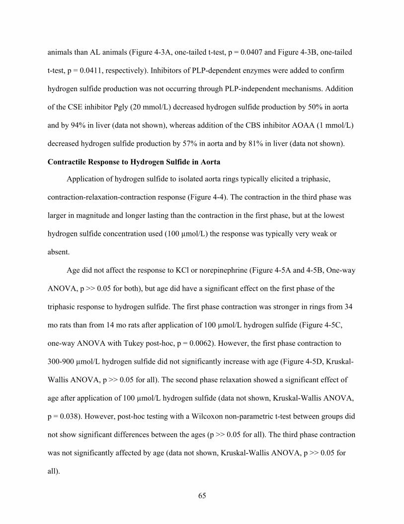
65
animals than AL animals (Figure 4-3A, one-tailed t-test, p = 0.0407 and Figure 4-3B, one-tailed
t-test, p = 0.0411, respectively). Inhibitors of PLP-dependent enzymes were added to confirm
hydrogen sulfide production was not occurring through PLP-independent mechanisms. Addition
of the CSE inhibitor Pgly (20 mmol/L) decreased hydrogen sulfide production by 50% in aorta
and by 94% in liver (data not shown), whereas addition of the CBS inhibitor AOAA (1 mmol/L)
decreased hydrogen sulfide production by 57% in aorta and by 81% in liver (data not shown).
Contractile Response to Hydrogen Sulfide in Aorta
Application of hydrogen sulfide to isolated aorta rings typically elicited a triphasic,
contraction-relaxation-contraction response (Figure 4-4). The contraction in the third phase was
larger in magnitude and longer lasting than the contraction in the first phase, but at the lowest
hydrogen sulfide concentration used (100 µmol/L) the response was typically very weak or
absent.
Age did not affect the response to KCl or norepinephrine (Figure 4-5A and 4-5B, One-way
ANOVA, p >> 0.05 for both), but age did have a significant effect on the first phase of the
triphasic response to hydrogen sulfide. The first phase contraction was stronger in rings from 34
mo rats than from 14 mo rats after application of 100 µmol/L hydrogen sulfide (Figure 4-5C,
one-way ANOVA with Tukey post-hoc, p = 0.0062). However, the first phase contraction to
300-900 µmol/L hydrogen sulfide did not significantly increase with age (Figure 4-5D, Kruskal-
Wallis ANOVA, p >> 0.05 for all). The second phase relaxation showed a significant effect of
age after application of 100 µmol/L hydrogen sulfide (data not shown, Kruskal-Wallis ANOVA,
p = 0.038). However, post-hoc testing with a Wilcoxon non-parametric t-test between groups did
not show significant differences between the ages (p >> 0.05 for all). The third phase contraction
was not significantly affected by age (data not shown, Kruskal-Wallis ANOVA, p >> 0.05 for
all).

66
Overall, CR had a significant effect on all phases of the contract-relax-contract response to
hydrogen sulfide. Furthermore, the KCl and NE contractions were significantly stronger in rings
from CR rats than from AL rats (Figure 4-6A and 4-6B, one-tailed t-test, p = 0.0282 and <
0.0001, respectively). Compared to rings from AL rats, CR increased the magnitude of the first
phase contraction after application of 300 – 900 µmol/L hydrogen sulfide (Figure 4-7A, pooled t-
test, p = 0.0006, p = 0.0079 and 0.0192, respectively), the magnitude of the second phase
relaxation after application of 300 and 600 µmol/L hydrogen sulfide (Figure 4-7B, one-tailed t-
test, p = 0.0492 and Wilcoxon test, p = 0.0027, respectively), and the magnitude of the third
phase contraction after application of 300 - 900 µmol/L hydrogen sulfide (Figure 4-7C, pooled t-
test, p < 0.0001, p = 0.0025, and p = 0.0008, respectively).
Discussion
CSE and CBS Protein Expression
In aorta, CSE expression increased with age on an AL diet, but remained constant on a
CR diet. Aorta CBS expression showed a similar trend, but with lower expression levels. CBS
expression was increased at age 38 months for AL animals, while CR animals had significantly
lower CBS expression at 38 months. It is perhaps not surprising that changes with age were more
evident for CSE than for CBS, since CSE is the expressed at higher levels in the endothelium and
smooth muscle cells of aorta and is also believed to have the most functional contribution to
regulating vascular tone (Hosoki et al. 1997; Zhao et al. 2001; Zhao et al. 2003; Wang et al.
2008). Therefore, CSE would be the more likely of the two enzymes to exhibit measurable
effects of age and diet. We have been able to measure CBS protein in aorta even though previous
researchers were only able to detect CSE mRNA and concluded there was no CBS in these
tissues (Hosoki et al. 1997; Zhao et al. 2001). However, the protein expression level of CBS was

67
much less than that of CSE, consistent with the assumption that CBS has a less important role in
the vasculature.
In aorta, the increase in CSE expression with age is similar to changes reported with age in
eNOS expression in peripheral arteries (Cernadas et al. 1998; van der Loo et al. 2000; Goettsch
et al. 2001; van der Loo et al. 2005), in which eNOS increased up to sevenfold with age. This
was attributed to a homeostatic attempt to correct for a decrease in the bioavailability of NO
caused by superoxide reacting with NO to form a peroxynitrite (van der Loo et al. 2000; van der
Loo et al. 2005). Since NO and hydrogen sulfide have been reported to interact synergistically,
changes in one system may impact the other (Hosoki et al. 1997; Wang et al. 2008).
Additionally, hydrogen sulfide may also react with, and be quenched by, increasing peroxynitrite
levels resulting from NO reacting with superoxide (Whiteman et al. 2004b). Furthermore, there
is limited evidence that hydrogen sulfide reacts with superoxide, which may also directly impact
hydrogen sulfide bioavailability (Geng et al. 2004a). CR has been hypothesized to prevent this
age-related endothelial dysfunction by preventing increases in superoxide levels with age,
thereby maintaining a more youthful state (Gredilla et al. 2001; Barja 2002; Gredilla et al. 2004;
Castello et al. 2005; Taddei et al. 2006). Accordingly, CSE and, to a lesser extent, CBS
expression in CR animals did not significantly increase with age and were more similar to 8
month old AL animals, consistent with a protective effect of CR in aorta maintaining a youthful
state.
In liver, CSE expression increased with age in CR animals whereas CBS expression did
not change with age. Since both CBS and CSE are involved in cysteine metabolism in addition to
producing hydrogen sulfide (Zhao et al. 2001), they are present in relative abundance in the liver
compared to other tissues. However, CBS is the primary hydrogen sulfide producing enzyme in

68
the liver (Zhao et al. 2003), and therefore the increase in CSE expression with age may not
accurately reflect what is occurring to hydrogen sulfide levels as well as CBS expression does.
Liver enzymatic capacity does not change substantially with age, and this also appears to be true
for CBS (Kitani 1991; Kitani 1994; Anantharaju et al. 2002). Since the liver is functionally much
different than aorta, it is perhaps not surprising that age and diet do not affect the liver to the
same degree as the aorta.
The heterogeneity between tissue types observed between aorta and liver also occurs in the
NO signaling system. Even when comparing peripheral vessels to heart tissue in the circulatory
system, changes are seen in the vasculature even though no significant changes are observed in
the heart (van der Loo et al. 2005). Therefore, the age and diet affects on the hydrogen sulfide
signaling system may not be global events that occur during aging, but rather tissue-specific
effects.
CSE and CBS mRNA Expression
There were complex and dramatic changes with age and diet in CSE protein expression in
aorta and liver. There were no significant changes in the mRNA message for CSE or CBS with
age, although CR did increase CSE and CBS mRNA levels in aorta. This seems to contradict the
protein expression, in which CR reduced the expression of those proteins. Conversely, liver CSE
and CBS mRNA message did not significantly change with age or diet, which was more
consistent with the protein data. We cannot account for the discrepancy between mRNA and
protein data, except to note that mRNA message does not always translate directly into protein
(Gygi et al. 1999). We must conclude that while CR causes increases in mRNA for CSE and
CBS in the vasculature, this does not translate into the synthesis of more new protein.
Alternatively, CR may attenuate the need for new protein formation, but does not stop the
synthesis of mRNA, leading to the observed increase in mRNA expression.

69
Hydrogen Sulfide Production
Extracellular concentrations of total hydrogen sulfide (representing the sum of H2S, HS-
and S2-) have been reported in the range of 50-160 µmol/L in the brain (Goodwin et al. 1989;
Abe et al. 1996) and ~50 µmol/L in the plasma (Zhao et al. 2001). However, recent
measurements in plasma seem to indicate that true hydrogen sulfide concentrations are much
lower (<100 nmol/L) (Whitfield et al. 2008). This discrepancy is likely due to methodological
differences in how free hydrogen sulfide is differentiated from that which is bound or “acid-
labile” (Olson 2009).
Homogenates of aorta and liver both produced a consistent amount of hydrogen sulfide
regardless of age, but CR samples consistently showed a higher hydrogen sulfide production
rate. This is particularly interesting for the aorta, because our data indicate that it had lower
levels of the hydrogen sulfide producing enzymes. Therefore, it can produce more hydrogen
sulfide than the AL samples that have significantly more hydrogen sulfide producing enzymes.
Note that because the data were calculated as nmol hydrogen sulfide/hr/mg protein, production
rates for aorta and liver were very similar. When hydrogen sulfide production is instead
calculated as nmol hydrogen sulfide/hr/g tissue or nmol hydrogen sulfide/hr/ml of homogenate,
liver has a higher production rate than aorta.
Addition of AOAA and Pgly reduced hydrogen sulfide production in both aorta and liver,
but did not completely block production. This indicates that both CSE and CBS contribute to
hydrogen sulfide production in these tissues, but there may be a small contribution of “acid
labile” hydrogen sulfide contributing to these production rate measurements. It is important to
note that the recovery rate of hydrogen sulfide using the assay is not 100%, and is more likely
33-59% depending on the hydrogen sulfide production rate of the tissue (Julian et al. 2002).

70
Therefore, these rates should only be used as a comparison between the treatment groups in this
study and not as an absolute rate of hydrogen sulfide production from aorta and liver in the rat.
Contractile Response to Hydrogen Sulfide in Aorta
Although triphasic responses to hydrogen sulfide application have been reported in rat
pulmonary artery (Olson et al. 2006), previous studies of rat aorta have shown only a
monophasic relaxation or contraction with hydrogen sulfide (Zhao et al. 2002; Dombkowski et
al. 2005; Kubo et al. 2007b). We found that the development of all three phases typically
required 15-30 min to develop after a bolus of hydrogen sulfide. Consequently, the triphasic
response may have been masked in previous studies that did not allow sufficient time after
hydrogen sulfide addition. Additional methodological variations, such as tissue preparation (ring
vs. strip), method of euthanasia, and lag time between tissue dissection and hydrogen sulfide
application, may have also contributed to differences in aorta response.
It has been suggested that high O2 concentrations in the tissue bath may lead to formation
of vasoactive hydrogen sulfide oxidation products (Koenitzer et al. 2007). Since our
experiments were run at ambient O2 concentrations, oxidation products may have contributed to
the triphasic response. However, in dorsal aorta of cyclostomes hydrogen sulfide-induced
contractions are enhanced at low O2 concentrations, suggesting that hydrogen sulfide oxidation
products are not the only cause of hydrogen sulfide-induced contractions (Olson et al. 2008).
Additionally, in rat aorta rings we see the same triphasic response during hypoxia (Dissertation
Chapter 2). This response to hypoxia is presumably mediated in part by hydrogen sulfide that is
not readily oxidized due to the extremely low oxygen levels (Olson et al. 2006).
Our data indicate that the response of rat aortic rings to hydrogen sulfide increases in
magnitude with age, with significant changes observed in the first phase contraction. The
triphasic response in aorta has not been reported previously, and it is not known what causes the

71
initial contraction phase. A likely candidate is the reaction of hydrogen sulfide with NO to form
a nitrosothiol (Whiteman et al. 2006), which has been suggested to reduce free NO and lead to
contraction at low levels of hydrogen sulfide (Ali et al. 2006), whereas higher hydrogen sulfide
concentrations may cause relaxation by activating KATP channels (Zhao et al. 2001). It is likely
that after application of the hydrogen sulfide donor, NO reacts quickly with hydrogen sulfide
causing contraction, which is followed by relaxation as KATP channels are activated. We have
shown evidence of this in aorta during hypoxia-induced responses (Chapter 2). If indeed this
NO-scavenging mechanism does mediate the first phase contraction, then the reduced
bioavailability of NO observed with age (Cernadas et al. 1998; van der Loo et al. 2000; Goettsch
et al. 2001; van der Loo et al. 2005) could cause the increase in sensitivity to hydrogen sulfide.
That is, the less free NO is available, the larger the effect on vascular tone for a given amount of
hydrogen sulfide.
Caloric restriction dramatically increased the response to hydrogen sulfide in all three
phases, as well as the response to KCl. Therefore, rather than representing a global effect on
hydrogen sulfide -signaling, the effect of CR may represent an overall enhancement of smooth
muscle tissue health, as has been reported in skeletal muscle (Payne et al. 2003),.
Conclusions
Our data show that age affects the hydrogen sulfide signaling system in the aorta, on both
the contractile response to hydrogen sulfide and the protein expression of CSE. Aorta responds
to hydrogen sulfide with a triphasic contract-relax-contract response, which becomes sensitized
to hydrogen sulfide with age. There was no age effect in the liver on the main hydrogen sulfide
producing enzyme CBS. CR benefits the hydrogen sulfide signaling system in the aorta. All
phases of the triphasic response were enhanced by CR, and CR maintained lower expression of
CSE and CBS proteins, which could produce more hydrogen sulfide than AL animals. CR has no

72
significant affect on CBS protein expression in the liver. Overall, aorta shows the most impact of
age and CR. While the effects of CR are likely due to maintenance of healthier tissue/proteins,
the sensitization with age on hydrogen sulfide signaling in aorta may owe to impacts of age on
NO. Therefore, the interactions of H2S, NO and CO are key to fully elucidating the ramifications
of these changes.

73
A
Age (mo)
8 18 29 38 8 18 29 38
Exp
ere
ssio
n R
ela
tive
to A
ctin
0
1
2
3
4
5
6
7
8
9
**
*
*
**
B
Age (mo)
8 18 29 38 8 18 29 38
Exp
eres
sion
Rel
ativ
e to
Act
in
0
1
2
3
4
5
6
7
*
*
C
Age (mo)
8 18 29 38 8 18 29 38
Exp
eres
sion
Rel
ativ
e to
Act
in
0
1
2
3
4
*
*
D
Age (mo)
8 18 29 38 8 18 29 38
Exp
ere
ssio
n R
elat
ive
to A
ctin
0
2
4
6
8
10
Figure 4-1. Effect of age and diet on CSE and CBS protein expression. CSE expression is shown in aorta (A) and liver (B). CBS expression is shown in aorta (C) and liver (D). The Y-axis shows CSE (A and B) or CBS (C and D) protein expression relative to -actin. The X-axis shows the age in months (mo). AL data are in white columns and CR data are in grey columns. Representative blots are aligned along the top, above each column. Data are represented as mean SE and were analyzed by 2-way ANOVA with Tukey post-hoc tests. Means that are significantly different are indicated by different letters.
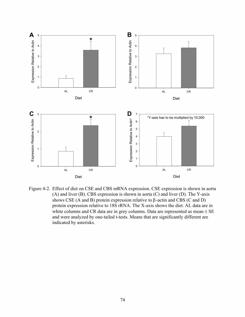
74
Diet
AL CR
Exp
ress
ion
Re
lativ
e to
Act
in
0
1
2
3
4
5
*A
Diet
AL CR
Exp
ress
ion
Rel
ativ
e to
Act
in
0
1
2
3
4
5B
Diet
AL CR
Exp
ress
ion
Rel
ativ
e to
Act
in
0
1
2
3
*C
Diet
AL CR
Exp
ress
ion
Re
lativ
e to
Act
in*
0
1
2
3
4
5
6
7*Y-axis has to be multiplied by 10,000
D
Figure 4-2. Effect of diet on CSE and CBS mRNA expression. CSE expression is shown in aorta (A) and liver (B). CBS expression is shown in aorta (C) and liver (D). The Y-axis shows CSE (A and B) protein expression relative to -actin and CBS (C and D) protein expression relative to 18S rRNA. The X-axis shows the diet. AL data are in white columns and CR data are in grey columns. Data are represented as mean SE and were analyzed by one-tailed t-tests. Means that are significantly different are indicated by asterisks.

75
Diet
AL CR
Pro
duc
tion
Ra
te n
mol
/hr/
mg
0.00
0.02
0.04
0.06
0.08
0.10
0.12
0.14
0.16
0.18
*A
Diet
AL CR
Pro
duct
ion
Rat
e nm
ol/h
r/m
g
0.00
0.05
0.10
0.15
0.20
*B
Figure 4-3. Effect of diet on hydrogen sulfide production. Hydrogen sulfide production rates are shown in aorta (A) and liver (B). The Y-axis shows production as nmol H2S/hr/mg protein. The X-axis shows the diet. AL data are in white columns and CR data are in grey columns. Data are represented as mean SE and were analyzed by one-tailed t-tests. Means that are significantly different are indicated by asterisks.

76
Figure 4-4. Representative tension tracing of a rat aorta ring pre-contracted with norepinephrine (NE) and exposed to hydrogen sulfide (H2S, 300 µmol/L). Scale bar shows time in min and tension in grams.

77
Age
14 27 34
Con
trac
tion
(g/m
g)
0.0
0.1
0.2
0.3
0.4A
Age
14 27 34
Con
trac
tion
(g/
mg)
0.00
0.05
0.10
0.15
0.20B
Age
14 27 34
Con
trac
tion
(g/m
g)
0.00
0.05
0.10
0.15
0.20
a b b
C
Age (months)
14 27 34 14 27 34 14 27 34 14 27 34
Co
ntr
act
ion
(g
/mg
)
0.0
0.1
0.2
0.3
0.4
0.5
a bb
100 umol/L 300 umol/L 600 umol/L 900 umol/LD
Figure 4-5. Effect of age on KCl- and norepinephrine (NE)-induced contractions and the first phase contraction to hydrogen sulfide. Age has no effect on KCl- (A) or NE-induced contractions (B). However, the first phase contraction to hydrogen sulfide (C) significantly increases with age at 100 µmol/L hydrogen sulfide addition. The first phase contractions did not significantly increase at 300-900 µmol/L hydrogen sulfideaddition (D). The Y-axis represents the hydrogen sulfide-induced tension of aortic rings (g/mg). The X-axis shows the age (mo) (A-C) for each of the four hydrogen sulfide concentrations, divided into four panels (D). The top labels represent the concentration of hydrogen sulfide used for that panel (D). Data are presented as means ± SE (A-C) or as box plots showing the median, quartiles and outliers (D). Data were analyzed by one-way ANOVA with Tukey post-hoc test. Means that are significantly different are indicated by letters.

78
Diet
AL CR
Con
trac
tion
(g/
mg)
0.0
0.1
0.2
0.3
0.4
*
A
Diet
AL CR
Con
tra
ctio
n (g
/mg)
0.00
0.05
0.10
0.15
0.20
*B
Figure 4-6. Effect of diet on KCl-and norepinephrine (NE)-induced contractions. CR causes an increase in contraction magnitude in response to KCl (A) and NE (B). The Y-axis represents the tension of aortic rings (g/mg). The X-axis represents age diet: ad libitum (AL) or caloric restricted (CR). AL data are in white columns and CR data are in grey columns. Data are represented as mean SE and were analyzed by one-tailed or pooled t-tests. Means that are significantly different are indicated by asterisks.

79
Figure 4-7. Effect of diet on hydrogen sulfide-induced contractions. CR causes an increase in contraction magnitude in response to hydrogen sulfide in the first phase contraction (A), second phase relaxation (B) and third phase contraction (C). The Y-axis represents the hydrogen sulfide-induced tension of aortic rings (g/mg). The X-axis represents diet: ad libitum (AL, white) or caloric restricted (CR, grey) for each of the four hydrogen sulfide concentrations, divided into four panels. The top labels represent the concentration of hydrogen sulfide used for that panel. Data are presented as medians, with error bars defining upper and lower quartiles. Data were analyzed by one-tailed or pooled t-tests. Medians that are significantly different are indicated by asterisks.
Diet
AL CR AL CR AL CR AL CR
Con
trac
tion
(g/m
g)
0.0
0.1
0.2
0.3
0.4
0.5 100 umol/L 300 umol/L 600 umol/L 900 umol/LA* * *
Diet
AL CR AL CR AL CR AL CR
Re
laxa
tion
(g
/mg)
-0.5
-0.4
-0.3
-0.2
-0.1
0.0
100 umol/L 300 umol/L 600 umol/L 900 umol/L
* *B

80
Diet
AL CR AL CR AL CR AL CR
Con
trac
tion
(g/m
g)
0.0
0.1
0.2
0.3
0.4
0.5
0.6
0.7C 100 umol/L 300 umol/L 600 umol/L 900 umol/L
* * *
Figure 4-7. Continued.

81
CHAPTER 5SYNTHESIS: UNDERSTANDING HYDROGEN SULFIDE SIGNALING IN VASCULAR
SMOOTH MUSCLE AND THE TRIPHASIC RESPONSE
Introduction
Through this work on hydrogen sulfide signaling it has become clear that there are many
pathways (potentially) involved in the vascular response. Some of these pathways have been
elucidated, others have not. When investigating in vessel preparations, predominantly aorta in
these studies, there is a distinct multiphasic contraction-relaxation-contraction response after
application of hydrogen sulfide to the tissue bath that takes 2-3 times longer than a majority of
the hydrogen sulfide actually remains in solution. Therefore hydrogen sulfide is most likely
activating a variety of pathways initially, which in turn modulate the phases after hydrogen
sulfide has been oxidized in the solution.
We also observe a similar contraction-relaxation-contraction response pattern in aorta rings
exposed to hypoxia. It has been hypothesized that hydrogen sulfide is mediating the hypoxia
response because hypoxia will allow the accumulation of hydrogen sulfide in tissues since there
is little oxygen to oxidize it. However, the time course is much slower in hypoxia responses, and
the mechanisms may not be the same.
Hydrogen sulfide also has a demonstrated effect on nitric oxide (NO) production from
endothelial nitric oxide synthase (eNOS). Within 30 min of application, hydrogen sulfide causes
an Akt-dependent increase in NO production via phosphorylation of Ser 1177 on eNOS. This
finding fits within a developing theme in the literature on gasotransmitters; that hydrogen sulfide
and NO having a synergistic relationship (Hosoki et al. 1997; Kimura 2002; Julian et al. 2005b;
Jeong et al. 2006). NO has also been demonstrated to increase hydrogen sulfide production from
cystathionine-lyase (Zhao et al. 2001; Zhao et al. 2003). Therefore, another signal must be
present to quench their production, such as a feedback inhibition, or carbon monoxide (CO),

82
which can inhibit iNOS (Oh et al. 2006). However, hydrogen sulfide and NO can also chemically
interact to form a nitrosothiol, so they may quench each other’s signal as a form of regulation.
Age does change the initial contraction phase of the triphasic response to hydrogen
sulfide, and this is likely due to this contraction being partially mediated by the reaction of
hydrogen sulfide with NO, reducing free NO levels. The mechanisms underlying the initial
contraction phase are discussed further below. Caloric restriction (CR) is an intervention that
appears to reduce the increase in oxidative stress that occurs with age, which includes increased
superoxide production. For the aging studies, we hypothesized that CR would ameliorate any
changes we saw with age. While this was true in the case of cystathionine-lyase (CSE) and
cystathionine-sythase (CBS) protein expression, it was not true of the functional response
measurements (smooth muscle tone) in aorta rings. Here we observed an increase in
responsiveness to all stimuli: KCl, norepinephrine, and hydrogen sulfide. It is believed that CR
enhances the contractile function of smooth muscle, as is the case in skeletal muscle (Payne et al.
2003), and that this explains the increased general responsiveness. Therefore it is not a hydrogen
sulfide-specific effect, but rather a global benefit of the CR intervention.
What is Modulating Phase 1 of the Triphasic Response?
The chemical reaction between NO and hydrogen sulfide likely contributes to the initial
contraction phase by quickly reducing NO bioavailability (Whiteman et al. 2006; Whiteman et
al. 2009). However, since removal of the endothelium from the aorta rings does not completely
block the initial contraction phase from occurring, loss of NO bioavailability does not entirely
explain what happens at this initial contraction. If this phase were completely NO-dependent,
then removal of the endothelium, eNOS would be removed with the endothelium, and therefore
the initial contraction should be completely abolished because there should be no NO to

83
scavenge. This also assumes that there would be no iNOS activity in smooth muscle. Application
of the eNOS inhibitor L-NAME produced mixed results, depending on how it was added
experimentally. Adding L-NAME prior to the PE precontraction and adjusting the tension after
its application (to maintain 1.5 g tension) does not significantly reduce the initial contraction
phase to hydrogen sulfide, further supporting the proposition that the reaction with NO is not
solely responsible for causing this initial contraction. However, adding L-NAME after the PE
precontraction and allowing the ring to further constrict (but not beyond the maximum tension
developed by KCl) did significantly reduce the initial contraction when hydrogen sulfide was
added. L-NAME also significantly reduced the initial contraction phase to hypoxia when added
before the PE precontraction. This suggests that the initial contraction to hypoxia may be more
dependent on NO/NOS than hydrogen sulfide, and that hydrogen sulfide may not play a large
role in hypoxia signaling during this initial contraction phase.
The partial dependence of hydrogen sulfide on NO during the initial contraction is
consistent with the observation that the initial contraction increases in sensitivity with age.
Bioavailable NO declines with age due to increases in formation of superoxide, which can
scavenge NO to form a peroxynitrite (van der Loo et al. 2000). If hydrogen sulfide is reducing
bioavailable NO as well as by chemically reacting with it, then more hydrogen sulfide would be
needed to reduce the NO pool in younger animals compared to older animals. Looking at this
another way, comparing small and large NO pools, if one were to add the same amount of
hydrogen sulfide to both pools, the smaller pool will show a larger effect than the larger pool
because the proportion of NO removed would be greater.
What is Modulating Phase 2 of the Triphasic Response?
The relaxation of blood vessels and smooth muscle in general, to hydrogen sulfide has an
accepted mechanism. Patch-clamping studies show that hydrogen sulfide activates ATP-sensitive

84
potassium channels (KATP) (Zhao et al. 2001; Zhao et al. 2002). Although it remains to be
determined how this activation occurs, it is most likely a direct interaction with the channels
rather than changing the ADP/ATP status (Whiteman et al. 2009). Using glibenclamide, a KATP
inhibitor, we have not been able to statistically show that these channels are causing the
relaxation in our observed triphasic response. It is clearly not endothelium-dependent, since
removal of the endothelium does not affect the relaxation phase, and neither does adding L-
NAME before addition of hydrogen sulfide. Other investigators have been unable to
satisfactorily show the action of KATP channels in the relaxation caused by hydrogen sulfide, and
others have proposed that KATP only partially mediates the relaxation (Kubo et al. 2007b).
We initially assumed that the discrepancy is related to dosage of glibenclimide. Our initial
studies used glibenclimide dosages that were comparatively high, which may affect not only
KATP, but chloride channels as well. Interestingly, this same dose did significantly reduce the
relaxation observed during hypoxia responses, while only causing a small, non-significant
decrease in the hydrogen sulfide treatments. This raises the question of what concentrations of
hydrogen sulfide are present during hypoxia, if at all? While a hydrogen sulfide-sensitive
electrode exists, no researchers have yet been able to perform these measurements. Therefore,
the true mechanism of the relaxation phase remains unknown, but it is likely at least partially
through KATP.
Recently however, a new mechanism of relaxation induced by hydrogen sulfide has been
uncovered: the Cl-/HCO3- anion exchanger (Kiss et al. 2008). I performed preliminary
experiments with an anion channel blocker, 4,4'-Diisothiocyanatostilbene-2,2'-disulfonate
(DIDS), to inhibit this exchanger. When adding DIDS, there was a complete blockade of the
relaxation phase in the response to hydrogen sulfide. Therefore, it is quite likely that these anion
85
exchangers are activated by hydrogen sulfide. This activation would lead to a reduction in
intracellular pH (Lee et al. 2007), which in itself will cause relaxation of the smooth muscle and
may also activate KATP channels leading to the observed relaxation phase to hydrogen sulfide
(Kiss et al. 2008).
What is Modulating Phase 3 of the Triphasic Response?
The third phase contraction is almost certainly not directly mediated by hydrogen sulfide,
and is likely endothelially-derived. This conclusion is drawn simply from the oxidation rate of
hydrogen sulfide from the tissue bath compared to the time this contraction phase starts.
Hydrogen sulfide will be at least 50% oxidized by ten minutes after application. By fifteen
minutes, which is the point at which the third phase contraction usually begins, most of the
hydrogen sulfide will have disappeared. Additionally, removal of the endothelium significantly
reduces the third phase contraction. It is likely, then, that hydrogen sulfide is causing a change in
the concentration of another vasoactive agent produced in the endothelial cells. When we
investigated the contribution of arachadonic acid metabolites (AAM) by inhibition of
cyclooxygenase, lypoxygenase, and cytochrome P-450, we saw a significant reduction in the
third phase contraction when all three enzymes were inhibited. Which AAM is causing this
contraction remains to be elucidated, but it is likely a combination since inhibition of
cyclooxygenase, lypoxygenase and cytochrome P-450 individually did not significantly reduce
the third phase contraction. Interestingly in this case, the third phase contraction to hypoxia was
not reduced with inhibition of AAM, suggesting another difference between the two signaling
events.
A Proposed Time-course and Mechanism of the Triphasic Response
We can now attempt to piece together a model of the chain of events that occurs in a
blood vessel (in this case a precontracted ring of rat thoracic aorta) following the addition of

86
hydrogen sulfide. Upon addition of Na2S, it rapidly dissociates into H2S gas and HS-. The H2S
diffuses into the aorta ring and the vascular smooth muscle cells (VSMC). Here H2S comes into
contact with free NO, and reacts to form a nitrosothiol (a vasoactive molecule in itself which
may go on to cause downstream events (Henderson et al. 2005; Que et al. 2005)). This reduces
NO bioavalability, which causes the VSMC to constrict, thereby starting the first phase
contraction. At the same time this contraction begins, H2S starts to activate KATP channels (or the
Cl-/HCO3- anion exchanger), causing the VSMC to hyperpolarize and begin to relax. This
relaxation continues for several minutes. Up to this point, the hydrogen sulfide has been
oxidizing steadily, and is by now at least half gone. However, from the very start, H2S has
caused a conformational change in cyclooxygenase, lipoxygenase and cytochrome P-450,
potentially by interacting with heme groups present in some of these enzymes, or by modifying
and/or making new thiol groups, causing conformational changes. This stimulates the production
of vasoconstricting AAMs, such as thromboxanes, leukotrienes, and prostaglandins (PGF2). As
these AAMs accumulate, they override the KATP channel (or Cl-/HCO3- anion exchanger)-
induced relaxation and start the third phase contraction, which eventually peaks, and comes back
to resting tension. This decrease to resting tension may also be aided by hydrogen sulfide
initiating the activation of eNOS via phosphorylation, which peaks at around 30 minutes. This
increases basal NO production, replenishing the NO that hydrogen sulfide initially scavenged,
thereby decreasing vascular tone.
Conclusions and Future Directions
While we have made a good start at understanding what is modulating the vascular
response to hydrogen sulfide, there is much to be learned. The triphasic response is likely the end
result of a multitude of changes resulting from the addition of hydrogen sulfide. Given the

87
extremely reactive nature of hydrogen sulfide, it is possible that many pathways are activated or
deactivated. This makes it difficult to isolate individual contributors within a given contraction or
relaxation phase. Even removal of the endothelium does not completely block any phase, despite
the majority of the response probably being endothelium-derived. Hydrogen sulfide likely acts
directly on the VSMCs as well. We also have not been able to completely block the relaxation
phase with KATP inhibitors, and therefore an additional mechanism probably contributes to the
relaxation phase.
Future work will be required to fully elucidate the mechanisms behind hydrogen sulfide
signaling in the cardiovascular system. A systematic, whole-animal approach would be helpful to
test the physiological relevance of the triphasic response. There is a consistent finding of a
decrease in blood pressure when hydrogen sulfide donors are injected into whole animals (Zhao
et al. 2001; Zhong et al. 2003; Wang et al. 2008; Webb et al. 2008; Yang et al. 2008; Zhao et al.
2008). However, one group has observed a biphasic response in blood pressure to hydrogen
sulfide injection, with blood pressure showing an initial decrease followed by a transient increase
(Kubo et al. 2007c). This is consistent with our observed triphasic response, which would
initially cause a drop in blood pressure (assuming the initial contraction is too short and small in
magnitude to be detected) and then a rise in blood pressure as the second contraction phase
begins. Whole animal studies may also be useful for resolving the seemingly contradictory data
that hydrogen sulfide activates eNOS, but causes contraction in aorta. The activation of eNOS
does not occur until 30 minutes after hydrogen sulfide addition, and therefore it may contribute
to the attenuation of the third phase contraction, and thus recovery of the vessel to base tension.
This activation should be tested in vivo to determine whether application of hydrogen sulfide

88
after 30 minutes significantly alters blood pressure or enhances the response to acetylcholine
compared to controls.

89
LIST OF REFERENCES
Abe, K., Kimura, H., 1996. The possible role of hydrogen sulfide as an endogenous neuromodulator. J Neurosci 16, 1066-1071.
Ali, M.Y., Ping, C.Y., Mok, Y.Y., Ling, L., Whiteman, M., Bhatia, M., Moore, P.K., 2006. Regulation of vascular nitric oxide in vitro and in vivo; a new role for endogenous hydrogen sulphide? Br J Pharmacol.
Anantharaju, A., Feller, A., Chedid, A., 2002. Aging Liver. A review. Gerontology 48, 343-353.
Aspnes, L.E., Lee, C.M., Weindruch, R., Chung, S.S., Roecker, E.B., Aiken, J.M., 1997. Caloric restriction reduces fiber loss and mitochondrial abnormalities in aged rat muscle. Faseb J 11, 573-581.
Attene-Ramos, M.S., Wagner, E.D., Gaskins, H.R., Plewa, M.J., 2007. Hydrogen sulfide induces direct radical-associated DNA damage. Mol Cancer Res 5, 455-459.
Bagarinao, T., 1992. Sulfide as an environmental factor and toxicant: tolerance and adaptations in aquatic organisms. Aquatic Toxicol. 24, 21-62.
Bagarinao, T., Vetter, R.D., 1992. Sulfide-hemoglobin interactions in the sulfide-tolerant salt-marsh resident, the California killifish Fundulus parvipinnis. J. Comp. Physiol. B 162, 614-624.
Barja, G., 2002. Endogenous oxidative stress: relationship to aging, longevity and caloric restriction. Ageing Res Rev 1, 397-411.
Beauchamp, R.O., Bus, J.S., Popp, J.S., Boreiko, C.J., Andjelkovich, D.A., 1984. A critical review of the literature on hydrogen sulfide toxicity. CRC Crit Rev Toxicol 13, 25-97.
Besse, S., Tanguy, S., Boucher, F., Bulteau, A.L., Riou, B., de Leiris, J., Swynghedauw, B., 2002. Aortic vasoreactivity during prolonged hypoxia and hypoxia-reoxygenation in senescent rats. Mech Ageing Dev 123, 275-285.
Blackstone, E., Morrison, M., Roth, M.B., 2005. H2S induces a suspended animation-like state in mice. Science 308, 518.
Cardounel, A.J., Cui, H., Samouilov, A., Johnson, W., Kearns, P., Tsai, A.L., Berka, V., Zweier, J.L., 2007. Evidence for the Pathophysiological Role of Endogenous Methylarginines in Regulation of Endothelial NO Production and Vascular Function. J Biol Chem 282, 879-887.
Cardounel, A.J., Zweier, J.L., 2002. Endogenous methylarginines regulate neuronal nitric-oxide synthase and prevent excitotoxic injury. J Biol Chem 277, 33995-34002. Epub 32002 Jun 33928.

90
Castello, L., Froio, T., Cavallini, G., Biasi, F., Sapino, A., Leonarduzzi, G., Bergamini, E., Poli, G., Chiarpotto, E., 2005. Calorie restriction protects against age-related rat aorta sclerosis. Faseb J 19, 1863-1865.
Cavallini, D., Mondovi, B., De Marco, C., Sciosciasantoro, A., 1962. Inhibitory effect of mercaptoethanol and hypotaurine on the desulfhydration of cysteine by cystathionase. Arch Biochem Biophys 96, 456-457.
Cernadas, M.R., Sanchez de Miguel, L., Garcia-Duran, M., Gonzalez-Fernandez, F., Millas, I., Monton, M., Rodrigo, J., Rico, L., Fernandez, P., de Frutos, T., Rodriguez-Feo, J.A., Guerra, J., Caramelo, C., Casado, S., Lopez, F., 1998. Expression of constitutive and inducible nitric oxide synthases in the vascular wall of young and aging rats. Circ Res 83, 279-286.
Chen KY, M.J., 1972. Kinetics of oxidation of aqueous sulfide by oxygen. Environ. Sci. Technol. 6, 529-537.
Chen, K.Y., Morris, J.C., 1972. Oxidation of sulfide by O2: catalysis and inhibition. J. San. Eng. Div. Proc. Am. Soc. Civ. Eng. 98, 215-227.
Chen, Y.H., Yao, W.Z., Geng, B., Ding, Y.L., Lu, M., Zhao, M.W., Tang, C.S., 2005. Endogenous hydrogen sulfide in patients with COPD. Chest 128, 3205-3211.
Cheng, Y., Ndisang, J.F., Tang, G., Cao, K., Wang, R., 2004. Hydrogen sulfide-induced relaxation of resistance mesenteric artery beds of rats. Am J Physiol Heart Circ Physiol 287, H2316-2323.
Chung, A.W., Au Yeung, K., Cortes, S.F., Sandor, G.G., Judge, D.P., Dietz, H.C., van Breemen, C., 2007. Endothelial dysfunction and compromised eNOS/Akt signaling in the thoracic aorta during the progression of Marfan syndrome. Br J Pharmacol 150, 1075-1083.
Chunyu, Z., Junbao, D., Dingfang, B., Hui, Y., Xiuying, T., Chaoshu, T., 2003. The regulatory effect of hydrogen sulfide on hypoxic pulmonary hypertension in rats. Biochem Biophys Res Commun 302, 810-816.
Claypool, G.E., Kvenvolden, K.A., 1983. Methane and Other Hydrocarbon Gases in Marine Sediment. Ann. Rev. Earth Planet Sci. 11, 299-327.
Cline, J.D., 1969. Spectrophotometric determination of hydrogen sulfide in natural waters. Limnol. Oceanogr. 14, 454-458.
d'Emmanuele di Villa Bianca, R., Sorrentino, R., Maffia, P., Mirone, V., Imbimbo, C., Fusco, F., De Palma, R., Ignarro, L.J., Cirino, G., 2009. Hydrogen sulfide as a mediator of human corpus cavernosum smooth-muscle relaxation. Proc Natl Acad Sci U S A.
Dello Russo, C., Tringali, G., Ragazzoni, E., Maggiano, N., Menini, E., Vairano, M., Preziosi, P., Navarra, P., 2000. Evidence that hydrogen sulphide can modulate hypothalamo-

91
pituitary-adrenal axis function: in vitro and in vivo studies in the rat. J Neuroendocrinol 12, 225-233.
Dieterle, A., Orth, R., Daubrawa, M., Grotemeier, A., Alers, S., Ullrich, S., Lammers, R., Wesselborg, S., Stork, B., 2009. The Akt inhibitor triciribine sensitizes prostate carcinoma cells to TRAIL-induced apoptosis. Int J Cancer.
Doeller, J.E., Grieshaber, M.K., Kraus, D.W., 2001. Chemolithoheterotrophy in a metazoan tissue: thiosulfate production matches ATP demand in ciliated mussel gills. J Exp Biol 204, 3755-3764.
Dombkowski, R.A., Doellman, M.M., Head, S.K., Olson, K.R., 2006. Hydrogen sulfide mediates hypoxia-induced relaxation of trout urinary bladder smooth muscle. J Exp Biol 209, 3234-3240.
Dombkowski, R.A., Russell, M.J., Olson, K.R., 2004. Hydrogen sulfide as an endogenous regulator of vascular smooth muscle tone in trout. Am J Physiol Regul Integr Comp Physiol 286, R678-685.
Dombkowski, R.A., Russell, M.J., Schulman, A.A., Doellman, M.M., Olson, K.R., 2005. Vertebrate phylogeny of hydrogen sulfide vasoactivity. Am J Physiol Regul Integr Comp Physiol 288, R243-252.
Dorman, D.C., Moulin, F.J.-M., McManus, B.E., Mahle, K.C., Arden James, R., Struve, M.F., 2002. Cytochrome oxidation inhibition induced by acute hydrogen sulfide inhalation: correlation with tissue sulfide concentrations in the rat brain, liver, lung, and nasal epithelium. Toxicol Sci 65, 18-25.
Du, J., Yan, H., Tang, C., 2003. Endogenous H2S is involved in the development of spontaneous hypertension. Beijing Da Xue Xue Bao 35, 102.
Ebrahimkhani, M.R., Mani, A.R., Moore, K., 2005. Hydrogen sulphide and the hyperdynamic circulation in cirrhosis: a hypothesis. Gut 54, 1668-1671.
Eghbal, M.A., Pennefather, P.S., O'Brien, P.J., 2004. H2S cytotoxicity mechanism involves reactive oxygen species formation and mitochondrial depolarisation. Toxicology 203, 69-76.
Elrod, J.W., Calvert, J.W., Morrison, J., Doeller, J.E., Kraus, D.W., Tao, L., Jiao, X., Scalia, R., Kiss, L., Szabo, C., Kimura, H., Chow, C.W., Lefer, D.J., 2007. Hydrogen sulfide attenuates myocardial ischemia-reperfusion injury by preservation of mitochondrial function. Proc Natl Acad Sci U S A 104, 15560-15565.
Eto, K., Kimura, H., 2002a. A novel enhancing mechanism for hydrogen sulfide-producing activity of cystathionine beta-synthase. J Biol Chem 277, 42680-42685.
Eto, K., Kimura, H., 2002b. The production of hydrogen sulfide is regulated by testosterone and S-adenosyl-L-methionine in mouse brain. J. Neurochem. 83, 80-86.

92
Fenga, C., Cacciola, A., Micali, E., 2002. [Cognitive sequelae of acute hydrogen sulphide poisoning. A case report]. Med Lav 93, 322-328.
Fiorucci, S., Antonelli, E., Distrutti, E., Rizzo, G., Mencarelli, A., Orlandi, S., Zanardo, R., Renga, B., Di Sante, M., Morelli, A., Cirino, G., Wallace, J.L., 2005a. Inhibition of hydrogen sulfide generation contributes to gastric injury caused by anti-inflammatory nonsteroidal drugs. Gastroenterology 129, 1210-1224.
Fiorucci, S., Antonelli, E., Mencarelli, A., Orlandi, S., Renga, B., Rizzo, G., Distrutti, E., Shah, V., Morelli, A., 2005b. The third gas: H2S regulates perfusion pressure in both the isolated and perfused normal rat liver and in cirrhosis. Hepatology 42, 539-548.
Fiorucci, S., Distrutti, E., Cirino, G., Wallace, J.L., 2006. The emerging roles of hydrogen sulfide in the gastrointestinal tract and liver. Gastroenterology 131, 259-271.
Fiorucci, S., Santucci, L., Distrutti, E., 2007. NSAIDs, coxibs, CINOD and H2S-releasing NSAIDs: what lies beyond the horizon. Dig Liver Dis 39, 1043-1051.
Fujii, K., Sakuragawa, T., Kashiba, M., Sugiura, Y., Kondo, M., Maruyama, K., Goda, N., Nimura, Y., Suematsu, M., 2005. Hydrogen sulfide as an endogenous modulator of biliary bicarbonate excretion in the rat liver. Antioxid Redox Signal 7, 788-794.
Furchgott, R.F., 1999. Endothelium-derived relaxing factor: discovery, early studies, and identification as nitric oxide. Biosci Rep 19, 235-251.
Gainey, L.F., Jr., Greenberg, M.J., 2003. Nitric oxide mediates seasonal muscle potentiation in clam gills. J Exp Biol 206, 3507-3520.
Gainey, L.F., Jr., Greenberg, M.J., 2005. Hydrogen sulfide is synthesized in the gills of the clam Mercenaria mercenaria and acts seasonally to modulate branchial muscle contraction. Biol Bull 209, 11-20.
Gallego, D., Clave, P., Donovan, J., Rahmati, R., Grundy, D., Jimenez, M., Beyak, M.J., 2008. The gaseous mediator, hydrogen sulphide, inhibits in vitro motor patterns in the human, rat and mouse colon and jejunum. Neurogastroenterol Motil 20, 1306-1316.
Gangopadhyay, R.K., Das, S.K., 2007. Accident due to release of hydrogen sulphide in a manufacturing process of cobalt sulphide - case study. Environ Monit Assess 129, 133-135.
Garcia-Bereguiain, M.A., Samhan-Arias, A.K., Martin-Romero, F.J., Gutierrez-Merino, C., 2008. Hydrogen sulfide raises cytosolic calcium in neurons through activation of L-type Ca2+ channels. Antioxid Redox Signal 10, 31-42.
Geng, B., Chang, L., Pan, C., Qi, Y., Zhao, J., Pang, Y., Du, J., Tang, C., 2004a. Endogenous hydrogen sulfide regulation of myocardial injury induced by isoproterenol. Biochem Biophys Res Commun 318, 756-763.
93
Geng, B., Cui, Y., Zhao, J., Yu, F., Zhu, Y., Xu, G., Zhang, Z., Tang, C., Du, J., 2007. Hydrogen sulfide downregulates the aortic L-arginine/nitric oxide pathway in rats. Am J Physiol Regul Integr Comp Physiol 293, R1608-1618.
Geng, B., Yang, J., Qi, Y., Zhao, J., Pang, Y., Du, J., Tang, C., 2004b. H2S generated by heart in rat and its effects on cardiac function. Biochem Biophys Res Commun 313, 362-368.
Gilboa-Garber, N., 1971. Direct spectrophotometric determination of inorganic sulfide in biological materials and in other complex mixtures. Analytical Biochemistry 43, 129-133.
Goettsch, W., Lattmann, T., Amann, K., Szibor, M., Morawietz, H., Munter, K., Muller, S.P., Shaw, S., Barton, M., 2001. Increased expression of endothelin-1 and inducible nitric oxide synthase isoform II in aging arteries in vivo: implications for atherosclerosis. Biochem Biophys Res Commun 280, 908-913.
Goodwin, L.R., Francom, D., Dieken, F.P., Taylor, J.D., Warenycia, M.W., Reiffenstein, R.J., Dowling, G., 1989. Determination of sulfide in brain tissue by gas dialysis/ion chromatography: postmortem studies and two case reports. J Anal Toxicol 13, 105-109.
Gredilla, R., Phaneuf, S., Selman, C., Kendaiah, S., Leeuwenburgh, C., Barja, G., 2004. Short-term caloric restriction and sites of oxygen radical generation in kidney and skeletal muscle mitochondria. Ann N Y Acad Sci 1019, 333-342.
Gredilla, R., Sanz, A., Lopez-Torres, M., Barja, G., 2001. Caloric restriction decreases mitochondrial free radical generation at complex I and lowers oxidative damage to mitochondrial DNA in the rat heart. Faseb J 15, 1589-1591.
Gygi, S.P., Rochon, Y., Franza, B.R., Aebersold, R., 1999. Correlation between protein and mRNA abundance in yeast. Mol Cell Biol 19, 1720-1730.
Han, Y., Qin, J., Chang, X., Yang, Z., Bu, D., Du, J., 2005. Modulating effect of hydrogen sulfide on gamma-aminobutyric acid B receptor in recurrent febrile seizures in rats. Neurosci Res 53, 216-219.
Hand, S.C., Somero, G.N., 1983. Energy Metabolism Pathways of Hydrothermal Vent Animals: Adaptations to a Food-Rich and Sulfide-Rich Deep-Sea Environment. Biol. Bull. 165, 167-181.
Haouzi, P., Notet, V., Chenuel, B., Chalon, B., Sponne, I., Ogier, V., Bihain, B., 2008. H2S induced hypometabolism in mice is missing in sedated sheep. Respir Physiol Neurobiol 160, 109-115.
Hecker, B., 1985. Fauna from a cold sulfur-seep in the Gulf of Mexico: comparison with hydrothermal vent communities and evolutionary implications. BULL. BIOL. SOC. WASH. 6, 465-473.
Henderson, E.M., Gaston, B., 2005. SNOR and wheeze: the asthma enzyme? Trends Mol Med.

94
Hosoki, R., Matsuki, N., Kimura, H., 1997. The possible role of hydrogen sulfide as anendogenous smooth muscle relaxant in synergy with nitric oxide. Biochem Biophys Res Commun 237, 527-531.
Hourdez, S., Lallier, F.H., De Cian, M.C., Green, B.N., Weber, R.E., Toulmond, A., 2000. Gas transfer system in Alvinella pompejana (Annelida polychaeta, Terebellida): functional properties of intracellular and extracellular hemoglobins. Physiol Biochem Zool 73, 365-373.
Hu, Y., Chen, X., Pan, T.T., Neo, K.L., Lee, S.W., Khin, E.S., Moore, P.K., Bian, J.S., 2007. Cardioprotection induced by hydrogen sulfide preconditioning involves activation of ERK and PI3K/Akt pathways. Pflugers Arch.
Hui, Y., Du, J., Tang, C., Bin, G., Jiang, H., 2003. Changes in arterial hydrogen sulfide (H2S) content during septic shock and endotoxin shock in rats. J Infect 47, 155-160.
Jeong, S.O., Pae, H.O., Oh, G.S., Jeong, G.S., Lee, B.S., Lee, S., Kim du, Y., Rhew, H.Y., Lee, K.M., Chung, H.T., 2006. Hydrogen sulfide potentiates interleukin-1beta-induced nitric oxide production via enhancement of extracellular signal-regulated kinase activation in rat vascular smooth muscle cells. Biochem Biophys Res Commun 345, 938-944.
Jha, S., Calvert, J.W., Duranski, M.R., Ramachandran, A., Lefer, D.J., 2008. Hydrogen sulfide attenuates hepatic ischemia-reperfusion injury: role of antioxidant and antiapoptotic signaling. Am J Physiol Heart Circ Physiol 295, H801-806.
Joyner, J.L., Peyer, S.M., Lee, R.W., 2003. Possible roles of sulfur-containing amino acids in a chemoautotrophic bacterium-mollusc symbiosis. Biol Bull 205, 331-338.
Julian, D., April, K.L., Patel, S., Stein, J.R., Wohlgemuth, S.E., 2005a. Mitochondrial depolarization following hydrogen sulfide exposure in erythrocytes from a sulfide-tolerant marine invertebrate. J Exp Biol 208, 4109-4122.
Julian, D., Dallia, W.E., Arp, A.J., 1998. Neuromuscular Sensitivity to Hydrogen Sulfide in the Marine Invertebrate Urechis caupo. The Journal of Experimental Biology 201, 1393-1403.
Julian, D., Gail, F., Wood, E., Arp, A.J., Fisher, C.R., 1999. Roots as a site of hydrogen sulfide uptake in the hydrocarbon seep vestimentiferan Lamellibrachia sp. The Journal of Experimental Biology 202, 2245-2257.
Julian, D., Statile, J., Roepke, T.A., Arp, A.J., 2005b. Sodium nitroprusside potentiates hydrogen-sulfide-induced contractions in body wall muscle from a marine worm. Biol Bull 209, 6-10.
Julian, D., Statile, J.L., Wohlgemuth, S.E., Arp, A.J., 2002. Enzymatic hydrogen sulfide production in marine invertebrate tissues. Comp Biochem Physiol A Mol Integr Physiol 133, 105-115.

95
Khan, A.A., Schuler, M.M., Prior, M.G., Yong, S., Coppock, R.W., Florence, L.Z., Lillie, L.E., 1990. Effects of hydrogen sulfide exposure on lung mitochondrial respiratory chain enzymes in rats. Toxicol Appl Pharmacol 103, 482-490.
Kimura, H., 2000. Hydrogen sulfide induces cyclic AMP and modulates the NMDA receptor. Biochem Biophys Res Commun 267, 129-133.
Kimura, H., 2002. Hydrogen sulfide as a neuromodulator. Mol Neurobiol 26, 13-19.
Kimura, H., Nagai, Y., Umemura, K., Kimura, Y., 2005. Physiological roles of hydrogen sulfide: synaptic modulation, neuroprotection, and smooth muscle relaxation. Antioxid Redox Signal 7, 795-803.
Kimura, Y., Dargusch, R., Schubert, D., Kimura, H., 2006. Hydrogen sulfide protects HT22 neuronal cells from oxidative stress. Antioxid Redox Signal 8, 661-670.
Kimura, Y., Kimura, H., 2004. Hydrogen sulfide protects neurons from oxidative stress. Faseb J 18, 1165-1167.
Kiss, L., Deitch, E.A., Szabo, C., 2008. Hydrogen sulfide decreases adenosine triphosphate levels in aortic rings and leads to vasorelaxation via metabolic inhibition. Life Sci.
Kitamura, K., Yamazaki, J., 2001. Chloride channels and their functional roles in smooth muscle tone in the vasculature. Jpn J Pharmacol 85, 351-357.
Kitani, K., 1991. Aging of the liver: facts and theories. Arch Gerontol Geriatr 12, 133-154.
Kitani, K., 1994. Aging and the liver: functional aspects. Arch Gerontol Geriatr 19, 145-158.
Koenitzer, J.R., Isbell, T.S., Patel, H.D., Benavides, G.A., Dickinson, D.A., Patel, R.P., Darley-Usmar, V.M., Lancaster, J.R., Jr., Doeller, J.E., Kraus, D.W., 2007. Hydrogen sulfide mediates vasoactivity in an O2-dependent manner. Am J Physiol Heart Circ Physiol 292, H1953-1960.
Kompanowska-Jezierska, E., Kuczeriszka, M., 2008. Cytochrome P-450 metabolites in renal circulation and excretion--interaction with the nitric oxide (NO) system. J Physiol Pharmacol 59 Suppl 9, 137-149.
Kraus, D.W., Doeller, J.E., 2004. Sulfide consumption by mussel gill mitochondria is not strictly tied to oxygen reduction: measurements using a novel polarographic sulfide sensor. J Exp Biol 207, 3667-3679.
Kraus, D.W., Doeller, J.E., Powell, C.S., 1996. Sulfide may directly modify cytoplasmic hemoglobin deoxygenation in Solemya reidi gills. J. Exp. Biol. 199, 1343-1352.
Kubo, S., Doe, I., Kurokawa, Y., Kawabata, A., 2007a. Hydrogen Sulfide Causes Relaxation in Mouse Bronchial Smooth Muscle. J Pharmacol Sci.

96
Kubo, S., Doe, I., Kurokawa, Y., Nishikawa, H., Kawabata, A., 2007b. Direct inhibition of endothelial nitric oxide synthase by hydrogen sulfide: contribution to dual modulation of vascular tension. Toxicology 232, 138-146.
Kubo, S., Kajiwara, M., Kawabata, A., 2007c. Dual modulation of the tension of isolated gastric artery and gastric mucosal circulation by hydrogen sulfide in rats. Inflammopharmacology 15, 288-292.
Kubo, S., Kurokawa, Y., Doe, I., Masuko, T., Sekiguchi, F., Kawabata, A., 2007d. Hydrogen sulfide inhibits activity of three isoforms of recombinant nitric oxide synthase. Toxicology 241, 92-97.
Labinskyy, N., Csiszar, A., Veress, G., Stef, G., Pacher, P., Oroszi, G., Wu, J., Ungvari, Z., 2006. Vascular dysfunction in aging: potential effects of resveratrol, an anti-inflammatory phytoestrogen. Curr Med Chem 13, 989-996.
Lakatta, E.G., 1993. Cardiovascular regulatory mechanisms in advanced age. Physiol Rev 73, 413-467.
Lee, S.W., Cheng, Y., Moore, P.K., Bian, J.S., 2007. Hydrogen sulphide regulates intracellular pH in vascular smooth muscle cells. Biochem Biophys Res Commun 358, 1142-1147.
Leeuwenburgh, C., Prolla, T.A., 2006. Genetics, redox signaling, oxidative stress, and apoptosis in Mammalian aging. Antioxid Redox Signal 8, 503-505.
Leeuwenburgh, C., Wagner, P., Holloszy, J.O., Sohal, R.S., Heinecke, J.W., 1997. Caloric restriction attenuates dityrosine cross-linking of cardiac and skeletal muscle proteins in aging mice. Arch Biochem Biophys 346, 74-80.
Leffler, C.W., Parfenova, H., Jaggar, J.H., Wang, R., 2006. Carbon monoxide and hydrogen sulfide: gaseous messengers in cerebrovascular circulation. J Appl Physiol 100, 1065-1076.
Levitt, M.D., Furne, J., Springfield, J., Suarez, F., DeMaster, E., 1999. Detoxification of hydrogen sulfide and methanethiol in the cecal mucosa. J Clin Invest 104, 1107-1114.
Li, L., Bhatia, M., Zhu, Y.Z., Zhu, Y.C., Ramnath, R.D., Wang, Z.J., Anuar, F.B., Whiteman, M., Salto-Tellez, M., Moore, P.K., 2005. Hydrogen sulfide is a novel mediator of lipopolysaccharide-induced inflammation in the mouse. Faseb J 19, 1196-1198.
Li, X.H., Du, J.B., Bu, D.F., Tang, X.Y., Tang, C.S., 2006. Sodium hydrosulfide alleviated pulmonary vascular structural remodeling induced by high pulmonary blood flow in rats. Acta Pharmacol Sin 27, 971-980.
Li, Y.F., Xiao, C.S., Hui, R.T., 2009. Calcium sulfide (CaS), a donor of hydrogen sulfide (H(2)S): A new antihypertensive drug? Med Hypotheses.

97
Lim, J.J., Liu, Y., Khin Sandar Win, E., Bian, J.S., 2008. Vasoconstrictive effect of hydrogen sulfide involves downregulation of cAMP in vascular smooth muscle cells. Am J Physiol Cell Physiol.
Lonsdale, P., 1977. Clustering of suspension-feeding macrobenthos near abyssal hydrothermal vents at oceanic spreading centers. Deep-Sea Res. 24, 857-863.
Lowicka, E., Beltowski, J., 2007. Hydrogen sulfide (H2S) - the third gas of interest for pharmacologists. Pharmacol Rep 59, 4-24.
Marin, E., Sessa, W.C., 2007. Role of endothelial-derived nitric oxide in hypertension and renal disease. Curr Opin Nephrol Hypertens 16, 105-110.
Martineu, P., Juniper, S.K., Fisher, C.R., Massoth, G.J., 1997. Sulfide binding in the body fluids of hydrothermal vent alvinellid polychaetes. Physiol Zool 70, 578-588.
McMullin, E.R., Bergquist, D.C., Fisher, C.R., 2000. Metazoans in Extreme Environments: Adaptations of Hydrothermal Vent and Hydrocarbon Seep Fauna. Gravitational and Space Biology Bulletin 12, 13-24.
Miller, D.L., Roth, M.B., 2007. Hydrogen sulfide increases thermotolerance and lifespan in Caenorhabditis elegans. Proc Natl Acad Sci U S A 104, 20618-20622.
Minamiyama, Y., Bito, Y., Takemura, S., Takahashi, Y., Kodai, S., Mizuguchi, S., Nishikawa, Y., Suehiro, S., Okada, S., 2007. Calorie restriction improves cardiovascular risk factors via reduction of mitochondrial reactive oxygen species in type II diabetic rats. J Pharmacol Exp Ther 320, 535-543.
Mount, P.F., Kemp, B.E., Power, D.A., 2007. Regulation of endothelial and myocardial NO synthesis by multi-site eNOS phosphorylation. J Mol Cell Cardiol 42, 271-279.
Muzaffar, S., Shukla, N., Bond, M., Newby, A.C., Angelini, G.D., Sparatore, A., Del Soldato, P., Jeremy, J.Y., 2008. Exogenous Hydrogen Sulfide Inhibits Superoxide Formation, NOX-1 Expression and Rac(1) Activity in Human Vascular Smooth Muscle Cells. J Vasc Res 45, 521-528.
Nicholls, P., 1975. The effect of sulphide on cytochrome aa3. Isosteric and allosteric shifts of the reduced alpha-peak. Biochim Biophys Acta 396, 24-35.
Nicholls, P., Kim, J.K., 1982. Sulphide as an inhibitor and electron donor for the cytochrome c oxidase system. Can J Biochem 60, 613-623.
Oh, G.S., Pae, H.O., Lee, B.S., Kim, B.N., Kim, J.M., Kim, H.R., Jeon, S.B., Jeon, W.K., Chae, H.J., Chung, H.T., 2006. Hydrogen sulfide inhibits nitric oxide production and nuclear factor-kappa B via heme oxygenase-1 expression in RAW264.7 macrophages stimulated with lipopolysaccharide. Free Radical Biology and Medicine 41, 106-119.

98
Olson, K.R., 2005. Vascular actions of hydrogen sulfide in nonmammalian vertebrates. Antioxid Redox Signal 7, 804-812.
Olson, K.R., 2008. Hydrogen sulfide and oxygen sensing: implications in cardiorespiratory control. J Exp Biol 211, 2727-2734.
Olson, K.R., 2009. Is hydrogen sulfide a circulating "Gasotransmitter" in vertebrate blood? Biochim Biophys Acta.
Olson, K.R., Dombkowski, R.A., Russell, M.J., Doellman, M.M., Head, S.K., Whitfield, N.L., Madden, J.A., 2006. Hydrogen sulfide as an oxygen sensor/transducer in vertebrate hypoxic vasoconstriction and hypoxic vasodilation. J Exp Biol 209, 4011-4023.
Olson, K.R., Donald, J.A., 2009. Nervous control of circulation - The role of gasotransmitters, NO, CO, and H(2)S. Acta Histochem.
Olson, K.R., Forgan, L.G., Dombkowski, R.A., Forster, M.E., 2008. Oxygen dependency of hydrogen sulfide-mediated vasoconstriction in cyclostome aortas. J Exp Biol 211, 2205-2213.
Palumbo, A., 2005. Nitric oxide in marine invertebrates: a comparative perspective. Comp Biochem Physiol A Mol Integr Physiol 142, 241-248.
Partlo, L.A., Sainsbury, R.S., Roth, S.H., 2001. Effects of repeated hydrogen sulfide (H2S) exposure on learning and memory in the adult rat. Neurotoxicology 22, 177-189.
Payne, A.M., Dodd, S.L., Leeuwenburgh, C., 2003. Life-long calorie restriction in Fischer 344 rats attenuates age-related loss in skeletal muscle-specific force and reduces extracellular space. J Appl Physiol 95, 2554-2562.
Pryor, W.A., Houk, K.N., Foote, C.S., Fukuto, J.M., Ignarro, L.J., Squadrito, G.L., Davies, K.J., 2006. Free radical biology and medicine: it's a gas, man! Am J Physiol Regul Integr Comp Physiol 291, R491-511.
Puranik, M., Weeks, C.L., Lahaye, D., Kabil, O., Taoka, S., Nielsen, S.B., Groves, J.T., Banerjee, R., Spiro, T.G., 2006. Dynamics of carbon monoxide binding to cystathionine beta-synthase. J Biol Chem 281, 13433-13438.
Qi, J., Du, J., Tang, X., Li, J., Wei, B., Tang, C., 2004. The upregulation of endothelial nitric oxide synthase and urotensin-II is associated with pulmonary hypertension and vascular diseases in rats produced by aortocaval shunting. Heart Vessels 19, 81-88.
Qingyou, Z., Junbao, D., Weijin, Z., Hui, Y., Chaoshu, T., Chunyu, Z., 2004. Impact of hydrogen sulfide on carbon monoxide/heme oxygenase pathway in the pathogenesis of hypoxic pulmonary hypertension. Biochem Biophys Res Commun 317, 30-37.
Qu, K., Chen, C.P., Halliwell, B., Moore, P.K., Wong, P.T., 2006. Hydrogen sulfide is a mediator of cerebral ischemic damage. Stroke 37, 889-893.

99
Que, L.G., Liu, L., Yan, Y., Whitehead, G.S., Gavett, S.H., Schwartz, D.A., Stamler, J.S., 2005. Protection from experimental asthma by an endogenous bronchodilator. Science 308, 1618-1621.
Robert, R., Norez, C., Becq, F., 2005. Disruption of CFTR chloride channel alters mechanical properties and cAMP-dependent Cl- transport of mouse aortic smooth muscle cells. J Physiol 568, 483-495.
Sharifi, A.M., Mohseni, S., Nekoparvar, S., Larijani, B., Fakhrzadeh, H., Oryan, S., 2008. Effect of caloric restriction on nitric oxide production, ACE activity, and blood pressure regulation in rats. Acta Physiol Hung 95, 55-63.
Shepherd, G., Velez, L.I., 2008. Role of hydroxocobalamin in acute cyanide poisoning. Ann Pharmacother 42, 661-669.
Sheppard, D.N., Robinson, K.A., 1997. Mechanism of glibenclamide inhibition of cystic fibrosis transmembrane conductance regulator Cl- channels expressed in a murine cell line. J Physiol 503 ( Pt 2), 333-346.
Shukla, N., Rossoni, G., Hotston, M., Sparatore, A., Del Soldato, P., Tazzari, V., Persad, R., Angelini, G.D., Jeremy, J.Y., 2009. Effect of hydrogen sulphide-donating sildenafil (ACS6) on erectile function and oxidative stress in rabbit isolated corpus cavernosum and in hypertensive rats. BJU Int.
Srilatha, B., Adaikan, P.G., Li, L., Moore, P.K., 2007. Hydrogen sulphide: a novel endogenous gasotransmitter facilitates erectile function. J Sex Med 4, 1304-1311.
Srilatha, B., Adaikan, P.G., Moore, P.K., 2006. Possible role for the novel gasotransmitter hydrogen sulphide in erectile dysfunction--a pilot study. Eur J Pharmacol 535, 280-282.
Stipanuk, M.H., Beck, P.W., 1982. Characterization of the enzymic capacity for cysteine desulphhydration in liver and kidney of the rat. Biochem J 206, 267-277.
Szabo, C., 2007. Hydrogen sulphide and its therapeutic potential. Nat Rev Drug Discov 6, 917-935.
Taddei, S., Virdis, A., Ghiadoni, L., Versari, D., Salvetti, A., 2006. Endothelium, aging, and hypertension. Curr Hypertens Rep 8, 84-89.
Tanaka, Y., Funabiki, M., Michikawa, H., Koike, K., 2006. Effects of aging on alpha1-adrenoceptor mechanisms in the isolated mouse aortic preparation. J Smooth Muscle Res 42, 131-138.
Tang, G., Wu, L., Liang, W., Wang, R., 2005. Direct stimulation of K(ATP) channels by exogenous and endogenous hydrogen sulfide in vascular smooth muscle cells. Mol Pharmacol 68, 1757-1764.

100
Taoka, S., Banerjee, R., 2001. Characterization of NO binding to human cystathionine beta-synthase: possible implications of the effects of CO and NO binding to the human enzyme. J Inorg Biochem 87, 245-251.
Tapley, D.W., Beuttner, G.R., Shick, J.M., 1999. Free radicals and chemiluminescence as products of the spontaneous oxidation of sulfide in seawater, and their biological implications. Biol. Bull. 196, 52-56.
Tatsuno, Y., Adachi, J., Mizoi, Y., Fujiwara, S., Nakanishi, K., Taniguchi, T., Yokoi, S., Shimizu, S., 1986. [Four cases of fatal poisoning by hydrogen sulfide. A study of greenish discoloration of the skin and formation of sulfhemoglobin]. Nihon Hoigaku Zasshi 40, 308-315.
Tvedt, B., Skyberg, K., Aaserud, O., Hobbesland, A., Mathiesen, T., 1991. Brain damage caused by hydrogen sulfide: a follow-up study of six patients. Am J Ind Med 20, 91-101.
van der Loo, B., Bachschmid, M., Labugger, R., Schildknecht, S., Kilo, J., Hahn, R., Palacios-Callender, M., Luscher, T.F., 2005. Expression and activity patterns of nitric oxide synthases and antioxidant enzymes reveal a substantial heterogeneity between cardiac and vascular aging in the rat. Biogerontology 6, 325-334.
van der Loo, B., Labugger, R., Skepper, J.N., Bachschmid, M., Kilo, J., Powell, J.M., Palacios-Callender, M., Erusalimsky, J.D., Quaschning, T., Malinski, T., Gygi, D., Ullrich, V., Luscher, T.F., 2000. Enhanced peroxynitrite formation is associated with vascular aging. J Exp Med 192, 1731-1744.
Verma, A., Hirsch, D.J., Glatt, C.E., Ronnett, G.V., Snyder, S.H., 1993. Carbon monoxide: a putative neural messenger. Science 259, 381-384.
Völkel, S., Berenbrink, M.K., 2000. Sulphaemoglobin formation in fish: A comparison between the haemoglobin of the sulphide-sensitive rainbow traout (Oncorhynchus mykiss) and of the sulphide-tolerant common carp (Cyprinus carpio). J. Exp. Biol. 203, 1047-1058.
Wallace, J.L., 2007a. Building a better aspirin: gaseous solutions to a century-old problem. Br J Pharmacol 152, 421-428.
Wallace, J.L., 2007b. Hydrogen sulfide-releasing anti-inflammatory drugs. Trends Pharmacol Sci 28, 501-505.
Wallace, J.L., Dicay, M., McKnight, W., Martin, G.R., 2007. Hydrogen sulfide enhances ulcer healing in rats. Faseb J 21, 4070-4076.
Wang, R., 2002. Two's company, three's a crowd: can H2S be the third endogenous gaseous transmitter? Faseb J 16, 1792-1798.
Wang, R., 2003. The gasotransmitter role of hydrogen sulfide. Antioxid Redox Signal 5, 493-501.

101
Wang, Y.F., Mainali, P., Tang, C.S., Shi, L., Zhang, C.Y., Yan, H., Liu, X.Q., Du, J.B., 2008. Effects of nitric oxide and hydrogen sulfide on the relaxation of pulmonary arteries in rats. Chin Med J (Engl) 121, 420-423.
Webb, G.D., Lim, L.H., Oh, V.M., Yeo, S.B., Cheong, Y.P., Ali, M.Y., El Oakley, R., Lee, C.N., Wong, P.S., Caleb, M.G., Salto-Tellez, M., Bhatia, M., Chan, E.S., Taylor, E.A., Moore, P.K., 2008. Contractile and vasorelaxant effects of hydrogen sulfide and its biosynthesis in the human internal mammary artery. J Pharmacol Exp Ther 324, 876-882.
Whiteman, M., Armstrong, J.S., Chu, S.H., Jia-Ling, S., Wong, B.S., Cheung, N.S., Halliwell, B., Moore, P.K., 2004a. The novel neuromodulator hydrogen sulfide: an endogenous peroxynitrite 'scavenger'? J Neurochem 90, 765-768.
Whiteman, M., Armstrong, J.S., Chu, S.H., Jia-Ling, S., Wong, B.S., Cheung, N.S., Halliwell, B., Moore, P.K., 2004b. The novel neuromodulator hydrogen sulfide: an endogenous peroxynitrite 'scavenger'? Journal of Neurochemistry 90, 765-768.
Whiteman, M., Li, L., Kostetski, I., Chu, S.H., Siau, J.L., Bhatia, M., Moore, P.K., 2006. Evidence for the formation of a novel nitrosothiol from the gaseous mediators nitric oxide and hydrogen sulphide. Biochem Biophys Res Commun 343, 303-310.
Whiteman, M., Moore, P.K., 2009. Hydrogen Sulfide and the Vasculature: a Novel Vasculoprotective Entity and Regulator of Nitric Oxide Bioavailability? J Cell Mol Med.
Whitfield, N.L., Kreimier, E.L., Verdial, F.C., Skovgaard, N., Olson, K.R., 2008. A Reappraisal of H2S/sulfide concentration in vertebrate blood and its potential significance in ischemic preconditioning and vascular signaling. Am J Physiol Regul Integr Comp Physiol.
Wood, C.E., Chen, G.F., Keller-Wood, M., 2005. Expression of nitric oxide synthase isoforms is reduced in late-gestation ovine fetal brainstem. Am J Physiol Regul Integr Comp Physiol 289, R613-R619.
Xiao, L., Wu, Y.M., Wang, R., Liu, Y.X., Wang, F.W., He, R.R., 2007. Hydrogen sulfide facilitates carotid sinus baroreceptor activity in anesthetized male rats. Chin Med J (Engl) 120, 1343-1347.
Xiaohui, L., Junbao, D., Lin, S., Jian, L., Xiuying, T., Jianguang, Q., Bing, W., Hongfang, J., Chaoshu, T., 2005. Down-regulation of endogenous hydrogen sulfide pathway in pulmonary hypertension and pulmonary vascular structural remodeling induced by high pulmonary blood flow in rats. Circ J 69, 1418-1424.
Yalamanchili, C., Smith, M.D., 2008. Acute hydrogen sulfide toxicity due to sewer gas exposure. Am J Emerg Med 26, 518 e515-517.
Yan, H., Du, J., Tang, C., 2004. The possible role of hydrogen sulfide on the pathogenesis of spontaneous hypertension in rats. Biochem Biophys Res Commun 313, 22-27.

102
Yan, S.K., Chang, T., Wang, H., Wu, L., Wang, R., Meng, Q.H., 2006. Effects of hydrogen sulfide on homocysteine-induced oxidative stress in vascular smooth muscle cells. Biochem Biophys Res Commun 351, 485-491.
Yang, G., Wu, L., Jiang, B., Yang, W., Qi, J., Cao, K., Meng, Q., Mustafa, A.K., Mu, W., Zhang, S., Snyder, S.H., Wang, R., 2008. H2S as a physiologic vasorelaxant: hypertension in mice with deletion of cystathionine gamma-lyase. Science 322, 587-590.
Yang, H., Shi, M., Story, J., Richardson, A., Guo, Z., 2004. Food restriction attenuates age-related increase in the sensitivity of endothelial cells to oxidized lipids. J Gerontol A Biol Sci Med Sci 59, 316-323.
Yang, W., Yang, G., Jia, X., Wu, L., Wang, R., 2005. Activation of KATP channels by H2S in rat insulin-secreting cells and the underlying mechanisms. J Physiol 569, 519-531.
Yonezawa, D., Sekiguchi, F., Miyamoto, M., Taniguchi, E., Honjo, M., Masuko, T., Nishikawa, H., Kawabata, A., 2007. A protective role of hydrogen sulfide against oxidative stress in rat gastric mucosal epithelium. Toxicology 241, 11-18.
Yong, Q.C., Lee, S.W., Foo, C.S., Neo, K.L., Chen, X., Bian, J.S., 2008a. Endogenous hydrogen sulphide mediates the cardioprotection induced by ischemic postconditioning. Am J Physiol Heart Circ Physiol 295, H1330-H1340.
Yong, Q.C., Pan, T.T., Hu, L.F., Bian, J.S., 2008b. Negative regulation of beta-adrenergic function by hydrogen sulphide in the rat hearts. J Mol Cell Cardiol 44, 701-710.
Zal, F., Leize, E., Lallier, F.H., Toulmond, A., Van Dorsselaer, A., Childress, J.J., 1998. S-Sulfohemoglobin and disulfide exchange: the mechanisms of sulfide binding by Riftia pachyptila hemoglobins. Proc Natl Acad Sci U S A 95, 8997-9002.
Zal, F., Suzuki, T., Kawasaki, Y., Childress, J.J., Lallier, F.H., Toulmond, A., 1997. Primary structure of the common polypeptide chain b from the multi-hemoglobin system of the hydrothermal vent tube worm Riftia pachyptila: an insight on the sulfide binding-site. Proteins 29, 562-574.
Zanardo, R.C., Brancaleone, V., Distrutti, E., Fiorucci, S., Cirino, G., Wallace, J.L., 2006. Hydrogen sulfide is an endogenous modulator of leukocyte-mediated inflammation. Faseb J 20, 2118-2120.
Zhang, H., Zhi, L., Moochhala, S.M., Moore, P.K., Bhatia, M., 2007. Endogenous hydrogen sulfide regulates leukocyte trafficking in cecal ligation and puncture-induced sepsis. J Leukoc Biol 82, 894-905.
Zhao, W., Ndisang, J.F., Wang, R., 2003. Modulation of endogenous production of H2S in rat tissues. Can J Physiol Pharmacol 81, 848-853.
Zhao, W., Wang, R., 2002. H(2)S-induced vasorelaxation and underlying cellular and molecular mechanisms. Am J Physiol Heart Circ Physiol 283, H474-480.

103
Zhao, W., Zhang, J., Lu, Y., Wang, R., 2001. The vasorelaxant effect of H2S as a novel endogenous gaseous KATP channel opener. Embo J 20, 6008-6016.
Zhao, X., Zhang, L.K., Zhang, C.Y., Zeng, X.J., Yan, H., Jin, H.F., Tang, C.S., Du, J.B., 2008. Regulatory effect of hydrogen sulfide on vascular collagen content in spontaneously hypertensive rats. Hypertens Res 31, 1619-1630.
Zhong, G., Chen, F., Cheng, Y., Tang, C., Du, J., 2003. The role of hydrogen sulfide generation in the pathogenesis of hypertension in rats induced by inhibition of nitric oxide synthase. J Hypertens 21, 1879-1885.

104
BIOGRAPHICAL SKETCH
Benjamin Predmore was born in Allentown, Pennsylvania in 1983. An only child, he grew
up in the rural area outside of Bangor, Pennsylvania, graduating from Bangor Area Senior High
School in 2001. He earned his bachelor’s degree in Biology from the Pennsylvania State
University (PSU) in 2005. While at PSU, he was active in research working in the lab of Dr.
Charles Fisher beginning in 2002.
While in the Fisher lab he participated in community ecology studies of the hydrocarbon
seep communities in the Gulf of Mexico. This afforded him the opportunity to participate in two
research cruises to the Gulf of Mexico during the summers of 2003 and 2004 onboard the R/V
Seward Johnson II. During these cruises he was able to make a total of dives on a variety of
hydrocarbon seep sites onboard the DSV Johnson Sea-link I. In addition to community ecology
work, he was also responsible for sampling the hydrogen sulfide concentrations around the water
column of these sites. Due to the volatility of hydrogen sulfide and it’s rapid oxidation in
seawater, this assay required strict attention to detail and had to be performed in very in a
nitrogen-filled glove bag in oxygen-free conditions. This experience primed him for his doctoral
work with hydrogen sulfide, and also allowed him to meet with Dr. David Julian of the
University of Florida who also worked extensively with hydrogen sulfide and gave Ben some
key troubleshooting techniques for his assay.
In 2005, Ben joined the Doctor of Philosophy program in the Department of Zoology at the
University of Florida under the supervision of Dr. Julian working on the mechanisms of
hydrogen sulfide signaling in mammals. Upon completion of his doctorate, Ben will work as a
post-doctoral member in the laboratory of Dr. David Lefer at Emory University. There he will
learn new surgical techniques on murine, and porcine models, continuing investigation of
hydrogen sulfide signaling.