2008 national osteoporosis foundation clinical practice guidelines
TRANSCRIPT
NATIONAL OSTEOPOROSIS FOUNDATION
CLINICIAN’S GUIDE TO PREVENTION AND TREATMENT OF OSTEOPOROSIS
Developed by the National Osteoporosis Foundation (NOF) in collaboration with:American Association of Clinical Endocrinologists (AACE)American College of Radiology (ACR)American Osteopathic Association (AOA)International Society for Physical Medicine and Rehabilitation (ISPRM)
It is expected that additional endorsements will be made as other medical societies completetheir final review of the document.
Attention Clinicians:It is important to note that the recommendations developed in this report are intended toserve as a reference point for clinical decision making with individual patients. They are notintended to be rigid standards, limits, or rules. They can be tailored to individual cases toincorporate personal facts that are beyond the scope of this guide. Because these arerecommendations and not rigid standards, they should not be interpreted as quality standards.Nor should they be used to limit coverage for treatments.
This guide was developed by an expert committee of the National Osteoporosis Foundation(NOF) in collaboration with a multi-specialty council of medical experts in the field of bonehealth convened by the NOF. Readers are urged to consult current prescribing information onany drug, device, or procedure discussed in this publication.
Development Committee and Organization RepresentedBess Dawson-Hughes, MD, chair, National Osteoporosis FoundationRobert Lindsay, MD, PhD, co-chair, National Osteoporosis FoundationSundeep Khosla, MD, National Osteoporosis FoundationL. Joseph Melton, III, MD, National Osteoporosis FoundationAnna N.A. Tosteson, ScD, National Osteoporosis FoundationMurray Favus, MD, American Society for Bone and Mineral ResearchSanford Baim, MD, International Society for Clinical Densitometry
Interspecialty Medical Council ReviewersWilliam C. Andrews, MD, American College of Obstetricians and GynecologistsCarolyn Beth Becker, MD, The Endocrine Society
© National Osteoporosis Foundation, 2008 2
Chad Deal, MD, American College of RheumatologyWendi El-Amin, MD, National Medical AssociationF. Michael Gloth, III, MD, American College of PhysiciansMartin Grabois, MD, American Academy of Pain ManagementPatricia Graham, MD, American Academy of Physical Medicine and RehabilitationCol. Richard W. Kruse, DO, MD, American Academy of PediatricsE. Michael Lewiecki, MD, International Society for Clinical DensitometryKenneth W. Lyles, MD, American Geriatrics SocietyJohn L. Melvin, MD, International Society for Physical Medicine and Pain RehabilitationSteven Petak, MD, JD, American Association of Clinical EndocrinologistsHelena W. Rodbard, MD, American Medical AssociationStuart Silverman, MD, American Society for Bone and Mineral ResearchRonald Bernard Staron, MD, American College of RadiologyKedrin Van Steenwyk, DO, American Osteopathic AssociationAndrew D. Bunta, MD, American Orthopaedic AssociationLaura L. Tosi, MD, American Academy of Orthopaedic Surgeons
DisclosureNo member of the Guide Development Committee has a relevant financial relationship withany commercial interest.
Note to ReadersThis revised guide is designed to serve as a basic reference on the prevention, diagnosis,and treatment of osteoporosis in the USA. It is based largely on the World HealthOrganization (WHO) 10-year fracture risk model and an accompanying economic analysisprepared by the National Osteoporosis Foundation (NOF) in collaboration with the WHO (Dr.J. Kanis), the American Society of Bone and Mineral Research, the International Society forClinical Densitometry, and a broad multidisciplinary coalition of clinical experts. The purposeof the revision is to encourage more appropriate testing and treatment of those at risk offractures attributable to osteoporosis.
This guide is intended for use by clinicians as a tool for clinical decision making in thetreatment of individual patients. While the guidance for testing and risk evaluation comesfrom an analysis of available epidemiological and economic data, the treatment information inthis guide is based mainly on evidence from randomized, controlled clinical trials. Theefficacy (fracture risk reduction) of medications was used in the analysis to help definerecommended levels of risk for intervention.
The guide addresses postmenopausal women and men age 50 and older. The guide alsoaddresses secondary causes of osteoporosis which should be excluded by clinical evaluation.Furthermore, all individuals should follow the universal recommendations for osteoporosisprevention outlined in this guide.
The recommendations herein reflect an awareness of the cost and effectiveness of bothdiagnostic and treatment modalities. Some effective therapeutic options that would beprohibitively expensive on a population basis might remain a valid choice in individual casesunder certain circumstances. This guide cannot and should not be used to govern healthpolicy decisions about reimbursement or availability of services. Its recommendations are not
© National Osteoporosis Foundation, 2008 3
intended as rigid standards of practice. Clinicians should tailor their recommendations and, inconsultation with their patients, devise individualized plans for osteoporosis prevention andtreatment.
TABLE OF CONTENTS
OSTEOPOROSIS: IMPACT AND OVERVIEWExecutive SummarySynopsis of Major Recommendations to the ClinicianScope of the ProblemMedical ImpactEconomic Toll
BASIC PATHOPHYSIOLOGY
APPROACH TO THE DIAGNOSIS AND TREATMENT OF OSTEOPOROSISRisk AssessmentClinical EvaluationDiagnosis
BMD Measurement and ClassificationWho Should Be Tested?Additional Skeletal Health Assessment Techniques
Use of World Health Organization Fracture Risk Algorithm
UNIVERSAL RECOMMENDATIONS FOR ALL PATIENTSAdequate Intake of Calcium and Vitamin DRegular Weight-Bearing ExerciseFall PreventionAvoidance of Tobacco Use and Alcohol Abuse
PHARMACOLOGIC THERAPYWho Should Be Treated?US FDA-Approved Drugs for Osteoporosis
BisphosphonatesCalcitoninEstrogen/Hormone TherapyEstrogen Agonist/AntagonistParathyroid HormoneMonitoring Effectiveness of TreatmentBone Mineral DensityBiochemical Markers
© National Osteoporosis Foundation, 2008 4
PHYSICAL MEDICINE AND REHABILITATION
CONCLUSIONS AND REMAINING QUESTIONS
GLOSSARY
KEY REFERENCES
OSTEOPOROSIS: IMPACT AND OVERVIEW
Executive SummaryOsteoporosis is a silent disease until it is complicated by fractures - fractures that can occurfollowing minimal trauma. These fractures are common and place an enormous medical andpersonal burden on aging individuals and a major economic toll on the nation. Osteoporosiscan be prevented and can be diagnosed and treated before any fracture occurs. Importantly,even after the first fracture has occurred, there are effective treatments to decrease the riskof further fractures. Prevention, detection, and treatment of osteoporosis should be amandate of primary care providers. This updated guide offers concise recommendationsregarding prevention, risk assessment, diagnosis and treatment of osteoporosis inpostmenopausal women and men age 50 and older. It includes indications for bonedensitometry and fracture risk thresholds for intervention with pharmacologic agents. Sincethe NOF first published the guide in 1999, it has become increasingly clear that manypatients are not being given appropriate information about prevention; many patients are nothaving appropriate testing to diagnose osteoporosis or establish osteoporosis risk; and, oncediagnosed (by testing or by the occurrence of a fracture), too many patients are not beingprescribed any of the FDA-approved, effective therapies.
Synopsis of Major Recommendations to the ClinicianFor postmenopausal women and men age 50 and older:
1. Counsel on the risk of osteoporosis and related fractures.
2. Check for secondary causes.
3. Advise on adequate amounts of calcium (at least 1200 mg/d, including supplements ifnecessary) and vitamin D (800 to 1000 IU per day of vitamin D3 for individuals at risk ofinsufficiency).
4. Recommend regular weight-bearing and muscle-strengthening exercise to reduce the riskof falls and fractures.
5. Advise avoidance of tobacco smoking and excessive alcohol intake.
6. In women age 65 and older and men age 70 and older, recommend BMD testing.
© National Osteoporosis Foundation, 2008 5
7. In postmenopausal women and men age 50-70, recommend BMD testing when you haveconcern based on their risk factor profile.
8. Recommend BMD testing to those who have suffered a fracture, to determine degree ofdisease severity.
9. Initiate treatment in those with hip or vertebral (clinical or morphometric) fractures.
10. Initiate therapy in those with BMD T-scores < -2.5 at the femoral neck, total hip, or spineby DXA, after appropriate evaluation.
11. Initiate treatment in postmenopausal women and in men age 50 and older with low bonemass (T-score -1 to -2.5, osteopenia) at the femoral neck, total hip, or spine and 10-yearhip fracture probability≥3% or a 10-yr all major osteoporosis-related fracture probabilityof≥ 20% based on the US-adapted WHO absolute fracture risk model.
12.Current FDA-approved pharmacologic options for osteoporosis prevention and/ortreatment are bisphosphonates (alendronate, ibandronate, risedronate, and zoledronate),calcitonin, estrogens and/or hormone therapy, raloxifene and parathyroid hormone (PTH1-34).
13.BMD testing performed in DXA centers using accepted quality assurance measures isappropriate for monitoring bone loss (recommendation every 2 years). For patients onpharmacotherapy, it is typically performed two years after initiating therapy and at 2-yearintervals thereafter.
Scope of the ProblemOsteoporosis is the most common bone disease in humans and represents a major publichealth problem as outlined in the Surgeon General’s Report on Bone Health and Osteoporosis (1). It is characterized by low bone mass, deterioration of bone tissue anddisruption of bone architecture, compromised bone strength, and an increase in the risk offracture. According to the World Health Organization (WHO) diagnostic classification,osteoporosis is defined by bone mineral density (BMD) at the hip or spine that is less than orequal to 2.5 standard deviations below the young normal mean reference population.Osteoporosis is an intermediate outcome for fractures and is a risk factor for fracture just ashypertension is for stroke. The majority of fractures, however, occur in patients with low bonemass rather than osteoporosis.
Osteoporosis affects an enormous number of people, of both sexes and all races, and itsprevalence will increase as the population ages. Based on data from the National Health andNutrition Examination Survey III, the NOF has estimated that more than 10 million Americanshave osteoporosis and an additional 33.6 million have low bone density of the hip (2). Aboutone out of every two white women will experience an osteoporosis-related fracture at somepoint in her lifetime, as will one in five men (1). Although osteoporosis is less frequent inAfrican Americans, those with osteoporosis have the same elevated fracture risk as whitepersons.
Medical Impact
© National Osteoporosis Foundation, 2008 6
Fractures and their complications are the relevant clinical sequelae of osteoporosis. The mostcommon fractures are those of the vertebrae (spine), proximal femur (hip), and distal forearm(wrist). However, most fractures in older adults are due in part to low bone mass, even whenthey result from considerable trauma. Fractures may be followed by full recovery or bychronic pain, disability, and death (2). These fractures can also cause psychologicalsymptoms, most notably depression and loss of self-esteem, as patients grapple with pain,physical limitations, and lifestyle and cosmetic changes. Anxiety, fear, and anger may alsoimpede recovery. The high morbidity and consequent dependency associated with thesefractures strain interpersonal relationships and social roles for patients and their families.
In particular, hip fractures result in 10% to 20% excess mortality within one year; additionally,about 10% of patients with a hip fracture will have another osteoporosis-related fracturewithin a year. Up to 25% of hip fracture patients may require long-term nursing home care,and only 40% fully regain their pre-fracture level of independence. Mortality is also increasedfollowing vertebral fractures, which cause significant complications including back pain,height loss and kyphosis. Postural changes associated with kyphosis may limit activity,including bending and reaching. Multiple thoracic fractures may result in restrictive lungdisease, and lumbar fractures may alter abdominal anatomy, leading to constipation,abdominal pain, distention, reduced appetite, and premature satiety. Wrist fractures are lessglobally disabling but can interfere with specific activities of daily living as much as hip orspine fractures.
Economic TollOsteoporosis-related fractures create a heavy economic burden, causing over 432,000hospital admissions, almost 2.5 million medical office visits, and about 180,000 nursing homeadmissions annually in the United States (1). The cost to the health care system associatedwith osteoporosis-related fractures has been estimated at $17 billion in 2005; hip fracturesaccount for 14% of incident fractures and 72% of fracture costs (3). Due to the agingpopulation, the Surgeon General estimates that the number of hip fractures and theirassociated costs could double or triple by the year 2040.
BASIC PATHOPHYSIOLOGY
Bone mass in older adults equals the peak bone mass achieved by age 25-30 years minusthe amount of bone subsequently lost. Peak bone mass is determined largely by geneticfactors, with contributions from nutrition, endocrine status, physical activity, and health duringgrowth (4). The process of bone remodeling that maintains a healthy skeleton may beconsidered a preventive maintenance program, continually removing older bone andreplacing it with new bone. Bone loss occurs when this balance is altered, resulting in greaterbone removal than replacement. The imbalance occurs with menopause and advancing age.With the onset of menopause, the rate of bone remodeling increases, magnifying the impactof the remodeling imbalance. The loss of bone tissue leads to disordered skeletal architectureand an increase in fracture risk.
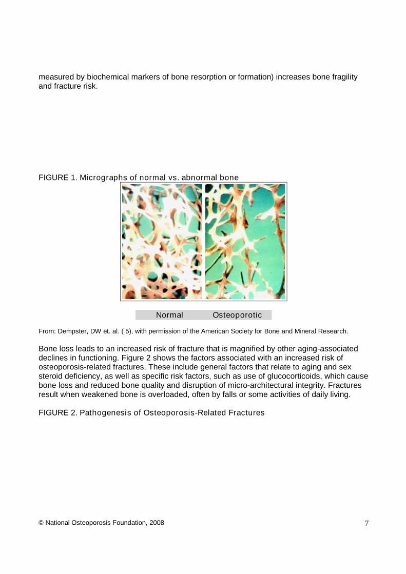
Figure 1 shows the changes within cancellous bone as a consequence of bone loss.Individual trabecular plates of bone are lost, leaving an architecturally weakened structurewith significantly reduced mass. Increasing evidence suggests that rapid bone remodeling (as
© National Osteoporosis Foundation, 2008 7
measured by biochemical markers of bone resorption or formation) increases bone fragilityand fracture risk.
FIGURE 1. Micrographs of normal vs. abnormal bone
Normal Osteoporotic
From: Dempster, DW et. al. ( 5), with permission of the American Society for Bone and Mineral Research.
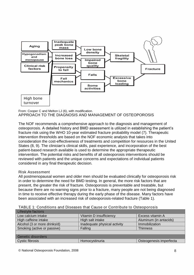
Bone loss leads to an increased risk of fracture that is magnified by other aging-associateddeclines in functioning. Figure 2 shows the factors associated with an increased risk ofosteoporosis-related fractures. These include general factors that relate to aging and sexsteroid deficiency, as well as specific risk factors, such as use of glucocorticoids, which causebone loss and reduced bone quality and disruption of micro-architectural integrity. Fracturesresult when weakened bone is overloaded, often by falls or some activities of daily living.
FIGURE 2. Pathogenesis of Osteoporosis-Related Fractures
© National Osteoporosis Foundation, 2008 8
From: Cooper C and Melton LJ (6), with modification.APPROACH TO THE DIAGNOSIS AND MANAGEMENT OF OSTEOPOROSIS
The NOF recommends a comprehensive approach to the diagnosis and management ofosteoporosis.A detailed history and BMD assessment is utilized in establishing the patient’s fracture risk using the WHO 10-year estimated fracture probability model (7). Therapeuticintervention thresholds are based on the NOF economic analysis that takes intoconsideration the cost-effectiveness of treatments and competition for resources in the UnitedStates (8, 9). The clinician’sclinical skills, past experience, and incorporation of the bestpatient-based research available is used to determine the appropriate therapeuticintervention. The potential risks and benefits of all osteoporosis interventions should bereviewed with patients and the unique concerns and expectations of individual patientsconsidered in any final therapeutic decision.
Risk AssessmentAll postmenopausal women and older men should be evaluated clinically for osteoporosis riskin order to determine the need for BMD testing. In general, the more risk factors that arepresent, the greater the risk of fracture. Osteoporosis is preventable and treatable, butbecause there are no warning signs prior to a fracture, many people are not being diagnosedin time to receive effective therapy during the early phase of the disease. Many factors havebeen associated with an increased risk of osteoporosis-related fracture (Table 1).
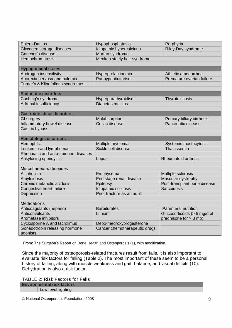
TABLE 1: Conditions and Diseases that Cause or Contribute to OsteoporosisLifestyle factorsLow calcium intake Vitamin D insufficiency Excess vitamin AHigh caffeine intake High salt intake Aluminum (in antacids)Alcohol (3 or more drinks/d) Inadequate physical activity ImmobilizationSmoking (active or passive) Falling Thinness
Genetic disordersCystic fibrosis Homocystinuria Osteogenesis imperfecta
High boneturnover
© National Osteoporosis Foundation, 2008 9
Ehlers-Danlos Hypophosphatasia PorphyriaGlycogen storage diseases Idiopathic hypercalciuria Riley-Day syndromeGaucher’s disease Marfan syndromeHemochromatosis Menkes steely hair syndrome
Hypogonadal statesAndrogen insensitivity Hyperprolactinemia Athletic amenorrheaAnorexia nervosa and bulemia Panhypopituitarism Premature ovarian failureTurner’s & Klinefelter’s syndromes
Endocrine disordersCushing’s syndrome Hyperparathyroidism ThyrotoxicosisAdrenal insufficiency Diabetes mellitus
Gastrointestinal disordersGI surgery Malabsorption Primary biliary cirrhosisInflammatory bowel disease Celiac disease Pancreatic diseaseGastric bypass
Hematologic disordersHemophilia Multiple myeloma Systemic mastocytosisLeukemia and lymphomas Sickle cell disease ThalassemiaRheumatic and auto-immune diseasesAnkylosing spondylitis Lupus Rheumatoid arthritis
Miscellaneous diseasesAlcoholism Emphysema Multiple sclerosisAmyloidosis End stage renal disease Muscular dystrophyChronic metabolic acidosis Epilepsy Post-transplant bone diseaseCongestive heart failure Idiopathic scoliosis SarcoidosisDepression Prior fracture as an adult
MedicationsAnticoagulants (heparin) Barbiturates Parenteral nutritionAnticonvulsantsAromatase inhibitors
Lithium Glucocorticoids (> 5 mg/d ofprednisone for > 3 mo)
Cyclosporine A and tacrolimus Depo-medroxyprogesteroneGonadotropin releasing hormoneagonists
Cancer chemotherapeutic drugs
From: The Surgeon’s Report on Bone Health and Osteoporosis (1), with modification.
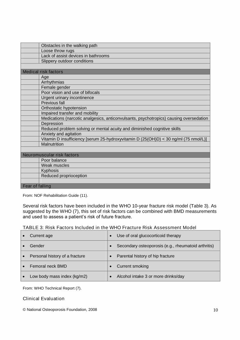
Since the majority of osteoporosis-related fractures result from falls, it is also important toevaluate risk factors for falling (Table 2). The most important of these seem to be a personalhistory of falling, along with muscle weakness and gait, balance, and visual deficits (10).Dehydration is also a risk factor.
TABLE 2: Risk Factors for FallsEnvironmental risk factors
Low level lighting
© National Osteoporosis Foundation, 2008 10
Obstacles in the walking pathLoose throw rugsLack of assist devices in bathroomsSlippery outdoor conditions
Medical risk factorsAgeArrhythmiasFemale genderPoor vision and use of bifocalsUrgent urinary incontinencePrevious fallOrthostatic hypotensionImpaired transfer and mobilityMedications (narcotic analgesics, anticonvulsants, psychotropics) causing oversedationDepressionReduced problem solving or mental acuity and diminished cognitive skillsAnxiety and agitationVitamin D insufficiency [serum 25-hydroxyvitamin D (25(OH)D) < 30 ng/ml (75 nmol/L)]Malnutrition
Neuromuscular risk factorsPoor balanceWeak musclesKyphosisReduced proprioception
Fear of falling
From: NOF Rehabilitation Guide (11).
Several risk factors have been included in the WHO 10-year fracture risk model (Table 3). Assuggested by the WHO (7), this set of risk factors can be combined with BMD measurementsand used to assess a patient’s risk of future fracture.
TABLE 3: Risk Factors Included in the WHO Fracture Risk Assessment Model
Current age Use of oral glucocorticoid therapy
Gender Secondary osteoporosis (e.g., rheumatoid arthritis)
Personal history of a fracture Parental history of hip fracture
Femoral neck BMD Current smoking
Low body mass index (kg/m2) Alcohol intake 3 or more drinks/day
From: WHO Technical Report (7).
Clinical Evaluation
© National Osteoporosis Foundation, 2008 11
Consider the possibility of osteoporosis and fracture risk in men and women, based on thepresence of the risk factors and conditions outlined in Tables 1 and 3.
Metabolic bone diseases other than osteoporosis, such as hyperparathyroidism orosteomalacia, may be associated with a low BMD. Many of these diseases have very specifictherapies, and it is appropriate to complete a history and physical examination before makinga diagnosis of osteoporosis on the basis of a low BMD alone. In patients in whom a specificsecondary, treatable cause of osteoporosis is being considered (Table 1), relevant blood andurine studies (such as serum and urine calcium, serum thyrotropin, protein electrophoresis,cortisol or antibodies associated with gluten-sensitive enteropathy) should be obtained priorto initiating therapy. For instance, elderly patients with recent fractures should be evaluatedfor secondary etiologies and, when considering osteomalacia or vitamin D insufficiency, aserum 25(OH)D level should be obtained. In general, biochemical testing (such as serumcalcium, creatinine, etc.) should be considered in patients with documented osteoporosisprior to initiation of treatment.
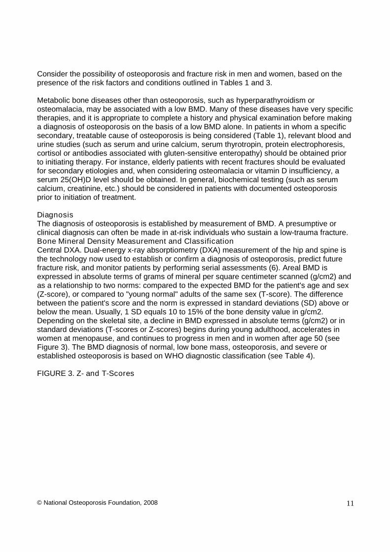
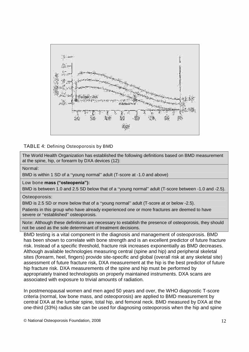
DiagnosisThe diagnosis of osteoporosis is established by measurement of BMD. A presumptive orclinical diagnosis can often be made in at-risk individuals who sustain a low-trauma fracture.Bone Mineral Density Measurement and ClassificationCentral DXA. Dual-energy x-ray absorptiometry (DXA) measurement of the hip and spine isthe technology now used to establish or confirm a diagnosis of osteoporosis, predict futurefracture risk, and monitor patients by performing serial assessments (6). Areal BMD isexpressed in absolute terms of grams of mineral per square centimeter scanned (g/cm2) andas a relationship to two norms: compared to the expected BMD for the patient's age and sex(Z-score), or compared to "young normal" adults of the same sex (T-score). The differencebetween the patient's score and the norm is expressed in standard deviations (SD) above orbelow the mean. Usually, 1 SD equals 10 to 15% of the bone density value in g/cm2.Depending on the skeletal site, a decline in BMD expressed in absolute terms (g/cm2) or instandard deviations (T-scores or Z-scores) begins during young adulthood, accelerates inwomen at menopause, and continues to progress in men and in women after age 50 (seeFigure 3). The BMD diagnosis of normal, low bone mass, osteoporosis, and severe orestablished osteoporosis is based on WHO diagnostic classification (see Table 4).
FIGURE 3. Z- and T-Scores
© National Osteoporosis Foundation, 2008 12
TABLE 4: Defining Osteoporosis by BMD
The World Health Organization has established the following definitions based on BMD measurementat the spine, hip, or forearm by DXA devices (12):
Normal:BMD is within 1 SD of a “young normal”adult (T-score at -1.0 and above)
Low bone mass (“osteopenia”):BMD is between 1.0 and 2.5 SD below that of a “young normal”adult (T-score between -1.0 and -2.5).
Osteoporosis:BMD is 2.5 SD or more below that of a “young normal”adult (T-score at or below -2.5).Patients in this group who have already experienced one or more fractures are deemed to havesevere or “established”osteoporosis.
Note: Although these definitions are necessary to establish the presence of osteoporosis, they shouldnot be used as the sole determinant of treatment decisions.
BMD testing is a vital component in the diagnosis and management of osteoporosis. BMDhas been shown to correlate with bone strength and is an excellent predictor of future fracturerisk. Instead of a specific threshold, fracture risk increases exponentially as BMD decreases.Although available technologies measuring central (spine and hip) and peripheral skeletalsites (forearm, heel, fingers) provide site-specific and global (overall risk at any skeletal site)assessment of future fracture risk, DXA measurement at the hip is the best predictor of futurehip fracture risk. DXA measurements of the spine and hip must be performed byappropriately trained technologists on properly maintained instruments. DXA scans areassociated with exposure to trivial amounts of radiation.
In postmenopausal women and men aged 50 years and over, the WHO diagnostic T-scorecriteria (normal, low bone mass, and osteoporosis) are applied to BMD measurement bycentral DXA at the lumbar spine, total hip, and femoral neck. BMD measured by DXA at theone-third (33%) radius site can be used for diagnosing osteoporosis when the hip and spine
© National Osteoporosis Foundation, 2008 13
cannot be measured. In premenopausal women, men less than 50 years of age, andchildren, the WHO BMD diagnostic classification should not be applied. In these groups,the diagnosis of osteoporosis should not be made on the basis of densitometric criteria alone.The International Society for Clinical Densitometry (ISCD) recommends that instead of T-scores, ethnic or race adjusted Z-scores should be used, with Z-scores of -2.0 or lowerdefined as either “low bone mineral density for chronological age” or “below the expected range for age” and those above -2.0 being “within the expected range for age.” (13).
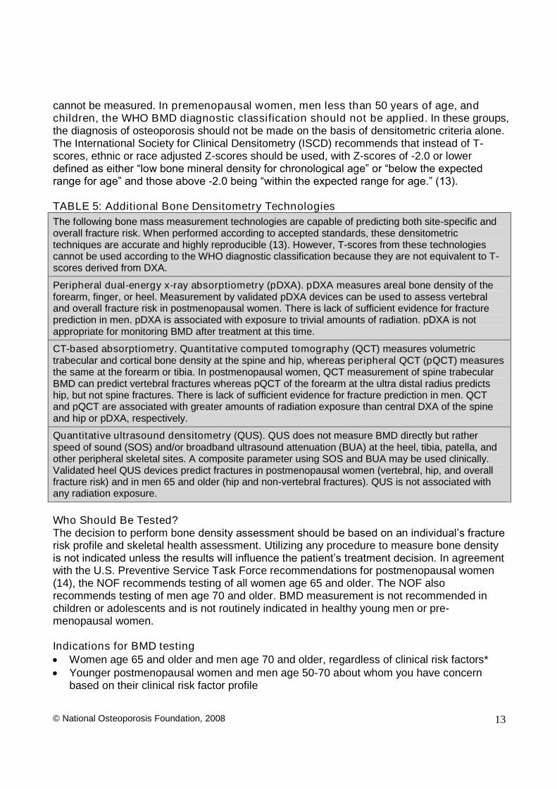
TABLE 5: Additional Bone Densitometry TechnologiesThe following bone mass measurement technologies are capable of predicting both site-specific andoverall fracture risk. When performed according to accepted standards, these densitometrictechniques are accurate and highly reproducible (13). However, T-scores from these technologiescannot be used according to the WHO diagnostic classification because they are not equivalent to T-scores derived from DXA.
Peripheral dual-energy x-ray absorptiometry (pDXA). pDXA measures areal bone density of theforearm, finger, or heel. Measurement by validated pDXA devices can be used to assess vertebraland overall fracture risk in postmenopausal women. There is lack of sufficient evidence for fractureprediction in men. pDXA is associated with exposure to trivial amounts of radiation. pDXA is notappropriate for monitoring BMD after treatment at this time.
CT-based absorptiometry. Quantitative computed tomography (QCT) measures volumetrictrabecular and cortical bone density at the spine and hip, whereas peripheral QCT (pQCT) measuresthe same at the forearm or tibia. In postmenopausal women, QCT measurement of spine trabecularBMD can predict vertebral fractures whereas pQCT of the forearm at the ultra distal radius predictship, but not spine fractures. There is lack of sufficient evidence for fracture prediction in men. QCTand pQCT are associated with greater amounts of radiation exposure than central DXA of the spineand hip or pDXA, respectively.
Quantitative ultrasound densitometry (QUS). QUS does not measure BMD directly but ratherspeed of sound (SOS) and/or broadband ultrasound attenuation (BUA) at the heel, tibia, patella, andother peripheral skeletal sites. A composite parameter using SOS and BUA may be used clinically.Validated heel QUS devices predict fractures in postmenopausal women (vertebral, hip, and overallfracture risk) and in men 65 and older (hip and non-vertebral fractures). QUS is not associated withany radiation exposure.
Who Should Be Tested?The decision to perform bone density assessment should be based on an individual’s fracture risk profile and skeletal health assessment. Utilizing any procedure to measure bone densityis not indicated unless the results will influence the patient’s treatment decision. In agreementwith the U.S. Preventive Service Task Force recommendations for postmenopausal women(14), the NOF recommends testing of all women age 65 and older. The NOF alsorecommends testing of men age 70 and older. BMD measurement is not recommended inchildren or adolescents and is not routinely indicated in healthy young men or pre-menopausal women.
Indications for BMD testing Women age 65 and older and men age 70 and older, regardless of clinical risk factors* Younger postmenopausal women and men age 50-70 about whom you have concern
based on their clinical risk factor profile
© National Osteoporosis Foundation, 2008 14
Women in the menopausal transition if there is a specific risk factor associated withincreased fracture risk such as low body weight, prior low-trauma fracture, or high riskmedication.
Adults who have a fracture after age 50 Adults with a condition (e.g., rheumatoid arthritis) or taking a medication (e.g.,
glucocorticoids,≥5 mg/day for≥3 months) associated with low bone mass or bone loss Anyone being considered for pharmacologic therapy for osteoporosis Anyone being treated for osteoporosis, to monitor treatment effect Anyone not receiving therapy in whom evidence of bone loss would lead to treatment Postmenopausal women discontinuing estrogen should be considered for bone density
testing.
* Footnote: Medicare covers BMD testing for many individuals age 65 and older, including butnot limited to: Estrogen deficient women at clinical risk for osteoporosis Individuals with vertebral abnormalities Individuals receiving, or planning to receive, long-term glucocorticoid (steroid) therapy≥ 5
mg/d of prednisone or an equivalent dose for≥ 3 months Individuals with primary hyperparathyroidism Individuals being monitored to assess the response or efficacy of an approved
osteoporosis drug therapy
Additional Skeletal Health Assessment Techniques
Biochemical markers of bone turnoverBone remodeling (or turnover) occurs throughout life to repair fatigue damage andmicrofractures in bone. Biochemical markers of bone remodeling (resorption and formation)can be measured in the serum and urine in untreated patients to assess risk of fracture. Theymay predict bone loss and, when repeated after 3-6 months of treatment with FDA approvedantiresorptive therapies, may be predictive of fracture risk reduction (15).
Vertebral fracture assessment (VFA)Independent of BMD, age and other clinical risk factors, radiographically confirmed vertebralfractures are a strong predictor of new vertebral fractures. VFA imaging of the thoracic andlumbar spine using central DXA densitometers should be considered at the time of BMDassessment when the presence of a spine fracture not previously identified may influenceclinical management of the patient. ISCD indications for VFA in postmenopausal women andmen are available on their website (13).
Use of WHO Fracture Risk AlgorithmThe WHO algorithm (FRAX®) was developed to calculate the 10-yr probability of a hipfracture and the 10-yr probability of any major osteoporotic fracture (defined as vertebral, hip,forearm, or humerus fracture) taking into account femoral neck BMD and the clinical riskfactors shown in Table 3. In the absence of femoral neck BMD, total hip BMD may besubstituted; however use of BMD from non-hip sites in the algorithm is not recommendedbecause such use has not been validated.
© National Osteoporosis Foundation, 2008 15
The WHO algorithm pertains only to previously untreated patients.
For this guide, the WHO algorithm was applied to U.S. fracture and mortality rates; hence thefracture risk figures herein are specific for the U.S. population. Economic modeling wasperformed to identify the 10-yr hip fracture risks above which it is cost-effective, from thesocietal perspective, to treat with pharmacologic agents (8). The generic 10-yr absolutefracture risk algorithm is described in the WHO Technical Report (7). The U.S.-adapted WHOfracture risk algorithm uses hip fracture risk in the calculation of intervention thresholds, andalso expresses fracture risk as the 10-yr probability of any major osteoporosis-related fracture.The U.S.-based economic modeling is described in one report (8), and the U.S.-adaptedWHO algorithm and its clinical application are illustrated in a companion report (9). The latteranalyses confirm the previous NOF conclusion (16) that it is cost-effective to treat individualswith a prior fracture and those with DXA femoral neck (or total hip) T scores≤ -2.5. Previousanalyses have established that a spine T -score≤ -2.5 also warrants treatment (16). TheU.S.-adapted WHO algorithm is most useful in identifying the subset of patients in the lowbone mass (T-score -1.0 to -2.5, osteopenia) category most likely to benefit from treatment.The WHO algorithm is not ideally suited to address the patient with risk factors and low bonemass at the spine but normal bone mass at the hip (since spine bone mass does not enterthe algorithm and its use therein has not been validated); consequently clinicians will need touse clinical judgement in this situation. Previous NOF guidance is to treat postmenopausalwomen with a spine T -score of -2.0 and below (or -1.5 with risk factors) (17).
UNIVERSAL RECOMMENDATIONS FOR ALL PATIENTS
Several interventions to reduce fracture risk can be recommended to the general population.These include an adequate intake of calcium and vitamin D, lifelong participation in regularweight-bearing and muscle-strengthening exercise, avoidance of tobacco use, identificationand treatment of alcoholism, and treatment of other risk factors for fracture such as impairedvision.
Adequate Intake of Calcium and Vitamin DControlled clinical trials have demonstrated that the combination of supplemental calcium andvitamin D can reduce the risk of fracture. Providing adequate daily calcium and vitamin D is asafe and inexpensive way to help reduce fracture risk.
Advise all individuals to obtain an adequate intake of dietary calcium (at least 1200 mg perday, including supplements if necessary). Lifelong adequate calcium intake is necessary forthe acquisition of peak bone mass and subsequent maintenance of bone health. Theskeleton contains 99% of the body's calcium stores; when the exogenous supply isinadequate, bone tissue is resorbed from the skeleton to maintain serum calcium at aconstant level. The NOF supports the National Academy of Sciences (NAS) recommendationthat women over age 50 consume at least 1200 mg per day of elemental calcium (18).Intakes in excess of 1200 to 1500 mg/d have limited potential for benefit and may increasethe risk of developing kidney stones or cardiovascular disease.
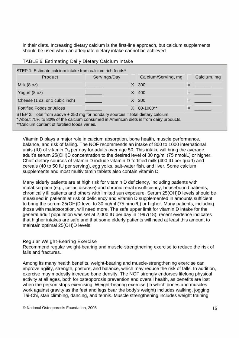
Table 6 illustrates a simple method for estimating the calcium content of a patient's diet. Menand women age 50 and older typically consume only about 600 to 700 mg per day of calcium
© National Osteoporosis Foundation, 2008 16
in their diets. Increasing dietary calcium is the first-line approach, but calcium supplementsshould be used when an adequate dietary intake cannot be achieved.
TABLE 6. Estimating Daily Dietary Calcium Intake
STEP 1: Estimate calcium intake from calcium rich foods*Product Servings/Day Calcium/Serving, mg Calcium, mg
Milk (8 oz) _______ X 300 = _______
Yogurt (8 oz) _______ X 400 = _______
Cheese (1 oz, or 1 cubic inch) _______ X 200 = _______
Fortified Foods or Juices _______ X 80-1000** = _______STEP 2: Total from above + 250 mg for nondairy sources = total dietary calcium* About 75% to 80% of the calcium consumed in American diets is from dairy products.**Calcium content of fortified foods varies.
Vitamin D plays a major role in calcium absorption, bone health, muscle performance,balance, and risk of falling. The NOF recommends an intake of 800 to 1000 internationalunits (IU) of vitamin D3 per day for adults over age 50. This intake will bring the averageadult’s serum 25(OH)D concentration to the desired level of 30 ng/ml (75 nmol/L) or higher. Chief dietary sources of vitamin D include vitamin D-fortified milk (400 IU per quart) andcereals (40 to 50 IU per serving), egg yolks, salt-water fish, and liver. Some calciumsupplements and most multivitamin tablets also contain vitamin D.
Many elderly patients are at high risk for vitamin D deficiency, including patients withmalabsorption (e.g., celiac disease) and chronic renal insufficiency, housebound patients,chronically ill patients and others with limited sun exposure. Serum 25(OH)D levels should bemeasured in patients at risk of deficiency and vitamin D supplemented in amounts sufficientto bring the serum 25(OH)D level to 30 ng/ml (75 nmol/L) or higher. Many patients, includingthose with malabsorption, will need more. The safe upper limit for vitamin D intake for thegeneral adult population was set at 2,000 IU per day in 1997(18); recent evidence indicatesthat higher intakes are safe and that some elderly patients will need at least this amount tomaintain optimal 25(OH)D levels.
Regular Weight-Bearing ExerciseRecommend regular weight-bearing and muscle-strengthening exercise to reduce the risk offalls and fractures.
Among its many health benefits, weight-bearing and muscle-strengthening exercise canimprove agility, strength, posture, and balance, which may reduce the risk of falls. In addition,exercise may modestly increase bone density. The NOF strongly endorses lifelong physicalactivity at all ages, both for osteoporosis prevention and overall health, as benefits are lostwhen the person stops exercising. Weight-bearing exercise (in which bones and muscleswork against gravity as the feet and legs bear the body's weight) includes walking, jogging,Tai-Chi, stair climbing, dancing, and tennis. Muscle strengthening includes weight training
© National Osteoporosis Foundation, 2008 17
and other resistive exercises. Before an individual with osteoporosis initiates a new vigorousexercise program, such as running or heavy weight lifting, a clinician’sevaluation isappropriate.
Fall PreventionMajor risk factors for falling are shown in Table 2. In addition to maintaining adequate vitaminD levels and physical activity, as described above, strategies to reduce falls include, but arenot limited to, checking and correcting vision and hearing, evaluating any neurologicalproblems, reviewing prescription medications for side effects that may affect balance andproviding a check list for improving safety at home. Wearing undergarments with hip padprotectors may protect an individual from injuring the hip in the event of a fall. Hip protectorsmay be considered for patients who have significant risk factors for falling or for patients whohave previously fractured a hip.
Avoidance of Tobacco Use and Excessive Alcohol IntakeAdvise patients to avoid tobacco smoking. The use of tobacco products is detrimental to theskeleton as well as to overall health. The NOF strongly encourages a smoking cessationprogram as an osteoporosis intervention.
Recognize and treat patients with excessive alcohol intake. Moderate alcohol intake has noknown negative effect on bone and may even be associated with slightly higher bone densityand lower risk of fracture in postmenopausal women. However, alcohol intake of 3 or moredrinks per day is detrimental to bone health, increases the risk of falling, and requirestreatment when identified.
PHARMACOLOGIC THERAPY
All patients being considered for drug treatment of osteoporosis should also be counseled onrisk factor reduction. Patients should be counseled specifically on the importance of calcium,vitamin D, and exercise as part of any treatment program for osteoporosis. Prior to initiatingtreatment, patients should be evaluated for secondary causes of osteoporosis and have BMDmeasurements by central DXA, when available.
The percentage risk reductions for vertebral and non-vertebral fractures cited below are thosecited in the FDA-approved Prescribing Information. In the absence of head-to-head trials,direct comparisons of risk reduction of one drug with another should be avoided.
Who Should Be Treated?Postmenopausal women and men age 50 and older presenting with the following should betreated: A hip or vertebral (clinical or morphometric) fracture Other prior fractures and low bone mass (T -score between -1.0 and -2.5 at the femoral
neck, total hip, or spine) T -score < -2.5 at the femoral neck, total hip or spine after appropriate evaluation to
exclude secondary causes
© National Osteoporosis Foundation, 2008 18
Low bone mass (T -score between -1.0 and -2.5 at the femoral neck, total hip, or spine)and secondary causes associated with high risk of fracture (such as glucocorticoid use ortotal immobilization)
Low bone mass (T -score between -1.0 and -2.5 at the femoral neck, total hip, or spine)and 10-yr probability of hip fracture≥3% or a 10-yr probability of any major osteoporosis-related fracture≥ 20% based on the U.S.-adapted WHO algorithm.
TABLE 7: Clinical Assessment of Osteoporosis in Postmenopausal Women and MenAge 50 and Older Obtain a detailed patient history pertaining to clinical risk factors for osteoporosis-related fracture Modify diet/supplements and other clinical risk factors for fracture Estimate patient’s 10-year probability of hip and any major osteoporosis-related fracture using the
US-adapted WHO algorithm Decisions on whom to treat and how to treat should be based on clinical judgment using this guide
and all available clinical information Consider FDA-approved medical therapies based on the following:
o A vertebral or hip fractureo A DXA hip (femoral neck or total site) or spine T score≤ -2.5o Low bone mass and a U.S.-adapted WHO 10-yr probability of a hip fracture≥ 3% or probability
of any major osteoporosis-related fracture≥ 20%o Patient preferences may indicate treatment for people with 10-yr fracture probabilities below
these levels Consider non-medical therapeutic interventions
o Modify risk factors related to fallingo Consider physical and occupational therapy including walking aids and hip pad protectorso Weight-bearing activities daily
Patients not requiring medical therapies at the time of initial evaluation should be clinically re-evaluated when medically appropriate
Patients taking FDA-approved medications should have laboratory and bone density re-evaluationafter two years or when medically appropriate
US FDA-Approved Drugs for OsteoporosisCurrent FDA-approved pharmacologic options for the prevention and/or treatment ofpostmenopausal osteoporosis include, in alphabetical order: bisphosphonates (alendronate,alendronate plus D, ibandronate, risedronate, and risedronate with 500 mg of calciumcarbonate, zoledronate), calcitonin, estrogens (estrogen and/or hormone therapy), estrogenagonist/antagonist (raloxifene), and parathyroid hormone [PTH (1-34), teriparatide]. The anti-fracture benefits of FDA-approved drugs have mostly been studied in women withpostmenopausal osteoporosis, and for bisphosphonates, only with daily administration. Thereare limited fracture data in glucocorticoid osteoporosis, and no fracture data in men.Treatment decisions should be based on clinical information as well as interventionthresholds.
Bisphosphonates
Alendronate, Brand name: Fosamax® or Fosamax® plus DAlendronate sodium is approved by the FDA for the prevention (5 mg daily and 35 mg weekly)and treatment (10 mg daily and 70 mg weekly [Tablet or liquid formulation] or 70 mg weekly
© National Osteoporosis Foundation, 2008 19
with 2,800 IU and 5600 IU of vitamin D3) of osteoporosis in postmenopausal women.Alendronate reduces the incidence of spine, hip and wrist fractures by about 50% over 3years in patients with a prior spine fracture. It reduces the incidence of spine fractures by48% over 3 years in patients without a prior spine fracture. Alendronate is also approved toincrease bone mass in men with osteoporosis and for the treatment of men and womenreceiving glucocorticoids in a daily dose of 5 mg or greater of prednisone and who have lowbone mass. In addition to tablet formulation, alendronate is available as a liquid with 70 mg in75ml, to be followed by at least 2 oz of plain water.
Ibandronate, Brand name: Boniva®Ibandronate sodium as 2.5 mg per day orally, 150 mg per month orally, and 3 mg every 3months by intravenous injection are approved by the FDA for the treatment ofpostmenopausal osteoporosis. The oral preparations are also approved for the prevention ofpostmenopausal osteoporosis. Ibandronate reduces the incidence of spine fractures by about50% over 3 years.
Risedronate, Brand name: Actonel® or Actonel® with CalciumRisedronate sodium (5 mg daily dose; 35 mg weekly dose; 35 mg weekly dose packagedwith 6 tablets of 500 mg calcium carbonate, or 75 mg on two consecutive days every month)is approved by the FDA for the prevention and treatment of postmenopausal osteoporosis.Risedronate reduces the incidence of spine fractures by 41-49% and non-spine fractures by36% over 3 years in patients with a prior spine fracture. Risedronate is approved fortreatment to increase bone mass in men with osteoporosis and for the prevention andtreatment of osteoporosis in men and women who are either initiating or continuing systemicglucocorticoid treatment (daily dose equivalent to 5 mg prednisone or greater) for chronicdisease.
Zoledronate, Brand name: Reclast®Zoledronate (5 mg by intravenous infusion over at least 15 minutes once yearly) is approvedby the FDA for the treatment of osteoporosis in postmenopausal women. Zoledronatereduces the incidence of spine fractures by 70%, hip fractures by 41%, and non-vertebralfractures by 25% over 3 years.
Side Effects and Administration of BisphosphonatesSide effects are similar for all oral bisphosphonate medications and include gastrointestinalproblems such as difficulty swallowing, inflammation of the esophagus and gastric ulcer.There have been reports of osteonecrosis of the jaw (particularly following intravenousbisphosphonate treatment for patients with cancer) and of visual disturbances, which shouldbe reported to the healthcare provider as soon as possible. The level of risk for osteonecrosisin patients being treated for osteoporosis with bisphosphonates is not known, but appearsextremely small for at least up to 5 years (19).
Alendronate and risedronate must be taken on an empty stomach, first thing in the morning,with 8 ounces of plain water (no other liquid), at least 30 minutes before eating or drinking.Patients should remain upright (sitting or standing) during this interval as well. Ibandronateshould be taken on the same day each month, at least 60 minutes before first food, drink(other than plain water) or medication of the day. Patients using the liquid formulation shouldswallow one bottle (75 ml) and follow with at least 2 oz of plain water. Other instructions
© National Osteoporosis Foundation, 2008 20
remain the same. Ibandronate tablets must be taken on an empty stomach, first thing in themorning, with a glass of plain water. Patients must remain upright for at least one hour aftertaking the medication. Ibandronate, by intravenous injection over 15 to 30 seconds, should begiven once every 3 months. Serum creatinine should be checked before each injection.Zoledronate, 5 mg in 100 ml, is given once yearly by intravenous infusion over at least 15minutes. Patients may be pre-treated with acetaminophen to reduce the risk of an acutephase reaction (arthralgia, headache, myalgia, fever). These symptoms occurred in 32% ofpatients after the first dose, 7% after the second dose, and 3% after the third dose.
Calcitonin
Calcitonin, Brand name: Miacalcin®, Calcimar® or Fortical®Salmon calcitonin is FDA-approved for the treatment of osteoporosis in women who are atleast 5 years postmenopausal. It is delivered as a single daily intranasal spray that provides200 IU of the drug. Subcutaneous administration by injection also is available. The effect ofnasal calcitonin on fracture risk is not stated in the Prescribing Information. Intranasalcalcitonin is generally considered safe although some patients experience rhinitis and, rarely,epistaxis. Oral calcitonin has recently been approved by the FDA for the treatment ofosteoporosis.
Estrogen/Hormone Therapy
Estrogen/Hormone Therapy (ET/HT), ET brand names: e.g. Climara®, Estrace®,Estraderm®, Estratab®, Ogen®, Ortho-Est®, Premarin®, Vivelle®); HT brand names:e.g. Activella™, Femhrt®, Premphase®, Prempro®Estrogen/hormone therapy is approved by the FDA for the prevention of osteoporosis, reliefof vasomotor symptoms and vulvovaginal atrophy associated with menopause. Women whohave not had a hysterectomy require HT, which contains progestin to protect the uterinelining. The Woman’s Health Initiative (WHI) found that 5 years of HT (Prempro®) reduced the risk of clinical vertebral fractures and hip fractures by 34% and other osteoporotic fractures by23% (20).
TheWomen’s Health Initiative (WHI) reported increased risks of myocardial infarction, stroke,invasive breast cancer, pulmonary emboli and deep vein phlebitis during 5 years of treatmentwith Premarin and medroxyprogesterone (20). Subsequent analysis of these data showed noincrease in cardiovascular disease in women starting treatment within 10 years ofmenopause. In the estrogen only arm of WHI, no increase in breast cancer incidence wasnoted over 7.1 years of treatment. Other doses and combinations of estrogen and progestinswere not studied and, in the absence of comparable data, their risks should be assumed tobe comparable. Because of the risks, ET/HT should be used in the lowest effective doses forthe shortest duration to meet treatment goals. When ET/HT use is considered solely forprevention of osteoporosis, the FDA recommends that approved non-estrogen treatmentsshould first be carefully considered.
Estrogen Agonist/Antagonist
Raloxifene, Brand name: Evista®
© National Osteoporosis Foundation, 2008 21
Raloxifene is approved by the FDA for both prevention and treatment of osteoporosis inpostmenopausal women. Raloxifene reduces the risk of spine fracture by 30% in patientswith and by 55% in patients without a prior spine fracture, over 3 years. Raloxifene does notreduce the risk of coronary heart disease, but it appears to have an effect similar to tamoxifenin the prevention of breast cancer. Raloxifene increases the risk of deep vein thrombosis to adegree similar to that observed with estrogen. It also increases hot flashes (6% over placebo).
Parathyroid Hormone
Parathyroid hormone [PTH(1-34), teriparatide], Brand name: Forteo®PTH(1-34) is approved by the FDA for the treatment of osteoporosis in postmenopausalwomen at high risk for fracture. PTH (1-34) is an anabolic (bone-building) agent whenadministered by daily subcutaneous injection. PTH (1-34) in a dose of 20 µg daily was shownto decrease the risk of spine fractures by 65% and non-spine fractures by 53% in patientswith osteoporosis, after an average of 18 months of therapy. PTH(1-34) is indicated toincrease bone mass in men with primary or hypogonadal osteoporosis who are at high risk offracture.
PTH (1-34) is well tolerated, although some patients experience leg cramps and dizziness.Because PTH (1-34) caused an increase in the incidence of osteosarcoma in rats, patientswith an increased risk of osteosarcoma (e.g., patients with Paget’s disease of bone) and those having prior radiation therapy of the skeleton, bone metastases, hypercalcemia, or ahistory of skeletal malignancy should not receive PTH (1-34) therapy. The safety and efficacyof PTH (1-34) has not been demonstrated beyond 2 years of treatment. Since PTH (1-34) isused for a maximum of 2 years, it is common practice to follow PTH (1-34) treatment with ananti-resorptive agent, usually a bisphosphonate, to maintain or further increase BMD.
Combination therapy (usually a bisphosphonate with a non-bisphosphonate) can provideadditional small increases in BMD when compared with mono-therapy; however, the impactof combination therapy on fracture rates is unknown. The added cost and potential sideeffects should be weighed against potential gains.
TABLE 8: Non-FDA-Approved Drugs for Osteoporosis
The NOF does not advocate the use of drugs not approved by the FDA for prevention or treatment ofosteoporosis. These drugs are listed for information only. These non-approved agents include:
Calcitriol. This synthetic vitamin D analogue, which promotes calcium absorption, has been approved
© National Osteoporosis Foundation, 2008 22
by the FDA for managing hypocalcemia and metabolic bone disease in renal dialysis patients. It isalso approved for use in hypoparathyroidism, both surgical and idiopathic, andpseudohypoparathyroidism. No reliable data demonstrate a reduction of risk for osteoporotic fracture.
Other bisphosphonates (etidronate, pamidronate, tiludronate). These medications varychemically from alendronate, ibandronate, risedronate and zoledronate but are in the same drugclass. At the time of publication, none is approved for prevention or treatment of osteoporosis. Most ofthese medications are currently approved for other conditions including Paget's disease,hypercalcemia of malignancy, and myositis ossificans.
Parathyroid hormone (1-84PTH). This medication is approved in some countries in Europe fortreatment of osteoporosis in women. In one clinical study 1-84hPTH effectively reduced the risk ofvertebral fractures at a dose of 100mcg/day.
Sodium fluoride. Through a process that is still unclear, sodium fluoride stimulates the formation ofnew bone. The quality of bone mass thus developed is uncertain, and the evidence that fluoridereduces fracture risk is conflicting and controversial.
Strontium ranelate. This medication is approved for the treatment of osteoporosis in some countriesin Europe. Strontium ranelate reduces the risk of both spine and non-spine fractures, but themechanism is unclear. Incorporation of strontium into the crystal structure replacing calcium may bepart of the mechanism of effect.
Tibolone. Tibolone is a tissue-specific, estrogen-like agent that may prevent bone loss and reducemenopausal symptoms but it does not stimulate breast or uterine tissue. It is indicated in Europe forthe treatment of vasomotor symptoms of menopause and for prevention of osteoporosis, but it is notapproved for use in the United States.
Monitoring Effectiveness of TreatmentIn addition to important lifestyle changes and institution of non-pharmacologic interventions,patients often require the use of FDA-approved pharmacologic therapies for the preventionand treatment of osteoporosis. With use of all therapeutic interventions, it is imperative to askpatients about adherence to their therapy and encourage continued and appropriatecompliance with their osteoporosis therapies to reduce fracture risk.
Bone Mineral DensitySerial BMD testing is an essential component of osteoporosis management and thereforehigh quality and cost-effective patient care requires that healthcare providers receive validBMD reports from adequately trained interpreters.
Central DXA. The purpose of monitoring medical therapies is to ensure reduction of futurefracture risk, stabilize or increase bone mass, and preserve or improve bone quality andstrength. Central DXA assessment of the hip or spine is currently the “gold standard” for serial assessment of BMD. However, biological changes in bone density are small comparedto the inherent error in the test itself, and interpretation of serial bone density studies dependson appreciation of the smallest change in BMD that is beyond the range of error of the test.This least significant change (LSC) varies with the specific instrument used, patientpopulation being assessed, measurement site, technologist’s skill with patient positioning and test analysis, and the confidence intervals used. Changes less than 3 to 6% at the hip and 2
© National Osteoporosis Foundation, 2008 23
to 4% at the spine from test to test may be due to the precision error of the testing itself.Information on how to assess precision and calculate the LSC is available at www.ISCD.org.
Serial bone density measurements for monitoring patients should be performed inaccordance with medical necessity, expected response, and in consideration of localregulatory requirements, usually after 2 years. Medicare permits repeat BMD testing,although the frequency depends upon criteria set by local Medicare carriers.
QCT. Trabecular BMD of the lumbar spine can be used to monitor age-, disease-, andtreatment- related BMD changes in men and women. Precision of acquisition should beestablished by phantom data and analysis precision by re-analysis of patient data.
pDXA, pQCT and QUS. Peripheral skeletal sites do not respond in the same magnitude asthe spine and hip to medications and thus are not appropriate for monitoring response totherapy at this time.
Biochemical Markers of Bone TurnoverSuppression of biochemical markers of bone turnover after 3-6 months of specificantiresorptive osteoporosis therapies, and biochemical marker increases after 1-3 months ofspecific anabolic therapies, have been predictive of greater BMD responses in studiesevaluating large groups of patients. Because of the high degree of biological and analyticalvariability in measurement of biochemical markers, changes in individuals must be large inorder to be clinically meaningful. It is critical to appreciate the LSC associated with thebiomarker being utilized, which is calculated by multiplying the “precision error” of the specific biochemical marker (laboratory provided) by 2.77 (95% confidence level). Biologicalvariability can be reduced by obtaining samples in the early morning after an overnight fast.Serial measurements should be made at the same time of day and preferably during thesame season of the year.
PHYSICAL MEDICINE AND REHABILITATION
Physical medicine and rehabilitation can reduce disability, improve physical function, andlower the risk of subsequent falls in patients with osteoporosis. Rehabilitation and exerciseare recognized means to improve function, such as activities of daily living. Psychosocialfactors also impact strongly on functional ability of the osteoporotic patient.Recommendations from the 2002 NOF Rehabilitation Guide (11) include:
Evaluate and consider the patient’s physical andfunctional safety as well as psychologicaland social status, medical status, nutritional status, and medication use before prescribinga rehabilitation program. Strive for an active lifestyle, starting in childhood.
Evaluate the patient and her/his current medication use and consider possible interactionsand risk for altered mental status. Intervene as appropriate.
Provide training for the performance of safe movement and safe activities of daily living,including posture, transfers, lifting and ambulation in populations with or at high risk forosteoporosis. Intervene as appropriate, e.g., with prescription for assistive device forimproved balance with mobility.
Evaluate home environment for risk factors for falls and intervene as appropriate.

© National Osteoporosis Foundation, 2008 24
Implement steps to correct underlying deficits whenever possible, i.e., improve postureand balance and strengthen quadriceps muscle to allow a person to rise unassisted froma chair; promote use of assistive devices to help with ambulation, balance, lifting, andreaching.
Based on the initial condition of the patient, provide a complete exercise recommendationthat includes weight-bearing aerobic activities for the skeleton, postural training,progressive resistance training for muscle and bone strengthening, stretching for tight softtissues and joints, and balance training. As long as principles of safe movement arefollowed, walking and daily activities, such as housework and gardening, are practicalways to contribute to maintenance of fitness and bone mass. Additionally, progressiveresistance training and increased loading exercises, within the parameter of the person'scurrent health status, are beneficial for muscle and bone strength. Proper exercise mayimprove physical performance/function, bone mass, muscle strength, and balance, as wellas reduce the risk of falling.
Avoid forward bending and exercising with trunk in flexion, especially in combination withtwisting.
Avoid long-term immobilization and recommend partial bed rest (with periodic sitting andambulating) only when required and for the shortest periods possible.
In patients with acute vertebral fractures or chronic pain after multiple vertebral fractures,the use of trunk orthoses (e.g., back brace, corset, Posture Training Support) may providepain relief by reducing the loads on the fracture sites and aligning the vertebra. However,long-term bracing may lead to muscle weakness and further de-conditioning.
Effective pain management is a cornerstone in rehabilitation from vertebral fractures. Painrelief may be obtained by the use of a variety of physical, pharmacological and behavioraltechniques with the caveat that the benefit of pain relief should not be outweighed by therisk of side effects such as disorientation or sedation which may result in falls.
Individuals with painful vertebral fractures that fail conservative management may becandidates for emerging interventions, such as kyphoplasty or vertebroplasty, whenperformed by experienced practitioners.
The 2003 NOF Health Professional's Guide to Rehabilitation of Patients with Osteoporosisprovides additional information on this topic and can be accessed at www.nof.org.
CONCLUSIONS AND REMAINING QUESTIONSThe guide has focused on the prevention, diagnosis, and treatment of osteoporosis inpostmenopausal women and men age 50 and older using the most common existingdiagnostic and treatment methods available. Much is known about osteoporosis in thispopulation. However, many additional issues urgently need epidemiologic, clinical, andeconomic research. Pressing issues include:
How can we better assess bone strength using non-invasive technologies and thusimprove identification of patients at high risk for fracture?
There is the need to expand the WHO algorithm to incorporate information on spine BMD. How can children, adolescents, and young adults maximize peak bone mass? What are the precise components (type, intensity, duration, frequency) of an effective
exercise program for osteoporosis prevention and treatment?
© National Osteoporosis Foundation, 2008 25
What should be done to identify and modify risk factors for falling, and what would be themagnitude of effect on fracture risk in a population?
How effective are different FDA-approved treatments in preventing fractures in patientswith moderately low bone mass?
What approaches are most effective in treating osteoporosis in disabled populations? How long should anti-resorptive therapies be continued, and are there long-term side
effects as yet unknown? Are combination therapies useful and, if so, which are the useful drug combinations and
when should they be used? Can we identify agents that will significantly increase bone mass and return bone
structure to normal?
The NOF is committed to continuing the effort to answer these and other questions related tothis debilitating disease, with the goal of eliminating osteoporosis as a threat to the health ofpresent and future generations.
For additional resources on osteoporosis and bone health visit www.nof.org or call 1-800-231-4222.
GLOSSARY
Alendronate (Fosamax®): A bisphosphonate approved by the US Food and DrugAdministration for prevention and treatment of osteoporosis; accumulates and persists in thebone. Studies indicate about a 50% reduction in vertebral, hip, and all non-vertebral fracturesin patients with osteoporosis.
Bone mineral density (BMD): A risk factor for fractures. Usually expressed as the amount ofmineralized tissue in the area scanned (g/cm2); with some technologies, expressed as theamount per volume of bone (g/cm3). Hip BMD, considered the best predictor of hip fracture,appears to predict other types of fractures as well as measurements made at other skeletalsites. Spine BMD may be preferable to assess changes early in menopause and afterbilateral ovariectomy.
Calcitonin (Miacalcin®, Calcimar® or Fortical®): A polypeptide hormone that inhibits theresorptive activity of osteoclasts.
Calcitriol: A synthetic form of 1,25-dihydroxyvitamin D3, a hormone that aids calciumabsorption and mineralization of the skeleton. Its effectiveness as a treatment forosteoporosis is still uncertain.
Calcium: A mineral that plays an essential role in development and maintenance of a healthyskeleton. If intake is inadequate, calcium is mobilized from the skeleton to maintain a normalblood calcium level. In addition to being a substrate for bone mineralization, calcium has aninhibitory effect on bone remodeling through suppression of circulating parathyroid hormone.
Cancellous bone: The spongy, or trabecular, tissue in the middle of bone (e.g., vertebrae)and at the end of the long bones.
© National Osteoporosis Foundation, 2008 26
Cortical bone: The dense outer layer of bone.
Dual-energy x-ray absorptiometry (DXA): A diagnostic test used to assess bone density atvarious skeletal sites using radiation exposure about one tenth that of a standard chest x-ray.Central DXA (spine, hip) is the preferred measurement for definitive diagnosis ofosteoporosis and for monitoring the effects of therapy.
Estrogen: One of a group of steroid hormones that control female sexual development;directly affects bone mass through estrogen receptors in bone, reducing bone turnover andbone loss. Indirectly increases intestinal calcium absorption and renal calcium conservationand, therefore, improves calcium balance. See hormone therapy.
Estrogen agonists/antagonists: A group of compounds that are selective estrogen receptormodulators, formerly known as SERMs.
Exercise: An intervention long associated with “strong bones,” despite limited evidence forsignificant beneficial effect on bone mineral density or fracture rates. Studies evaluatingexercise are ongoing; however, enough is known about the positive effect of exercise on fallprevention to support its inclusion in a comprehensive fracture prevention program.
Family history: A risk factor for osteoporotic fractures; defined here as a maternal and/orpaternal history of hip, wrist, or spine fracture when the parent was age 50 years or older.
Fluoride: A compound that stimulates the formation of new bone by enhancing therecruitment and differentiation of osteoblasts. Studies show varying effects on BMDdepending upon the preparation, dose, measurement site, and outcomes assessed.
Fracture: Breakage of a bone, either complete or incomplete. Most studies of osteoporosisfocus on hip, vertebra, and/or distal forearm fractures. Vertebral fractures includemorphometric as well as clinical fractures.
Hormone/estrogen therapy (HT, ET) (HT–ActivellaTM, Femhrt®, Premphase®,Prempro®; ET–Climara®, Estrace®, Estraderm®, Estratab®, Ogen®, Ortho-Est®,Premarin®, Vivelle®): HT is a general term for all types of estrogen replacement therapywhen given along with progestin, cyclically or continuously. HT is generally prescribed forwomen after natural menopause or bilateral ovariectomy. ET is prescribed for women whohave had a hysterectomy, after menopause or bilateral ovariectomy. Studies indicate that 5years of HT may decrease vertebral fractures by 35 to 50% and nonvertebral fractures byabout 25%. Ten or more years of use might be expected to decrease the rate of all fracturesby about 50%.
Ibandronate (Boniva®): A bisphosphonate approved by the FDA for the prevention andtreatment of postmenopausal osteoporosis. Ibandronate reduces the incidence of spinefractures by about 50% over 3 years.
Low bone mass (osteopenia): The designation for bone density between 1.0 and 2.5standard deviations below the mean for young normal adults (T-score between -1 and -2.5).
© National Osteoporosis Foundation, 2008 27
Modeling: The term for processes that occur during growth (e.g., linear growth, corticalapposition, and cancellous modification) and increase bone mass.
National Osteoporosis Foundation (NOF): America’s only nonprofit voluntary health organization dedicated to overcoming the widespread prevalence of osteoporosis throughresearch, public policy, professional and public education, and patient advocacy. NationalOsteoporosis Foundation, 1232 22nd Street, NW Washington, DC 20037. Web site:http/www.nof.org.
Normal bone mass: The designation for bone density within 1 standard deviation of themean for young normal adults (T-score at -1.0 and above).
Osteopenia: See low bone mass.
Osteoporosis: A chronic, progressive disease characterized by low bone mass,microarchitectural deterioration of bone tissue and decreased bone strength, bone fragilityand a consequent increase in fracture risk; bone density 2.5 or more standard deviationsbelow the young normal mean (T-score at or below -2.5).
Peak bone mass: The maximum bone mass accumulated during young adult life.
Peripheral DXA: A DXA test used to assess bone density in the forearm, finger, and heel.
“Peripheral” fractures: Nonvertebral fractures, for example, those of the hip, wrist, forearm,leg, ankle, foot, and other sites.
Physiatrist: A physician who specializes in medicine and rehabilitation, or physiatry.
Prevention of osteoporosis: The practice of preventing BMD from dropping lower than 2.5standard deviations below the mean for young normal adult women; commonly used todescribe the prevention of osteoporosis-related fractures.
Previous fracture: A risk factor for future fractures, defined here as a history of a previousfracture after age 40.
PTH(1-34) (teriparatide, Forteo®): An anabolic therapy approved for the treatment ofosteoporosis. The pivotal study indicates a 65% reduction in spine fractures and a 54%reduction in non-spine fractures after 18 months of therapy in patients with osteoporosis.
Quantitative computed tomography (QCT): A diagnostic test used to assess bone density;reflects three-dimensional BMD. Usually used to assess the lumbar spine, but has beenadapted for other skeletal sites. It is also possible to measure trabecular and cortical bonedensity in the periphery by peripheral QCT (pQCT).
Quantitative ultrasound densitometry (QUS): A diagnostic test used to assess bonedensity at the calcaneus or patella. Ultrasound measurements correlate only modestly with
© National Osteoporosis Foundation, 2008 28
other assessments of bone density in the same patient, yet some prospective studies indicatethat ultrasound may predict fractures as well as other measures of bone density.
Radiographic absorptiometry (RA): A diagnostic test used to assess bone density at aperipheral site, usually the hand. Such techniques are referred to as aluminum equivalence,photodensitometry, and radiographic densitometry.
Raloxifene (Evista®): an estrogen agonist/antagonist (or selective estrogen receptormodulator) approved by the FDA for prevention and treatment of osteoporosis. It lowers riskof vertebral fracture by 40%.
Remodeling: The ongoing dual processes of bone formation and bone resorption aftercessation of growth.
Resorption: The loss of substance (in this case, bone) through physiological or pathologicalmeans.
Risedronate (Actonel®): a bisphosphonate approved by the FDA for prevention andtreatment of osteoporosis. It lowers risk of vertebral fracture by 40% and non-vertebralfractures by 30%.
Risk factors: For osteoporotic fractures, include low BMD, parental history of hip fracture,low body weight, previous fracture, smoking, excess alcohol intake, glucocorticoid use, andsecondary osteoporosis (e.g., rheumatoid arthritis), and history of falls. These readilyaccessible and commonplace factors are associated with the risk of hip fracture and, in mostcases, with that of vertebral and other types of fracture as well.
“Secondary” osteoporosis: Osteoporosis that is drug-induced or caused by disorders suchas hyperthyroidism, renal disease, or chronic obstructive pulmonary disease.Severe or “established” osteoporosis: Osteoporosis characterized by bone density that is 2.5standard deviations or more below the young normal mean (T-score at or below -2.5),accompanied by the occurrence of at least one fragility-related fracture.
Single x-ray absorptiometry (SXA): A diagnostic test used to assess bone density. Limitedto peripheral sites, it cannot measure bone density in the hip or spine.
Standard deviation (SD): A measure of variation of a distribution.
T-score: In describing BMD, the number of standard deviations above or below the mean foryoung normal adults of the same sex.
Vitamin D: A group of fat-soluble sterol compounds that includes ergocalciferol (vitamin D2)and cholecalciferol (vitamin D3). These compounds are ingested from plant and animalsources; cholecalciferol is also formed in skin on exposure to ultraviolet light. When activatedin the liver and then the kidney, vitamin D promotes calcium absorption and bone mass.Vitamin D replacement also increases muscle strength and lowers risk of falling. A 25(OH)Dlevel of≥ 30 ng/ml (75 nmol/L) is considered by many to be optimal.
© National Osteoporosis Foundation, 2008 29
Zoledronate (Reclast®): A bisphosphonate approved by the FDA for treatment ofpostmenopausal osteoporosis. It lowers risk of spine fractures by 70%, hip fractures by 41%,and non-vertebral fractures by 25%.
Z-score: In describing BMD, the number of standard deviations above or below the mean forpersons of the same age and sex.
KEY REFERENCES
1. U.S. Department of Health and Human Services. Bone Health and Osteoporosis: AReport of the Surgeon General. Rockville, MD: U.S. Department of Health and HumanServices, Office of the Surgeon General, 2004.
2. National Osteoporosis Foundation.America’s Bone Health:The State of Osteoporosisand Low Bone Mass in Our Nation. National Osteoporosis Foundation, Washington,DC, pp. 1-55, 2002.
3. Burge RT, Dawson-Hughes B, Solomon D, Wong JB, King AB, Tosteson ANA.Incidence and economic burden of osteoporotic fractures in the United States, 2005-2025. J Bone Min Res 2007; 22(3):465-475.
4. Khosla S, Riggs BL 2005 Pathophysiology of age-related bone loss and osteoporosis.Endocrinol Metab Clin N Am 34:1015-1030.
5. Dempster DW. J Bone and Miner Res 1:15-21, 1986.6 . Cooper C, Melton LJ. Epidemiology of osteoporosis. Trends Endocrinol Metab 3:224-
229, 1992.7. The World Health Organization Fracture Risk Assessment Tool.www.shef.ac.uk/FRAX8. Tosteson ANA, Melton LJ, Dawson-Hughes B, Baim S, Favus MJ, Khosla S, Lindsay
RL. Cost-effective osteoporosis treatment thresholds: The U.S. perspective from theNational Osteoporosis Foundation Guide Committee. Osteoporos IntDOI1007/s00198-007-0544-4
9. Dawson-Hughes B, Tosteson ANA, Melton LJ, Baim S, Favus MJ, Khosla S, Lindsay L.Implications of absolute fracture Risk assessment for osteoporosis practice guidelinesin the U.S. Osteoporos Int DOI 10.1007/s000198-007-0559-5
10. Anonymous. Guideline for the prevention of falls in older persons. J Am Geriatr Soc49:664-672, 2001.
11. National Osteoporosis Foundation. Health Professional’s Guide toRehabilitation ofPatients with Osteoporosis. 2003. Copyright NOF, Washington, DC.
12. Kanis JA, Melton LJ III, Christiansen C, Johnston CC, Khaltaev N. The diagnosis ofosteoporosis J Bone Miner Res 9:1137-1141, 1994.
13. ISCD Official Positions. www.ISCD.org14. U.S. Preventive Services Task Force. Screening for osteoporosis in postmenopausal
women: Recommendations and rationale. Ann Intern Med 137:526-528, 2002.15. Garnero P, Delmas PD. 2001. Biochemical markers of bone turnover in osteoporosis.
In: Marcus M, Feldman D, Kelsey J (eds.) Osteoporosis, vol 2. Academic Press, pp459-477.
16. Osteoporosis: Review of the Evidence for Prevention, Diagnosis, and Treatment andCost-Effectiveness Analysis. Osteoporos Int 8 (Supplement 4), 1998.
17. National Osteoporosis Foundation.Clinician’sguide to prevention and treatment ofosteoporosis. Copyright NOF, Washington, DC. 2005
© National Osteoporosis Foundation, 2008 30
18. Standing Committee on the Scientific Evaluation of Dietary Reference Intakes. Dietaryreference intakes for calcium, phosphorus, magnesium, vitamin D, and fluoride.Washington, D.C.: National Academy Press; 1997
19. Khosla S. (chair). Bisphosphonate-associated osteonecrosis of the jaw: report of atask force of the American Society for Bone and Mineral Research. J Bone MinerRes. 22:1470-1489, 2007.
20. Writing Group for the Women’s Health Initiative Investigators. Risks and benefits of estrogen plus progestin in healthy postmenopausal women. JAMA 288:321-333, 2002.
Copyright © 2008. National Osteoporosis Foundation,1232 22nd Street NW, Washington, D.C. 20037-1292All rights reserved. No part of this guide may be reproduced in any form without advancewritten permission from the National Osteoporosis Foundation.