2008 an exciting photopigment
TRANSCRIPT

Melanopsin: an exciting photopigmentMark W. Hankins, Stuart N. Peirson and Russell G. Foster
Circadian and Visual Neuroscience Group, Nuffield Laboratory of Ophthalmology, University of Oxford, Wellcome Trust Centre for
Human Genetics, Roosevelt Drive, Oxford OX3 7BN, UK
Review TRENDS in Neurosciences Vol.31 No.1
The discovery that mice lacking rods and cones arecapable of regulating their circadian rhythms by lightprovided the conceptual framework for the discovery ofan entirely new photoreceptor system within the mam-malian eye. We now know that a small subset of retinalganglion cells are directly photosensitive and utilize anopsin/vitamin A-based photopigment called melanopsinmaximally sensitive in the blue part of the spectrum. Wealso know that these photosensitive retinal ganglioncells mediate a broad range of physiological responsesto light, ranging from the regulation of circadianrhythms to pupil constriction. Most recently, it hasbecome clear that the melanopsins are only distantlyrelated to visual pigments and in terms of their biochem-istry share more in common with invertebrate photo-pigments. Here we outline the discovery of thisremarkable new photoreceptor system, review thestructure of melanopsin and conclude with a workingmodel of melanopsin phototransduction.
IntroductionThe discovery of a third photoreceptor system in themammalian retina, quite separate from rods and cones,arose from attempts to understand how endogenous 24 hbody clocks (circadian clocks) are regulated by light.Circadian clocks are not exactly 24 h (hence the term:circa/about and dies/day) and must be synchronized toensure that internal and local time coincide. This regula-tion is usually mediated by photoreceptors which detectchanges in the quantity and quality of light over the24 h dawn/dusk cycle, a process that has been termedphotoentrainment [1].
The sensory task of photoentrainment differs markedlyfrom image detection, requiring light stimuli of high irra-diance and long duration. For example, the circadiansystem of the hamster requires light intensities some200 times brighter than the intensities that would elicita visual response. Furthermore, the clock is largely insen-sitive to stimulus durations of less than 30 s [2]. The non-mammalian vertebrates use a broad range of photo-receptors for photoentrainment including intracranialpineal organs and even photoreceptors buried deep withinthe brain. Although considerable amounts of light pene-trate deep into the brain, the light reaching these extra-retinal photoreceptors is filtered and scattered byoverlaying tissues. This would preclude any form of imagedetection but make them ideally suited to detect gross
Corresponding authors: Hankins, M.W. ([email protected]);Foster, R.G. ([email protected]).
Available online 4 December 2007.
www.sciencedirect.com 0166-2236/$ – see front matter � 2007 Elsevier Ltd. All rights reserve
changes in environmental irradiance and hence time ofday [3] By contrast, photoentrainment in mammals reliesexclusively upon ocular photoreceptors [4]. Why the mam-malian lineage lost its extraocular photoreceptors remainsspeculative, but has been correlated with the early evol-utionary history of mammals and their passage throughwhat has been termed a nocturnal bottleneck [5,6]. Itseems likely that the relatively poor colour vision, well-developed olfactory and auditory systems and loss of extra-retinal photoreceptors in modern mammals all reflect thisnocturnal ancestry. Because multiple studies had shownthat eye loss blocks photoentrainment in mammals, andbecause rods and cones were the only known ocular photo-receptors, all light responses were attributed to these cells.This gave rise to the straightforward question that framedmuch of the early research: how can rods and cones, whichare so exquisitely adapted to build a visual representationof the world, also act as dawn/dusk detectors?
Irradiance detectionIn mammals, light information reaches the master circa-dian pacemaker, the suprachiasmatic nuclei (SCN),through a dedicated monosynaptic pathway originatingin the retina and called the retinohypothalamic tract(RHT) [7]. Although the RHT had been defined by theearly 1970s, the photoreceptor inputs to the SCN hadnot. Disentangling which retinal cells mediate photoen-trainment was first addressed using animals with natu-rally occurring retinal mutations, such as micehomozygous for retinal degeneration (rd/rd). Althoughall functional rods are lost in the rd/rd retina, a smallpercentage (�5%) of cone cells survive beyond 18 months[8]. Despite this loss of photoreceptors, rd/rd mice showcircadian responses to light that are indistinguishablefrom those of congenic mice with phenotypically normalretinas (rd/+; wild-type) [9]. These findings, along withstudies on other rodent models such as the blind molerat (Spalax) [10] and retinally degenerate humans [11,12],raised the possibility that perhaps the retina contained anadditional specialized class of photoreceptor, analogous tothe pineal and deep brain photoreceptors of non-mammals.Initially, this suggestion was greeted with considerableopposition based upon the argument that only a verysmall number of rods and/or cones are sufficient fornormal photoentrainment [13]. To resolve this issue,mice were generated that lacked all functional rod andcone photoreceptors (rd/rd cl). Such animals wereshown to be capable of normal photoentrainment of beha-vioural [14] and neuroendocrine rhythms [15]. Collectively,these results showed unambiguously that a third class
d. doi:10.1016/j.tins.2007.11.002

Box 1. The pupillary light response
The rd/rd cl mouse model also proved to be important in
demonstrating that a range of other irradiance detection tasks are
mediated by a non-rod, non-cone retinal photoreceptor. For
example, in mammals, light-induced pupil constriction is regulated
by both rod and cone photoreceptors, but is not abolished in
animals that show a significant loss of these photoreceptors as a
result of inherited retinal disease. Not unreasonably, it was assumed
that the residual pupillary light response (PLR) was a result of the
survival of a small number of rod and/or cone photoreceptors [58].
Detailed studies in rd/rd cl mice have shown that although there is
diminished sensitivity, a full PLR can still occur in mice lacking rods
and cones [19]. This suggests a complex interaction between visual
and nonvisual photoreceptors in the regulation of pupil size. One
hypothesis is that the PLR is a two-stage process: rapid and dim-
light PLR is mediated by rods and cones, whereas nonvisual
photoreceptors mediate the sustained PLR under relatively high
environmental irradiances. A sensory task would be poorly suited to
the rapidly adapting responses typical of rods and cones [19].
Table 1. A range of the derived opsin lmax values for irradiancedetection tasks reported in various mammalian species
Species Measure lmax Refs
Mouse rd cl Pupillometry 479 nm [19]
Mouse rd cl Circadian phase shifting 481 nm [27]
Rat WT pRGC light response 484 nm [16]
Macaque pRGC light response 482 nm [20]
Macaque Pupillometry 482 nm [59]
Human Melatonin suppression 446–477 nm [60]
Human Melatonin suppression 459 nm [61]
Human Regulation of cone
electroretinogram
483 nm [62]
Human Heterologous expression 420–440 nm [38]
Mouse Heterologous expression 420 nm [37]
Mouse Heterologous expression 479 nm [39]
Mouse Heterologous expression 480 nm [40]
28 Review TRENDS in Neurosciences Vol.31 No.1
of photoreceptor must reside within the mammalian eye(Box 1).
Melanopsin-based photosensitive retinal ganglion cellsIf not the rods and cones, then what other retinal neuronsare photoreceptive? Parallel studies in the rat and mouseaddressed this question and identified a subset of photo-sensitive retinal ganglion cells (pRGCs) (Figure 1). In therat, the approach involved injecting fluorescent micro-spheres into the SCN which then traveled down the axonsof the RHT to retrogradely label retinal ganglion cells(RGCs). These RGCs showed a light-evoked depolarisationthat persisted in the presence of a cocktail of drugs thatblocked all retinal intercellular communication. Further-more, the labeled RGCs still showed intrinsic lightresponses when dissected and isolated from the surround-ing retinal tissue [16]. In mice, the approach utilized theisolated rd/rd cl retinae loaded with the Ca2+-sensitiveFURA-2AM dye. Fluorescent imaging identified light-induced Ca2+ changes in �3.0% of neurons within theRGC layer (Figure 1). Significantly, the gap junctionblocker carbenoxolone reduced the number of RGCsresponding to light to �1.0%. This suggests that pRGCsare coupled via gap junctions and form a syncitium ofphotosensitive and nonphotosensitive neurons. Three dis-crete classes of light-induced Ca2+ change have been ident-ified in pRGCs, but the basis for these different responsesremains unclear and it remains unknown whether specificpRGCs project differentially to all retinorecipient regionsof the brain [17,18].
The rd/rd cl mouse also proved valuable in character-izing the photopigment of pRGCs. The known photopig-ments of animals consist of an opsin protein linked to achromophore which is a specific form of vitamin A called11-cis retinal. All opsin/vitamin A-based photopigmentshave a characteristic absorption profile that allows thesephotopigments to be identified on the basis of their spectralresponses to light or action spectra. The process of gen-erating an action spectrum involves the protracted task ofmeasuring a series of full dose–response curves for a rangeof monochromatic stimuli. The first full action spectrum todefine the nature of the non-rod, non-cone photopigmentstudied pupil constriction in rd/rd cl mice. The results
www.sciencedirect.com
revealed a previously uncharacterized, opsin/vitaminA-based photopigment with peak sensitivity at 479 nm,which was tentatively named OP479 (opsin photopigmentlmax 479 nm) [19] (Figure 2). Subsequently, action spectrafrom mice to human have been deduced for a range ofresponses including the direct recording from pRGCs inrats [16] and macaque [20]. Additional action spectra arelisted in Table 1. A clear consensus has emerged from thesestudies demonstrating the existence of a common singlenovel opsin photopigment with a lmax of around 480 nm.Although the biochemistry of the photopigment wasdeduced by action spectroscopy, the molecular identity ofOP479 remained unknown.
The discovery that the orphan opsin gene, melanopsin(now officially designated Opn4), is expressed within asubpopulation of RGCs generated considerable excitementbecause the anatomy and distribution of these melanopsinRGCs was remarkably similar to the RGCs that form theRHT [18,21–23]. Melanopsin gene ablation studies pro-vided the definitive link between melanopsin and thecapacity of the pRGCs to respond to light. Melanopsinknockout mice (Opn4�/�) show attenuated phase shifting;fail to show full pupil constriction; and show loss of directphotosensitivity in pRGCs [24–26]. Furthermore, ifOpn4�/� mice are crossed with mice lacking functionalrods and cones, all responses to light are lost [27,28]. Thesestudies demonstrate that rods, cones and pRGCs can fullyaccount for all light detection within the eye, and stronglyimplicate melanopsin as the photopigment molecule ofpRGCs. These triple knockout studies also addressed thesuggestion that cryptochrome (CRY) might act as a photo-pigment for photoentrainment [29,30]. The complete loss oflight responses in these animals leaves little room for aCRY-base photopigment, and there is now a broad con-sensus that the CRYs do not form photopigments in mam-mals. For further discussion, see Refs [31,32].
Rod, cone and pRGC interactionsThe results from rd/rd cl mice demonstrate that rods andcones are not required for photoentrainment [14]. How-ever, one cannot conclude from this that rods and conesplay no role. Indeed, multiple lines of evidence have impli-cated an input from rods and cones [10,33–35], not least thefinding that Opn4�/� mice still show circadian entrain-ment, albeit in an attenuated form [24–26]. Thus, mela-nopsin ablation studies show that rods and cones can
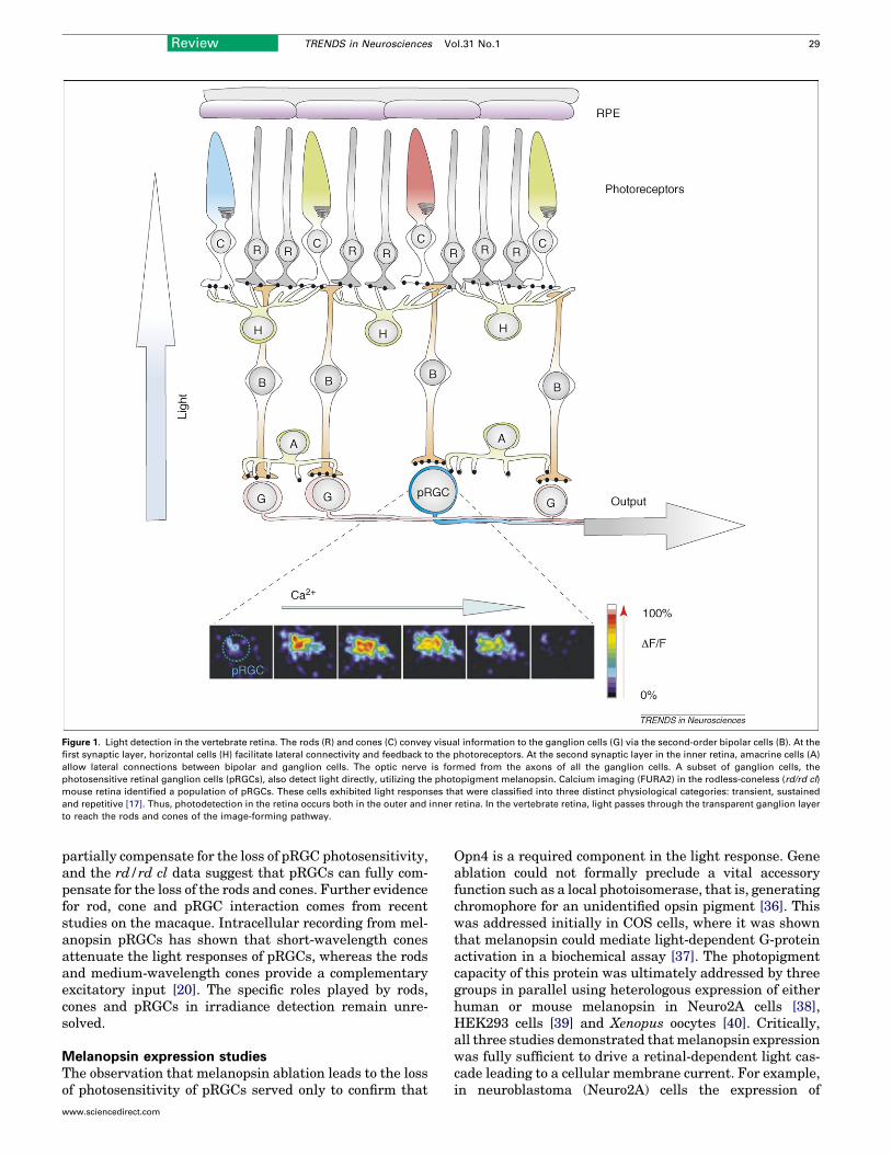
Figure 1. Light detection in the vertebrate retina. The rods (R) and cones (C) convey visual information to the ganglion cells (G) via the second-order bipolar cells (B). At the
first synaptic layer, horizontal cells (H) facilitate lateral connectivity and feedback to the photoreceptors. At the second synaptic layer in the inner retina, amacrine cells (A)
allow lateral connections between bipolar and ganglion cells. The optic nerve is formed from the axons of all the ganglion cells. A subset of ganglion cells, the
photosensitive retinal ganglion cells (pRGCs), also detect light directly, utilizing the photopigment melanopsin. Calcium imaging (FURA2) in the rodless-coneless (rd/rd cl)
mouse retina identified a population of pRGCs. These cells exhibited light responses that were classified into three distinct physiological categories: transient, sustained
and repetitive [17]. Thus, photodetection in the retina occurs both in the outer and inner retina. In the vertebrate retina, light passes through the transparent ganglion layer
to reach the rods and cones of the image-forming pathway.
Review TRENDS in Neurosciences Vol.31 No.1 29
partially compensate for the loss of pRGC photosensitivity,and the rd/rd cl data suggest that pRGCs can fully com-pensate for the loss of the rods and cones. Further evidencefor rod, cone and pRGC interaction comes from recentstudies on the macaque. Intracellular recording from mel-anopsin pRGCs has shown that short-wavelength conesattenuate the light responses of pRGCs, whereas the rodsand medium-wavelength cones provide a complementaryexcitatory input [20]. The specific roles played by rods,cones and pRGCs in irradiance detection remain unre-solved.
Melanopsin expression studiesThe observation that melanopsin ablation leads to the lossof photosensitivity of pRGCs served only to confirm that
www.sciencedirect.com
Opn4 is a required component in the light response. Geneablation could not formally preclude a vital accessoryfunction such as a local photoisomerase, that is, generatingchromophore for an unidentified opsin pigment [36]. Thiswas addressed initially in COS cells, where it was shownthat melanopsin could mediate light-dependent G-proteinactivation in a biochemical assay [37]. The photopigmentcapacity of this protein was ultimately addressed by threegroups in parallel using heterologous expression of eitherhuman or mouse melanopsin in Neuro2A cells [38],HEK293 cells [39] and Xenopus oocytes [40]. Critically,all three studies demonstrated thatmelanopsin expressionwas fully sufficient to drive a retinal-dependent light cas-cade leading to a cellular membrane current. For example,in neuroblastoma (Neuro2A) cells the expression of

Figure 2. Action spectra. The functional properties of photopigments have been characterized through action spectroscopy. Irradiance-dependent responses from a range
of species suggest a consensus opsin template with a lmax (peak sensitivity) of around 480 nm (blue line). This is compared to the typical representative power spectra for
natural daylight (black) and artificial fluorescent lighting (red). The range of the visible spectrum between the UV and IR is depicted on the x axis.
30 Review TRENDS in Neurosciences Vol.31 No.1
melanopsin, in the presence of retinal chromophore(9-cis-retinal or 11-cis-retinal), transformed a nonphoto-sensitive neuron into a functional irradiance detector[38]. Furthermore, these studies demonstrated thatmelanopsin appears to function as a bistable pigment,able to regenerate its chromophore utilizing all-trans-retinal and long-wavelength light in a manner reminis-cent of the photoreversal observed in invertebrates [38].In this respect, melanopsin is unique among the mam-malian photopigments in forming a stable associationwith all-trans-retinal. The photoreversal capacity of mel-anopsin has also been observed with spectroscopicapproaches in the case of amphioxus melanopsin [41].Collectively, all of the expression studies have led to aconsensus view that melanopsin forms a photopigmentfully capable of coupling to ubiquitous cell signallingpathways. The ability to regenerate its own chromophoreis most likely an important adaptation to its expressionin retinal ganglion cells distant from the centre ofretinoid recycling in the retinal pigment epithelium(Figure 1) [38].
Although all three expression studies showed broadlythe same result, inconsistencies relating to the spectralsensitivity of expressed human and mouse melanopsinemerged. The spectral maxima of the pigments variedbetween 420 and 440 nm (human) [38] and 480 nm (mouse)[39,40]. The reason for this discrepancy still remains to befully resolved, but presumably relates to varied experimen-tal procedures, including the host cell environment andperhaps cell retinoid status. Classical spectroscopicapproaches should resolve this issue.
www.sciencedirect.com
Melanopsin structureAlthough melanopsin almost certainly forms the photopig-ment of pRGCs, very little is known about how this proteinfunctions. Some deductions can be inferred from its struc-ture and below we address a few of the structure/functionrelationships of the melanopsins. The first melanopsingene isolated was from Xenopus dermal melanophores(hence the name, melan opsin), and it was recognizedimmediately that this opsin shared greater homology withinvertebrate opsins, such as octopus rhodopsin (39%), thanwith the classical visual pigments (�27%) [42,43]. Inaddition, melanopsin shows several other features whichresemble invertebrate opsins. These include the presenceof a tyrosine instead of a glutamate counterion (E113); aninsertion in the third cytoplasmic loop increasing itslength; and, finally, an extended intracellular C terminus.Phylogenetic analysis also supported an affiliation withthe invertebrate opsins, placing melanopsin in a cladecloser to the invertebrate rather than vertebrate opsins[42]. The genomic structure of the melanopsins also differsmarkedly from the vertebrate visual pigments, possessingnine intron insertion sites. This strongly suggests that themelanopsin gene family has a different evolutionary lin-eage from the rod, cone, pineal and VA-opsin photopig-ments [43].
Recent studies have also shown that there are twomelanopsin genes in the vertebrate lineage, the mamma-lian-like Opn4m and Xenopus-like Opn4x genes [44]. Non-mammalian vertebrates possess both Opn4m and Opn4xgenes in separate chromosomal loci. However, the Opn4xgene is not present in either eutherian or marsupial

Review TRENDS in Neurosciences Vol.31 No.1 31
mammals [44,45], and preliminary analysis of the platypusand echidna suggests that the Opn4x gene is also missingfrom the monotremes (Pires, Halford, and Foster, unpub-lished). These results indicate that the Opn4x gene waslost as a result of chromosomal reorganisation relativelyearly in mammalian evolution. Both genes seem capable offorming photosensory pigments in the chicken, althoughthe expression pattern ofOpn4m is restricted to a subset ofretinal ganglion cells whereas Opn4x appears to beexpressed throughout the chicken retina [44]. The comp-lementary function of these two forms of melanopsinremains completely unknown.
Whole amino acid sequence alignments of the variousvisual opsin classes show high levels of conservation (�40%identity [43]). By contrast, a comparison of melanopsinswith the visual opsins produces very low levels of aminoacid identity as a result of the highly variable N and Ctermini. When the more conserved transmembranedomains are considered alone, melanopsin still only shares�27% identity with the visual opsins [43]. However, oneproblem with an overall comparison of sequence identitiesacross the transmembrane regions is that it does not takeinto account the functional position of individual aminoacids, in particular whether they are involved in the for-mation of the retinal/ligand binding pocket. To addressthis, a 35 amino acid vector termed the ligand pocket vector(LPV) of various melanopsins was deduced and alignedwith other opsin sequences [46]. The LPV is shown sche-matically in Figure 3a. These core amino acids of the ligandbinding pocket were then compared in rod, cone and mel-anopsins. The LPVs of visual pigment classes are highlyconserved across species (Figure 3b, left). For example,within the rods, the sequences of the LPVs are 94%–100%identical. By contrast, the LPVs of the melanopsins acrossmultiple species are considerably more variable, with77%–100% identity (Figure 3b, right, top). However, whenmelanopsin sequences are segregated into Opn4x andOpn4m, these interspecies differences are markedlyreduced, with Opn4x genes showing 89%–100% identityand Opn4m showing an even greater identity of 94%–100%. Collectively, this analysis shows that when theretinal binding pocket is compared within an opsin class,there is a remarkable degree of conservation. Indeed, themelanopsins are no more divergent in their LPV than theother opsin classes. This suggests that the melanopsinsacross species will interact with retinal in a similar man-ner, which may account for the conserved spectral maxima(�480 nm; Table 1). Based upon consensus sequences foreach opsin class, Opn4x sequences share 40% amino acididentity with rod opsins, whereas Opn4m sequences share46% identity (Figure 3b, lower right). These values areconsiderably higher than the level of overall sequenceidentity between rod opsins and melanopsins (�27%). Thisanalysis emphasizes the importance of comparing func-tionally conserved regions of the different opsins ratherthan a simple comparison of overall sequence identity.
A consideration of the individual residues of the ligandbinding pocket is beyond the scope of this review, but twokey features are outlined below. (i) As noted above, themelanopsins have a glutamate-to-tyrosine substitution inthe Schiff base counterion position (E113Y). It has been
www.sciencedirect.com
suggested that E181 may serve as the counterion in mel-anopsin [47], as in amphioxus rhodopsin [48]. If true, thenone would predict a corresponding change in the aminoacids forming the binding pocket around this region. Thisseems to be the case: helices 1–4 show amuch lower level ofidentity compared to helices 5–7 (Figure 3b, lower right).(ii) The second extracellular loop folds into the helicalbundle and forms the extracellular boundary of the retinalbinding pocket [49] and in melanopsin shows several non-conserved substitutions compared to the visual opsins.Differences in this region could influence the retinal bind-ing site and/or the release of retinal following photoisome-risation [38].
Melanopsin phototransductionThe deduced structure of the melanopsins immediatelysuggested strong parallels with invertebrate light signal-ling systems. Invertebrate phototransduction has beencharacterized most extensively in Drosophila [50], andvery broadly the rhabdomeric cascade involves activationof a Gq/G11-type G protein, activation of phospholipase C(PLC) and subsequent opening of transient receptor poten-tial channels (TRPCs) resulting in the depolarisation of thecell membrane. By contrast, the mechanisms of photo-transduction utilized by the rod and cone opsins are quitedifferent, involving the activation of transducin (Gi/o),phosphodiesterase, hydrolysis of cGMP, closure of cyclicnucleotide gated (CNG) channels and a hyperpolarisationof the plasma membrane [50,51] (Figure 4).
In contrast to invertebrate and vertebrate visual photo-transduction, our understanding of melanopsin signallingis only beginning to emerge. Nonnative heterologous cellsystems have been used to express melanopsin andexamine possible G-protein binding partners. The problemhere is that G-protein-coupled receptors (GPCRs) can binda variety of G proteins promiscuously and so the findingthat a particular G protein can bind to a GPCR does notmean that this necessarily occurs in the native cell. Forexample, although it appears that melanopsin can activatethe a subunit of rod transducin in biochemical assays [37],this is unlikely to be a significant mode of action in pRGCsbecause ablation of this gene has no effect on cellular lightresponses [27].
Another approach has explored signalling in the pRGCsby combining pharmacology with electrophysiology or ima-ging in native cells. Although there have been advancesusing these techniques, progress has been limited becauseof the lack of a comprehensive pharmacological toolkit withwhich to probe this uncharacterized system. A furthercomplication arises because melanopsin may activatemultiple signalling channels in a semiredundant manner.Thus, the interpretation of drug action on pRGCs has to besuitably cautious.
The most recent approach, which has the advantage ofassuming very little about the signalling systems, hasutilized a microarray-based technique to investigate thetranscriptional realignment that occurs in the rd/rd clmouse eye following a light pulse. This has identified anumber candidate genes/proteins that might be associatedwith the melanopsin cascade. This approach is also limitedin that it tends to generate large numbers of candidates

Figure 3. Ligand pocket vector. Analysis of the putative retinal binding site of rod opsins, cone opsins and melanopsins. (a) Opsin peptide sequence illustrating major
structural features. The amino acids of the ligand pocket vector (LPV) are highlighted in yellow. The 35 amino acid sequence (below) of the LPV is colour coded according to
helix location. Amino acid numbering and sequence are based upon the crystal structure of bovine rhodopsin [49]. (b) The LPV of visual pigments (left column) and
32 Review TRENDS in Neurosciences Vol.31 No.1
www.sciencedirect.com
Review TRENDS in Neurosciences Vol.31 No.1 33
and investigation of each individual gene is still relativelyslow and laborious.
The consensus view is that melanopsin indeed signalsthrough a G-protein-coupled system. For example, thedisruption of GPCR coupling using suramin affects bothexpressed melanopsin and native cell responses to light.Primarily by analogy to rhabdomeric transduction, it wasfirst proposed that melanopsin might be specificallycoupled to the Gq/G11 class of G protein and PLC [42].There is support for this proposal from the heterologousexpression studies. For example, melanopsin responsesappear to be attenuated by antibodies to a subunits ofGq/G11 [40]. In Neuro2A cells, the use of Gi/G0 blockers failsto inhibit melanopsin-dependent light responses [38],whereas putative Gq/G11 agonists fully blocked melanop-sin-dependent light responses in HEK293-TRPC3 cells[39]. Although there is a degree of congruity in these data,it is important to remember that they only explore thebroad potential of cascade coupling in a diverse range ofhost environments. PLC antagonists, such as U73122 orET-18-OCH3, may be effective in some expression systems[39] but are without effect in Neuro2A cells [38]. It is hardto form solid conclusions from the current data sets, com-plicated by the real possibility of parallel transductioncascades in the native cells with some degree of functionalredundancy. This possibility is supported by the findingthat the light responses of these cells show a mixture ofcalcium currents that can be both sustained and transient[17].
Investigations of the channel, or indeed channels,involved in gating the melanopsin light response quicklycentred upon the TRPC family. This was partially throughanalogy to the invertebrate cascades, but also because ofthe biophysical characteristics of the light current in nativemelanopsin-expressing cells [52]. Light induces a conduc-tance increase in native pRGCs, and the photocurrentreverses close to zero mV, consistent with a nonspecificcationic channel. The mammalian canonical TRPCs can bedivided into four families based upon sequence homologyand pharmacological properties: TRPC1, TRPC2,TRPC3+6+7 and TRPC4+5 [53]. Furthermore, their func-tional diversity is increased because these channels canform heteromeric complexes with members of the samefamily (except TRPC1). TRPC proteins form nonspecificcationic channels with substantial Ca2+ permeability,matching known features of the pRGC light-activatedchannel [17]. Coexpression of melanopsin with TRPCshas been shown to result in a functional cascade [39,40].Furthermore, evidence suggests that the light-evoked cur-rents in rat pRGCs can be suppressed by TRPC blockers[54]. The expression profile of TRPC family members in
melanopsins (right column). Shaded regions show the consensus LPV sequence with
species. lmax values for the various pigments are taken from [43]. Consensus sequences
amino acid identity of the LPV with respect to the rod opsin consensus. GenBank access
U49742; Frog Rod, S79840; Goldfish Rod, L11863; Anolis RH2, AF134191; Chicken Green
Chicken Blue, M92037; Frog Green Rod, AB010085; Goldfish Blue, L11864; Budgerigar
Salamander UV, AF038948; Xenopus Violet, U23463; Goldfish UV, D85863; Goldfish R
Human Red, M13305; Gecko P521, M92036; Xenopus Opn4x, AF014797; Chicken Opn4
DQ384639; Chicken Opn4m, AY882944; ZF Opn4m1, AY882945; ZF Opn4m2, AY078161
Dunnart Opn4m, DQ383281; Cat Opn4m, NM_001009325; Dog Opn4m, XM_848642; Bov
NM_033282; Human Opn4m isoform 2, NM_001030015.
www.sciencedirect.com
mouse pRGCs suggests the presence of both TRPC6 andTRPC7 (but not TRPC3) in the same cells that expressedmelanopsin [55]. It has also been shown that 2-ami-noethoxydiphenylborane (2-APB), which is both anantagonist at IP3 receptors and an inhibitor of TRP ionchannels, is an extremely potent in vitro inhibitor of thelight responses of pRGCs and that its effect is independentof store-dependent Ca2+ release [55]. Significantly, 2-APBis also effective in vivo, where it induces an acute knock-down of the pupillary light reflex, consistent with thesilencing of melanopsin-dependent light detection [55].
A working model of melanopsin phototransductionBased upon both expression studies and pharmacologicalapproaches, an outline model of the melanopsin photo-transduction cascade can be devised (Figure 4, right).Light-activated melanopsin seems to interact withGq/G11, which in turn activates a PLC-b. PLC-b generatesIns(1,4,5)P3 and diacylglycerol (DAG), which may ulti-mately modulate a TRPC6 or TRPC7 channel, possiblyvia a PKC. However, given the limits of the experimentaldata, virtually none of these components are confirmedbeyond reasonable doubt. Furthermore, unlike the rod/cone andDrosophila cascades, we have virtually no knowl-edge regarding the likely modulatory and adaptation-de-pendent regulation of melanopsin signalling. Similarly,the deactivation stages of light current and receptor-specific issues such as potential phosphorylation sites waitto be explored.
This model of melanopsin phototransduction can beaugmented substantially by very recent microarray-basedapproaches [56]. These experiments utilizing the rd/rd clmouse demonstrated that �30% of the ocular transcrip-tome is transiently regulated in response to nocturnal lightexposure. From a selection of genes, laser capture micro-dissection demonstrated that Gnas, Gnb2l1, Gnaq, Prkcz,Pik3r1, Inadl, Slc9a3r1 and Drd1a colocalized with mela-nopsin. The impact of genetic ablation of one of these genes,protein kinase C zeta (Prkcz), was assessed. Quite remark-ably, Prkcz�/�mice were found to precisely phenocopy theextensively characterized melanopsin-ablated transgeniclines. They show attenuated circadian phase-shiftingresponses to light, reduced period lengthening under con-stant light and attenuated pupillary responses at highirradiances, as well as impaired light-induced gene expres-sion in the suprachiasmatic nuclei (Figure 5). By analogy tothe Drosophila phototransduction cascade, Prkcz mayinfluence TRP ion-channel activity via participation inan INAD-like signalling complex (including PLC-, PKC-,and PDZ-domain-containing scaffolding proteins; Figure 4,right). Alternatively, it could act by regulating the activity
in each opsin class, highlighting the remarkable degree of conservation between
for the various opsin classes (lower right) are shown. Grey shading illustrates the
ion numbers are as follows: Anolis RH1, L31503; Chicken Rod, D00702; Human Rod,
, M92038; Gecko P467, M92035; Goldfish Green1, L11865; Anolis SWS2, AF133907;
UV, Y11787; Chicken Violet, M92039; Mouse Blue, U92562; Human Blue, U53874;
ed, L11867; Xenopus Red, U90895; Chicken Red, M62903; Human Green, K03494;
x, AY036061; Cod Opn4x-a, AF385823; Cod Opn4x-b, AY126448; Xenopus Opn4m,
; Mouse Opn4m, AF147789; Rat Opn4m, NM_138860; Hamster Opn4m, Q5XXP2;
ine Opn4m, XM_593123; Chimp Opn4m, XM_001135445; Human Opn4m isoform 1,
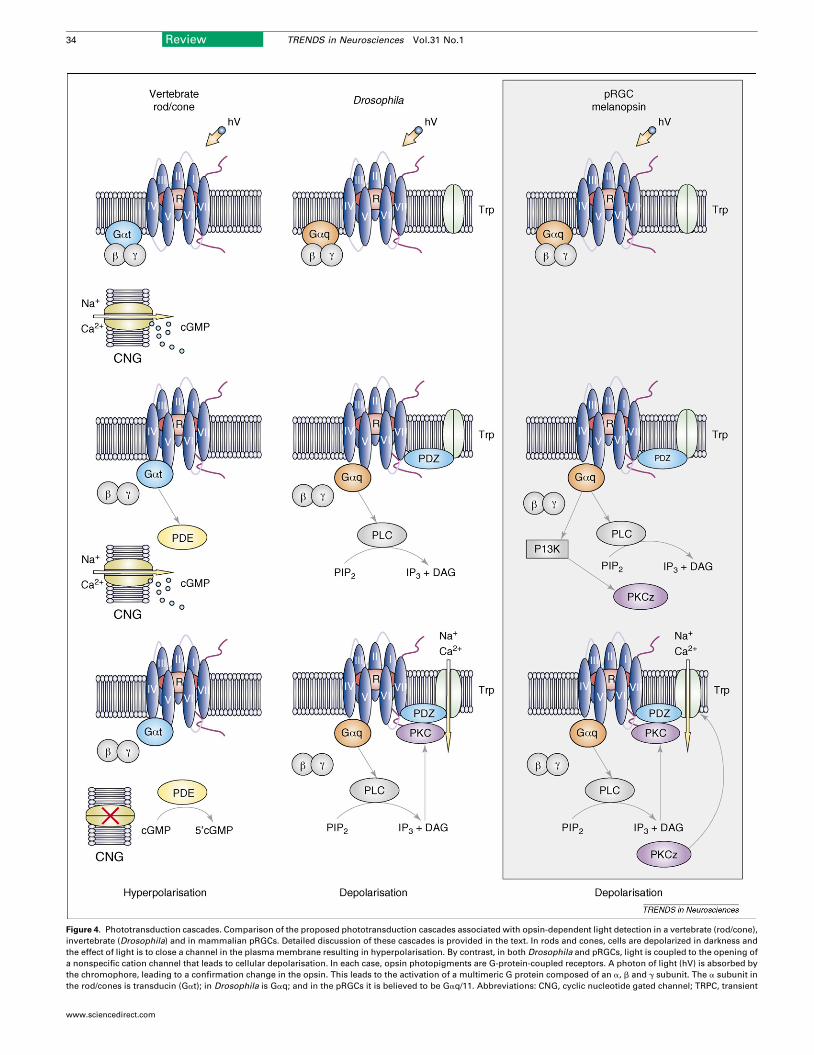
Figure 4. Phototransduction cascades. Comparison of the proposed phototransduction cascades associated with opsin-dependent light detection in a vertebrate (rod/cone),
invertebrate (Drosophila) and in mammalian pRGCs. Detailed discussion of these cascades is provided in the text. In rods and cones, cells are depolarized in darkness and
the effect of light is to close a channel in the plasma membrane resulting in hyperpolarisation. By contrast, in both Drosophila and pRGCs, light is coupled to the opening of
a nonspecific cation channel that leads to cellular depolarisation. In each case, opsin photopigments are G-protein-coupled receptors. A photon of light (hV) is absorbed by
the chromophore, leading to a confirmation change in the opsin. This leads to the activation of a multimeric G protein composed of an a, b and g subunit. The a subunit in
the rod/cones is transducin (Gat); in Drosophila is Gaq; and in the pRGCs it is believed to be Gaq/11. Abbreviations: CNG, cyclic nucleotide gated channel; TRPC, transient
34 Review TRENDS in Neurosciences Vol.31 No.1
www.sciencedirect.com

Figure 5. Protein kinase C zeta. Deletion of the Prkcz gene results in a
comprehensive phenocopy of the Opn4�/� mouse, demonstrating that this gene
is critical for melanopsin signalling. Prkcz�/� animals show attenuated phase-
shifting responses to light and period lengthening under constant light (LL). These
animals also show attenuated pupil responses to bright light but normal responses
to lower-intensity stimuli. Finally, gene induction in the SCN in response to light is
also reduced. The deficits in Prkcz�/� mice mirror those of Opn4�/� animals,
suggesting that the lesion occurs at the level of the pRGCs. Data are taken from
[56].
Review TRENDS in Neurosciences Vol.31 No.1 35
of some other critical component of the melanopsin signal-ling cascade.
Conclusions and future studiesThe study of the irradiance signalling system of the mam-malian retina has advanced dramatically over a relativelyshort period of time. However, several critical areas remainonly poorly understood. Although there is a general con-sensus regarding pRGC-dependent action spectra, thereare some significant disparities in the spectral sensitivityof heterologous expressed melanopsin. The basis for thisdifference is obscure and classical spectroscopic analysisof the melanopsin photopigment is urgently required.We also know that non-mammalian vertebrates possesstwo melanopsin genes and that each encodes a fullyfunctional photopigment [44]. Their differential roles
receptor potential channel; cGMP, cyclic guanosine monophosphate; 50GMP, 50-guano
kinase; PtdIns(4,5)P2, phosphatidylinositol bisphosphate; Ins(1,4,5)P3, inositol triphosph
postsynaptic density protein complex.
www.sciencedirect.com
and the selection pressures that led to the loss of one ofthese genes inmammals remain entirely unclear. Previouspredictions ofmelanopsin function based upon its sequencehave suggested that this opsin family is only poorly con-served. This in turn led to the suggestion that melanopsinsmay subserve several different roles [43]. However, whenthe sequence analysis is restricted to the predicted ligandbinding pocket, the results suggest that the apparentdiversity has been considerably overestimated.
Clear parallels are also emerging between melanopsinand invertebrate phototransduction cascades; however, itwould be unwise, based on analogy alone, to assume thatthe two systems map precisely onto one another. Ourcurrent knowledge of the cascade remains a skeleton workin progress. Microarray-based techniques have generatedsome additional candidates, but validating these genes inthe absence of robust pharmacological tools remains aserious challenge. Although these issues can be partiallyapproached with transgenesis and gene ablation, the studyof this complex system will surely benefit from the appli-cation of in vivo RNA-interference approaches.
Finally, there remain many largely unexplored ques-tions that relate to the modulatory and regulatory stepsassociated with melanopsin phototransduction. Insightinto this sphere may possibly explain some of the uniqueproperties of melanopsin signalling, including its relativeinsensitivity to light and a remarkable capacity to inte-grate photons over many minutes [57].
AcknowledgementsThe authors would like to thank Martin Ebeling, Nicole Kratochwil andSilvia Gatti-McArthur for their valuable input relating to GPCRmodelling. The authors would also like to acknowledge the generoussupport given by the Wellcome Trust (UK), Biotechnology and BiologicalSciences Research Council (BBSRC; UK) and Hoffmann-La Roche(Switzerland).
References1 Roenneberg, T. and Foster, R.G. (1997) Twilight times: light and the
circadian system. Photochem. Photobiol. 66, 549–5612 Nelson, D.E. and Takahashi, J.S. (1991) Sensitivity and integration in
a visual pathway for circadian entrainment in the hamster(Mesocricetus auratus). J. Physiol. 439, 115–145
3 Shand, J. and Foster, R.G. (1999) The extraretinal photoreceptors ofnon-mammalian vertebrates. InAdaptiveMechanisms in the Ecology ofVision (Archer, S.N. et al., eds), pp. 197–222, Kluwer Academic
4 Foster, R.G. (1998) Shedding light on the biological clock. Neuron 20,829–832
5 Young, J.Z. (1962) The Life of the Vertebrates, The Clarendon Press6 Foster, R.G. and Menaker, M. (1993) Circadian photoreception in
mammals and other vertebrates. In Light and Biological Rhythmsin Man (Wetterberg, L., ed.), pp. 73–91, Pergamon
7 Moore, R.Y. and Lenn, N.J. (1972) A retinohypothalamic projection inthe rat. J. Comp. Neurol. 146, 1–14
8 Carter-Dawson, L.D. et al. (1978) Differential effect of the rd mutationon rods and cones in the mouse retina. Invest. Ophthalmol. Vis. Sci. 17,489–498
9 Foster, R.G. et al. (1991) Circadian photoreception in the retinallydegenerate mouse (rd/rd). J. Comp. Physiol. A 169, 39–50
10 David-Gray, Z.K. et al. (1998) Light detection in a ’blind’ mammal.Nat.Neurosci. 1, 655–656
sine monophosphate; PDE, phosphodiesterase; PLC, phospholipase C; P13K, P13
ate; DAG, diacylglycerol; PKCz, protein kinase C zeta; PKC, protein kinase C; PDZ,

36 Review TRENDS in Neurosciences Vol.31 No.1
11 Czeisler, C.A. et al. (1995) Suppression of melatonin secretion in someblind patients by exposure to bright light. N. Engl. J. Med. 332, 6–11
12 Lockley, S.W. et al. (1995) Assessment of 6-sulphatoxymelatonin, sleepand activity rhythms in visually impared subjects. Biol. Rhythm Res.26, 413
13 Foster, R.G. et al. (1993) Photoreceptors regulating circadian behavior:a mouse model. J. Biol. Rhythms 8, 17–25
14 Freedman, M.S. et al. (1999) Regulation of mammalian circadianbehavior by non-rod, non-cone, ocular photoreceptors. Science 284,502–504
15 Lucas, R.J. et al. (1999) Regulation of the mammalian pineal by non-rod, non-cone, ocular photoreceptors. Science 284, 505–507
16 Berson, D.M. et al. (2002) Phototransduction by retinal ganglion cellsthat set the circadian clock. Science 295, 1070–1073
17 Sekaran, S. et al. (2003) Calcium imaging reveals a network ofintrinsically light-sensitive inner-retinal neurons. Curr. Biol. 13,1290–1298
18 Hattar, S. et al. (2006) Central projections of melanopsin-expressingretinal ganglion cells in the mouse. J. Comp. Neurol. 497, 326–349
19 Lucas, R.J. et al. (2001) Characterization of an ocular photopigmentcapable of driving pupillary constriction in mice. Nat. Neurosci. 4,621–626
20 Dacey, D.M. et al. (2005) Melanopsin-expressing ganglion cells inprimate retina signal colour and irradiance and project to the LGN.Nature 433, 749–754
21 Provencio, I. et al. (1998) Retinal projections in mice with inheritedretinal degeneration: implications for circadian photoentrainment.J. Comp. Neurol. 395, 417–439
22 Provencio, I. et al. (2000) A novel human opsin in the inner retina.J. Neurosci. 20, 600–605
23 Provencio, I. et al. (2002) Photoreceptive net in the mammalian retina.Nature 415, 493
24 Lucas, R.J. et al. (2003) Diminished pupillary light reflex at highirradiances in melanopsin-knockout mice. Science 299, 245–247
25 Ruby, N.F. et al. (2002) Role of melanopsin in circadian responses tolight. Science 298, 2211–2213
26 Panda, S. et al. (2002) Melanopsin (Opn4) requirement for normallight-induced circadian phase shifting. Science 298, 2213–2216
27 Hattar, S. et al. (2003) Melanopsin and rod-cone photoreceptivesystems account for all major accessory visual functions in mice.Nature 424, 76–81
28 Panda, S. et al. (2003) Melanopsin is required for non-image-formingphotic responses in blind mice. Science 301, 525–527
29 Sancar, A. (2000) Cryptochrome: the second photoactive pigment in theeye and its role in circadian photoreception. Annu. Rev. Biochem. 69,31–67
30 Van Gelder, R.N. and Sancar, A. (2003) Cryptochromes and innerretinal non-visual irradiance detection. Novartis Found. Symp. 253,31–42 discussion 42–55, 102–109, 281–284
31 Peirson, S.N. et al. (2005) Mammalian photoentrainment: results,methods, and approaches. Methods Enzymol. 393, 697–726
32 Foster, R.G. andHelfrich-Forster, C. (2001) The regulation of circadianclocks by light in fruitflies andmice.Philos. Trans. R. Soc. Lond. B Biol.Sci. 356, 1779–1789
33 David-Gray, Z.K. et al. (1999) Spectral tuning of a circadianphotopigment in a subterranean ‘‘blind’’ mammal (Spalax ehrenbergi).FEBS Lett. 461, 343–347
34 David-Gray, Z.K. et al. (2002) Adaptive loss of UVS/VS cone opsin in theblind mole rat (Spalax ehrenbergi). Eur. J. Neurosci. 16, 1186–1194
35 Foster, R.G. et al. (2003) Non-rod, non-cone photoreception in rodentsand teleost fish. Novartis Found. Symp. 253, 3–23 discussion 23–30,52–55, 102–109
36 Foster, R.G. and Bellingham, J. (2002) Opsins and melanopsins. Curr.Biol. 12, R543–R544
37 Newman, L.A. et al. (2003) Melanopsin forms a functional short-wavelength photopigment. Biochemistry 42, 12734–12738
www.sciencedirect.com
38 Melyan, Z. et al. (2005) Addition of human melanopsin rendersmammalian cells photoresponsive. Nature 433, 741–745
39 Qiu, X. et al. (2005) Induction of photosensitivity by heterologousexpression of melanopsin. Nature 433, 745–749
40 Panda, S. et al. (2005) Illumination of the melanopsin signalingpathway. Science 307, 600–604
41 Koyanagi, M. et al. (2005) Cephalochordate melanopsin: evolutionarylinkage between invertebrate visual cells and vertebratephotosensitive retinal ganglion cells. Curr. Biol. 15, 1065–1069
42 Provencio, I. et al. (1998) Melanopsin: an opsin in melanophores, brainand eye. Proc. Natl. Acad. Sci. U. S. A. 95, 340–345
43 Bellingham, J. and Foster, R.G. (2002) Opsins and mammalianphotoentrainment. Cell Tissue Res. 309, 57–71
44 Bellingham, J. et al. (2006) Evolution of melanopsin photoreceptors:discovery and characterization of a newmelanopsin in nonmammalianvertebrates. PLoS Biol. 4, e254
45 Pires, S.S. et al. (2007) Isolation and characterization of melanopsin(Opn4) from the Australian marsupial Sminthopsis crassicaudata(fat-tailed dunnart). Proc. Biol. Sci. 274, 2791–2799
46 Kratochwil, N.A. et al. (2005) An automated system for the analysis ofG protein-coupled receptor transmembrane binding pockets:alignment, receptor-based pharmacophores, and their application.J. Chem. Inf. Model. 45, 1324–1336
47 Yan, E.C. et al. (2003) Retinal counterion switch in the photoactivationof the G protein-coupled receptor rhodopsin. Proc. Natl. Acad. Sci.U. S. A. 100, 9262–9267
48 Terakita, A. et al. (2004) Counterion displacement in the molecularevolution of the rhodopsin family. Nat. Struct. Mol. Biol. 11, 284–289
49 Palczewski, K. et al. (2000) Crystal structure of rhodopsin: a G protein-coupled receptor. Science 289, 739–745
50 Hardie, R.C. and Raghu, P. (2001) Visual transduction in Drosophila.Nature 413, 186–193
51 Arshavsky, V.Y. et al. (2002) G proteins and phototransduction. Annu.Rev. Physiol. 64, 153–187
52 Warren, E.J. et al. (2003) Intrinsic light responses of retinal ganglioncells projecting to the circadian system. Eur. J. Neurosci. 17,1727–1735
53 Montell, C. (2005) The TRP superfamily of cation channels. Sci. STKE2005, re3
54 Warren, E.J. et al. (2006) The light-activated signaling pathwayin SCN-projecting rat retinal ganglion cells. Eur. J. Neurosci. 23,2477–2487
55 Sekaran, S. et al. (2007) 2-Aminoethoxydiphenylborane is an acuteinhibitor of directly photosensitive retinal ganglion cell activity in vitroand in vivo. J. Neurosci. 27, 3981–3986
56 Peirson, S.N. et al. (2007) Microarray analysis and functional genomicsidentify novel components of melanopsin signaling. Curr. Biol. 17,1363–1372
57 Sekaran, S. et al. (2005) Melanopsin-dependent photoreceptionprovides earliest light detection in the mammalian retina. Curr.Biol. 15, 1099–1107
58 Trejo, L.J. and Cicerone, C.M. (1982) Retinal sensitivity measuredby the pupillary light reflex in RCS and albino rats. Vision Res. 22,1163–1171
59 Gamlin, P.D. et al. (2007) Human and macaque pupil responses drivenby melanopsin-containing retinal ganglion cells. Vision Res. 47,946–954
60 Brainard, G.C. et al. (2001) Action spectrum formelatonin regulation inhumans: evidence for a novel circadian photoreceptor. J. Neurosci. 21,6405–6412
61 Thapan, K. et al. (2001) An action spectrum for melatonin suppression:evidence for a novel non-rod, non-cone photoreceptor system inhumans. J. Physiol. 535, 261–267
62 Hankins, M.W. and Lucas, R.J. (2002) The primary visual pathway inhumans is regulated according to long-term light exposure through theaction of a nonclassical photopigment. Curr. Biol. 12, 191–198