2005class 7b 8 - massachusetts institute of technology · neural crest, dorsal and ventral ... g....
TRANSCRIPT
9.14 - Brain Structure and its OriginsSpring 2005 Massachusetts Institute of Technology Instructor: Professor Gerald Schneider
9.14 MIT 2005
Class 7 completion

Migration: three types
• Nuclear translocation • Guidance of cell movement by radial glia cells
• Guidance of cell movement by other substrate factors

A definitive demonstration of nuclear translocation as a mechanism of cell “migration” in the CNS
• Development of the Shepherd’s Crook Cell in Chick Optic Tectum
• Why was this important? Think about the techniques being used, and the nature of the controversy about the mechanism of cell migration in the developing CNS.
Domesick V.B. and Morest D.K. (1977) Migration and differentiation of shepherd's crook cells in the optic tectum of the chick embryo. Neuroscience 2: 477-492

Ventricle
1 2 3 4
Midbrain surface
• Other mechanisms of cell migration in the CNS will be considered later.
• After – or even during -- neuronal migration in the spinal cord, the neurons are starting to differentiate.

Neuroepithelium, chick spinal cord, day 3 (Cajal)SHOWN EARLIER
Figure removed due to copyright reasons.
Please see:Cajal, S., and Ramón Y. Histology of the Nervous System of Man and Vertebrates.
Translated from the French by Neely Swanson and Larry W. Swanson. 2 vols.
New York, NY: Oxford University Press, 1995. ISBN: 0195074017.
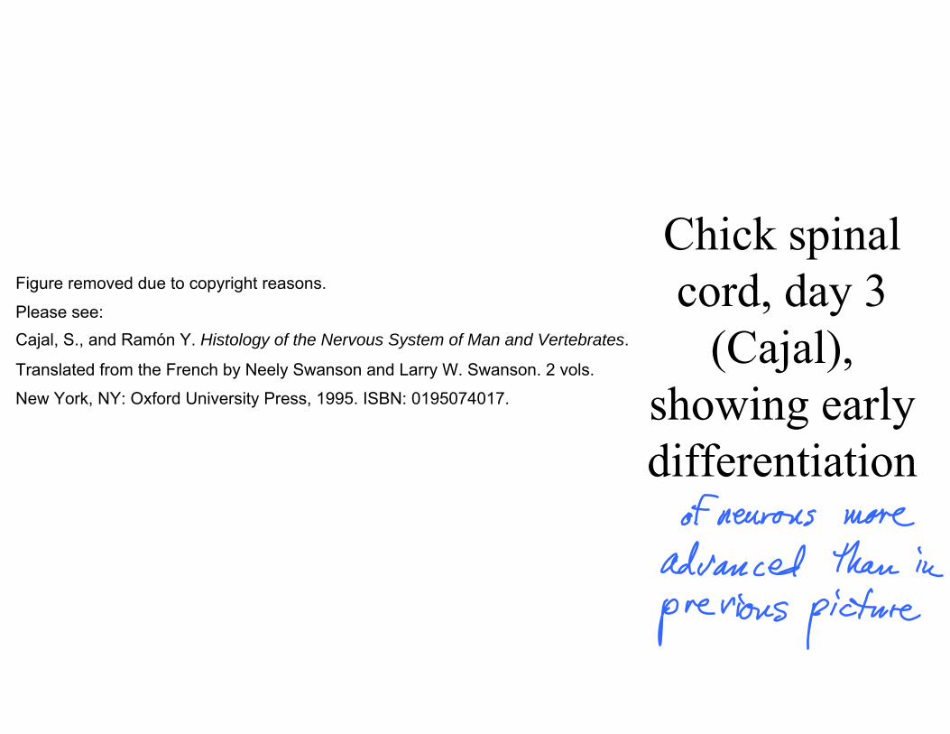
Figure removed due to copyright reasons.
Please see:Cajal, S., and Ramón Y. Histology of the Nervous System of Man and Vertebrates.
Translated from the French by Neely Swanson and Larry W. Swanson. 2 vols.
New York, NY: Oxford University Press, 1995. ISBN: 0195074017.
Chick spinal cord, day 3
(Cajal), showing early differentiation
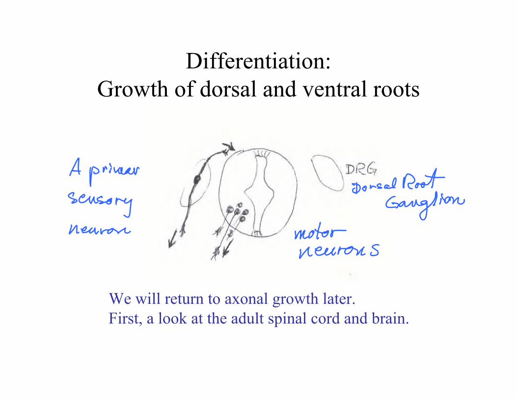
Differentiation: Growth of dorsal and ventral roots
We will return to axonal growth later. First, a look at the adult spinal cord and brain.
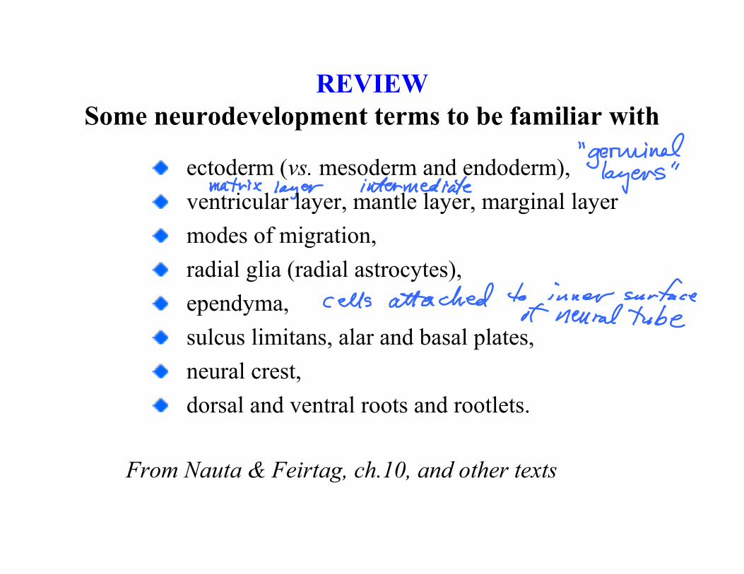
REVIEWSome neurodevelopment terms to be familiar with
(vs.ectoderm mesoderm and endoderm), ventricular layer, mantle layer, marginal layer modes of migration, radial glia (radial astrocytes), ependyma, sulcus limitans, alar and basal plates, neural crest,dorsal and ventral roots and rootlets.
From Nauta & Feirtag, ch.10, and other texts
Survey of adult human spinal cord
• Different levels, illustrated
• The sensory channels (reflex, spinocerebellar and spinothalamic tracts, origin of dorsal column axons)
• Major descending pathways (cortico-, rubro-, reticulo-, and vestibulo-spinal)
• “Propriospinal” fibers.
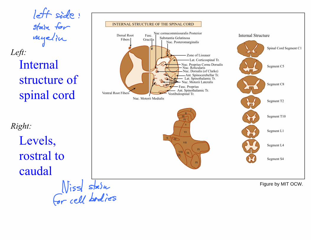
Internal structure of spinal cord
Levels,
caudal rostral to
Left:
Right:
INTERNAL STRUCTURE OF THE SPINAL CORD
Dorsal RootFibers
Fasc.Gracilis
Nuc cornucommissuralis PosteriorSubstantia Gelatinosa
Nuc. Posteromarginalis
Zone of LissauerLat. Corticospinal Tr.
Nuc. Proprius Cornu DorsalisNuc. ReticularisNuc. Dorsalis (of Clarke)
Ant. Spinocerebellar Tr.Lat. Spinothalamic Tr.
Nuc. Motorii LateralisFasc. Proprius
Ant. Spinothalamic Tr.Vestibulospinal Tr.
Nuc. Motorii MedialisVentral Root Fibers
Internal Structure
Spinal Cord Segment C1
Segment C5
Segment C8
Segment T2
Segment T10
Segment L1
Segment L4
Segment S4
III
IIIIV
V
VI
VII
VIIIIX
IXIX
IX
X M
Figure by MIT OCW.
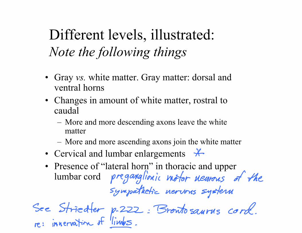
Different levels, illustrated:Note the following things
• Gray vs. white matter. Gray matter: dorsal and ventral horns
• Changes in amount of white matter, rostral to caudal – More and more descending axons leave the white
matter – More and more ascending axons join the white matter
• •
Cervical and lumbar enlargements Presence of “lateral horn” in thoracic and upper lumbar cord
Comparisons, speculations
• Role of myelin – “A crucial vertebrate innovation” (Allman p. 78):
Why? – Not found in any invertebrate or in the jawless
vertebrates (hagfish, lampreys) • How does spinal cord of humans differ from
spinal cords of other mammals?
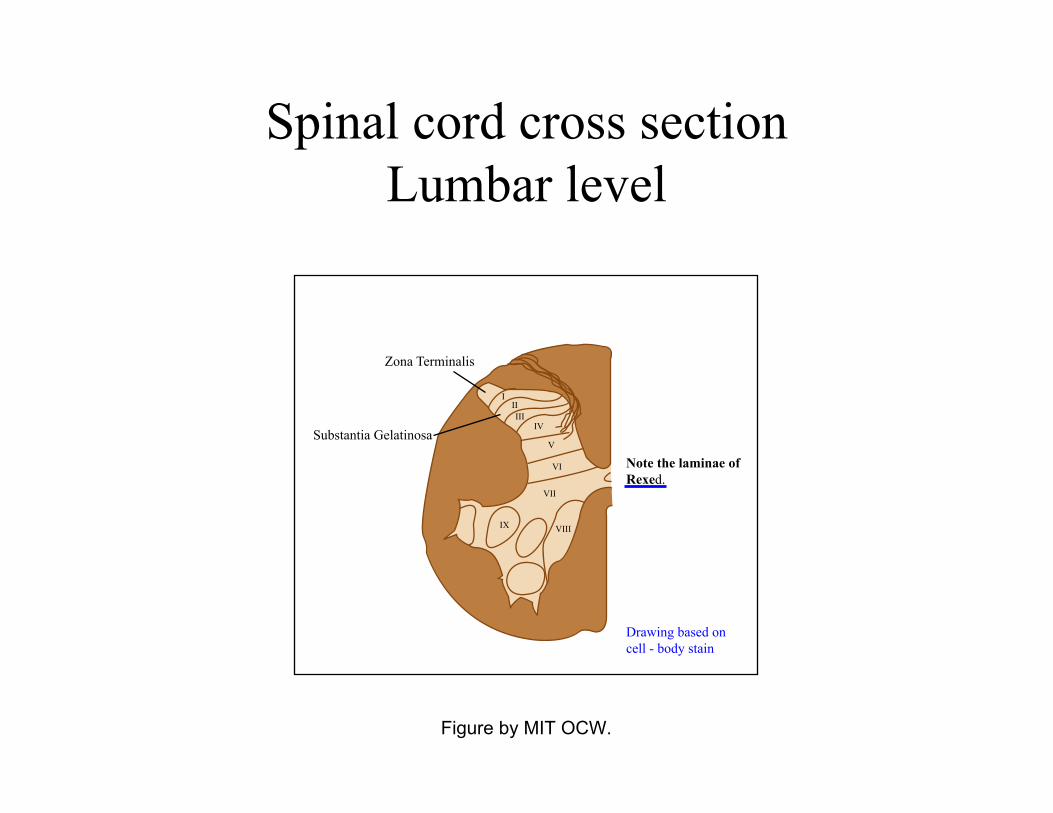
Spinal cord cross sectionLumbar level
Figure by MIT OCW.
Zona Terminalis
Substantia Gelatinosa
IIIIII
IV
V
VI
VII
VIIIIX
Drawing based on cell - body stain
Note the laminae ofRexed.
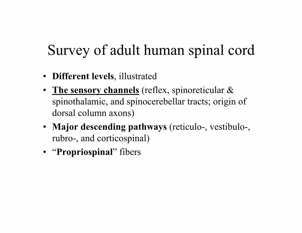
Survey of adult human spinal cord
• Different levels, illustrated
• The sensory channels (reflex, spinoreticular & spinothalamic, and spinocerebellar tracts; origin of dorsal column axons)
• Major descending pathways (reticulo-, vestibulo-, rubro-, and corticospinal)
• “Propriospinal” fibers
A sketch of the central nervous system and its origins
G. E. Schneider 2005 Part 4: Development and differentiation, spinal level
MIT 9.14 Class 8
CNS structure at the spinal level; autonomic and enteric nervous systems

Survey of adult human spinal cord
• Different levels, illustrated
• The sensory channels (reflex, spinoreticular & spinothalamic, and spinocerebellar tracts; origin of dorsal column axons)
• Major descending pathways (reticulo-, vestibulo-, rubro-, and corticospinal)
• “Propriospinal” fibers

Termination of Dorsal Root Fibers
Again, note the laminae of Rexed.
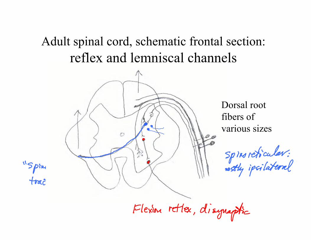
Adult spinal cord, schematic frontal section:reflex and lemniscal channels
Dorsal root fibers of various sizes
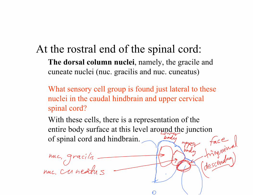
At the rostral end of the spinal cord:The dorsal column nuclei, namely, the gracile and cuneate nuclei (nuc. gracilis and nuc. cuneatus)
What sensory cell group is found just lateral to these nuclei in the caudal hindbrain and upper cervical spinal cord? With these cells, there is a representation of the entire body surface at this level around the junction of spinal cord and hindbrain.
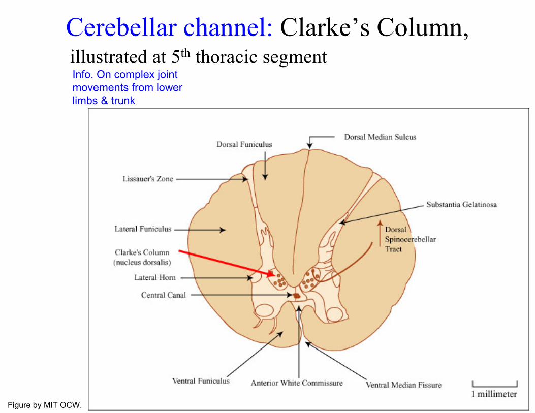
Cerebellar channel: Clarke’s Column, illustrated at 5th thoracic segment
limbs & trunk
Info. On complex joint movements from lower
Figure by MIT OCW.
Survey of adult human spinal cord
• Different levels, illustrated
• The sensory channels (reflex, spinocerebellar and spinothalamic tracts, origin of dorsal column axons)
• Major descending pathways (cortico-, rubro-, reticulo-, and vestibulospinal)
• “Propriospinal” fibers.

Adult spinal cord:some descending and intrinsic axons
Propriospinal axons
axons
Rubrospinal axons
(from Cb), Tectospinal
Corticospinal
Reticulospinal, Vestibulospinal, Fastigiospinal
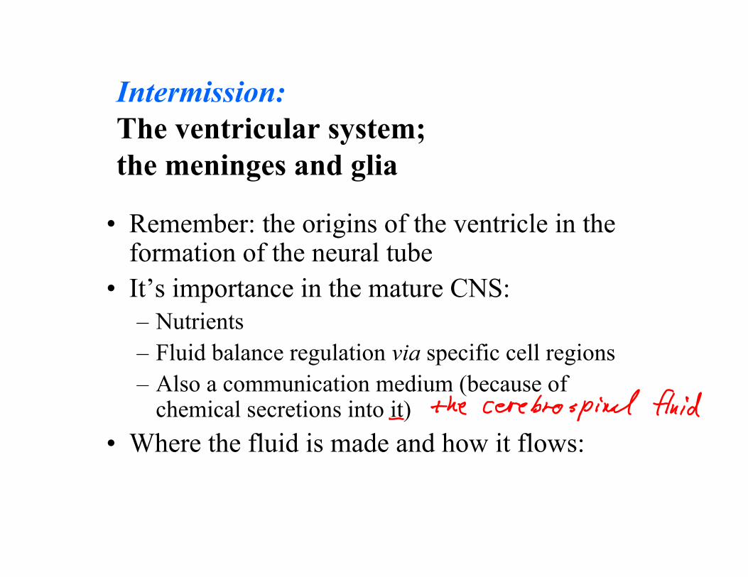
Intermission: The ventricular system; the meninges and glia
• Remember: the origins of the ventricle in the formation of the neural tube
• It’s importance in the mature CNS: – Nutrients – Fluid balance regulation via specific cell regions– Also a communication medium (because of
)chemical secretions into it• Where the fluid is made and how it flows:
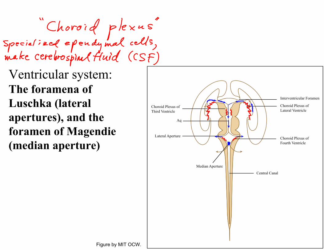
Ventricular system: The foramena of Luschka (lateral apertures), and the foramen of Magendie (median aperture)
Figure by MIT OCW.
Interventricular Foramen
Choroid Plexus of Lateral Ventricle
Choroid Plexus ofFourth Ventricle
Central Canal
Median Aperture
Lateral Aperture
Aq
Choroid Plexus of Third Ventricle

The Meninges
1.
2."subarachnoid space".
Define " " and " ":
Define " " and
dura mater pia mater meaning of the Latin terms, and basic anatomy.
arachnoid membrane
See Nauta & Feirtag, ch. 10; also P. Brodal, ch. 1, and other texts
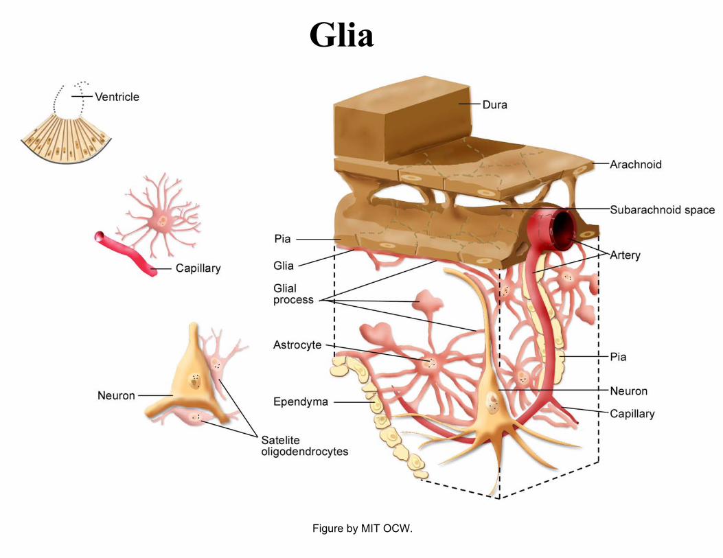
Glia
Figure by MIT OCW.
Picture taken with transmission electron microscope (EM): Astroctyes, pial cells, subarachnoid space Photo removed due to copyright reasons.
(Peters, Palay & Webster, 1976)
SS = subarachnoid space PM = pial membrane Col = collagen fibers SM = smooth muscle GL = glia limitans (astrocyte processes) B = basal lamina As = astrocyte arrows, lower fig: attachment points
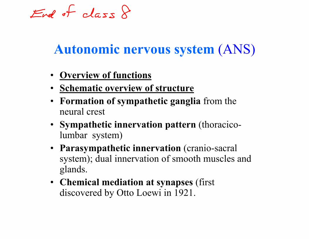
Autonomic nervous system (ANS)
• Overview of functions • Schematic overview of structure• Formation of sympathetic ganglia from the
neural crest • Sympathetic innervation pattern (thoracico
lumbar system) • Parasympathetic innervation (cranio-sacral
system); dual innervation of smooth muscles and glands.
• Chemical mediation at synapses (first discovered by Otto Loewi in 1921.
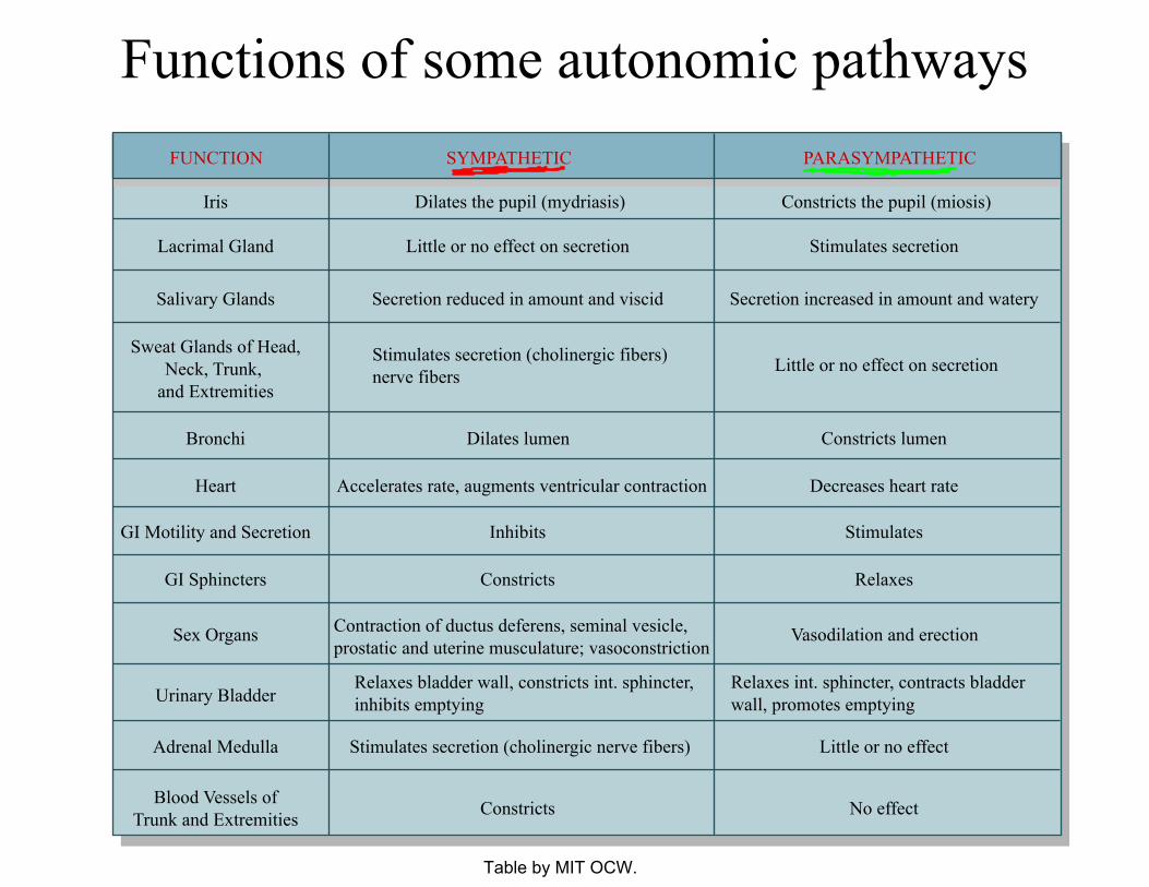
Functions of some autonomic pathways FUNCTION SYMPATHETIC PARASYMPATHETIC
Iris
Lacrimal Gland
Salivary Glands
Sweat Glands of Head,Neck, Trunk,
and Extremities
Bronchi
Heart
GI Motility and Secretion
GI Sphincters
Sex Organs
Urinary Bladder
Adrenal Medulla
Blood Vessels ofTrunk and Extremities
Dilates the pupil (mydriasis)
Little or no effect on secretion
Secretion reduced in amount and viscid
Stimulates secretion (cholinergic fibers)nerve fibers
Dilates lumen
Accelerates rate, augments ventricular contraction
Inhibits
Constricts
Contraction of ductus deferens, seminal vesicle,prostatic and uterine musculature; vasoconstriction
Relaxes bladder wall, constricts int. sphincter,inhibits emptying
Constricts
Stimulates secretion (cholinergic nerve fibers)
Constricts the pupil (miosis)
Stimulates secretion
Secretion increased in amount and watery
Little or no effect on secretion
Constricts lumen
Decreases heart rate
Stimulates
Relaxes
Vasodilation and erection
Relaxes int. sphincter, contracts bladder wall, promotes emptying
Little or no effect
No effect
Table by MIT OCW.

Autonomic pathways: schematic of structural arrangements
Note the CNS locations of the preganglionic motor neurons of the two divisions of the ANS.
Figure by MIT OCW.

Another schematic view of ANS
Figure by MIT OCW.
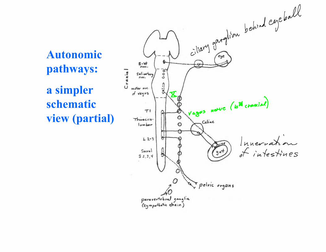
Autonomic pathways:
a simpler schematic view (partial)

Step back a moment:Arrangement of motor neurons in the three major divisions of the motor system
Somatic: Synaptic Autonomic: Paracrine Neuroendocrine: Endocrine
S
E
S P
Figure by MIT OCW.
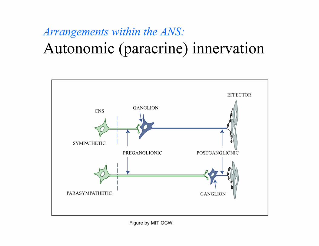
Arrangements within the ANS:Autonomic (paracrine) innervation
GANGLION
GANGLION
PREGANGLIONIC POSTGANGLIONIC
CNS
EFFECTOR
SYMPATHETIC
PARASYMPATHETIC
Figure by MIT OCW.

Autonomic innervation of the intestine in several vertebrate classes.
Fish
Fish
Reptiles
Amphibians Mammals
CholinergicAdrenergic
8th
Figure by MIT OCW.
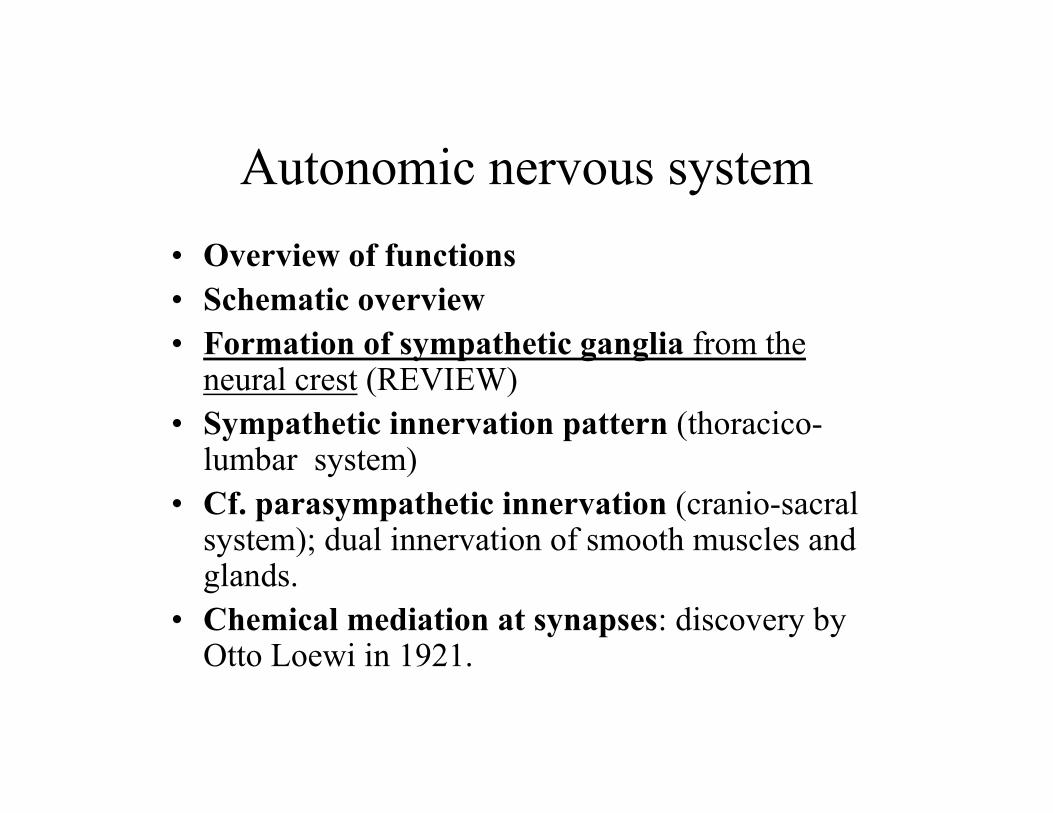
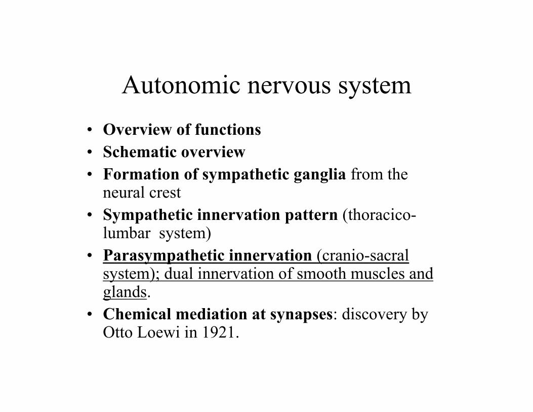
Autonomic nervous system
• Overview of functions• Schematic overview • Formation of sympathetic ganglia from the
neural crest (REVIEW) • Sympathetic innervation pattern (thoracico
lumbar system) • Cf. parasympathetic innervation (cranio-sacral
system); dual innervation of smooth muscles and glands.
• Chemical mediation at synapses: discovery by Otto Loewi in 1921.
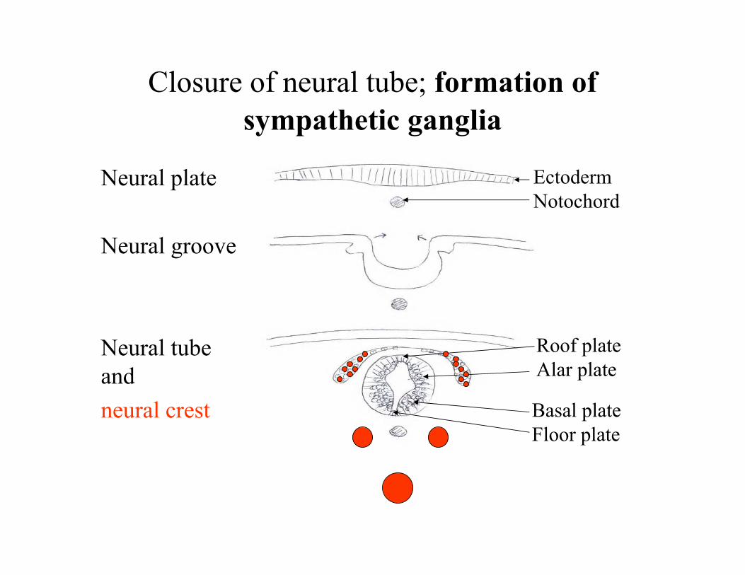
Closure of neural tube; formation ofsympathetic ganglia
Neural plate
Neural groove
Neural tube and
neural crest Basal plate Floor plate
Roof plateAlar plate
Ectoderm Notochord
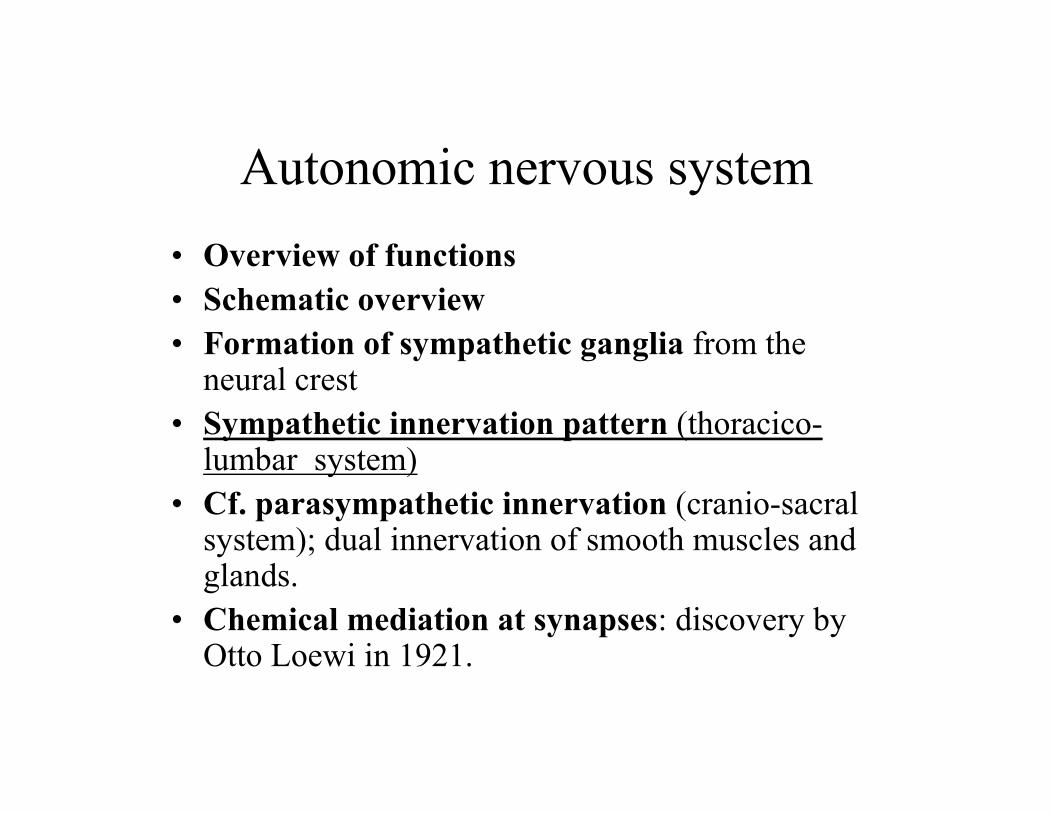
Autonomic nervous system
• Overview of functions• Schematic overview • Formation of sympathetic ganglia from the
neural crest • Sympathetic innervation pattern (thoracico
lumbar system) • Cf. parasympathetic innervation (cranio-sacral
system); dual innervation of smooth muscles and glands.
• Chemical mediation at synapses: discovery by Otto Loewi in 1921.
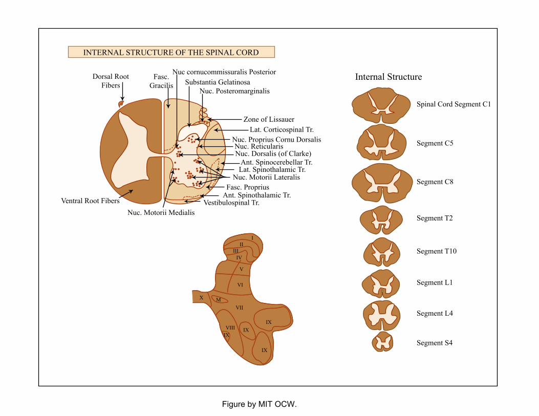
Figure by MIT OCW.
INTERNAL STRUCTURE OF THE SPINAL CORD
Dorsal RootFibers
Fasc.Gracilis
Nuc cornucommissuralis PosteriorSubstantia Gelatinosa
Nuc. Posteromarginalis
Zone of LissauerLat. Corticospinal Tr.
Nuc. Proprius Cornu DorsalisNuc. ReticularisNuc. Dorsalis (of Clarke)
Ant. Spinocerebellar Tr.Lat. Spinothalamic Tr.
Nuc. Motorii LateralisFasc. Proprius
Ant. Spinothalamic Tr.Vestibulospinal Tr.
Nuc. Motorii MedialisVentral Root Fibers
Internal Structure
Spinal Cord Segment C1
Segment C5
Segment C8
Segment T2
Segment T10
Segment L1
Segment L4
Segment S4
III
IIIIV
V
VI
VII
VIIIIX
IXIX
IX
X M
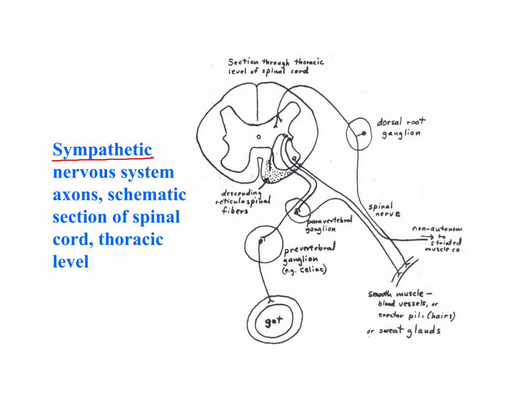
Sympathetic nervous system axons, schematic section of spinal cord, thoracic level
xxxxx
xxxxx
xx
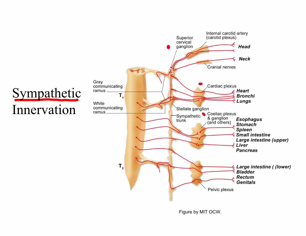
Sympathetic Innervation
Figure by MIT OCW.
Autonomic nervous system
• Overview of functions• Schematic overview • Formation of sympathetic ganglia from the
neural crest • Sympathetic innervation pattern (thoracico
lumbar system) • Parasympathetic innervation (cranio-sacral
system); dual innervation of smooth muscles and glands.
• Chemical mediation at synapses: discovery by Otto Loewi in 1921.
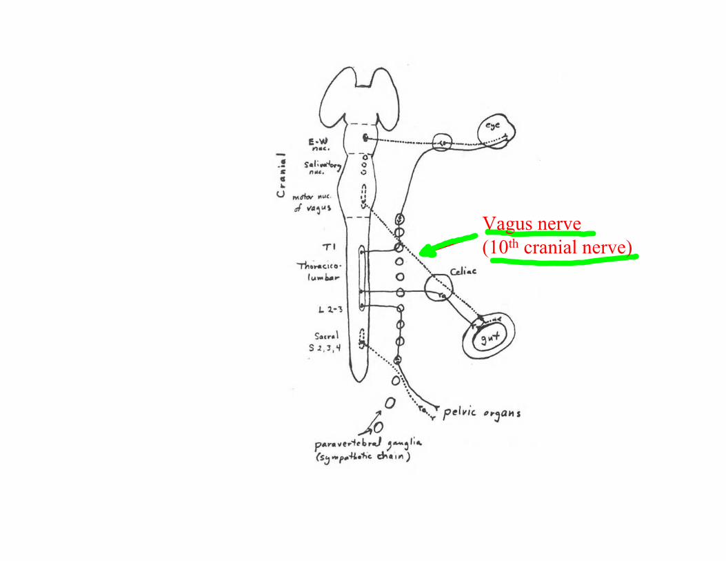
(10th Vagus nerve
cranial nerve)
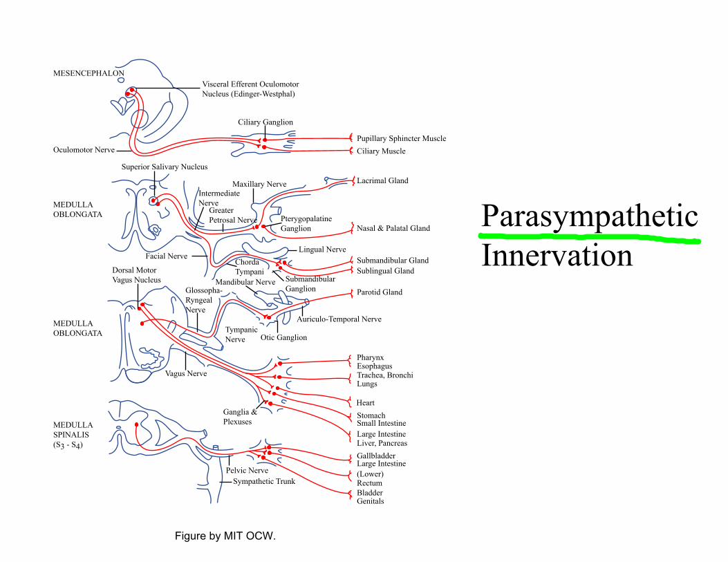
Figure by MIT OCW.
Parasympathetic Innervation
MESENCEPHALONVisceral Efferent OculomotorNucleus (Edinger-Westphal)
Oculomotor Nerve
Superior Salivary Nucleus
MEDULLAOBLONGATA
MEDULLAOBLONGATA
MEDULLASPINALIS(S3 - S4)
Pupillary SphinCiliary Muscle
Lacrimal Gland
Nasal & Palatal
Submandibular Sublingual Gla
Parotid Gland
PharynxEsophagusTrachea, BroncLungs
HeartStomachSmall IntestineLarge IntestineLiver, PancreasGallbladderLarge Intestine(Lower)RectumBladderGenitals
Ciliary Ganglion
Maxillary NerveIntermediateNerve
GreaterPetrosal Nerve Pterygopalatine
Ganglion
Facial Nerve
Dorsal MotorVagus Nucleus
Glossopha-RyngealNerve
ChordaTympani
Mandibular Nerve
Lingual Nerve
SubmandibularGanglion
Auriculo-Temporal Nerve
Otic GanglionTympanicNerve
Vagus Nerve
Ganglia &Plexuses
Pelvic NerveSympathetic Trunk
cter Muscle
Gland
Glandnd
hi
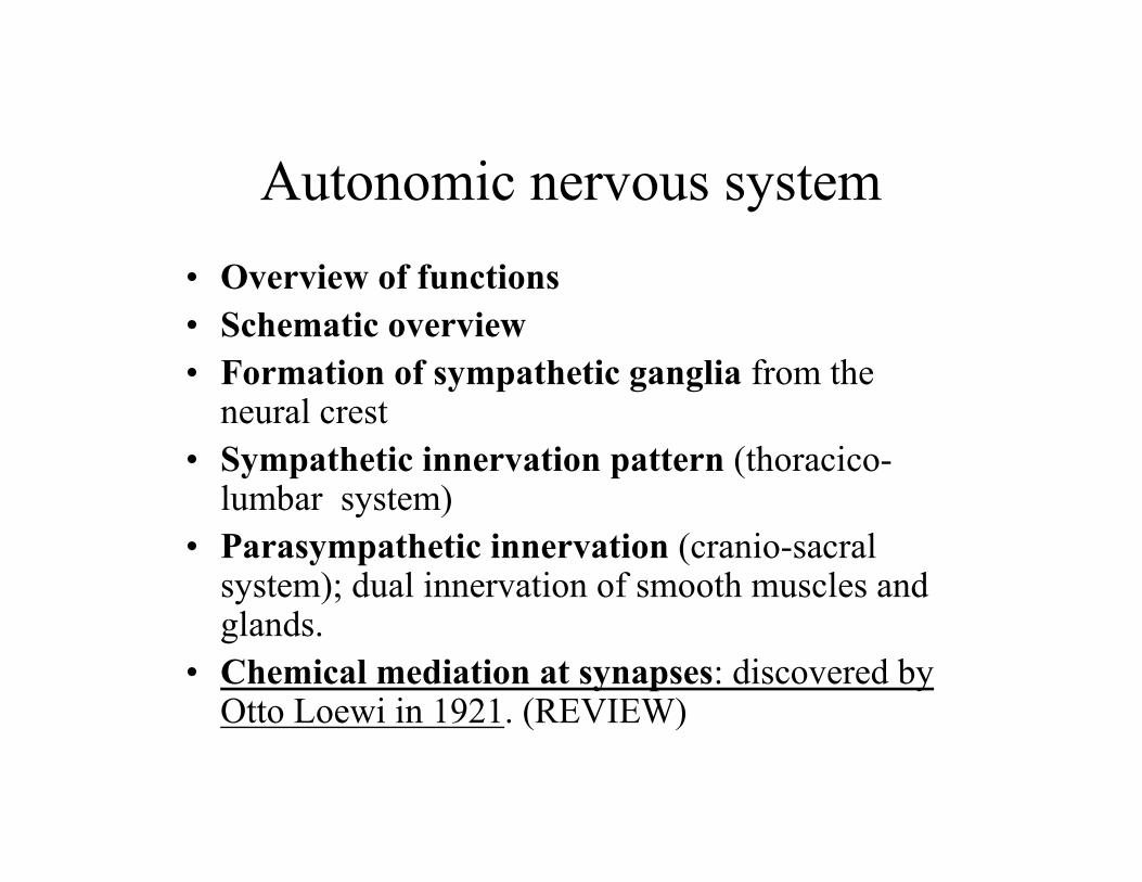
Autonomic nervous system
• Overview of functions• Schematic overview • Formation of sympathetic ganglia from the
neural crest • Sympathetic innervation pattern (thoracico
lumbar system) • Parasympathetic innervation (cranio-sacral
system); dual innervation of smooth muscles and glands.
• Chemical mediation at synapses: discovered by Otto Loewi in 1921. (REVIEW)
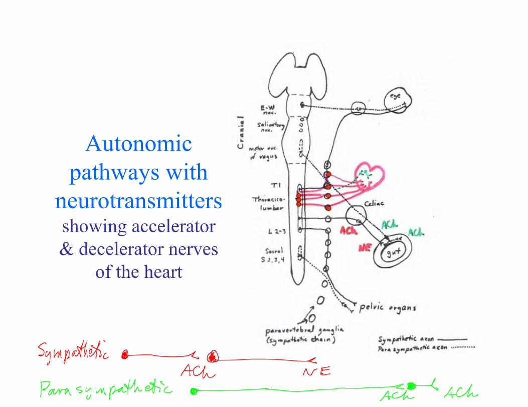
Autonomic pathways with
neurotransmitters
& decelerator nerves of the heart
showing accelerator

Enteric nervous systemThe “little brain” in the gut: Perhaps as many neurons as in the entire spinal cord.
In the wall of the intestine:•Myenteric plexus (the outer plexus) •Submucous plexus (the middle plexus) •Villous plexus (inner plexus) •Periglandular plexus (inner plexus)
Innervation by vagus nerve
Cardiac ganglion: Does the heart have a brain?

Levels of control in the ANS: the temperature regulation systems
• See reviews by Evelyn Satinoff
Selected References
Slide 21: Nauta, Walle J. H., and Michael Feirtag. Fundamental Neuroanatomy. New York, NY: Freeman, 1986, chapter 1, fig. 64. ISBN: 0716717239. Slide 27: Brodal, Per. The Central Nervous System, Structure and Function. 3rd ed. New York, NY: Oxford University Press, 2003. ISBN: 0195165608. Slide 31: Truex, Raymond C. and Malcolm B. Carpenter. Human neuroanatomy. 6th ed. Baltimore, MD: Williams & Wilkins, 1969. Slide 32: Nolte, John. The human brain: an introduction to its functional anatomy. St. Louis, Mo: Mosby, 2002. Slide 34: Swanson, Larry W. Brain Architecture, Understanding the Basic Plan. Oxford; New York, NY: Oxford University Press, 2003, p. 99. ISBN: 0195105052. Slide 42: Brodal, Per. The Central Nervous System, Structure and Function. 3rd ed. New York, NY: Oxford University Press, 2003. ISBN: 0195165608. Slide 45: Brodal, Per. The Central Nervous System, Structure and Function. 3rd ed. New York, NY: Oxford University Press, 2003. ISBN: 0195165608.