2.0 review of literature 2.1 about fungi -...
TRANSCRIPT
6
2.0 REVIEW OF LITERATURE
2.1 About fungi
Of the five kingdoms of the living world; Planta, Animalia, Fungi, Protista and Monera the
kingdom fungi contains a diversity of organisms which include both macroscopic and
filamentous or yeast-like microscopic structures. Unlike plants and algae, fungi are not
autotrophic since they lack chlorophyll. They are often referred to as “saprophytes” or
“saprobes” as they draw their nourishment from decomposing organic matter. These organisms
play a vital role in the recycling of organic matter (Rippon et al., 1988; Kwon-Chung and
Bennett, 1992). Structurally, fungi are eukaryotes which may be unicellular or multi-cellular.
These organisms are surrounded by a rigid cell wall which is made up of chitin and can
reproduce by sexual and asexual means (Alexopoulos et al., 1996, St-Germanin and Summerbell,
1996). The fungi are present almost in every ecological niche. There are about 1.5 million
species of fungi worldwide out of which, approximately 120000 species have been described till
date, though the number is steadily increasing (Hawksworth et al., 1991). Of these, about 200
fungal species are known to cause human infections termed as mycoses (Mc-Ginnis, 1991).
2.2 Historical background
2.2.1 Mycology and mycoses
The era of mycology began with the observation of some filamentous organisms on the yellow
spots on the leaf of Desmark rose by Hooks in 1677 with the help of magnifying lens. Studies
were later conducted by several other workers. Some of the fungi are pathogenic and can cause
infections in humans and animals. The term mycoses refer to the various infections caused by
fungi. Augustino Bassi (1835) was the first to explain that a mould, Beauveria bassiana was
responsible for the deadly disease of silkworms (Bombyx mori). This was the first organism to be
recognized as a fungal pathogen (Emmons et. al., 1977). Subsequently, several fungal species
have been implicated in different disease conditions.
2.2.2 Dermatophytosis
The human medical mycology started with the discovery of etiologic agents of dermatophytosis
referred to as dermatophytes. These fungi are implicated in superficial skin infections. According
to Seeliger (1985), Remak in 1835 was first to observe microscopic structures appearing as rods
and buds in the crust of skin from a patient suffering from tinea favosa peculiar to what were
7
later termed as dermatophytes. The mycotic nature of these structures causing tinea favosa was
described by Schonlein (1839).
The work of David Gruby (1841-44) laid down the foundation of dermatomycology. He first
described the clinical entity caused by dermatophytes and also demonstrated their contagious
nature. He also recognized ectothrix and endothrix hair invasion of a dermatophyte species and
named it as Microsporum audouinii (Chander, 1995). Raymond Sabouraud (1892), a renowned
mycologists initiated work on dermatophytosis and published his monumental work in his classic
volume, Les Teignes in 1910. He classified the dermatophytes into four genera; Achorion,
Epidermophyton, Microsporum and Trichophyton. Saboraud also described the methods of
culturing dermatophytes and suggested the therapeutic measures for dermatophytosis. In 1925,
Wood invented a lamp, named after his name as Wood‟s lamp. He used this lamp for the
detection of dermatophytic infection of hair (Kwon Chung and Bennett, 1992). Emmons (1934)
modified the taxonomic scheme of Sabouraud and other scientists. He eliminated the genus
Achorion and established the current classification of dermatophytes on the basis of spore
morphology and accessory structures, such shape of hyphae, growth rate and color of the fungal
growth (obverse and reverse).
Vanbreuseghem (1952) described hair bait technique for the culturing the dermatophytes.
Dawson and Gentles (1959) cultured the ascomycetous teleomorphs of Trichophyton ajjelloi
(Collier et al., 1998). The teleomorphs of the Microsporum gypseum were independently
obtained by other workers (Griffin, 1960, Stockdale, 1961). Taplin et al., (1969) developed
dermatophyte test medium (DTM) for isolating and differentiating dermatophytes from
fungal/bacterial contaminants. It is a selective medium which is routinely being used for isolation
of dermatophytes.
2.3 Classification of dermatophytes
Dermatophytes are hyaline and well septate moulds which include more than 100 species. Of
these, only 42 species have been considered valid and about less than half of these species are
pathogenic. Emmons (1934) classified dermatophytes into three anamorphic (asexual or
imperfect) genera, namely, Microsporum, Trichophyton and Epidermophyton of the class
Hyphomycetes of the deuteromycota (Fungi imperfecti). The classification of dermatophytes is
based on the formation of conidia and their morphology and is updated with the discovery of
8
new species (Ajello, 1977, Matsumoto and Ajello, 1987). Each genus comprises many species,
the description of these genera is given below:
2.3.1 Microsporum
There are about 16 valid species belonging to the genus Microsporum which are associated with
the skin and hair infections (Chander, 1995). However, they are not associated with nail
infections. Microsporum audouinii is the prototype of this genus (Gruby, 1843). The shape of
macroconia varies from spindle or fusiform to obovate (egg shaped) in M. nanum and
cylindrofusiform in M. vanbruseghemii. (Fuentes, 1956; Georg et al., 1962). Macroconida may
be septate having 1-15 septa, the size of which may vary from 6µm-160µm by 6µm-25µm. Some
of the commonly observed species of Microsporum are; M. audouinii M. canis, M. gypseum, M.
nanum, M. ferrugineum, M. cookie, M. vanbreuseghemii, M. persicolor.
2.3.2 Trichophyton
Twenty four valid species of the genus Trichophyton have been identified. T. tonsurans is the
type species of genus Trichophyton (Malmsten, 1845). Trichophyton spp. usually infects skin,
hair and nails (Chander, 1995). Well septate, pencil-fusiform or cylindrical macroconida having
1-12 septa with smooth and thin wall may be observed on microscopic examination. The
macroconidia may be present singly or in clusters, each macroconidium ranging from 8µm-
86µm x 4µm-14µm in size. Micoconidia are numerous and their shape may vary from globose,
pyriform to spherical. Most common species of Trichophyton are; T. tonsurans, T.
mentagrophyte, T. rubrum, T. schoenleinii, T. verrucosum, T. violaceum, T. concentricum.
2.3.3 Epidermophyton
This genus Epidermophyon has only two known species, E. floccosum and E. stockdaleae. The
former is the type species of this genus which is pathogenic (Sabouraud, 1910). These fungi
produce thin to thick, smooth walled and septate macroconida having septa 1-9 in number which
may be observed on microscopic examinations. The size of each macroconidia ranges from
20µm-60µm x 4µm-13µm. The microconidia are absent (Chander, 1995).
2.4 Ecology of dermatophytes
Dermatophytes are among few fungi which cause communicable disease i.e. disease may
acquired from the infected animals or birds to humans or by the fomites. Based on the ecology
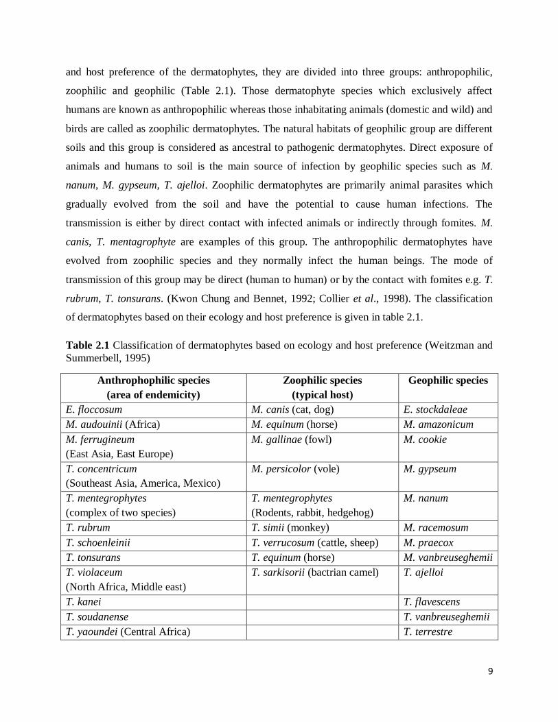
9
and host preference of the dermatophytes, they are divided into three groups: anthropophilic,
zoophilic and geophilic (Table 2.1). Those dermatophyte species which exclusively affect
humans are known as anthropophilic whereas those inhabitating animals (domestic and wild) and
birds are called as zoophilic dermatophytes. The natural habitats of geophilic group are different
soils and this group is considered as ancestral to pathogenic dermatophytes. Direct exposure of
animals and humans to soil is the main source of infection by geophilic species such as M.
nanum, M. gypseum, T. ajelloi. Zoophilic dermatophytes are primarily animal parasites which
gradually evolved from the soil and have the potential to cause human infections. The
transmission is either by direct contact with infected animals or indirectly through fomites. M.
canis, T. mentagrophyte are examples of this group. The anthropophilic dermatophytes have
evolved from zoophilic species and they normally infect the human beings. The mode of
transmission of this group may be direct (human to human) or by the contact with fomites e.g. T.
rubrum, T. tonsurans. (Kwon Chung and Bennet, 1992; Collier et al., 1998). The classification
of dermatophytes based on their ecology and host preference is given in table 2.1.
Table 2.1 Classification of dermatophytes based on ecology and host preference (Weitzman and
Summerbell, 1995)
Anthrophophilic species
(area of endemicity)
Zoophilic species
(typical host)
Geophilic species
E. floccosum M. canis (cat, dog) E. stockdaleae
M. audouinii (Africa) M. equinum (horse) M. amazonicum
M. ferrugineum
(East Asia, East Europe)
M. gallinae (fowl) M. cookie
T. concentricum
(Southeast Asia, America, Mexico)
M. persicolor (vole) M. gypseum
T. mentegrophytes
(complex of two species)
T. mentegrophytes
(Rodents, rabbit, hedgehog)
M. nanum
T. rubrum T. simii (monkey) M. racemosum
T. schoenleinii T. verrucosum (cattle, sheep) M. praecox
T. tonsurans T. equinum (horse) M. vanbreuseghemii
T. violaceum
(North Africa, Middle east)
T. sarkisorii (bactrian camel) T. ajelloi
T. kanei T. flavescens
T. soudanense T. vanbreuseghemii
T. yaoundei (Central Africa) T. terrestre
10
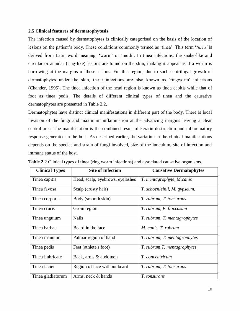
2.5 Clinical features of dermatophytosis
The infection caused by dermatophytes is clinically categorised on the basis of the location of
lesions on the patient‟s body. These conditions commonly termed as „tinea‟. This term „tinea’ is
derived from Latin word meaning, „worm‟ or „moth‟. In tinea infections, the snake-like and
circular or annular (ring-like) lesions are found on the skin, making it appear as if a worm is
burrowing at the margins of these lesions. For this region, due to such centrifugal growth of
dermatophytes under the skin, these infections are also known as „ringworm‟ infections
(Chander, 1995). The tinea infection of the head region is known as tinea capitis while that of
foot as tinea pedis. The details of different clinical types of tinea and the causative
dermatophytes are presented in Table 2.2.
Dermatophytes have distinct clinical manifestations in different part of the body. There is local
invasion of the fungi and maximum inflammation at the advancing margins leaving a clear
central area. The manifestation is the combined result of keratin destruction and inflammatory
response generated in the host. As described earlier, the variation in the clinical manifestations
depends on the species and strain of fungi involved, size of the inoculum, site of infection and
immune status of the host.
Table 2.2 Clinical types of tinea (ring worm infections) and associated causative organisms.
Clinical Types Site of Infection Causative Dermatophytes
Tinea capitis Head, scalp, eyebrows, eyelashes T. mentagrophyte, M.canis
Tinea favosa Scalp (crusty hair) T. schoenleinii, M. gypseum.
Tinea corporis Body (smooth skin) T. rubrum, T. tonsurans
Tinea cruris Groin region T. rubrum, E. floccosum
Tinea unguium Nails T. rubrum, T. mentagrophytes
Tinea barbae Beard in the face M. canis, T. rubrum
Tinea manuum Palmar region of hand T. rubrum, T. mentagrophytes
Tinea pedis Feet (athlete's foot) T. rubrum,T. mentagrophytes
Tinea imbricate Back, arms & abdomen T. concentricum
Tinea faciei Region of face without beard T. rubrum, T. tonsurans
Tinea gladiatorum Arms, neck & hands T. tonsurans
11
Tinea infections are discussed below:
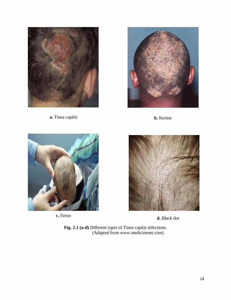
2.5.1 Tinea capitis
This is the infection of shaft of the scalp hairs which is of two types.
a. Inflammatory: Kerion, Favus
b. Non inflammatory: Black Dot, grey patch
The infected hairs in tinea capitis appear dull and gray. The base of the hair shaft and hair
follicles also get invaded leading to the patch formation with broken hairs and ring formation
(Fig. 2.1 a). The predominant causative fungal species of tinea capitis belongs to the genus
Trichophyton. Different types of tinea capitis are discussed as under:
i. Kerion (German word for Honeycomb): This is a painful inflammatory condition with raised,
bounded boggy mass on the scalp (Fig. 2.1 b). The hair follicles may be seen discharging pus.
Kerion in usually caused by zoophilic dermatophytes such as T. verrucosum and T.
mentagrophyte.
ii. Favus (Tinea Favosa): „Favus‟ is a latin word for the honeycomb. This infection is caused by
T. schoenleinii in which cup like crusts around infected follicles are formed. The fungal growth
within and around the hair follicles produces waxy, honeycomb-like crust on scalp which may
lead to alopecia and scarring (Fig. 2.1 c). Tinea favosa is becoming very rare because of the
increased hygienic life style of the public at large.
iii. Black Dot: T. tonsurans and T. violaceum are commonly associated with this type of tinea
infection. These dermatophytes attack hair shafts by endothrix type invasion with abundant
sporulation inside hair and breakage of the hair near the surface of scalp. This results in black dot
appearance within the area of smooth scalp surface (Fig. 2.1 d).
iv. Ectothrix infection: In this type of infection, the arthrospores of the fungi construct sheath or
chains on the surface of the hair shaft. The cuticle of the hair remains intact. The hyphae invade
the shaft at mid-follicles. As the hair grows out of the follicle, the hyphae brust out of the shaft
and cover the hair surface with small arthrospores. T. mentagrophyte, M.canis, M. gypseum and
M. audouinii are usually implicated in this type of infection.
v. Endothrix infection: In endothrix infection, the arthrospores invade the hair shaft and their
hyphae weaken the hair because of the destruction of the cuticle. The infected hair becomes
12
grayish white, breakes off to give „black dot‟ like appearance. T. violaceum and T. tonsurans are
commonly involved in this type of invasion.
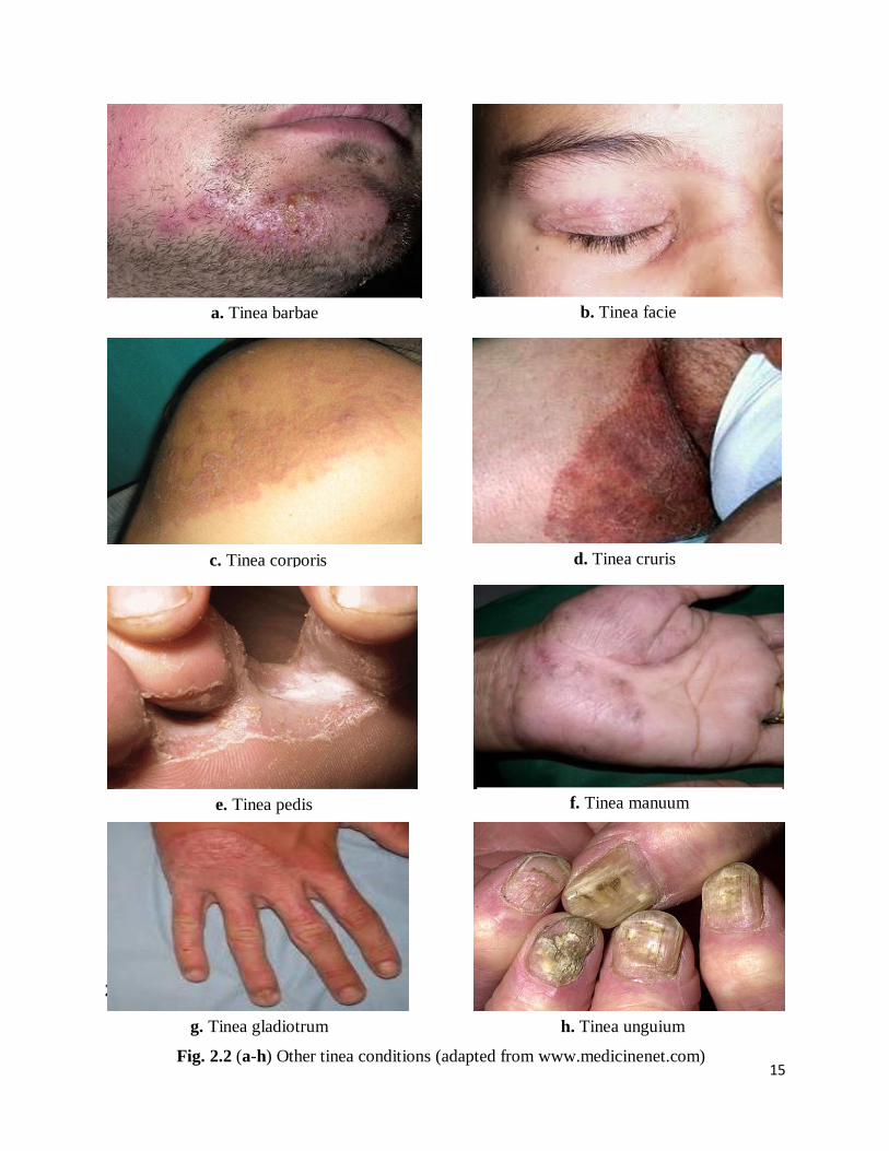
2.5.2 Tinea barbae
It is ringworm infection of coarse hairs of beard and moustache (Fig. 2.2 a) and is popularly
known as „barber‟s itch‟. The inflammatory and pustular lesions with fragile and lusterless hairs
are clearly visible in this tinea condition. T. verrucosum and T. mentagrophyte are commonly
implicated. T. rubrum and M. canis are also occasionally associated with tinea barbae.
2.5.3 Tinea faciei
This dermatophytic infection is seen on the non-bearded region of the face and is characterized
by erythematous annular plaques with central clearing (Fig.2.2 b). It is common in the patients
usually having history of photosensitivity. Tinea faciei is sometimes, and treated with topical
steroids because of wrong diagnosis.
2.5.4 Tinea corporis
It is the infection of glabrous (non-hairy) skin which may result from extension of the infection
from scalp, groin or beard region. The erythematous raised lesions, annular, sharply marginated
single or multiple plaques are common clinical features of tinea corporis (Fig.2.2 c). Most
common species associated with this condition are; T. rubrum, T. mentagrophyte, T. tonsurans.
2.5.5 Tinea cruris (Jock itch)
This is a worldwide ringworm infection of the inguinal area involving groin, perianal, perineal
areas often involving upper thigh region also. The infection is more prevalent in tropical
countries. Men are more affected than women due to prolonged use of tight-fitting
undergarments. The borders of the lesions are well delineated, erythematous plaques, arciform
lesions with sharp margins (Fig.2.2 d). T. rubrum and E. floccosum are commonly involved
dermatophyte species. Tinea cruris is commonly known as jock itch.
2.5.6 Tinea pedis (Athlete’s foot)
Popularly known as athlete‟s foot, tinea pedis is infection of feet involving interdigital webs and
sole. This condition is clinically characterized by scaling, fissuring, maceration and erythematous
toe webs. The small vesicles may discharge fluid on rupture. The maceration and peeling results
13
into cracks which may further lead to secondary bacterial infection (Fig.2.2 e). Tenia pedis is
common among the individuals wearing shoes for prolonged periods. Anthropophilic species (T.
mentagrophyte and E. floccosum) are common causative agents of this condition.
2.5.7 Tinea manuum
In this condition, the diffused hyperkeratotic lesions typical of ring worm infections can be seen
on the palms and interdigital areas of hands (Fig. 2.2 f). Anthropophilic species (T. rubrum, T.
mentagrophyte and E. floccosum) are mainly responsible for this condition.
2.5.8 Tinea gladiatorum
This infection is common in wrestlers and other athletes depending upon their playing habits
which spread as a result of skin to skin contact rather than via fomites. T. tonsurans is the
commonest causative agent of this infection. The clinical features are sililar to that seen in tinea
corporis (Fig.2.2 g).
2.5.9 Tinea unguium
The ringworm infection of nail plates commonly affecting adults is known as tinea unguium.
Distal subungual infection is the commonest pattern which involves nail bed and underside of
nail in distal portion (Fig.2.2 h). The nail plate become brittle, friable, thickened and may crack
due to piling up of subungual debris. The color of the nail often turns to yellow-brown or brown-
black. T. rubrum, T. mentagrophyte and E. floccosum are the most common species involved in
tinea unguium.
2.5.10 Tinea imbricata
This is an unusual form of the tinea corporis which is caused by anthropophilic dermatophyte T.
concentricum and is restricted to limited geographical regions (China, India, Fiji, Samoa, Papua
and Central and South America). Tinea imbricata is characterized by polycyclic, concentrically
arranged rings, papulosquamous patches of scales scattered over and often covering most of the
body.
14
Fig. 2.1 (a-d) Different types of Tinea capitis infections.
(Adapted from www.medicinenet.com)
a. Tinea capitis b. Kerion
c. Favus d. Black dot
15
2.6 Chronic dermatophytic infections
c. Tinea corporis d. Tinea cruris
e. Tinea pedis f. Tinea manuum
h. Tinea unguium
Fig. 2.2 (a-h) Other tinea conditions (adapted from www.medicinenet.com)
a. Tinea barbae b. Tinea facie
g. Tinea gladiotrum
16
2.6 Chronic dermatophytosis
Many patients have continuous and recurrent dermatophytic infections which are regarded as
chronic dermatophytosis. The infections of feet and groin regions in particular, are chronic in
nature. Also, the chronic cases of scalp are mostly observed in children. Certain factors are
responsible for such types of infections: i. the nature of patient provides the favourable
environment for the growth and persistence of dermatophytes e.g. immunocompromised patients
such as organ transplant recipients, diabetic patients or HIV infected patients are more prone to
chronic dermatophytosis ii. recurrence of the infections are also seen in the patients receiving
treatment who frequently stop applying topical antifungals. Most of the topical antifungals are
fungistatic and short term therapy may not remove the pathogen completely iv. the development
of resistance against antifungal agents by the dermatophytic strains is also responsible for
chronic dermatophytosis. Such strains emerge due to indiscriminate use of antifungal drugs.
2.7 Cultivation of dermatophytes and their colony characteristics
A number of culture media are used for the isolation of the dermatophytes. Sabouraud‟s dextrose
agar (SDA) supplemented with chloramphenicol and cyclohexamide, is the most commonly used
medium for isolation of fungal species. Dermatophyte test medium (DTM), Corn meal agar
(CMA), Potato dextrose agar (PDA) are other media used for this purpose.
2.7.1 Commonly used growth media
2.7.1.1 Sabouraud’s dextrose agar (SDA) with chloramphenicol and cyclohexamide
This is a standard medium for the isolation of the dermatophyte species. It contains
chloramphenicol (0.05%) which inhibits the bacterial growth while the cyclohexamide at the rate
of 0.1 to 0.4 mg per ml suppresses the growth of saprophytic fungi (aspergillus, candida,
scytalidium).
2.7.1.2 Dermatophyte test medium (DTM)
DTM is a selective medium which contain phenol red as pH indicator. The change of color from
yellow to red indicates the growth of dermatophytes in the medium. DTM contains antibiotics,
chlortetracycline, gentamycin and cyclohexamide which inhibit growth of most of the bacteria
and several contaminating fungi. The reason for the color change is the release alkaline
metabolites by the growing dermatophytes into the medium. This medium is generally used for
screening of dermatophyte species and not for diagnosis.
17
2.7.1.3 Corn meal agar (CMA)
CMA is a nutritionally deficient medium which induces the sporulation by suppressing the
vegetative growth of the fungi. This medium is used with dextrose to differentiate T. rubrum
from T. mentagrophyte on the basis of pigments produced.
2.7.1.4 Potato dextrose agar (PDA)
PDA induces sporulation in dermatophytes and can be used in slide cultures.
Different dermatophyte species grow differently on SDA medium and can be differentiated on
the basis of their peculiar morphology on SDA, urease test and hair perforation test as detailed in
Table 2.3 and briefly mentioned below.
i. Growth rate: Majority of the dermatophytes grow within 7 to 10 days but it may take longer
time in cases of T. tonsurans and T. verrucosum.
ii. Colony obverse: The colonies are observed for color (white, pearl, ivory, cream, brown etc.),
texture of the surface (glabrous or waxy, powdery, granular, suede-like velvety, downy or fluffy
etc.).
iii. Colony reverse: The colonies on the reverse are observed for pigmentation (deep red, rusty
brown, yellow etc.), topography (flat, raised, heaped etc.). Generally, T. rubrum produce a dark
cherry red color under the colony. Confusion exists in differentiation of many slow pigment
producing varieties of T. mentagrophyte. For this, stimulation of pigment production is achieved
by growing them on potato-dextrose agar.
2.7.2 Other differential features
2.7.2.1 Nutritional requirements
Nicotinic acid for T. equinum, histidine for T. megninii, thiamine for T. violaceum and T.
tonsurans are growth requirements which might be helpful in identification of these
dermatophytes.
2.7.2.2 Temperature
Although some of the dermatophyte species tolerate a wide range of temperature, most grow best
at 25°C to 35°C. At this temperature range, T. rubrum induces a red pigment on serum albumin
agar within 7 days, whereas T. mentagrophyte is not able to form it. T. verrucosum grows better
at 37°C.
18
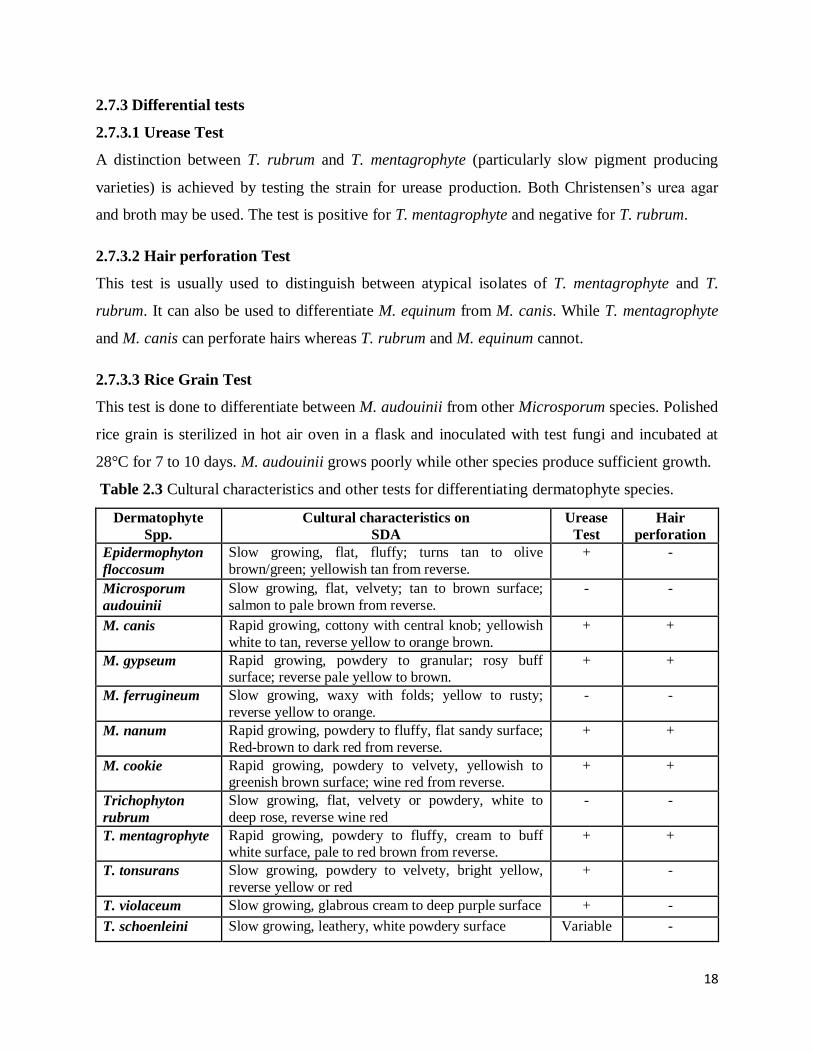
2.7.3 Differential tests
2.7.3.1 Urease Test
A distinction between T. rubrum and T. mentagrophyte (particularly slow pigment producing
varieties) is achieved by testing the strain for urease production. Both Christensen‟s urea agar
and broth may be used. The test is positive for T. mentagrophyte and negative for T. rubrum.
2.7.3.2 Hair perforation Test
This test is usually used to distinguish between atypical isolates of T. mentagrophyte and T.
rubrum. It can also be used to differentiate M. equinum from M. canis. While T. mentagrophyte
and M. canis can perforate hairs whereas T. rubrum and M. equinum cannot.
2.7.3.3 Rice Grain Test
This test is done to differentiate between M. audouinii from other Microsporum species. Polished
rice grain is sterilized in hot air oven in a flask and inoculated with test fungi and incubated at
28°C for 7 to 10 days. M. audouinii grows poorly while other species produce sufficient growth.
Table 2.3 Cultural characteristics and other tests for differentiating dermatophyte species.
Dermatophyte
Spp.
Cultural characteristics on
SDA
Urease
Test
Hair
perforation
Epidermophyton
floccosum
Slow growing, flat, fluffy; turns tan to olive
brown/green; yellowish tan from reverse.
+ -
Microsporum
audouinii
Slow growing, flat, velvety; tan to brown surface;
salmon to pale brown from reverse.
- -
M. canis Rapid growing, cottony with central knob; yellowish
white to tan, reverse yellow to orange brown.
+ +
M. gypseum Rapid growing, powdery to granular; rosy buff surface; reverse pale yellow to brown.
+ +
M. ferrugineum Slow growing, waxy with folds; yellow to rusty;
reverse yellow to orange.
- -
M. nanum Rapid growing, powdery to fluffy, flat sandy surface;
Red-brown to dark red from reverse.
+ +
M. cookie Rapid growing, powdery to velvety, yellowish to greenish brown surface; wine red from reverse.
+ +
Trichophyton
rubrum
Slow growing, flat, velvety or powdery, white to
deep rose, reverse wine red
- -
T. mentagrophyte Rapid growing, powdery to fluffy, cream to buff white surface, pale to red brown from reverse.
+ +
T. tonsurans Slow growing, powdery to velvety, bright yellow,
reverse yellow or red
+ -
T. violaceum Slow growing, glabrous cream to deep purple surface + -
T. schoenleini Slow growing, leathery, white powdery surface Variable -
19
2.8 Epidemiology of dermatophytosis
Epidemiology is important in control of infection and public health issues related to different
types of dermatophytosis. A detailed information of the various epidemiological factors (i.e. age,
sex, occupation of patient, cultural, environmental and geographical conditions etc.) facilitate in
better management of the diseases conditions. Although age, sex and the occupation have a little
impact on the frequency of dermatophytosis yet, these factors have correlation with the incidence
of the dermatophytosis. These factors are discussed below.
Age: Tinea capitis is common in children and rarely seen in adults. Higher incidence of
dermatophytosis have been reported in the age group of 20-40 years also (Sarma and Borthakur,
2007; Patel et al., 2010).
Sex: In countries like India males are more affected by dermatophytosis as compared to females.
This may be due to the occupational exposure of both the sexes (Singh and Beena, 2003a).
Occupational factors: Some studies have shown that occupation of the people also affect the
incidence of dermatophytosis. Laborers, farmers and industrial workers who carryout strenuous
physical works tend to be more prone to the dermatophytic infections.
Cultural factors: Large incidence of tinea corporis has been reported in Indian women as they
wear sarees. The relatively high frequency of tinea capitis due to poor scalp hygine has been
reported from South India. Tinea pedis is more common among the people who wear occlusive
footwear and use community washing facilities such as army camp, boarding schools etc.
Environmental conditions: These conditions play an important role in the incidence and
prevalence of dermatophytes in a particular region. The hot and humid climatic conditions of the
tropical and sub-tropical regions are best suited for the growth of the dermatophytes (Deshmukh
et al., 2010).
The prevalence and endemicity of a dermatophyte species in a particular region is the combined
effect of all the aforesaid factors as a specific pattern of the disease cannot be predicted on the
basis of a single factor. Also, it is difficult to establish overall incidence and prevalence of
various dermatophyte infections in different parts of the world because the studies of one region
20
of the country may not be a true reflection of the overall disease pattern of that country (Ameen,
2010).
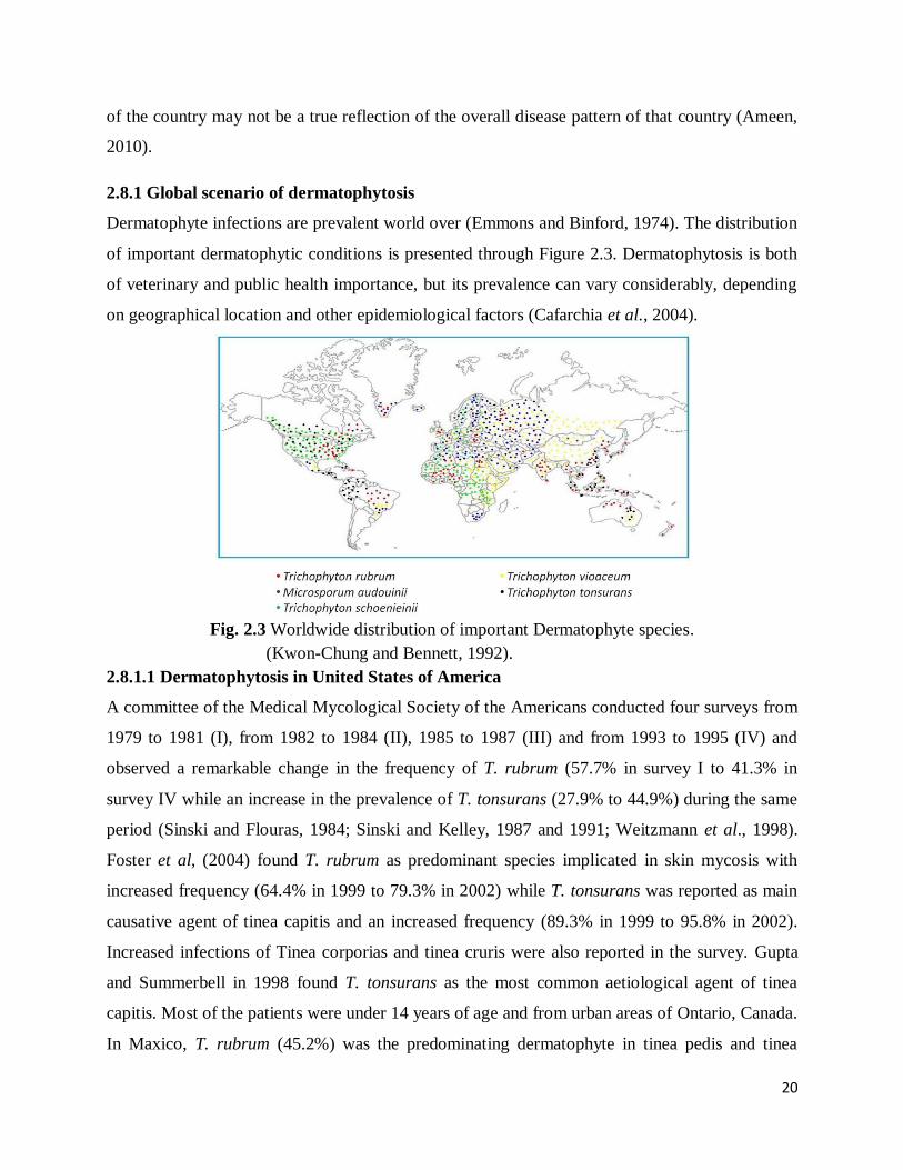
2.8.1 Global scenario of dermatophytosis
Dermatophyte infections are prevalent world over (Emmons and Binford, 1974). The distribution
of important dermatophytic conditions is presented through Figure 2.3. Dermatophytosis is both
of veterinary and public health importance, but its prevalence can vary considerably, depending
on geographical location and other epidemiological factors (Cafarchia et al., 2004).
Fig. 2.3 Worldwide distribution of important Dermatophyte species.
(Kwon-Chung and Bennett, 1992).
2.8.1.1 Dermatophytosis in United States of America
A committee of the Medical Mycological Society of the Americans conducted four surveys from
1979 to 1981 (I), from 1982 to 1984 (II), 1985 to 1987 (III) and from 1993 to 1995 (IV) and
observed a remarkable change in the frequency of T. rubrum (57.7% in survey I to 41.3% in
survey IV while an increase in the prevalence of T. tonsurans (27.9% to 44.9%) during the same
period (Sinski and Flouras, 1984; Sinski and Kelley, 1987 and 1991; Weitzmann et al., 1998).
Foster et al, (2004) found T. rubrum as predominant species implicated in skin mycosis with
increased frequency (64.4% in 1999 to 79.3% in 2002) while T. tonsurans was reported as main
causative agent of tinea capitis and an increased frequency (89.3% in 1999 to 95.8% in 2002).
Increased infections of Tinea corporias and tinea cruris were also reported in the survey. Gupta
and Summerbell in 1998 found T. tonsurans as the most common aetiological agent of tinea
capitis. Most of the patients were under 14 years of age and from urban areas of Ontario, Canada.
In Maxico, T. rubrum (45.2%) was the predominating dermatophyte in tinea pedis and tinea
21
unguium followed by T. mentagrophyte and T. tonsurans. The latter was the predominant cause
of tinea capitis particularly in children (Welsh et al., 2006).
2.8.1.2 Dermatophytosis in Europe
T. rubrum was the most common dermatophyte mainly associated with tines corporis and tinea
cruris infections in north and central Europe (Tietz et al., 1995; Kostanje and Staas, 1995;
Seebacher et al., 2008). Also, M. canis remains the most common causative agent of tinea capitis
in Europe (Ameen , 2010).
Slovakia: In Slovakia an increase in anthropophilic dermatophytes, especially T. rubrum and M.
canis and rapid decline of zoophilic dermatophytes like T. mentagrophytes and T. verrucosum
during 1956-1992 has been reported (Buchvald and Simaljakova, 1995).
France: T. rubrum was the predominant dermatophyte. Zoophilic dermatophytes such as M.
canis, T. mentagrophyte and T. verrucosum were reported with high frequency in rural regions of
north-east France while the involvement of anthropophiilic dermatophytes like T. tonsurans, and
T. soudanense were observed in urban areas especially in tinea capitis infections (Ginter-
Hanselmayer et al., 2007; Foulet et al., 2007).
Germany: E. floccosum and M. audouinii dominated among human dermatophytoses earlier but
after the Second World War T. rubrum emerged as predominant species with tinea pedis and
onychomycosis infections (Karrenberg, 1928; Gotz, 1952). T. rubrum accounted for the 80-90%
isolated dermatophyte species in Germany followed by T. mentagrophyte var. interdigitale and
E. floccosum respectively. Tinea pedis and tinea unguium are nowadays most frequently
diagnosed dermatophytosis (Seebacher et al., 2008). M. canis has been found as predominating
causative agent of tinea capitis followed by T. tonsurans, T. violaceum and T. mentegrophyte in
Germany (Tietz et al., 1999).
United Kingdom: The anthropophilic dermatophyte, T. tonsurans has been reported as leading
cause of tinea capitis in UK. Other anthropophilic dermatophytes isolated were: M. audouinii
and M. rivalieri (Fuller et al., 2003). The prevalence of tinea capitis in young school children in
south-east London has been reported (Hay et al., 1996). These workers also suggested guidelines
for treating this condition. In another study T. rubrum has been found to be the predominating
dermatophyte in U.K. followed by T. mentagrophyte and E. floccosum in UK along with the
tinea pedis as most prevalent skin infection (Borman et al., 2007).
22
Finland: T. rubrum (66%) has been reported as most common dermatophyte involved in various
skin infections followed by T. mentagrophyte (26%) and E. floccosum (6%) in northern Finland
during the period 1982-1990 (Lehenkari and Selvennoienen-Kassinen, 1995). During 1987-
1990, T. verrucosum infection was epidemic in cattle keepers. M. canis, T. violaceum and T.
terrestre were other dermatophytes which could be isolated on rare occasions only. Among the
tinea conditions in Finland, tinea pedis was the most common infection followed by tinea cruris.
Belgium, Netherlands and Sweden: An increase in the incidence of anthropophilic tinea capitis
caused by T. violaceum, T. soudanense and T. tonsurans has been reported (Kolivras et al.,
2003). Likewise, rise in the zoophilic dermatophytes like M. canis has also been reported
(Hallgren et al., 2004). A higher frequency of M. canis (9% to 40%) was reported during 1960-
1990. Kostanje and Staats (1994) reported the beginning of the period of T. verrucosum which
declined in mid 1970s.
Switzerland: T. rubrum most frequently isolated species (62.5%) followed by T. mentagrophyte
(24.5%) and M. canis (5.0%) (Monod et al., 2002). M. gypseum, T. soudanense, T. verrucosum,
T. tonsurans E. floccosum were other less frequently isolated dermatophytes.
Mediterranean countries: In Greece, T. rubrum was the most prevalent species (44.4%)
followed by M. canis (25%), T. mentagrophyte (3.4%) and T. verrucosum (1.8%) while among
tinea conditions, tinea pedis (25.7%) was most common infection followed by tinea corporis
(24.7%), tinea unguium (19.9%) and tinea capitis (11.3%) (Maraki and Tselentis, 1998). T.
rubrum was the main causative agent of onycomycosis (87.1%) in Italy while the prevalence of
T. mentagrophyte was to the extent of 10% (Romano et al., 2005). M. canis was the predominant
dermatophyte in human patients in Rome followed by M. audouinii during the period 2002 -2004
(Panasiti et al., 2007).
Poland: During a 12 years survey (1984-1985) it was observed that the most common
dermatophytosis was tinea cutis glabrae (32.3%) followed by tinea pedis (24%), onycomycosis
(16.5%), tinea capitis (11.7%), tinea inguinalis (8.9%) and tinea manus (4.0%) were other
conditions reported (Nowicki, 1996). Lange et al., (2004) reported M. canis (62%) and T.
rubrum (12%) as the most common dermatophytes involved during the period 1999- 2001. Tinea
cutis glabrae (42%) was the most prevalent dermatophytosis followed by tinea capitis (30%).
These workers also reported that T. rubrum and T. mentagrophyte were mainly involved in tinea
pedis and scalp lesions mostly in children of 4-7 years (Lange et al., 2004).
23
Russia: T. rubrum (71.3%) and T. interdigitale (28.7%) were the main fungal pathogens
involved in foot mycoses in workers at metallurgical plants (Mikhasik et al., 1990). Up to 83%
cases of tinea pedis were recovered in the Russian population (Katsambas et al., 2003). About
70% toe nail infections were caused by dermatophytes and the T. rubrum was the predominating
dermatophyte involved in these infections while 10% infections were due to yeasts and 7% by
moulds (Jarv et al., 2004). T. rubrum was reported as predominat dermatophyte causing 65-75%
of skin mycoses in the Russian Federation (Khaldin et al., 2005).
Solvenia: M. canis (46.8%) was the most frequently isolated dermatophyte followed by T.
rubrum (36.7%), T. mentagtrophyte var. interdigitale (7.9%) and T. mentagrophyte (4.9%)
during the period 1995-2002 in Solvenia. Tinea corporis, onycomycosis, tinea pedis and tinea
faciei were among the common skin infections (Dolenc-Voljc, 2005).
2.8.1.3 Dermatophytosis in Africa
Different types of clinical manifestations of dermatophytosis were reported in African
subcontinent in which different dermatophyte species were implicated as compared to Europe. T.
audouinii, T. violaceum and T. soudanense were the common dermatophytes in some African
countries. T. gourvilii is endemic in Africa and is mainly involved in tinea capitis. The black dot
infection caused by T. tonsurans and T. violaceum is also endemic in Africa. T. schoenleinii is
found in closely dense population of Africa. T. soudanense is commonly associated with tinea
capitis especially in north-western Africa. T. rubrum and T. mentegrophyte are less commonly
isolated and are particularly associated with tinea corporis and tinea cruris. In some parts of
Africa, tinea faciei and tinea barbae are caused by M. gypseum and M. ferrugineum (Havlickova
et al., 2008).
South Africa: T. violaceum (90% cases) was reported as the most common causative agent of
tinea capitis in school children of average age of 4.6 years in Kwa-Zulu in South Africa while
Black Dot was the most common clinical condition seen in 50% of patients (Morar et al., 2004).
In a study on HIV patients at a South African clinic demonstrated that herpes zoster (19%) and
tinea corporis (7%) were the most common cutaneous infections observed in HIV infected
patients (Morar et al., 2006).
Nigeria: M. audouinii (46.8%), T. mentegrophyte (25.5%), T. rubrum (21.3%), E. floccosum
(4.3%) and T. tounsurans (2.1%) were isolated from the different tinea conditions in school
children in Nigeria (Enweani et al., 1996).
24
Malawi: Tinea faciei (2.5%) followed by tinea corporis or cruris (1.5%) were diagnosed among
the northern Malawi population during the period, 1987-1989. M. audouinii (57%) was the most
predominant dermatophyte isolated while E. floccosum (56%) was commonly associated with
tinea cruris infection. T. rubrum (1%) was rarely involved (Ponnighaus et al., 1996).
Libya: 45.9% cases of tinea corporis and 8.1% cases of tinea pedis were reported from Libya.
Other infections included candidiasis and pityrisis versicolor (Ellabib et al., 2002). T. violaceum
was the most common dermatophyte responsible for 44% of dermatophyte infections. M. canis
(8.1%), E. floccosum (6.6%) and T. mentegrophyte (3.1%) were other dermatophyte species
involved.
Jordan: Tinea pedis (35%) and tinea cruris (10%) were more frequent during the summers
whereas tinea capitis (23%), tinea unguium (22%) and tinea corporis (10%) in spring and winter
seasons in Jordan (Abu-Elteen and Malek, 1999). T. mentegrophyte, E. floccosum, T. rubrum and
M. canis were the most common dermatophytes implicated in these dermatophytic conditions.
2.8.1.4 Dermatophytosis in Australia
20 isolates of T. viollaceum has been reported which were associated with tinea capitis and tinea
corporis at Melbourne in Australia during a period of 32 years (Malsen and Andrew, 1997).
Among school students, prevalence of tinea pedis was reported mainly in males than females. T.
mentegrophyte and T. rubrum were the most common dermatophytes isolated from these
conditions (Merlin et al., 1999).
2.8.1.5 Dermatophytosis in Asia
T. rubrum and T. mentagrophyte have been the most common dermatophyte species isolated
from different dermatophytic conditions. T. violaceum is the main causative agent of tinea capitis
and tinea corporis which are frequent in children and adolescents.
Iran: Tinea capitis (54.1%) most prevalent clinical form, followed by tinea corporis, tinea pedis
(8.9% each), tinea cruris (6.8%), tinea unguiumm (3.5%), tinea manuum (2.6%) and tinea barbae
(0.3%) in patients in Iran (Chadeganipour et al., 1997). T. verrucosum, E. floccosum, T.
mentegrophytes M. canis, T. violaceum and T. schoenleinii and T. rubrum species were isolated
from the dermatophytic infections.
In another study in Tehran (Iran) E. floccosum (31.4%) was found to be the most frequently
isolated dermatophyte followed by T. rubrum (18.3%), T. mentagrophyte (17.2%), T. violaceum
25
(16.6%), M. canis (6.5%), T. verrucosum (4.7%) and M. gypseum (4.1%) (Falahati et al., 2003).
In this study among the tinea conditions, tinea corporis (31.4%) was the most common
dermatophytic infection, followed by tinea cruris (20.7%), tinea manum (15.4%), tinea capitis
(12.4%), tinea pedis (10.6%), tinea faciei (7.1%) and tinea unguium (2.4%). Lari et al., (2005)
reported T. violaceum (28.3%), M. canis (15.1%), E. floccosum (15.1%), T. rubrum 13.2%, T.
mentagrophyte (11.3%) and T. verrucosum (5.7%) dermatophytes isolated from tinea capitis
(39.6%), tinea corporis (30.2%), tinea faciei (18.9%) and tinea manuum (7.5%) fron children
aged ≤ 16 years.
Turkey: In Turkey, 185/195 soldiers (20%) were found infected with superficial mycoses. Of
the infected soldiers 151 (60%) had dermatophytosis (Sasmaz et al., 2003). Celik et al., (2003)
observed superficial mycoses in 73 (16.9%) in a textile factory workers. Of these, 76.7% cases
were of dermatophytosis in which T. rubrum and T. mentagrophyte were the most common
dermatophytes implicated. Another study reported T. rubrum (56%) and T. mentagrophye (38%)
common dermatophytes isolated from various dermatophytic conditions while T. verrucosum, M.
canis and E. floccosum were the other dermatophytes involved at a university Hospital in
Turkish (Ozkutuk et al., 2007). Tinea pedis (47%) was the most common dermatophytosis
followed by tinea unguium (29%), tinea inguinails (15%), tinea corporis (7.4%) and tinea capitis
(1.6%) in this study.
Japan: In the year 2003, tinea pedis was observed in 25% of cases in Japan. Of these, 50% cases
had tinea unguium infection simultaneously (Ogasawara, 2002).
China: Increased incidence of zoophilic dermatophytes especially in tinea capitis has been
reported from China by Zhu et al., (2004) as is the cases in Europe. M. canis was identified as
the main pathogen involved (65% cases) in Shanghai. While T. violaceum (18%) and T.
tonsurans (9%) were other important anthropophilic dermatophytes involved. In northwestern
China, 15 % of suspected cases of fungal infection in a ten year study were found positive for
fungi (Tao-Xiang et al., 2005). T. rubrum (43.9%), T. mentagrophyte (29.4%) and Candida spp.
(14.0%) were the predominant fungi involved. T. pedis was the predominant clinical condition
followed by onychomycosis and tinea manuum. T. violaceum was observed as the most common
causative agent of tinea capitis in western China especially in rural communities (Deng et al.,
2008).
26
Korea: T. rubrum and other non-dermatophytic fungi like Trichosporon spp. and Candida spp.
were reported from various dermatomycoses in Korea. T. pedis was the most common clinical
condition (Kim et al., 2003). Increased incidence of tinea corporis was reported from Korea
(Jang et al., 2004). T. rubrum was the predominant species involved followed by T.
mentagrophyte and M. canis.
Singapore: The superficial fungal infections in Singapore during 1999-2003, tinea pedis
(27.3%) was the most common clinical condition caused by T. interdigitale while T. rubrum was
reported as most common causative agent in other tinea conditions (Tan, 2005).
2.8.2 Dermatophytosis in India
A study on clinically diagnosed cases of dermatophytosis revealed that, tinea corporis (34.62%)
was the commonest clinical type encountered, followed by tinea cruris (32.31%), tinea capitis
(6.93%), tinea pedis (1.53%) and tinea manuum (1.53%) (Siddappa and Mahipal, 1982).T.
rubrum was the predominant species (81.82%) isolated, followed by E. floccosum (9.09%), T.
violaceum (4.54%), M. audouinii (3.03%) and T. mentagrophytes (1.51%). Kumar and Lakshmi
in 1990 observed that the incidence of tinea capitis was more common in males (58%) than in
females (42%) and was more prevalent in children aged between 6-12 years (44%) at Tirupati in
Andhra Pradesh. T. violaceum was the predominant species isolated (63.15%) followed by M.
audouinii (13.15%), T. verrucosum (7.89%) and T. mentagrophytes (5.26%). Tinea capitis was
predominant in the age group of 1-10 years. Girls (60.3%) were more commonly affected than
boys. T. violaceum was the most common isolate (66.2%) followed by T. tonsurans (4.4%)
(Reddy et al., 1991). Kalla et al., (1995) from Jodhpur (Rajasthan) reported male to female ratio
in tinea capitis as 1.8:1. On culturing T. violaceum was the predominant (88.5%) fungus
recovered. In another study from Rajasthan, majority of the infections (64%) were found in the
age group of 0-30 years. Male to female ratio was 2:1. T. violaceum (55.7%) was the most
common isolate from all clinical types followed by T. rubrum (42.3%) (Karmakar et al., 1995).
In Tamil Nadu, the prevalence of dermatophytosis has been correlated to socio-economic status
of the patients. Majority of patients belonged to the very low income groups (35% and 34.2%
respectively) followed by middle income group (23.2%). T. rubrum was the most commonly
isolated species (52.2%), followed by T. mentagrophyte (29.35%) and E. floccosum (6.11%)
(Ranganathan et al., 1995). Most common isolate was T. rubrum (88.15%) in superficial
mycoses in upper Assam and male to female ratio was 1.86:1 (Huda et al., 1995). Mishra et al.,
27
(1998) observed maximum incidence of dermatophytosis in patients in the age group 15-35
years. Male to female ratio was 5.2:1. They found tinea versicolor (33.95%) as the most common
clinical type, followed by tinea corporis (24.55%) and tinea cruris (16.33%). T. rubrum was the
commonest fungus isolated in their study. Studies of Bindu and Pavithran (2002) at Calicut also
studied epidemiological factors in clinically diagnosed cases of dermatophytosis. Male to female
ratio was 2.06:1. Tinea corporis (54.6%) was the commonest clinical type followed by tinea
cruris (38.6%). T. rubrum was the predominant species isolated (66.2%) in all clinical types
followed by T. mentagrophytes (25%), T. tonsurans (5.9%) and E. floccosum (2.9%). Tinea
capitis was predominantly observed in male children. Madhuri et al., (2002) from Andhra
Pradesh reported majority of the cases of dermatophytosis between 21-40 years. Females
(51.96%) were slightly more than males (48.04%). Housewives (33.33%) were most frequently
affected and 48.04% had occupations associated with wet work, 21.57% had occupations
associated with increased physical activity. In Gujarat, Singh & Beena in 2003 observed that,
tinea corporis was the most common clinical presentation followed by tinea cruris. T. rubrum
(73.27%) was the most common isolate, followed by T. mentagrophytes (17.24%), E. floccosum
(7.75%) and T. violaceum (1.72%). Grover (2003) conducted a study on clinically diagnosed
patients of onychomycosis in which male to female ratio was 1.63:1. Among dermatophytes, T.
rubrum was the commonest isolate (42.9%), followed by E. floccosum (35.7%), T. tonsurans
(14.3%) and T. schoenleinii (7.1%). Vijaya et al., in 2004 from Karnataka reported male to
female rato of 51:49 in patients of onychomycosis. Toe nail infection (78.43%) was more
common in males than females while finger nail infection was more common in females
(85.71%) in their study. Also, T. rubrum (68.75%) was the commonest dermatophytes, followed
by T. mentagrophytes (25%) and E. floccosum (6.25%). In a similar clinical study of clinical
study of onychomycosis in central India, male to female ratio 3:1 was observed by Garg et al.,
(2004) in which the mean age was 29. Among the dermatophytes, T. rubrum (23.07%) was the
commonest isolate, followed by T. verrucosum. In a study on clinically diagnosed cases of
dermatophytosis, Kannan et al., (2006) observed that T. rubrum (70.83%) was the commonest
dermatophyte involved especially in tinea corporis followed by T. mentagrophytes and E.
floccosum. Sen and Rasul, (2006) demonstrated that the most common clinical type was tinea
corporis (48%) followed by tinea cruris (19%) and tinea unguium (11%) infection in Guwahati
(Assam). T. rubrum was found to be the most common causative agent (68.63%) followed by T.
28
mentagrophyte (23.53%) and E. floccosum (3.92%). T. rubrum (57.6%) was the most common
dermatophyte involved followed by T. mentagrophyte (42.3%) among clinical cases of
onychomycosis in Aurangabad (Veer et al., 2007). The commonest age group was 31 to 40 years
followed by 41 to 50 years. Male to female ratio was 1.8:1. The ratio of finger nail to toe nail
infection was 3:1. In Himachal Pradesh, the prevalence of onycomycosis among the age group of
8-76 years was observed by Gupta et al., (2007). The higher prevalence of onycomycosis was
observed among farmers and office workers. T. rubrum (32.6%), T. mentagrophytes (6.1%) and
T. verrucosum (2.1%) were the dermatophytes implicated in these conditions. Venkatesan et al.,
(2007) conducted a study on 90 clinically suspected cases of dermatophytosis in Chennai and
found T. rubrum (73.3%) as the commonest isolate followed by T. mentagrophyte (19.7%), E.
floccosum (4.2%) and M. gypseum (2.8%). Tinea corporis was the most common clinical type
(64.8%) followed by tinea cruris (26.8%), tinea pedis (5.6%) and Onychomycosis (2.8%). T.
rubrum was the predominant species responsible for chronic dermatophytosis (81.8%). T.
rubrum was the commonest isolate (84.21%) followed by E. floccosum (10.52%) and M.
gypseum (5.26%) in tinea cruris while T. rubrum was the commonest isolate (50%) followed by
T. mentagrophytes (25%) and E. floccosum (25%) in tinea pedis. Deshmukh and co-workers
(2010) isolated 14 species of keratinophilic fungi including dermatophytes from soil samples
from Ladakh (Himachal Pradesh). The prevalence was thus seen even in the cold desert of
Ladakh, there is prevalence of keratinophilic fungi as these fungi were tolerant and time adaptive
to various biotic and abiotic factors.
2.8.2.1 Miscellaneous dermatophytosis reported in India
Siddaramappa et al., (1991) reported a case of tinea favus of scalp due to T. schoenleinii in a 60
year old farmer in Belgaum (Maharashtra). Rai (1992) reported a case of tinea capitis in a 40
year old woman due to T. rubrum. Ghorpade and Ramanan (1995) reported a case of tinea capitis
in a 12 day old infant due to T. tonsurans in Bhilai (Chhattisgarh). Mittal and Shivali (1996)
reported a case of tinea faciei and tinea capitis in a 15-day old male infant caused by T.
tonsurans at Patiala, Punjab.
2.9 Pathogenesis of dermatophytosis
The dermatophytes grow within dead, keratinized tissue only. The fungal cells produce
keratinolytic proteases in vivo and in vitro which provide means of entry into living cells. The
29
fungal metabolic products diffuse through the Malphigian layer of epidermis to cause erythema,
vesicles and pustules. The fungal hyphae grow rapidly, become old, break into arthrospores and
are shed off from the epidermis. Dermatophytes colonize the horny layers of the skin, hair and
nails initially. The outcome of the disease depends on several other factors such as nature of the
host, strain of the dermatophyte, species variation and the site of infection. The dermatophytic
infection spreads centrifugally in all directions on the glabrous skin showing the classical
ringworm pattern (Chander, 1995). However, it is still not clear how dermatophytic fungi
regulate the utilization of different proteases against various cornified layers of the host in
addition to the other roles played by these proteins especially in adherence or
immunomodulation etc. When compared to the other fungal diseases, dermatophytes infect the
immunocompetent patients irrespective of the other fungal diseases where the incidence is more
in immunocopromised patients. The dermatophytic infection can be acute and may rapidly be
eliminated through efficient host immune responses (Vermount et al., 2007).
2.10 Virulence factors of dermatophytes
2.10.1 Adherence to host superficial skin tissues and their invasion
Spores adhere to the host tissue and germinate into hyphae which grow in multiple directions
invading the stratum corneum (Aljabre et al., 1992). The ability to adherence in case of T.
rubrum to epithelial cells has been associated with carbohydrate-specific adhesions expressed on
the surface of the microconidia. The fibrillar projections have also been observed in T.
mentegrophyte during the phase of adhesion (Esquenazi et al., 2004; Kaufman et al., 2007).
2.10.2 Proteases and growth of dermatophytes on keratinized substrate
Different dermatophyte species have different proteases which digest the keratin network into
assimilable oligopeptides and aminoacids. These include: multiple serine (subtilisins), metallo-
endoproteases, lipases and ceramidases. A direct relationship exists between keratinases and
pathogenicity (Viani et al., 2001). High levels of fungal proteases are produced when the sole
available carbon and nitrogen source is made of complex proteins as opposed to glucose or
peptidic digests (Jousson et al., 2004).
2.10.3. Factors encountering immune mechanisms
Dermatophyte infection induces a specific humoral as well and cellular immune response. The
cell mediated response is known as main protective and efficient response against dermatophytic
30
infection which is a kind of delayed type hypersensitivity (DTH) characterized by effector
macrophage cells and cytokines like interferon-γ (INF-γ). Different dermatophytes adopt
different ways to counter the immune defenses. They include lymphocyte inhibition by cell wall
mannans, alteration of macrophage function, differential activation of keratinocytes and
differential secretion of proteases (Giddey et al., 2007). Some dermatophyte species produce
mannans, a glycoprotein constituent of cell wall, (T. rubrum cell wall mannans (TRM) with
interfere RNA synthesis or RNA functions that are necessary for presentation of antigen to
appropriate T cells (MacCarthy et al., 1994).
2.11 Diagnosis
The Laboratory diagnosis of dermatophytosis is based on of clinical history, signs, and
symptoms, typical lesions and laboratory investigations. The history of the patient (e.g. age, sex,
occupation, living style, duration of infection, previous treatment if taken, any etc.) plays an
important role in the diagnosis of the dermatophytosis.
2.11.1 Clinical examination
The clinical examination includes recording of the type, shape, size and site of lesions and other
signs and symptoms related to the infection. These observations help clinicians to diagnose the
type of tinea condition which is further supported by laboratory investigations.
Wood’s lamp-direct examination
Wood‟s lamp is used for the detection of fungal infection of hairs. Wood‟s glass consists of
barium silicate with 9% nickel oxide. It transmits ultraviolet light of longer wavelength
(365 nm). The pathogenic fungi commonly Microsporum species shows florescence when this
light falls on them. The presence of chemical substance pteridine, in the fungi is responsible for
this florescence. Different fungi produce different types of fluorescence a. Bright green: M.
audouinii, M. canis and M. ferrugineum b. Dull green: T. schoenleinii c. Golden yellow:
Pityriasis versicolor (Malessezia furfur) d. No florescence: all other dermatophytes. The Wood‟s
lamp is helpful for rapid detection of pathogenic fungi especially in the scalp region on a large
population. However, it has become less useful nowadays because of the increased involvement
of non-fluorescing fungi such as T. tonsurans, T. verrucosum and other dermatophyte species
implicated in tinea capitis (Chander, 1995).
31
2.11.2 Laboratory examinations
The laboratory examination involves the isolation of the dermatophyte species on suitable media
and its identification which is based on morphology and microscopic examination of the fungal
pathogen. The molecular methods further support the diagnosis. The samples of skin and nail
scrapings, infected hair follicles are collected in sterile containers and analyzed in the laboratory.
2.11.2.1 Direct potassium Hydroxide (KOH) preparation
The collected samples are mounted on a clean glass side with a drop of 10% KOH solution in
case of skin scrapings and 40% in case of nail samples. The slides are examined under light
microscope for the presence of fungi. The details of the procedure are explained in research
methodology under section 3.2.
2.11.2.2 Gross morphology of dermatophyte species
The morphology of different dermatophyte species has been detailed in Table 2.3.
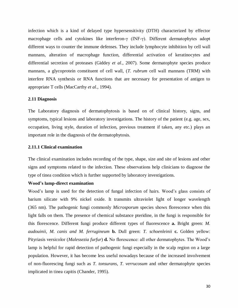
2.11.2.3 Microscopic examination of lactophenol cotton blue stained preparations
A small portion of aerial mycelium is stained in lactophenol cotton blue (LCB), covered with
cover slip and observed under microscope for looking at the microscopic morphology of the
dermatophytes of different genera (Fig. 2.4 a-c).
i. Trichophyton species: Both microconidia as well as macroconidia are found in Trichophyton
species. Microconidia are predominant spore forms and are found in abundance. They are
arranged singly or in clustures along the hyphae. The macroconidia are sparse, smooth walled,
pencil-shaped with blunt ends. They may have 1-12 septations (Fig.2.4, a).
ii. Microsporum species: the hyphae are large and rough-walled with multicellular, spindle
shaped conidia which are formed at the ends of hyphae. The macroconidia are abundant and may
have 1-15 septations. Microconida are lesser in number (Fig.2.4 b).
iii. Epidermophyton species: The members of this genus do not produce microconidia. The
macroconidia are smooth thin walled, pear or club shaped. The hyphae are septate and bifurcated
at the ends (Fig.2.4 c).
32
Fig. 2.4 (a-c) Microscopic view of hyphal arrangement in different dermatophyte species
a. Trichophyton species (Large, smooth, thin walled, septate, pencil shaped
hyphae are visible) b. Thick walled, spindle shaped, multicellular hyphae of
Microsporum species c. Bifurcated hyphae with multiple, smooth, club
shaped macroconidia of Epidermophyton species.
Species within each genus has peculiar microscopic morphology with features that differentiate
between them (Fig. 2.5 to Fig. 2.9).
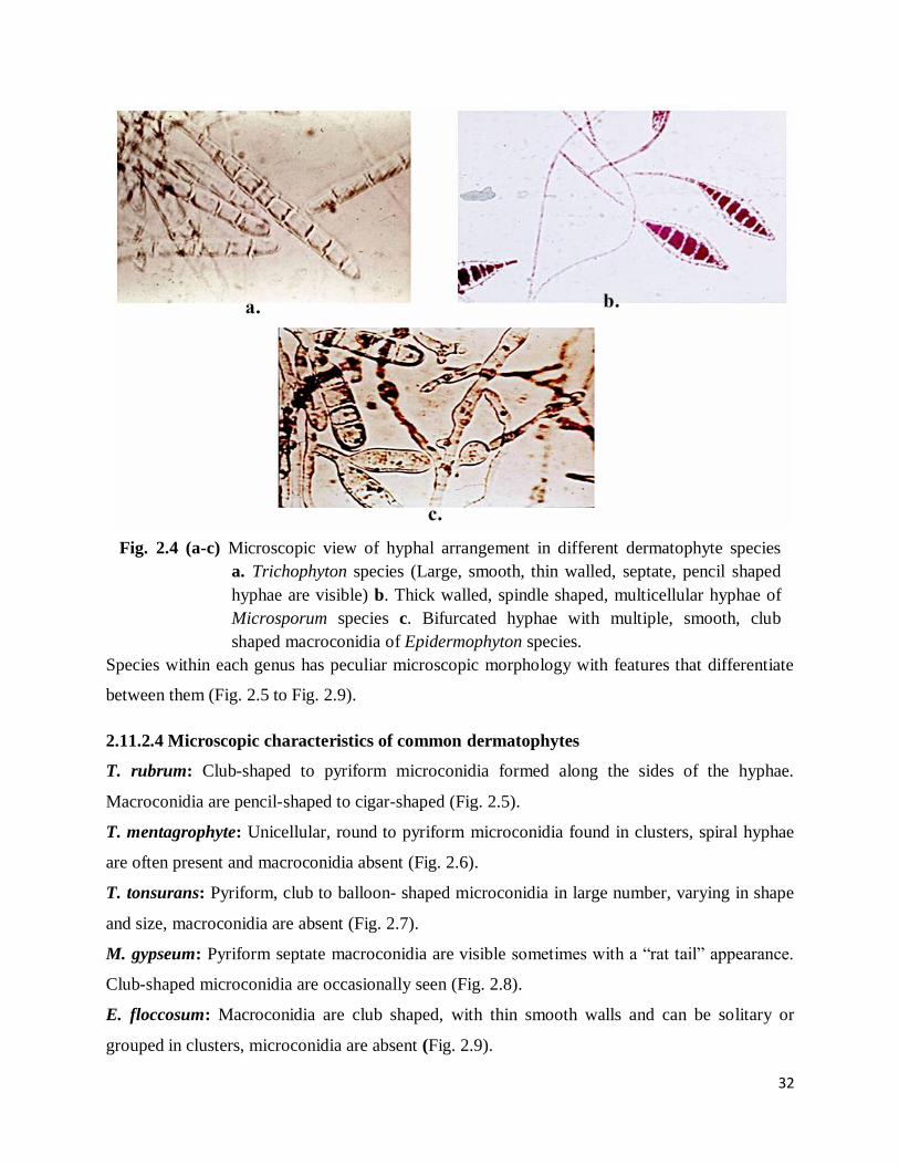
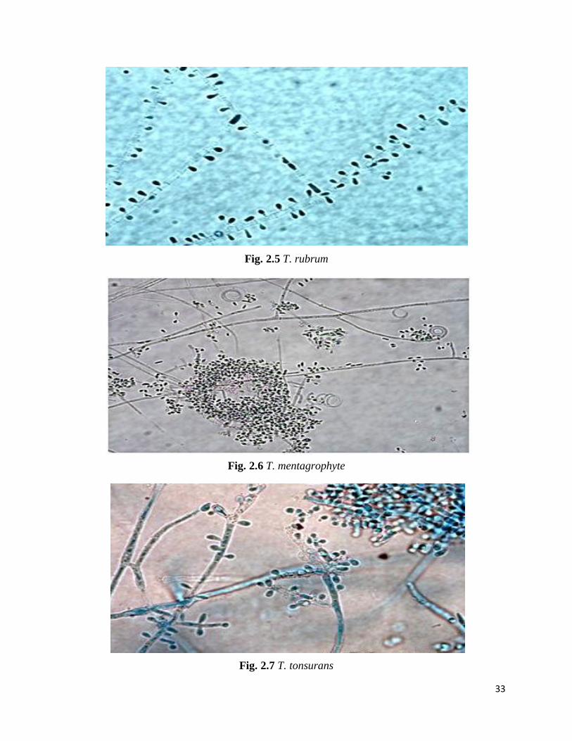
2.11.2.4 Microscopic characteristics of common dermatophytes
T. rubrum: Club-shaped to pyriform microconidia formed along the sides of the hyphae.
Macroconidia are pencil-shaped to cigar-shaped (Fig. 2.5).
T. mentagrophyte: Unicellular, round to pyriform microconidia found in clusters, spiral hyphae
are often present and macroconidia absent (Fig. 2.6).
T. tonsurans: Pyriform, club to balloon- shaped microconidia in large number, varying in shape
and size, macroconidia are absent (Fig. 2.7).
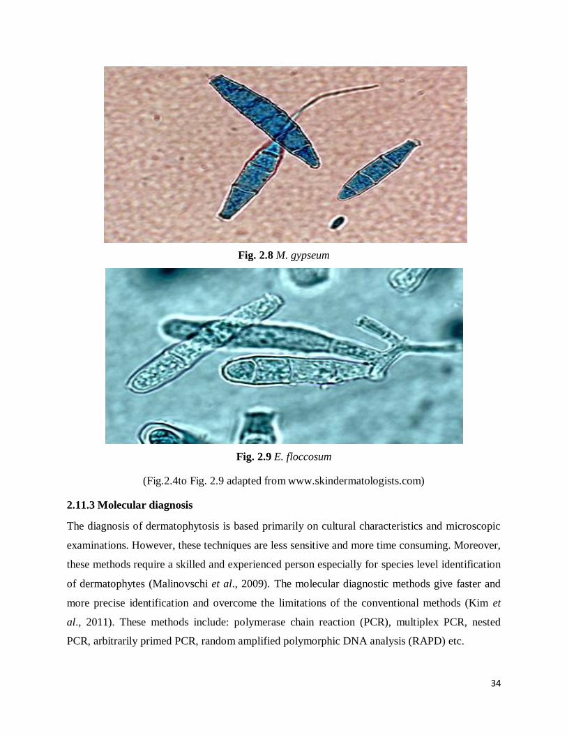
M. gypseum: Pyriform septate macroconidia are visible sometimes with a “rat tail” appearance.
Club-shaped microconidia are occasionally seen (Fig. 2.8).
E. floccosum: Macroconidia are club shaped, with thin smooth walls and can be solitary or
grouped in clusters, microconidia are absent (Fig. 2.9).
33
Fig. 2.5 T. rubrum
Fig. 2.6 T. mentagrophyte
Fig. 2.7 T. tonsurans
34
Fig. 2.8 M. gypseum
Fig. 2.9 E. floccosum
(Fig.2.4to Fig. 2.9 adapted from www.skindermatologists.com)
2.11.3 Molecular diagnosis
The diagnosis of dermatophytosis is based primarily on cultural characteristics and microscopic
examinations. However, these techniques are less sensitive and more time consuming. Moreover,
these methods require a skilled and experienced person especially for species level identification
of dermatophytes (Malinovschi et al., 2009). The molecular diagnostic methods give faster and
more precise identification and overcome the limitations of the conventional methods (Kim et
al., 2011). These methods include: polymerase chain reaction (PCR), multiplex PCR, nested
PCR, arbitrarily primed PCR, random amplified polymorphic DNA analysis (RAPD) etc.
35
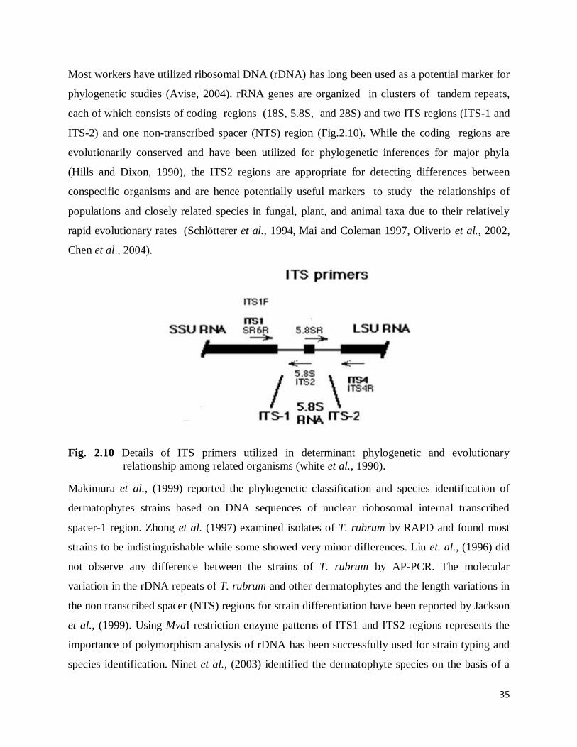
Most workers have utilized ribosomal DNA (rDNA) has long been used as a potential marker for
phylogenetic studies (Avise, 2004). rRNA genes are organized in clusters of tandem repeats,
each of which consists of coding regions (18S, 5.8S, and 28S) and two ITS regions (ITS-1 and
ITS-2) and one non-transcribed spacer (NTS) region (Fig.2.10). While the coding regions are
evolutionarily conserved and have been utilized for phylogenetic inferences for major phyla
(Hills and Dixon, 1990), the ITS2 regions are appropriate for detecting differences between
conspecific organisms and are hence potentially useful markers to study the relationships of
populations and closely related species in fungal, plant, and animal taxa due to their relatively
rapid evolutionary rates (Schlötterer et al., 1994, Mai and Coleman 1997, Oliverio et al., 2002,
Chen et al., 2004).
Fig. 2.10 Details of ITS primers utilized in determinant phylogenetic and evolutionary
relationship among related organisms (white et al., 1990).
Makimura et al., (1999) reported the phylogenetic classification and species identification of
dermatophytes strains based on DNA sequences of nuclear riobosomal internal transcribed
spacer-1 region. Zhong et al. (1997) examined isolates of T. rubrum by RAPD and found most
strains to be indistinguishable while some showed very minor differences. Liu et. al., (1996) did
not observe any difference between the strains of T. rubrum by AP-PCR. The molecular
variation in the rDNA repeats of T. rubrum and other dermatophytes and the length variations in
the non transcribed spacer (NTS) regions for strain differentiation have been reported by Jackson
et al., (1999). Using MvaI restriction enzyme patterns of ITS1 and ITS2 regions represents the
importance of polymorphism analysis of rDNA has been successfully used for strain typing and
species identification. Ninet et al., (2003) identified the dermatophyte species on the basis of a
36
DNA sequence encoding a part of the large sub-unit (LSU) rRNA (28S rRNA) by using the
MicroSeq D2 LSU rRNA fungal sequencing kit. Two taxa causing distinct dermatophytosis were
clearly distinguishable among isolates of the T. mentagrophytes species complex. Li et al.,
(2008), identified dermatophyte species using sequencing of ribosomal internal transcribed
spacer (ITS1) and (ITS2) regions. The identification rate in their study was > 97%. ITS
sequencing was found as very accurate and useful method for the identification of
dermatophytes. The use of PCR based single-strand conformation polymorphism (SSCP)
analysis was made by Cafarchia et al., (2009). This mutation scanning approach proved very
effective, together with the potential of ITS1, ITS2 and chs-1 as markers for the specific or
genomic identification of dermatophytes. Microsporum canis and Trichophyton tonsurans were
identified using rapid PCR based analysis by Malinovschi et al., (2009). The primers used were
ITS1 and ITS4 which amplify the variable ITS1 and ITS2 sequences surrounding the 5.8S-
coding sequence and situated between the small subunit-coding sequence and large subunit-
coding sequence of the ribosomal operon. The reaction was performed on 17 samples of
dermatophytes; T. tonsurans, T. rubrum (9), T. mentagrophyte (3), M. canis (2), M. gypseum (1)
and one unidentified fungi. The reproducible results were observed for both M. canis and T.
tonsurans without cross reaction with other fungi showing the species specificity of the method.
Kim et al., (2011) utilized multiplex polymerase chain reaction for identifying different
dermatophyte species. This method allows more than two target DNA molecules to be amplified
with more than two primers. Specially designed primers containing ITS-1 and 2, 18S rRNA and
28SrRNA regions were used for the analysis. The reaction was performed on 11 standard strains
and directly on scales obtained from 73 patients. All standard strains were successfully identified
and among 73 samples 69 were identified as T. rubrum, while T. mentagrophyte (1), T. tonsurans
(2), M. gypseum (1) were also observed. This method is useful in early and precise identification
of the dermatophyte species.
2.12 Therapeutics
The confirmation of dermatophyte infections is very essential before prescribing suitable
therapy. The spontaneous healing of dermatophytosis is uncommon which makes the
implementation of treatment more important. A variety of antifungal agents are available in the
market. However, the selection of the appropriate antifungal is very important for the effective
treatment. Dermatophytes are located in the stratum corneum within the keratinocytes. The

37
lesions appear in infected individuals due to the acute and chronic inflammatory changes that
appear in the dermis. Therefore, in order to ensure that the topical antifungals work efficiently,
these agents should have the ability to penetrate the cells of stratum corneum. Also, the agents
must have non-irritant action and should be well-tolerable to the patients. Skin lesions located on
face, trunk and limbs usually require 2 to 3 weeks therapy. Inflammatory reaction of tinea pedis
and capitis should be treated for 4 to 6 weeks and the hyperkeratotic lesions of palms and soles,
and the infection of nails are best treated with oral antifungals as the etiological agents causing
nail infection do not respond to the topical treatment (del Palacio et al., 2008).
Azoles (ketoconazole), Triazoles (itraconazole, fluconazole, vericonazole), Allylamines
(terbinfine), Griseofulvin (From miscellaneous class) are some of the common classes of
antifungal agents used for the treatment of dermatophytosis.
Griseofulvin is the drug of choice against dermatophytosis since 1950s. This drug is more
effective than fluconazole and is equally effective as compared to itraconazole and terbinafine.
Griseofulvin has advantages over other antifungal agents in that, it is much cheaper and has no
side effect even at higher doses. The efficacy of this drug can be increased by simultaneous
ingestion of fatty food. Both oral as well as topical forms of griseofulvin are available
(Develoux, 2001; Gonzalez et al., 2007).
2.12.1 Oral Triazoles
Fluconazole is one of the triazoles and is generally used as third-line of treatment in cases of
dermatophytes especially the infection of hairs and nails. Itraconazole is second best antifungal
nearly in every fungal infection. However, this drug has some adverse effects such as nausea,
vomiting and headache (Yang et al., 2008; Elkeeb et al., 2010). Vericonazole is third generation
triazole. It is effective against dermatophytes and is available in oral formulations as well as
intravenous cyclodextrin suspension (Radford et al., 1998).
2.12.2 Topical Azoles
Topical azoles are cheaper, potent having broad spectrum activity against dermatophytes, moulds
and yeasts. In inflammatory tinea infections, the use of azoles in combination with
corticosteroids has been recommended. A large number of topical azole derivatives are available
in the market: bifonazole, butoconazole, clotrimazole, coroconazole, fenticonazole, flutrimazole,
38
isoconazole, ketoconazole, oxiconazole, omoconazole, econazole, terconazole (del Palacio et al.,
2008).
2.12.3 Allylamines
Both oral as well as topical formulations are used for treating tinea conditions. Terbinafine
belonging to this class is the drug of choice which is given orally in nail infections as well as in
tinea conditions that respond poorly with topical or first line of treatment. The use of oral
terbinafine is restricted in children below the age of 2 years. It is also an effective drug for the
treatment of tinea pedis in adults (Millikan, 2010). Naftifine, terbinafine and butinfine are topical
derivatives of allylamines and benzylamines. The topical derivatives of allyalmines possess
inherent anti-inflammatory activity. The butinfine is an effective topical agent against
dermatophytes as well as C. albicans (Brennan and Leyden, 1997).
2.12.4 Morpholines and ciclopirox
These drugs have a broad spectrum of antifungal activity and are capable of penetrating both
glaborous skin and nail plates. They are effective against tinea pedis and tinea unguium
infections but have certain side effects. Amorolfine is a derivative of morpholines and is
available in topical formulations (de Padua et al., 2008).
2.13 Antifungal susceptibility testing
Large number of antifungal agents has been introduced during past two decades for treating
dermatophytosis (Chadeganipor et al., 2004). Different susceptibility patterns of different
dermatophyte species to various antifungal agents have been reported. Different methods have
been employed by different groups of researchers for determining the in vitro antifungal
susceptibility to new and existing antifungal agents. These methods include; broth macro and
microdilution methods, agar dilution, E test, colorimetric microdilution, disk diffusion method
etc. (Perera et al., 2001; Fernandez-Torres et al., 2003, Karaca and Koc, 2004; Santos and
Hamdan, 2005). Clinical and Laboratory Standards Institute (CLSI) published a reference
method M38-A2 document in 2008, in which the protocol for determining the MICs of several
antifungal agents against filamentous fungi including dermatophytes was mentioned. Araujo et
al., (2009) tested antifungal activities of fluconazole, itraconazole, ketoconazole, terbinafine and
griseofulvin by broth microdilution technique, against dermatophyte species recovered from
nails and skin specimens from Goiania city in Brazil. The low MIC values 0.03 µg/ml were
39
found for 33.3, 31.6 and 15% of isolates for itraconazole, ketoconazole and terbinafne,
respectively. Adimi et al., (2013) and co workers evaluated the efficacy of ten antifungal agents
(fluconazole, itraconazole, ketoconazole, terbinafine and griseofulvin, voriconazole,
clotrimazole, ciclopirox olamine, amorolfine and naftifine) against large number of
dermatophyte strains using CLSI broth microdilution method (M38-A). Itraconazole and
terbinafine were found highly effective as compared to other antifungal agents while fluconazole
was found least effective in their study. Yadav et al., (2013) determined the susceptibility of the
clinical isolates of dermatophytes using commercially available antifungal disks (Himedia
10ug/disk) of griseofulvin, miconazole, terbinafine, clotrimazole, fluconazole and ketoconazole
in the disk diffusion method. Clotrimazole was found the best antimycotic agent against
dermatophytes followed by miconazole and ketoconazole. Similarly, Nweze et al., (2010) also
used disk diffusion method for determining the susceptibility of dermatophyte isolates against
eight antifungal agents. These researchers conducted that disk diffusion method was
reproducible, simple and could be used to determine the antifungal susceptibility of
dermatophytes. Robledo-Leal et al., (2012) followed M38-A2 protocol of CLSI for determining
the susceptibility to dermatophyte species against thiabendazole (TBZ) and fluconazole (FCZ).
TBZ showed a significantly greater potency than FCZ against all isolates tested. The MIC50 and
MIC90 values for the TBZ were lower and similar for all dermatophyte species tested the values
for FCZ were found to be higher.
2.14 Prevention and control
Prevention and control of dermatophytosis should be considered keeping in view the area
invaded by the dermatophyte species involved and the source of infection.
In case of tinea capitis caused by M. canis or M. audouinii, the infected hairs can be screened
with the help of wood‟s lamp as the fluorescent hairs would glow in the dark under UV light.
While in case of nonflurescent tinea capitis (T. tonsurans infection) detection is difficult. In such
cases, the scalp should be carefully examined checked for the presence of spotty alopecia and
lesions. In addition, culturing of the sample from infected area may be performed using hair
brush technique (Mackenzie, 1963). As tinea capitis is prevalent among young children, routine
inspection of their scalps should be performed at the beginning of the school. The outbreaks in
school or institutions if any should be reported to the proper authorities. Infected persons must be
40
instructed not to share their comb, hair brushes, scissors, hair bands etc. with others, employ
good hygiene and must be treated promptly to prevent further spread of the infection.
There must be the investigation of nosocomial outbreaks of dermatophyte infection (Arnow et
al., 1991; Kane et al., 1988; Shah et al., 1988). The personnel handling the infants should screen
the area for florescence using wood‟s lamp and the source of infection must be investigated. The
healthcare workers handling the infants infected during an outbreak must follow the infection
control measures such as wearing long sleeves, protective clothing, long gloves etc. (Mossovitch
et al., 1986).
Anthrophilic dermatophytes causing tinea corporis and tinea cruris infections can be transmitted
through the infected clothes, towels and bedding of the infected patient. Such items should be
disinfected after the use by the patient and should not be shared by others. The individuals
infected with tinea corporis should avoid the close contact sports such as wrestling (Stiller et al.,
1992).
The infections caused by the zoophilic dermatophytes such as M. canis and T. mentagrophyte
can be detected using woods lamp and the animal reservoir must be separated from the herd and
must be treated promptly. Good hygiene, sanitation and fungicidal spray are useful in controlling
such infections. Use of griseofulvin topically could be more effective in control of such
infection. The human infections with zoophilic dermatophytes may be prevented by wearing
protective cloths especially cloves while handling such animals (dogs, cats and other pets etc.).
Good foot hygiene (regular washing of feet, proper drying and application of foot powder),
wearing washed shocks, avoiding excessive moister in the feet, avoiding trauma to feet etc. may
enhance the prevention of tinea pedis.