2 -dependentdesensitizationoftrpv2channels ... · ca2-dependentdesensitizationoftrpv2channels...
TRANSCRIPT

Cellular/Molecular
Ca2�-Dependent Desensitization of TRPV2 ChannelsIs Mediated by Hydrolysis of Phosphatidylinositol4,5-Bisphosphate
Jose Mercado,1 Ariela Gordon-Shaag,3 William N. Zagotta,1,2 and Sharona E. Gordon1
1Department of Physiology and Biophysics, 2Howard Hughes Medical Institute, University of Washington, Seattle, Washington 98195, and 3Department ofOptometry and Vision Science, Hadassah Academic College, Jerusalem 91010, Israel
TRPV2 is a member of the transient receptor potential family of ion channels involved in chemical and thermal pain transduction. Unlikethe related TRPV1 channel, TRPV2 does not appear to bind either calmodulin or ATP in its N-terminal ankyrin repeat domain. Inaddition, it does not contain a calmodulin-binding site in the distal C-terminal region, as has been proposed for TRPV1. We have foundthat TRPV2 channels transiently expressed in F-11 cells undergo Ca 2�-dependent desensitization, similar to the other TRPVs, suggestingthat the mechanism of desensitization may be conserved in the subfamily of TRPV channels. TRPV2 desensitization was not altered inwhole-cell recordings in the presence of calmodulin inhibitors or on coexpression of mutant calmodulin but was sensitive to changes inmembrane phosphatidylinositol 4,5-bisphosphate (PIP2 ), suggesting a role of membrane PIP2 in TRPV2 desensitization. Simultaneousconfocal imaging and electrophysiological recording of cells expressing TRPV2 and a fluorescent PIP2-binding probe demonstrated thatTRPV2 desensitization was concomitant with depletion of PIP2. We conclude that the decrease in PIP2 levels on channel activationunderlies a major component of Ca 2�-dependent desensitization of TRPV2 and may play a similar role in other TRP channels.
IntroductionDesensitization of sensory neurons is particularly important con-sidering their crucial role in the physiological perception of andreaction to the external environment. A remarkable example ofthis type of mechanism is the capsaicin sensitivity of peripheralnociceptors mediated by TRPV1. Topical application of capsa-icin to skin causes desensitization of TRPV1 channels expressedin these neurons rendering them less responsive to noxious stim-uli (Szallasi and Blumberg, 1999). It is, therefore, through TRPV1desensitization that peripheral neurons can adapt to painful ther-mal and chemical stimuli.
TRPV1, as well as many other members of the TRP family,shows a Ca 2�-dependent desensitization, inhibition, or inactiva-tion (Zhu and Birnbaumer, 1998; Clapham, 2002). This conserva-tion of function is remarkable, suggesting a common molecularmechanism for desensitization among TRP receptors. Despitethe strikingly similar desensitization among members in the TRPsuperfamily and the importance of this form of regulation, recentstudies (Juvin et al., 2007; Phelps et al., 2010) have suggested that
TRPV2, a channel closely related to TRPV1, does not exhibitdesensitization. Consistent with this idea, a series of molecularstudies have shown that TRPV2 does not share the regulatorydomains suggested to be important for desensitization of otherTRPV channels (Fig. 1). For example, it has been reported thatCa 2�/calmodulin (Ca 2�/CaM), an ubiquitous calcium sensor,may play a role in TRPV1 Ca2�-dependent desensitization by (1)binding to the N-terminal ankyrin repeat domain (ARD) (Rosen-baum et al., 2004), (2) competing with ATP for a nucleotide-bindingsite in the ARD (Lishko et al., 2007), and (3) directly binding to adistal C-terminal region (Numazaki et al., 2003) (Fig. 1). In contrast,biochemical studies suggest that the TRPV2 ARD does not appear tobind either CaM or ATP (Lishko et al., 2007; Phelps et al., 2010).Furthermore, the TRPV2 C-terminal region does not show conser-vation of the proposed TRPV1 C-terminal CaM-binding site (Fig.1). It has also been shown that Ca 2� influx through TRPV1 causesdepletion of phosphatidylinositol 4,5-bisphosphate (PIP2) dur-ing desensitization, and the recovery of the channel from desen-sitization requires resynthesis of the lipid (Liu et al., 2005; Klein etal., 2008). A proximal and distal C-terminal region have beenproposed to mediate PIP2 binding in TRPV1 (Prescott and Julius,2003; Brauchi et al., 2007), but homologous PIP2-binding sites inTRPV2 are either absent (distal) or have not been studied (prox-imal) (Fig. 1).
Here, we report that TRPV2 undergoes Ca 2�-dependentdesensitization similar to TRPV1. This makes TRPV2 an idealexperimental model to study the molecular mechanism of Ca 2�-dependent desensitization since it does not contain the bindingsites for CaM, ATP, or PIP2 but still shows the prototypical Ca 2�-dependent desensitization. We show that, although CaM binds in
Received April 23, 2010; revised Aug. 6, 2010; accepted Aug. 11, 2010.This work was supported by National Institutes of Health (NIH) Grant R01EY017564 (S.E.G.), NIH Grant
2R01EY010329-16 (W.N.Z.), and National Eye Institute Core Grant for Vision Research P30EY01730. This work wasalso supported by the University of Washington Vision Training Grant 5T32EY007031-33 (J.M.) and the HowardHughes Medical Institute. We thank David Julius (University of California, San Francisco, San Francisco, CA) forproviding the TRPV2 cDNA, Dr. Mark Lemmon for providing the PLC�1-PH domain cDNA, and Dr. John Addelman forproviding the CaM1234 cDNA. We thank Mika Munari for her excellent technical assistance and Drs. Luis FernandoSantana and Manuel Navedo for technical assistance in the combined imaging and electrophysiology experiments.
Correspondence should be addressed to Dr. Sharona E. Gordon, University of Washington, 1705 Northeast PacificStreet, HSB I-312, Seattle, WA 98195. E-mail: [email protected].
DOI:10.1523/JNEUROSCI.2108-10.2010Copyright © 2010 the authors 0270-6474/10/3013338-10$15.00/0
13338 • The Journal of Neuroscience, October 6, 2010 • 30(40):13338 –13347

vitro to a novel binding site in a TRPV2 C-terminal fragment in aCa 2�-dependent manner, CaM binding may not be functionallycoupled to TRPV2 desensitization. In contrast, we determinedthat decreases in membrane PIP2 levels on channel activationunderlie a major component of the Ca 2�-dependent desensitiza-tion observed in TRPV2 channels. We propose that decreases inmembrane PIP2 levels are responsible for much, if not all, theCa 2�-dependent desensitization seen in TRPV2.
Materials and MethodsCell culture and transfection. F-11 and human embryonic kidney tsA201-cells were cultured and maintained as previously described (Klein et al.,2008). Cells were transiently transfected with Lipofectamine 2000 (In-vitrogen) according to the manufacturer’s instructions and used for elec-trophysiology 1– 4 d after transfection.
Electrophysiology. Whole-cell patch-clamp measurements were obtainedat a continuous holding potential of �60 mV. After obtaining stable whole-cell recordings, negative holding pressure was applied, and the cell was liftedfrom the coverslip and positioned in front of the perfusion pipette. Accessresistance was compensated �80% using amplifier circuitry. Whole-cell ca-pacitance values were obtained from the amplifier settings. Solutions wereapplied using a “sewer pipe” solution changer controlled by RSC-200 (Bio-Logic). The extracellular solution consisted of Hanks buffered salt solutioncontaining the following (in mM): 140 NaCl, 4 KCl, 1.8 CaCl2, 1 MgCl2, 10HEPES, 5 glucose, pH 7.4. For Ca2�-free solution measurements, the extra-cellular solution contained the following (in mM): 145 NaCl, 5 KCl, 1.5MgCl2, 1 EGTA, 10 HEPES, 10 glucose, pH 7.4.
Recording electrodes (1– 4 M�) were fabricated from filamented bo-rosilicate glass pipettes (Sutter Instrument) using a multistage puller(Flaming-Brown model P-97; Sutter Instrument). Recording pipetteswere filled with the following (in mM): 110 K-aspartate, 30 KCl, 10 NaCl,1 MgCl2, 0.050 EGTA, 10 HEPES, 3 Mg-ATP, 0.1 GTP, pH 7.2. EGTAwas replaced with 10 mM 1,2-bis(2-aminophenoxy)ethane- N, N,N�,N�-tetra-acetic acid (BAPTA) for Ca 2�-free solution measurements.
For the excised patch experiments, inside-out configuration was es-tablished and the patch was positioned in front of the application pipette.Patch pipettes (2– 4 M�) were filled with symmetrical recording solu-tions (in mM: 130 NaCl, 3 HEPES, and 0.2 EDTA). As discussed in Re-
sults, we found that TRPV2 currents in inside-out patches underwent anactivity-dependent rundown. As expected, this rundown was exacer-bated during potentiation of TRPV2 currents (see Fig. 8 A) (applicationof PIP2), making it difficult to obtain maximal values of activation/mod-ulation. Furthermore, because incorporation of natural PIP2 into patchmembranes was slower than incorporation of diC8-PIP2, more rundownwas apparent in the former case. This phenomenon likely explains thesmall, not statistically significant difference between the effects of diC8-PIP2 and natural PIP2. All currents were recorded with an Axopatch 200Bamplifier (Molecular Devices) interfaced to a computer.
Cloning of expression vectors. cDNA fragments encoding the ankyrinrepeat domain of rat TRPV1 (residues 147-266), rat TRPV2 (residues116-192), mouse TRPV3 (residues 163-264), rat TRPV4 (residues 184-287), human TRPV5 (residues 74-166), and human TRPV6 (residues74-166) were cloned into the EcoRI and XhoI sites of pGEX-6P-1 withthe addition of a FLAG epitope to the C terminus. For the TRPV2C-terminal construct lacking the last seven residues, rat TRPV2 cDNAencoding residues 645-746 were cloned into NcoI and HindIII sites ofpHmalC2T and pNGFP_BC (kindly provided by Eric Gouaux, OregonHealth and Science University, Portland, OR) vectors. Proximal (resi-dues 645-684) and distal (residues 704-753) TRPV2 C-terminal con-structs were cloned into NcoI and HindIII sites of pNGFP_BC. Allclones were verified by DNA sequencing.
Fusion protein expression and purification. Bacteria [BL21 (DE3)] wereeither transformed or cotransformed with the indicated constructs.Overnight starter cultures were used to inoculate cultures at 1:100; thesewere grown at 37°C until they reached OD600 � 0.6 – 0.8. IPTG (1 mM)was then added and induction proceeded overnight at 18°C.
After pelleting of bacteria, cells were resuspended in solutions containingthe following (in mM): 150 KCl, 1 or 10 CaCl2, and/or 10 EGTA as indicated,30 Tris, pH 8.5, and 0.5 tris(2-carboxyethyl)phosphine (TCEP). Large-scalebacterial cultures were lysed by two passages through an Avestin C-5instrument, whereas small-scale cultures were lysed with a probe sonica-tor set to 50% duty cycle and power output of 0.5. Lysates were spun at40,000 rpm for 45 min at 4°C in a Ti-45 ultracentrifuge rotor.
Glutathione S-transferase (GST) fusion proteins were purified using abatch method by incubation with 800 ml of a 50% glutathione (GSH)-agarose slurry for 30 min at room temperature, and then washed exten-sively with PBS. The proteins were eluted with the following (in mM): 50Tris, pH 8, 15 GSH, 0.1% Triton X-100 for 30 min. For affinity purifica-tion of MBP fusion proteins, a 100 ml column was packed with amyloseresin (New England BioLabs) and equilibrated with solution A (in mM:150 KCl, 10 CaCl2, 30 Tris, pH 8.5, and 0.5 TCEP). The cleared lysatefrom the ultracentrifuge was loaded onto the column, followed by awash with 2.5 column volumes solution A. The protein was theneluted with a one column volume step with solution B (solution Aplus 50 mM maltose).
Size exclusion chromatography. All protein samples were diluted inrunning buffer (in mM: 150 KCl, 1 or 10 CaCl2 and/or 10 EGTA asindicated, 30 Tris, pH 8.5, and 0.5 TCEP) followed by separation on aSuperdex 200 10/300 GL column (GE Healthcare) using the same run-ning buffer at room temperature.
CaM pull-down assay. CaM agarose pull-down assay was performed aspreviously described (Rosenbaum et al., 2004).
Simultaneous electrophysiology and confocal imaging. Simultaneouselectrophysiology-imaging experiments were performed as previouslydescribed (Klein et al., 2008). Briefly, perforated patch currents wererecorded during a step depolarization (200 ms) from 0 to �60 mV usinga MultiClamp 700A (Molecular Devices) interfaced to a computer con-trolled with Clampex 8.2. Analysis of perforated-patch currents was per-formed using Clampfit 8.2 (Molecular Devices). Confocal images duringsimultaneous electrophysiology-imaging experiments were obtained us-ing a Radiance 2100 confocal system (Bio-Rad Laboratories) controlledwith proprietary software (scanning rate of 500 lines/s). The confocalsystem was coupled to a Nikon TE300 inverted microscope using a Nikon60� oil-immersion objective (numerical aperture, 1.4). PH-PLC�1-GFPwas excited using the 488 nm line of an argon laser, and emission from515 to 530 nm was collected. Analysis of images was performed usingMetaMorph 7.0 (Molecular Devices).
Figure 1. Diagram of rat TRPV2 and rat TRPV1 primary sequence. The transmembrane do-mains (S1–S6) are shown in gray and the pore lining domain is shown in white. The N- andC-terminal regions are cytosolic. TRPV2 and TRPV1 ARDs (Jin et al., 2006; Lishko et al., 2007) areshown in blue, regions suggested to interact with CaM (Numazaki et al., 2003; Rosenbaum etal., 2004) are shown in green, and region suggested to interact with ATP (Kwak et al., 2000;Lishko et al., 2007) is shown in red. Regions in the C-terminal domain of TRPV1 suggested to beimportant for PIP2 modulation (proximal residues 686-752 and distal residues 777-820) (Pres-cott and Julius, 2003; Brauchi et al., 2007) and a potential PIP2 binding site in the C-terminaldomain (proximal residues 647-715) of TRPV2 are shown in orange.
Mercado et al. • PIP2 Regulation of TRPV2 Channels J. Neurosci., October 6, 2010 • 30(40):13338 –13347 • 13339

Reagents. All phosphoinositides were obtained from Avanti Polar Lip-ids. DiC8- and natural-PI(4,5)P2 solutions were prepared as previouslydescribed (Stein et al., 2006). Polylysine (PolyK) (70 –150 kDa) was dis-solved as a 2 mg/ml stock, aliquoted, and frozen at �20°C. All chemicalswere purchased from Sigma-Aldrich unless otherwise noted.
Data analysis. All electrophysiology data were analyzed with eitherIgor Pro (Wavemetrics) or GraphPad Prism 5 software. Desensitizingresponses [1 min; 1 mM 2-aminoethoxydiphenyl-borate (2-APB)] werefitted by up to two exponential components. Goodness of fit was deter-mined by visual inspection. Weighted time constants were calculated as�w � �(�n*An)/�An. Comparison of the desensitization in the absence orpresence of Ca 2� was analyzed using Student’s two-tailed unpaired t test.Changes in fluorescence and desensitization in the simultaneous imagingand electrophysiology experiments were analyzed using Student’s two-tailed paired t test. Changes in desensitization and current densities in thepresence of CaM modulators, intracellular BAPTA, intracellulardiC8PIP2, and PIP2 modulation of 2-APB currents on patches were an-alyzed using a one-way ANOVA, followed by post hoc Tukey’s test todetermine the level of significance.
ResultsCa 2�-dependent desensitization of TRPV2 channelsWe studied the mechanism of Ca 2�-dependent desensitizationof TRPV2 by measuring whole-cell currents recorded from F-11cells transiently transfected with TRPV2 (Fig. 2). F-11 cells arederived from dorsal root ganglion (DRG) neurons (Francel et al.,1987; Caterina et al., 1999) and therefore are an excellent systemto mimic TRPV2 function in native DRG neurons. TRPV2 cur-rents were evoked by the application of 1 mM 2-APB, a nonselec-tive TRPV2 agonist (Hu et al., 2004), at a constant holdingpotential of �60 mV. No 2-APB-activated currents were ob-served in nontransfected F-11 cells (data not shown). Acute de-sensitization of TRPV2 was studied with a prolonged application(60 s) of 2-APB. Application of 1 mM 2-APB induced a largecurrent (4.1 0.8 nA; n � 14), which, contrary to previousreports (Juvin et al., 2007; Phelps et al., 2010), showed robustdesensitization when 1.8 mM Ca 2� was present in the extracellu-lar solution (Fig. 2A). The rate of desensitization typically rangedin the order of seconds (�1/2 � 18 3 s; n � 8). The extent ofdesensitization was quantified as the residual steady-state currentafter 60 s relative to the peak current. In the presence of 1.8 mM
Ca 2�, TRPV2 currents were inhibited by 77 2.6% (n � 9) (Fig.2B). A similar, although more rapid, current inhibition (98 0.6%; n � 5) was observed for TRPV1 in F-11 cells after a pro-longed exposure to 100 �M 2-APB (Fig. 2) or 1 �M capsaicin(supplemental Fig. 1, available at www.jneurosci.org as supple-mental material) (83 4%; n � 7) in the presence of Ca 2�,indicating that desensitization was not agonist specific. In addi-tion, repeated agonist applications in the presence of Ca 2� sim-ilarly decreased 2-APB responsiveness in F-11 cells expressingeither TRPV2 or TRPV1 channels (Fig. 2A, inset; supplementalFig. 1, available at www.jneurosci.org as supplemental material).This diminution of the maximal current amplitude after succes-sive agonist application has been termed tachyphylaxis and issuggested to be phenomenologically different from acute desen-sitization. Thus, TRPV2 undergoes both acute desensitizationand tachyphylaxis similar to TRPV1 channels. Importantly, littlereduction of agonist-induced currents was observed when eitherthe bath contained 1.8 mM Ca 2� and the pipette contained 10 mM
BAPTA (supplemental Fig. 2, available at www.jneurosci.org assupplemental material) (33 2%; n � 3) or when the bath con-tained 1 mM EGTA with no added Ca 2� and the pipette con-tained 10 mM BAPTA for both TRPV2 (3 1.2%; n � 5) andTRPV1 (3 1.6%; n � 6) (Fig. 2A). Thus, Ca 2� flux through thechannels induced desensitization of TRPV2 channels. The strong
similarity between desensitization phenomena in the closely re-lated TRPV2 and TRPV1 channels, regardless of agonist used,suggests that the mechanism of Ca 2�-dependent desensitizationmight be conserved between both channels.
Ca 2�/CaM binds to a C-terminal but not an N-terminalfragment of TRPV2It has previously been reported that Ca 2�/CaM may play a role inCa 2�-dependent desensitization of TRPV1 by binding to theN-terminal ARD (Rosenbaum et al., 2004) or by competing withATP for a nucleotide-binding site in the ARD (Lishko et al.,2007). We wondered whether, like in TRPV1 and related TRPVchannels (Warr and Kelly, 1996; Scott et al., 1997; Niemeyer et al.,2001; Tang et al., 2001; Trost et al., 2001; Zhang et al., 2001; Singhet al., 2002; Phelps et al., 2010), Ca 2�/CaM could interact directlywith the ARD of TRPV2. We tested whether the TRPV2 ARDbinds Ca 2�/CaM using a pull-down assay. As expected, the
Figure 2. TRPV2 desensitizes in the presence of external Ca 2�. A, Representative whole-cellrecordings at a holding potential of�60 mV from F-11 cells transiently expressing either TRPV2(top) or TRPV1 (bottom). TRPV2 and TRPV1 currents were evoked by a prolonged exposure (60s) to 1 mM 2-APB or 100 �M 2-APB, respectively, either in the presence (left current traces) orabsence (right current traces) of Ca 2�. Each current represents a separate cell. A brief agoniststimulus (20 s) separated by 2 min washes with the standard bath solution containing Ca 2� isshown as an inset. B, Summary boxplot showing the amount of remaining steady-state current(60 s) relative to the peak response in the absence or presence of Ca 2�. Boxes encompass the25th through 75th percentile of the data, the horizontal bar represents the median, and thewhiskers extend to the 10th and 90th percentile of the data. The TRPV2 and TRPV1 mean valuesfor current remaining in the presence of Ca 2� are 0.2 0.03 and 0.02 0.006 and in theabsence of Ca 2� are 0.97 0.01 and 0.97 0.02, respectively. Data represent the mean SEM from at least five independent experiments. *p 0.0001.
13340 • J. Neurosci., October 6, 2010 • 30(40):13338 –13347 Mercado et al. • PIP2 Regulation of TRPV2 Channels

TRPV1-ARD GST-fusion protein bound to CaM-agarose beadsin a Ca 2�-dependent manner (Fig. 3). Consistent with previousreports (Phelps et al., 2010), at least some binding was also ob-served for TRPV3-ARD, TRPV4-ARD, TRPV5-ARD, andTRPV6-ARD, and this binding required the presence of Ca 2�.However, for TRPV2-ARD, binding to CaM-agarose was not ob-served, either in the presence or absence of Ca 2� (Fig. 3). Weconclude that Ca 2�/CaM does not interact with the ARD ofTRPV2 and thus that a role for a Ca 2�/CaM interaction with theARD of TRPV2 in Ca 2�-dependent desensitization seemsunlikely.
It has been proposed that binding of Ca 2�/CaM to the distalC-terminal region of TRPV1 plays a role in TRPV1 Ca 2�-dependent desensitization (Numazaki et al., 2003). We testedwhether a similar binding site might be present in TRPV2 bystudying a C-terminal fragment of TRPV2, corresponding toamino acids 684 –753, fused to maltose binding protein as anaffinity purification tag (TRPV2-C-MBP). We coexpressed thisconstruct in bacteria with a second plasmid containing the cDNAfor CaM. We found that, on affinity purification of the TRPV2-C-MBP fusion protein, CaM was purified as well (Fig. 4A).
To test whether the TRPV2 C-terminal fusion protein wasmonodispersed or aggregated, we ran an affinity-purified sampleon a size exclusion column. As shown in Figure 4B, the TRPV2-C-GFP fusion construct eluted as two peaks, one a proteolyticfragment corresponding to green fluorescent protein (GFP)alone and a second corresponding to the fusion protein (Fig. 4B,black trace). The monodispersion of the fusion protein indicatesthat its copurification with CaM was not attributable to nonspe-cific interactions with aggregated protein.
The binding of Ca 2�/CaM could be observed in size exclusionchromatography experiments. When added to TRPV2-C-GFP,Ca 2�/CaM produced a shift in the fusion peak toward lowerelution volumes (Fig. 4B, blue trace). Indeed, light scattering sizeexclusion chromatography showed a 1:1 stoichiometry for theTRPV2-C:CaM complex (data not shown). Coexpression ofTRPV2-C-GFP and CaM in the same bacteria not only produced
TRPV2-C-GFP:CaM complex but also increased the relative frac-tion of monodispersed fusion protein (Fig. 4B, red trace vs bluetrace). The complex dissociated on the addition of 10 mM EDTA(Fig. 4C). Thus, the TRPV2 C-terminal fusion protein boundCaM in a Ca 2�-dependent manner.
The CaM-binding domain of myosin light chain kinase(MLCK), a 17 aa peptide, binds CaM with high affinity (KD � 6pM) (Torok et al., 1998). To determine whether Ca 2�/CaM bindspecifically to the TRPV2 C-terminal fragment, we testedwhether the MLCK peptide could compete with TRPV2-C-MBPfor binding to CaM. We found that the addition of 1 �M of theMLCK peptide to the TRPV2-C-MBP protein coexpressed withCaM produced a shift in the TRPV2-C-MBP fusion peak (Fig.4D, green trace) similar to that produced by addition of EDTA(Fig. 4C, black trace). These data suggest that the binding of CaMto the C-terminal fragment of TRPV2 is specific and reversible.
To identify the region(s) in the C terminus of TRPV2 involvedin CaM binding, two TRPV2-C-GFP fusion constructs were gen-erated: a proximal construct containing the first 39 residues(TRPV2#645-684-C-GFP) and a distal construct containing thelast 49 residues of the TRPV2 C terminus (TRPV2#704-753-C-GFP). Both constructs were soluble and monodispersed as deter-mined by size exclusion chromatography (Fig. 4E,F, black trace).However, only TRPV2#645-684-C-GFP bound Ca 2�/CaM, asrevealed by the CaM-dependent shift in the elution volume froma size exclusion column (Fig. 4E, red trace) indicating that this 39residue fragment is sufficient for binding of CaM to the C termi-nal of TRPV2 in vitro.
Ca 2�/CaM is not a major player in TRPV2 desensitizationBased on our biochemistry results (Fig. 4), we hypothesized thatCaM binds to the TRPV2 proximal C-terminal region and acts asa Ca 2� sensor, modulating TRPV2 channel activity in response toincreases in intracellular Ca 2� concentration. We assessed thephysiological importance of CaM binding to the TRPV2C-terminal region by recording from intact TRPV2 channels us-ing the patch-clamp technique. Previously, we showed that theMLCK peptide could remove CaM from the TRPV2 C-terminalregion (Fig. 4D). We therefore applied the MLCK peptidethrough the patch pipette in the whole-cell configuration. Appli-cation of 1 mM 2-APB in the presence of 1.8 mM Ca 2� to cellsdialyzed with 1 �M MLCK caused TRPV2 currents to desensi-tize by 79 4% (Iss/Ipeak; n � 3) similar to TRPV2 currentswithout peptide (Iss/Ipeak, 77 2.6%; n � 9) (Fig. 5). Thus,intracellular infusion of 1 �M MLCK did not significantly af-fect TRPV2 desensitization.
Previous studies on TRPV1 channels have suggested that CaMis already associated with the channel even in the absence of Ca 2�
(Numazaki et al., 2003; Rosenbaum et al., 2004). Although ourbiochemistry results suggest that 1 �M MLCK peptide is sufficientto effectively compete with TRPV2 for binding to CaM, the neg-ative results with MLCK peptide might be explained by the in-ability of the MLCK peptide to compete with endogenous CaMpreassociated with TRPV2 channels. We, therefore, coexpressedTRPV2 channels with a nonfunctional CaM (CaM1234), a mu-tant in which all four Ca 2� binding sites have been crippled. Ifthere is a Ca 2�-free association, overexpressed CaM1234 oughtto compete with wild-type CaM for association with the chan-nels, but it should not be able to transduce the changes in Ca 2�
concentration into changes in channel function. Cotransfec-tion of TRPV2 with CaM1234 also failed to prevent TRPV2desensitization (Iss/Ipeak, 74 5%; n � 6) (Fig. 5). No signif-icant differences were observed in current densities between
Figure 3. TRPV2-ARD does not interact with CaM. Coomassie-stained gel of the six TRPVARDs showing the amount of each protein that was loaded on the CaM-agarose beads (inputlane), and protein bound to CaM-agarose beads in the presence of 2 mM Ca 2� (Ca 2� lane) or inthe presence of 2 mM EGTA with no added Ca 2� (EGTA lane). Molecular weights for TRPV-GSTare as follows: 76.4 kDa (TRPV1-ARD), 38.3 kDa (TRPV2-ARD), 38.6 kDa (TRPV3-ARD), 37 kDa(TRPV4-ARD), 35.4 kDa (TRPV5-ARD), and 38 kDa (TRPV6-ARD).
Mercado et al. • PIP2 Regulation of TRPV2 Channels J. Neurosci., October 6, 2010 • 30(40):13338 –13347 • 13341

cells coexpressing TRPV2 and CaM1234
(618 162 pA/pF; n � 6) and cells ex-pressing TRPV2 dialyzed with theMLCK peptide (611 307 pA/pF; n �3) when compared with TRPV2 alone(419 68 pA/pF; n � 7) ruling out dif-ferences in the amount of Ca 2� entry.Overall, these results suggest that CaMbinding to the proximal C-terminal re-gion of TRPV2 may not be functionallycoupled to desensitization of TRPV2.Alternatively, CaM might not bind tothe C terminus of intact TRPV2 chan-nels, as it does for the C-terminal frag-ments in vitro.
TRPV2 Ca 2�-dependentdesensitization is mediated byPIP2 depletionRecent evidence indicates that a growingnumber of mammalian TRP channels arefunctionally regulated by phosphoinosi-tide 4,5-bisphosphate (PIP2) (Runnels etal., 2002; Lee et al., 2005; Rohacs et al.,2005; Zhang et al., 2005; Nilius et al., 2006;Stein et al., 2006; Karashima et al., 2008;Klein et al., 2008; Thyagarajan et al.,2009). It has been proposed that Ca 2� en-try through TRPV1 channels can activatea calcium-sensitive phospholipase C (PLC)to reduce the concentration of PIP2 in theplasma membrane (Lukacs et al., 2007).We examined whether Ca 2� entrythrough TRPV2 channels would inducedegradation of PIP2 in the plasma mem-brane. As a marker for PIP2, we used afluorescent probe, GFP fused to thePLC�1-PH domain (GFP-PLC�1-PH).We transiently transfected F-11 cells witheither TRPV2 and GFP-PLC�1-PH orGFP-PLC�1-PH alone. Confocal imagingshows that GFP-PLC�1-PH was localizedto the plasma membrane (Fig. 6) as ex-pected if it bound to the relatively highconcentration of PIP2 in the membrane(Varnai and Balla, 1998). Application of 1mM 2-APB in the presence of Ca 2� in-duced a robust translocation of the GFP-PLC�1-PH from the plasma membrane tothe cytosol in F-11 cells cotransfected withTRPV2 (Fig. 6A; supplemental Movie 1, available at www.jneurosci.org as supplemental material). In contrast, transloca-tion did not occur in F-11 cells not transfected with TRPV2 (Fig.6B) or in the absence of extracellular Ca 2� (supplemental Movie1, available at www.jneurosci.org as supplemental material).Thus, Ca 2� entry through TRPV2 resulted in a decrease in PIP2
levels in the plasma membrane possibly via activation of acalcium-sensitive PLC.
If the reduction in plasma membrane PIP2 contributes di-rectly to Ca 2�-dependent desensitization of TRPV2, thenchanges in PIP2 levels should precede or coincide with desensiti-zation. We tested this hypothesis using simultaneous confocalimaging and whole-cell electrophysiology measurements. We co-
expressed TRPV2 and GFP-PLC�1-PH in tsA201 cells (Fig. 7A)and recorded the localization of the GFP-PLC�1-PH probe at thesame time as we measured TRPV2 current (Fig. 7A,B; supple-mental Movie 1, available at www.jneurosci.org as supplementalmaterial). We first activated the TRPV2 channels with 2-APB in aCa 2�-free bath (Fig. 7B, white bar). Neither the translocation ofthe GFP-PLC�1-PH PIP2 probe (Fig. 7B, red trace; C) (cytosolicF/F0, 0.9 0.02) nor desensitization of the TRPV2 currents (Fig.7B, black trace; C) (Iss/Ipeak, 97 1%) occurred in the absence ofextracellular Ca 2�. Next, we added 1.8 mM free Ca 2� in the pres-ence of 2-APB (Fig. 7B, gray bar; supplemental Movie 1, availableat www.jneurosci.org as supplemental material). Addition ofCa 2� to the bath caused a rapid increase in the cytosolic GFP-
Figure 4. CaM forms a Ca 2�-dependent complex with a TRPV2 C-terminal region. A, Coomassie-stained gel of TRPV2-C-MBP:CaM complex on affinity purification of the TRPV2-C-MBP fusion protein from bacteria expressing both CaM and TRPV2-C-MBP.B–D, When coexpressed with CaM (B–D, red trace) or mixed together with exogenous CaM (B, blue trace) in the presence of 10 mM
Ca 2�, the TRPV2-C-GFP fusion construct (B, black trace) elute as a higher molecular weight complex from a size exclusionchromatography column. This complex is disrupted by replacement of Ca 2� with 10 mM EDTA (C, black trace). D, TRPV2-MBPfusion construct coexpressed with CaM, purified as shown in A, was injected on a size exclusion chromatography column in theabsence (red trace) or presence of 1 �M MLCK peptide (green trace), all in the presence of 10 mM Ca 2�. E, F, When coexpressedwith CaM, a proximal C-terminal-C-GFP fusion construct (TRPV2#645-684-C-GFP) eluted as a higher molecular weight complexfrom a size exclusion column (E, red trace), but a distal C-terminal fusion construct (TRPV2#704-753-C-GFP) did not (F, red trace).
13342 • J. Neurosci., October 6, 2010 • 30(40):13338 –13347 Mercado et al. • PIP2 Regulation of TRPV2 Channels

PLC�1-PH fluorescence (Fig. 7B, red trace; C) (cytosolic F/F0,2 0.1) that coincided with TRPV2 channel desensitization (Fig.7B, black trace; C) (Iss/Ipeak, 20 3%). The coincidence betweenPIP2 degradation and TRPV2 desensitization indicates that thetwo processes, PIP2 hydrolysis and TRPV2 desensitization, arecoupled to Ca 2� flux through the channels. These data supportthe hypothesis that hydrolysis of PIP2 by PLC underlies Ca 2�-dependent desensitization of TRPV2 channels.
PIP2 inhibits desensitization in cells and potentiates TRPV2in excised patchesOur data suggest that hydrolysis of PIP2 may underlie desensiti-zation of TRPV2 channels. We propose that the mechanism in-volves a direct interaction of PIP2 and TRPV2, such that TRPV2activation is favored when PIP2 is bound, and deactivation (ordesensitization) is favored when PIP2 is degraded.
To test our hypothesis, we performed whole-cell patch-clampexperiments in F-11 cells expressing TRPV2 in which diC8-PIP2
(100 �M) was included in the whole-cell patch pipette (supple-mental Fig. 3, available at www.jneurosci.org as supplementalmaterial). If TRPV2 desensitization is favored when PIP2 is de-graded, replenishing the membrane with diC8-PIP2 dialyzed intothe cell via the patch pipette ought to decrease TRPV2 desensiti-zation. Indeed, inclusion of diC8-PIP2 in the whole-cell patchpipette caused a significant reduction in TRPV2 desensitization(Iss/Ipeak, 36 5%; n � 3) (supplemental Fig. 3B, available atwww.jneurosci.org as supplemental material) when comparedwith control conditions (Iss/Ipeak, 77 2.6%; n � 9). Theincomplete inhibition of TRPV2 desensitization by inclusionof diC8- PIP2 in the intracellular pipette solution suggest ei-ther an inefficient delivery of diC8-PIP2 to the plasma mem-brane compared with the efficiency of hydrolysis by PLC or theinvolvement of additional mechanisms in TRPV2 Ca 2�-dependent desensitization.
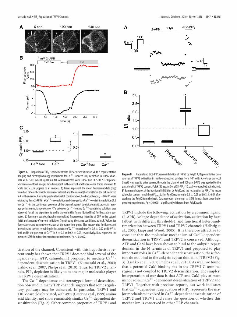
We next asked whether PIP2 can act directly on the channelsor, alternatively, requires an intact cellular environment. Ourstrategy involved application of the PIP2-sequestering agentPolyK to inside-out excised patches followed by application ofeither diC8-PIP2 or natural PIP2. We found that TRPV2 currentsin inside-out patches underwent an activity-dependent rundownduring 2-APB application (data not shown). To minimize run-down during our experiments, we used low concentrations(�100 �M) of 2-APB, in which the currents remained stable. Toexamine the direct effect of PIP2 on TRPV2 channels, we appliedPolyK, a polycation that sequesters PIP2 (Toner et al., 1988; Ga-bev et al., 1989; Ben-Tal et al., 1996), to the intracellular surface ofinside-out patches (Fig. 8A). Application of PolyK (30 �g/ml) tothe inner leaflet of the membrane inhibited 2-APB-induced cur-rents, suggesting that negatively charged lipids are required for2-APB-mediated activation of TRPV2 channels. The mean valuesfor current remaining (I/Iinitial) after PolyK treatment was 0.2 0.03 (Fig. 8). Although the currents did not recover on removal ofPolyK from the bath, they were rescued by the addition of eithernatural PIP2 (I/Iinitial, 0.8 0.2; n � 3) or diC8-PIP2 (I/Iinitial, 2 0.2; n � 7), respectively (Fig. 8) (see Materials and Methods). Weconclude that PIP2 can substitute for the activating molecule se-questered by PolyK. In sum, our data indicate that TRPV2 chan-nels is activated by the direct binding of PIP2, and thatdesensitization arises from the hydrolysis of PIP2 that resultsfrom Ca 2� entry through the channels.
DiscussionHere, we report that TRPV2 channels undergo Ca 2�-dependentdesensitization, similar to the closely related TRPV1 channel. Po-tential molecular mechanisms for Ca 2�-dependent desensitiza-tion of TRPV2 were studied using electrophysiology as well asfluorescence-based methods. We found that, although CaMbinds to a proximal C-terminal fragment of TRPV2 in our in vitroassay, CaM does not play a major role in TRPV2 Ca 2�-dependentdesensitization. In contrast, decreases in membrane PIP2 levelson channel activation underlie a major component of the Ca 2�-dependent desensitization observed on TRPV2 channels. To-gether, these results indicate that changes in membrane PIP2
levels are responsible for much, if not all, the TRPV2 Ca 2�-dependent desensitization and raise the question whether thismechanism is conserved among other TRP channels with similarCa 2�-dependent desensitization.
Historically, two types of desensitization have been describedfor TRPV1 channels: acute desensitization and tachyphylaxis.Acute desensitization refers to a decrease in the current ampli-tude seen in response to a long, continuous application of the
Figure 5. Inhibiting CaM does not prevent TRPV2 Ca 2�-dependent desensitization. A, Rep-resentative whole-cell TRPV2 currents in control, cells dialyzed with the MLCK peptide, or cellscotransfected with CaM1234. MLCK peptide, at a concentration of 1 �M, was dialyzed throughthe patch pipette. For MLCK peptide experiments, recordings were performed 5 min after theformation of whole-cell configuration. B, Summary boxplot showing the amount of remainingsteady-state current relative to the peak response. The TRPV2 mean value for current remainingin cells cotransfected with CaM1234 is 0.3 0.05 and 0.21 0.04 in cells dialyzed with 1 �M
MLCK peptide. TRPV2 mean values for current remaining in the presence and absence of Ca 2�
reported on Figure 2 are shown for comparison. Data represent the mean SEM from at leastthree independent experiments. *p 0.0001.
Mercado et al. • PIP2 Regulation of TRPV2 Channels J. Neurosci., October 6, 2010 • 30(40):13338 –13347 • 13343

ligand, whereas tachyphylaxis refers to thereduction in evoked current when verybrief applications of the ligand are madeseveral minutes apart (Koplas et al., 1997).With such protocols, Ca 2�-dependentdesensitization of TRPV1 has been foundto depend on a number of intracellularfactors, including ATP (Koplas et al.,1997), calcineurin (Docherty et al., 1996),PKA (Bhave et al., 2002; Mohapatra andNau, 2003), PKC (Mandadi et al., 2004),PIP2 (Liu et al., 2005; Rohacs et al., 2005;Lishko et al., 2007; Yao and Qin, 2009;Phelps et al., 2010), and CaM (Numazakiet al., 2003; Rosenbaum et al., 2004).However, the brief stimuli used in tachy-phylaxis present limitations on the mech-anistic interpretation of the data sincethey convolve multiple factors includingchannel activation, acute desensitization,and recovery from desensitization. In thisstudy, we used prolonged applications ofthe ligands at a relatively high concentra-tion to ensure maximal or near-maximaldesensitization of the channels. With thisprotocol, we found that the desensitiza-tion of TRPV2 channels (Fig. 2) is typicalof its subfamily in several aspects: (1) itdepends on extracellular Ca 2�, (2) the de-sensitization rate is on the order of sec-onds, and (3) the channel enters into arefractory period where channel activation has significantlydecreased (Fig. 2, inset). Thus, although TRPV2 channelsmight mediate a completely different range of physiological andsensory responses than other TRPV channels (Caterina et al.,1999; Muraki et al., 2003; Stokes et al., 2004; Link et al., 2010),they share a common structural architecture (Fig. 1) (Caterina etal., 1999) and a common Ca 2�-dependent desensitization withother TRPV channels (Fig. 2).
What is the molecular mechanism responsible for Ca 2�-dependent desensitization in TRPV2? The Ca 2� dependence ofCaM binding to the proximal TRPV2 C terminus (Fig. 4) pre-sents an attractive model where Ca 2� entering through the chan-nels can generate TRPV2-CaM/Ca 2� complexes, leading toTRPV2 channel desensitization. However, our negative resultswith the CaM-scavenging peptide (MLCK peptide) and the mu-tant CaM (CaM1234) (Fig. 5) suggest that CaM/Ca 2� binding tothe proximal TRPV2 C terminus is not coupled to TRPV2 desen-sitization. It is also unlikely that CaM/Ca 2� binding to theTRPV2 N terminus, another potential cytosolic domain for CaMbinding, is involved in TRPV2 desensitization since recent stud-ies (Lishko et al., 2007; Phelps et al., 2010) and our biochemistryresults (Fig. 3) indicate that the N terminus of TRPV2 does notinteract with CaM. Although we cannot rule out an indirect rolefor CaM in Ca 2�-dependent desensitization, a direct interactionwith the channels does not appear to regulate desensitization ofTRPV2.
Changes in the plasma membrane PIP2 concentration havebeen implicated in the regulation of many membrane proteins, inparticular, ion channels (for review, see Nilius et al., 2008; Ro-hacs, 2009). PIP2 depletion, however, occurs in many cases,through activation of a second messenger signaling pathwaymainly involving G-protein-coupled receptors (Suh et al., 2004;
Horowitz et al., 2005). In particular, TRPV2 channel traffickingto the plasma membrane has been shown to be regulated byinsulin-like growth factor-I (Kanzaki et al., 1999). Here, wehave shown that TRPV2 is also sensitive to changes in mem-brane PIP2 (Figs. 7, 8). The rapid depletion of PIP2 on TRPV2activation, however, suggests that Ca 2� entry through the chan-nel is sufficient to trigger PIP2 depletion without the need for anauxiliary surface receptor. In addition, we have shown that theeffect of PIP2 in TRPV2 channels is a direct modulation ratherthan through auxiliary mechanisms (Fig. 8; supplemental Fig. 3,available at www.jneurosci.org as supplemental material) such astrafficking of the channel in and out of the plasma membrane.Thus, TRPV2 acts as both the auxiliary receptor and PIP2 target.In this way, the entry of Ca 2� through open channels could feed-back on the channel within seconds to cause the channel to close.
TRPV2 recovery from desensitization would presumably re-quire resynthesis of PIP2. It has been suggested that resynthesis ofmembrane PIP2 determines recovery of TRPV1 channels fromdesensitization (Liu et al., 2005). The production of phospholip-ids by lipid kinases generally takes minutes and require a signifi-cant amount of intracellular ATP (�1 mM) (Varnai and Balla,1998; Suh and Hille, 2002), involving a series of steps includingphosphorylating phosphatidylinositol lipid on multiple posi-tions. The proposed slow PIP2-dependent recovery is consistentwith the observed failure of the channel to recover from desensi-tization during our “tachyphylaxis” protocol (Fig. 2, insets) as hasbeen suggested for TRPV1 (Liu et al., 2005).
Is PIP2 depletion solely responsible for TRPV2 desensitiza-tion? Although we cannot exclude a role for other molecularmechanisms in shaping various aspects of TRPV2 desensi-tization, our data suggest that PIP2 can directly regulate thechannel and that PIP2 depletion induces a significant desensi-
Figure 6. Ca 2� entry through TRPV2 channels induces PIP2 depletion. F-11 cells were transfected with either TRPV2 and a GFPfused to the PLC�1-PH domain (GFP-PLC�1-PH) that is targeted to the plasma membrane through binding to PIP2 (A) or withGFP-PLC�1-PH only (B). Shown are confocal images of GFP-PLC�1-PH fluorescence from representative cells before and afteraddition of 1 mM 2-APB in the constant presence of Ca 2�. PIP2 depletion was assessed by the translocation of the GFP-PLC�1-PHfrom the plasma membrane to the cytosol. Scale bar: 5 �m (applies to all images). The right panel shows summary boxplot ofnormalized fluorescence intensity of GFP in the cytosol for each condition. Data represent the mean SEM from at least threeindependent experiments. *p 0.0001.
13344 • J. Neurosci., October 6, 2010 • 30(40):13338 –13347 Mercado et al. • PIP2 Regulation of TRPV2 Channels
tization of the channel. Consistent with this hypothesis, a re-cent study has shown that TRPV2 does not bind several of theligands (e.g., ATP, calmodulin) proposed to mediate Ca 2�-dependent desensitization in TRPV1 (Numazaki et al., 2003;Lishko et al., 2007; Phelps et al., 2010). Thus, for TRPV2 chan-nels, PIP2 depletion is likely to be the major molecular playerin TRPV2 desensitization.
The Ca 2� dependence and stereotyped form of desensitiza-tion observed in many TRP channels suggests that some regula-tory pathways may be conserved. In particular, TRPV1 andTRPV2 are closely related, with 49% (Caterina et al., 1999) aminoacid identity, and show remarkably similar Ca 2�-dependent de-sensitization (Fig. 2). Other common properties of TRPV1 and
TRPV2 include the following: activation by a common ligand(2-APB), voltage dependence of activation, activation by heat(albeit with different thresholds), and functional heteromul-timerization between TRPV1 and TRPV2 channels (Hellwig etal., 2005; Liapi and Wood, 2005). It is therefore attractive toconsider that the molecular mechanism of Ca 2�-dependentdesensitization in TRPV1 and TRPV2 is conserved. AlthoughATP and CaM have been shown to bind to the ankyrin repeatdomain in the N terminus of TRPV1 and proposed to playimportant roles in Ca 2�-dependent desensitization, these fac-tors do not bind to the ankyrin repeat domain of TRPV2 (Fig.3) (Lishko et al., 2007; Phelps et al., 2010). As well, we foundthat a potential CaM binding site in the TRPV2 C-terminalregion is not coupled to TRPV2 desensitization. The simplestinterpretation of our data is that ATP and CaM play at mostminor roles in Ca 2�-dependent desensitization of TRPV2 andTRPV1. Together with previous reports, our work indicatesthat Ca 2�-dependent degradation of PIP2 represents the ma-jor mechanism involved in Ca 2�-dependent desensitization ofTRPV2 and TRPV1 and raises the question of whether thismechanism is conserved in other TRP channels.
Figure 7. Depletion of PIP2 is coincident with TRPV2 desensitization. A, B, A representativeimaging and electrophysiology experiment for Ca 2�-induced PIP2 depletion in TRPV2 chan-nels. A, GFP-PLC�1-PH signal in a tsA cell transfected with TRPV2 and GFP-PLC�1-PH probe.Shown are confocal images for a time point in the current and fluorescence traces shown in B.Scale bar: 5 �m (applies to all images). B, Traces represent the mean fluorescent data (top)from two different cytosolic regions of interest and the current (bottom) from the cell depictedin A with an arrow. Currents (perforated-patch configuration; holding potential,�60 mV) wereelicited by 1 mM 2-APB in a Ca 2�-free solution and changed to a Ca 2�-containing solution (1.8mM Ca 2�) in the continuous presence of the channel agonist to elicit desensitization. An aver-age perfusion exchange delay of 47 s between Ca 2�-free and Ca 2�-containing solutions wasobserved for all the experiments and is shown in this figure (dotted line) for illustration pur-poses. C, Summary boxplot showing normalized fluorescence intensity of GFP in the cytosol(left) and amount of current inhibition (right) using the same conditions as in B. Values forfluorescence and current were taken at the same time point. The mean value for fluorescentintensity and current remaining in the absence of Ca 2� (open boxes) is 0.9 0.02 and 0.97 0.01 and in the presence of Ca 2� is 2 0.1 and 0.2 0.03, respectively. Data represent themean SEM from four independent experiments. *p 0.0002.
Figure 8. Natural and diC8-PIP2 rescue inhibition of TRPV2 by PolyK. A, Representative timecourses of TRPV2 activation in inside-out excised patches from F-11 cells. A voltage protocol(inset) was used to drive current through the channel and 100 �M 2-APB was applied to thepatch to elicit TRPV2 current. PolyK (30 �g/ml) or diC8-PIP2 (10 �M) were applied as indicated.B, Summary boxplot of the fractional inhibition by PolyK and the restoration by PIP2. The meanvalues for current remaining (I/Iinitial) after PolyK treatment is 0.2 0.03 and 0.3 0.04 afterwashing the PolyK from the bath. Data represent the mean SEM from at least three inde-pendent experiments. *p 0.0001, significantly different from PolyK wash.
Mercado et al. • PIP2 Regulation of TRPV2 Channels J. Neurosci., October 6, 2010 • 30(40):13338 –13347 • 13345

ReferencesBen-Tal N, Honig B, Peitzsch RM, Denisov G, McLaughlin S (1996) Bind-
ing of small basic peptides to membranes containing acidic lipids: theo-retical models and experimental results. Biophys J 71:561–575.
Bhave G, Zhu W, Wang H, Brasier DJ, Oxford GS, Gereau RW 4th (2002)cAMP-dependent protein kinase regulates desensitization of the capsa-icin receptor (VR1) by direct phosphorylation. Neuron 35:721–731.
Brauchi S, Orta G, Mascayano C, Salazar M, Raddatz N, Urbina H, Rosenmann E,Gonzalez-Nilo F, Latorre R (2007) Dissection of the components for PIP2activation and thermosensation in TRP channels. Proc Natl Acad Sci U S A104:10246–10251.
Caterina MJ, Rosen TA, Tominaga M, Brake AJ, Julius D (1999) Acapsaicin-receptor homologue with a high threshold for noxious heat.Nature 398:436 – 441.
Clapham DE (2002) Sorting out MIC, TRP, and CRAC ion channels. J GenPhysiol 120:217–220.
Docherty RJ, Yeats JC, Bevan S, Boddeke HW (1996) Inhibition of cal-cineurin inhibits the desensitization of capsaicin-evoked currents in cul-tured dorsal root ganglion neurones from adult rats. Pflugers Arch431:828 – 837.
Francel PC, Harris K, Smith M, Fishman MC, Dawson G, Miller RJ (1987)Neurochemical characteristics of a novel dorsal root ganglion X neuro-blastoma hybrid cell line, F-11. J Neurochem 48:1624 –1631.
Gabev E, Kasianowicz J, Abbott T, McLaughlin S (1989) Binding of neomy-cin to phosphatidylinositol 4,5-bisphosphate (PIP2). Biochim BiophysActa 979:105–112.
Hellwig N, Albrecht N, Harteneck C, Schultz G, Schaefer M (2005)Homo- and heteromeric assembly of TRPV channel subunits. J Cell Sci118:917–928.
Horowitz LF, Hirdes W, Suh BC, Hilgemann DW, Mackie K, Hille B (2005)Phospholipase C in living cells: activation, inhibition, Ca 2� requirement,and regulation of M current. J Gen Physiol 126:243–262.
Hu HZ, Gu Q, Wang C, Colton CK, Tang J, Kinoshita-Kawada M, Lee LY,Wood JD, Zhu MX (2004) 2-Aminoethoxydiphenyl borate is a commonactivator of TRPV1, TRPV2, and TRPV3. J Biol Chem 279:35741–35748.
Jin X, Touhey J, Gaudet R (2006) Structure of the N-terminal ankyrin repeatdomain of the TRPV2 ion channel. J Biol Chem 281:25006 –25010.
Juvin V, Penna A, Chemin J, Lin YL, Rassendren FA (2007) Pharmacologi-cal characterization and molecular determinants of the activation of tran-sient receptor potential V2 channel orthologs by 2-aminoethoxydiphenylborate. Mol Pharmacol 72:1258 –1268.
Kanzaki M, Zhang YQ, Mashima H, Li L, Shibata H, Kojima I (1999) Trans-location of a calcium-permeable cation channel induced by insulin-likegrowth factor-I. Nat Cell Biol 1:165–170.
Karashima Y, Prenen J, Meseguer V, Owsianik G, Voets T, Nilius B (2008)Modulation of the transient receptor potential channel TRPA1 by phos-phatidylinositol 4,5-biphosphate manipulators. Pflugers Arch 457:77– 89.
Klein RM, Ufret-Vincenty CA, Hua L, Gordon SE (2008) Determinants ofmolecular specificity in phosphoinositide regulation. Phosphatidylinosi-tol (4,5)-bisphosphate (PI(4,5)P2) is the endogenous lipid regulatingTRPV1. J Biol Chem 283:26208 –26216.
Koplas PA, Rosenberg RL, Oxford GS (1997) The role of calcium in thedesensitization of capsaicin responses in rat dorsal root ganglion neurons.J Neurosci 17:3525–3537.
Kwak J, Wang MH, Hwang SW, Kim TY, Lee SY, Oh U (2000) IntracellularATP increases capsaicin-activated channel activity by interacting withnucleotide-binding domains. J Neurosci 20:8298 – 8304.
Lee J, Cha SK, Sun TJ, Huang CL (2005) PIP2 activates TRPV5 and releasesits inhibition by intracellular Mg 2�. J Gen Physiol 126:439 – 451.
Liapi A, Wood JN (2005) Extensive co-localization and heteromultimer for-mation of the vanilloid receptor-like protein TRPV2 and the capsaicinreceptor TRPV1 in the adult rat cerebral cortex. Eur J Neurosci 22:825– 834.
Link TM, Park U, Vonakis BM, Raben DM, Soloski MJ, Caterina MJ (2010)TRPV2 has a pivotal role in macrophage particle binding and phagocyto-sis. Nat Immunol 11:232–239.
Lishko PV, Procko E, Jin X, Phelps CB, Gaudet R (2007) The ankyrin repeatsof TRPV1 bind multiple ligands and modulate channel sensitivity. Neu-ron 54:905–918.
Liu B, Zhang C, Qin F (2005) Functional recovery from desensitization ofvanilloid receptor TRPV1 requires resynthesis of phosphatidylinositol4,5-bisphosphate. J Neurosci 25:4835– 4843.
Lukacs V, Thyagarajan B, Varnai P, Balla A, Balla T, Rohacs T (2007) Dualregulation of TRPV1 by phosphoinositides. J Neurosci 27:7070 –7080.
Mandadi S, Numazaki M, Tominaga M, Bhat MB, Armati PJ, Roufogalis BD(2004) Activation of protein kinase C reverses capsaicin-inducedcalcium-dependent desensitization of TRPV1 ion channels. Cell Calcium35:471– 478.
Mohapatra DP, Nau C (2003) Desensitization of capsaicin-activated cur-rents in the vanilloid receptor TRPV1 is decreased by the cyclic AMP-dependent protein kinase pathway. J Biol Chem 278:50080 –50090.
Muraki K, Iwata Y, Katanosaka Y, Ito T, Ohya S, Shigekawa M, Imaizumi Y(2003) TRPV2 is a component of osmotically sensitive cation channels inmurine aortic myocytes. Circ Res 93:829 – 838.
Niemeyer BA, Bergs C, Wissenbach U, Flockerzi V, Trost C (2001) Compet-itive regulation of CaT-like-mediated Ca 2� entry by protein kinase C andcalmodulin. Proc Natl Acad Sci U S A 98:3600 –3605.
Nilius B, Mahieu F, Prenen J, Janssens A, Owsianik G, Vennekens R, Voets T(2006) The Ca 2�-activated cation channel TRPM4 is regulated by phos-phatidylinositol 4,5-biphosphate. EMBO J 25:467– 478.
Nilius B, Owsianik G, Voets T (2008) Transient receptor potential channelsmeet phosphoinositides. EMBO J 27:2809 –2816.
Numazaki M, Tominaga T, Takeuchi K, Murayama N, Toyooka H, TominagaM (2003) Structural determinant of TRPV1 desensitization interactswith calmodulin. Proc Natl Acad Sci U S A 100:8002– 8006.
Phelps CB, Wang RR, Choo SS, Gaudet R (2010) Differential regulation ofTRPV1, TRPV3, and TRPV4 sensitivity through a conserved binding siteon the ankyrin repeat domain. J Biol Chem 285:731–740.
Prescott ED, Julius D (2003) A modular PIP2 binding site as a determinantof capsaicin receptor sensitivity. Science 300:1284 –1288.
Rohacs T (2009) Phosphoinositide regulation of non-canonical transientreceptor potential channels. Cell Calcium 45:554 –565.
Rohacs T, Lopes CM, Michailidis I, Logothetis DE (2005) PI(4,5)P2 regu-lates the activation and desensitization of TRPM8 channels through theTRP domain. Nat Neurosci 8:626 – 634.
Rosenbaum T, Gordon-Shaag A, Munari M, Gordon SE (2004) Ca 2�/cal-modulin modulates TRPV1 activation by capsaicin. J Gen Physiol 123:53– 62.
Runnels LW, Yue L, Clapham DE (2002) The TRPM7 channel is inactivatedby PIP2 hydrolysis. Nat Cell Biol 4:329 –336.
Scott K, Sun Y, Beckingham K, Zuker CS (1997) Calmodulin regulation ofDrosophila light-activated channels and receptor function mediates ter-mination of the light response in vivo. Cell 91:375–383.
Singh BB, Liu X, Tang J, Zhu MX, Ambudkar IS (2002) Calmodulin regu-lates Ca 2�-dependent feedback inhibition of store-operated Ca 2� influxby interaction with a site in the C terminus of TrpC1. Mol Cell 9:739 –750.
Stein AT, Ufret-Vincenty CA, Hua L, Santana LF, Gordon SE (2006) Phos-phoinositide 3-kinase binds to TRPV1 and mediates NGF-stimulatedTRPV1 trafficking to the plasma membrane. J Gen Physiol 128:509 –522.
Stokes AJ, Shimoda LM, Koblan-Huberson M, Adra CN, Turner H (2004) ATRPV2-PKA signaling module for transduction of physical stimuli inmast cells. J Exp Med 200:137–147.
Suh BC, Hille B (2002) Recovery from muscarinic modulation of M currentchannels requires phosphatidylinositol 4,5-bisphosphate synthesis. Neu-ron 35:507–520.
Suh BC, Horowitz LF, Hirdes W, Mackie K, Hille B (2004) Regulation ofKCNQ2/KCNQ3 current by G protein cycling: the kinetics of receptor-mediated signaling by Gq. J Gen Physiol 123:663– 683.
Szallasi A, Blumberg PM (1999) Vanilloid (Capsaicin) receptors and mech-anisms. Pharmacol Rev 51:159 –212.
Tang J, Lin Y, Zhang Z, Tikunova S, Birnbaumer L, Zhu MX (2001) Identi-fication of common binding sites for calmodulin and inositol 1,4,5-trisphosphate receptors on the carboxyl termini of trp channels. J BiolChem 276:21303–21310.
Thyagarajan B, Benn BS, Christakos S, Rohacs T (2009) PhospholipaseC-mediated regulation of transient receptor potential vanilloid 6 chan-nels: implications in active intestinal Ca 2� transport. Mol Pharmacol75:608 – 616.
Toner M, Vaio G, McLaughlin A, McLaughlin S (1988) Adsorption of cat-ions to phosphatidylinositol 4,5-bisphosphate. Biochemistry 27:7435–7443.
Torok K, Cowley DJ, Brandmeier BD, Howell S, Aitken A, Trentham DR (1998)Inhibition of calmodulin-activated smooth-muscle myosin light-chain ki-
13346 • J. Neurosci., October 6, 2010 • 30(40):13338 –13347 Mercado et al. • PIP2 Regulation of TRPV2 Channels

nase by calmodulin-binding peptides and fluorescent (phosphodiesterase-activating) calmodulin derivatives. Biochemistry 37:6188–6198.
Trost C, Bergs C, Himmerkus N, Flockerzi V (2001) The transient receptorpotential, TRP4, cation channel is a novel member of the family of cal-modulin binding proteins. Biochem J 355:663– 670.
Varnai P, Balla T (1998) Visualization of phosphoinositides that bind pleck-strin homology domains: calcium- and agonist-induced dynamic changesand relationship to myo-[ 3H]inositol-labeled phosphoinositide pools.J Cell Biol 143:501–510.
Warr CG, Kelly LE (1996) Identification and characterization of two dis-tinct calmodulin-binding sites in the Trpl ion-channel protein of Dro-sophila melanogaster. Biochem J 314:497–503.
Yao J, Qin F (2009) Interaction with phosphoinositides confers adaptationonto the TRPV1 pain receptor. PLoS Biol 7:e46.
Zhang Z, Tang J, Tikunova S, Johnson JD, Chen Z, Qin N, Dietrich A, StefaniE, Birnbaumer L, Zhu MX (2001) Activation of Trp3 by inositol 1,4,5-trisphosphate receptors through displacement of inhibitory calmodulinfrom a common binding domain. Proc Natl Acad Sci U S A 98:3168 –3173.
Zhang Z, Okawa H, Wang Y, Liman ER (2005) Phosphatidylinositol 4,5-bisphosphate rescues TRPM4 channels from desensitization. J Biol Chem280:39185–39192.
Zhu X, Birnbaumer L (1998) Calcium channels formed by mammalian Trphomologues. News Physiol Sci 13:211–217.
Mercado et al. • PIP2 Regulation of TRPV2 Channels J. Neurosci., October 6, 2010 • 30(40):13338 –13347 • 13347