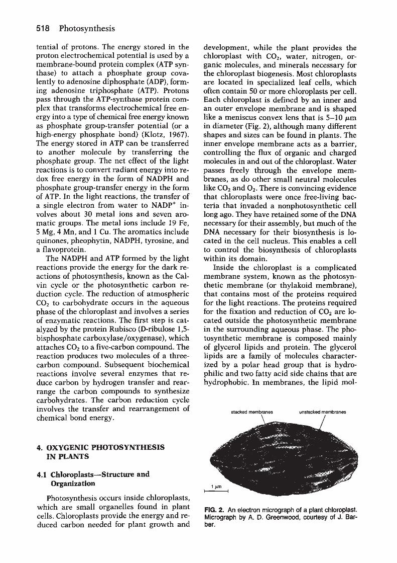
1995 photo whitmarsh - life.illinois.edu · photosystem ii john whitmarsh,university of illinois,...
TRANSCRIPT


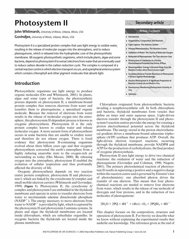
Photosystem IIJohn Whitmarsh, University of Illinois, Urbana, Illinois, USA
Govindjee, University of Illinois, Urbana, Illinois, USA
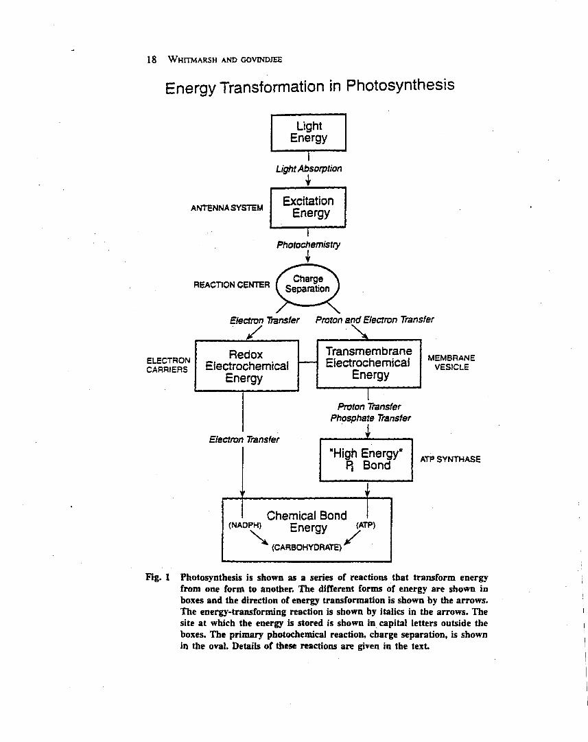
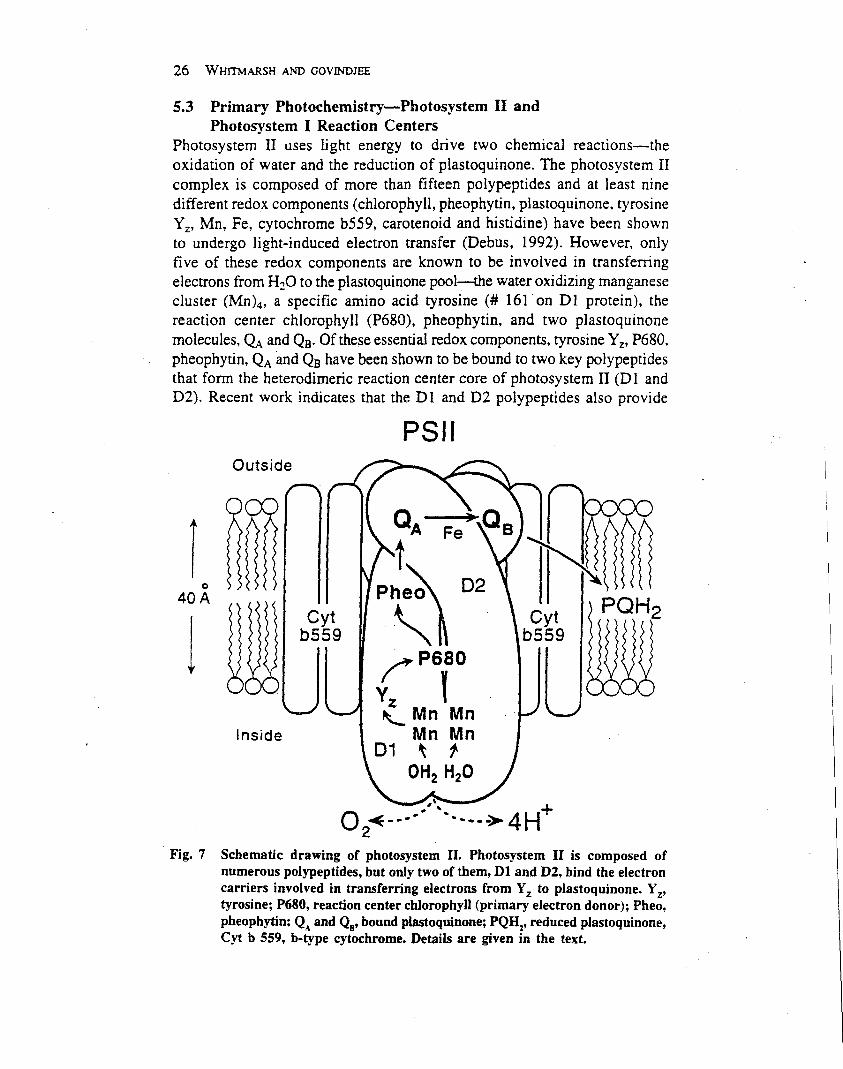
Photosystem II is a specialized protein complex that uses light energy to oxidize water,
resulting in the release of molecular oxygen into the atmosphere, and to reduce
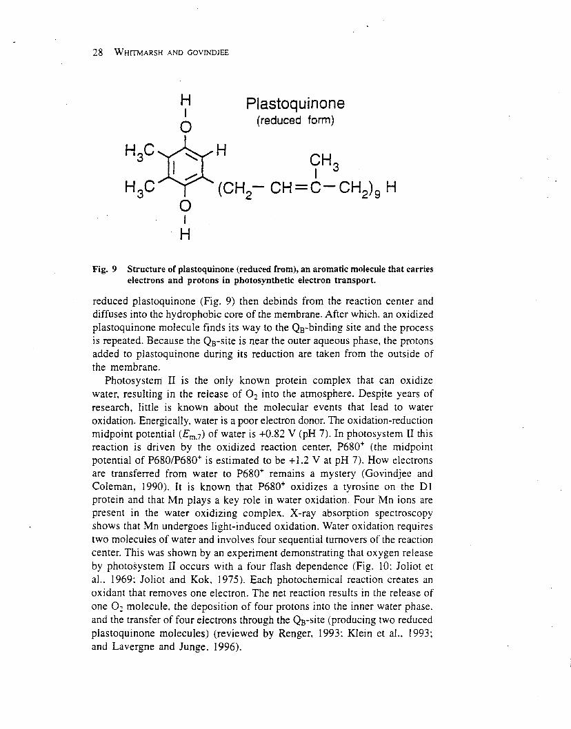
plastoquinone, which is released into the hydrophobic core of the photosynthetic
membrane. All oxygenic photosynthetic organisms, which include plants, algae and some
bacteria, depend on photosystem II to extract electrons from water that are eventually used
to reduce carbon dioxide in the carbon reduction cycle. The complex is composed of a
central reaction centre in which electron transport occurs, and a peripheral antenna system
which contains chlorophyll and other pigment molecules that absorb light.
Introduction
Photosynthetic organisms use light energy to produceorganic molecules (Ort and Whitmarsh, 2001). In plants,algae and some types of bacteria, the photosyntheticprocess depends on photosystem II, a membrane-boundprotein complex that removes electrons from water andtransfers them to plastoquinone, a specialized organicmolecule. Because the removal of electrons from waterresults in the release of molecular oxygen into the atmo-sphere, this photosystem II-dependent process is known asoxygenic photosynthesis. Photosystem II is the onlyprotein complex known to oxidize water and releasemolecular oxygen. A more ancient form of photosynthesisoccurs in some bacteria that are unable to oxidize waterand therefore do not release oxygen. There is fossilevidence that photosystem II-containing organismsevolved about three billion years ago and that oxygenicphotosynthesis converted the earth’s atmosphere from ahighly reducing anaerobic state to the oxygen-rich airsurrounding us today (Des Marais, 2000). By releasingoxygen into the atmosphere, photosystem II enabled theevolution of cellular respiration and thus profoundlyaffected the diversity of life.
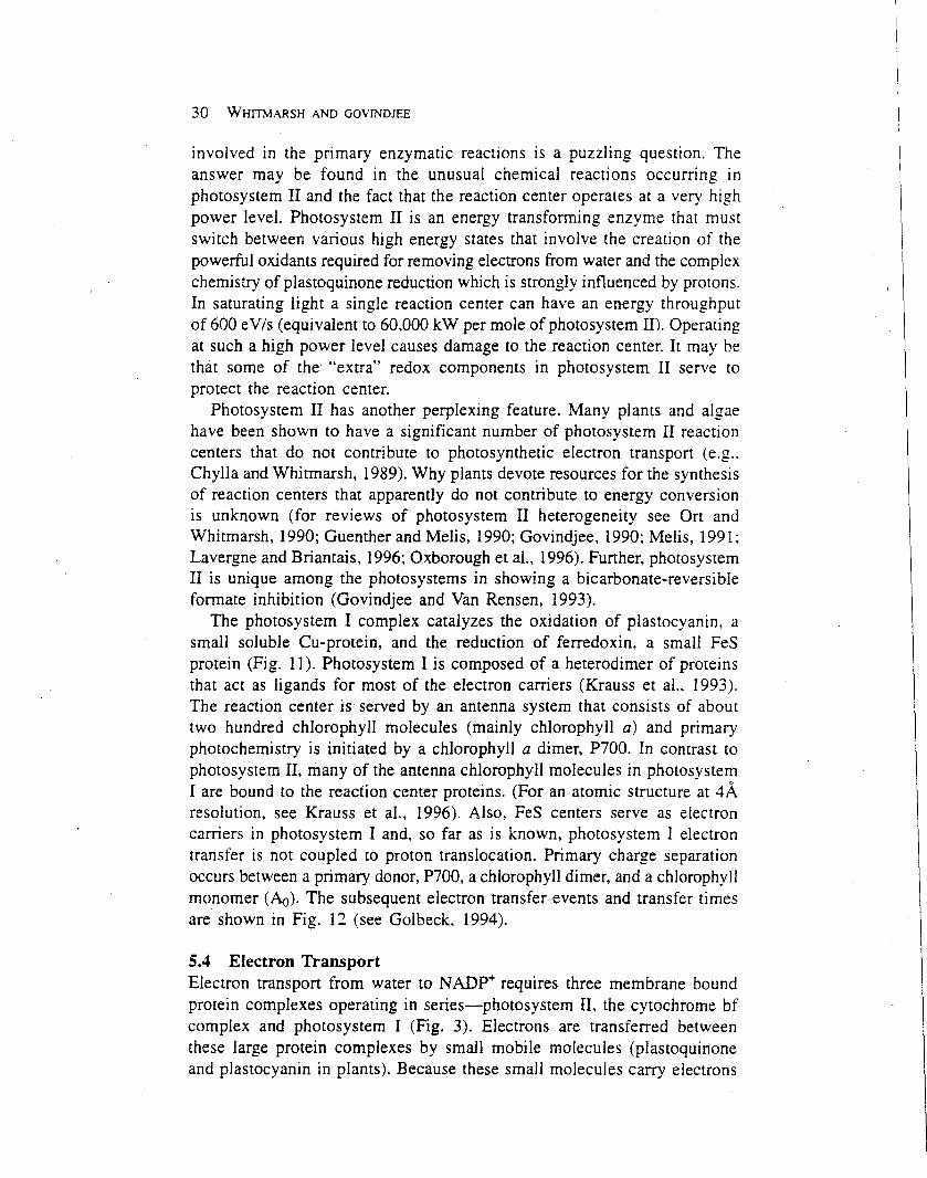
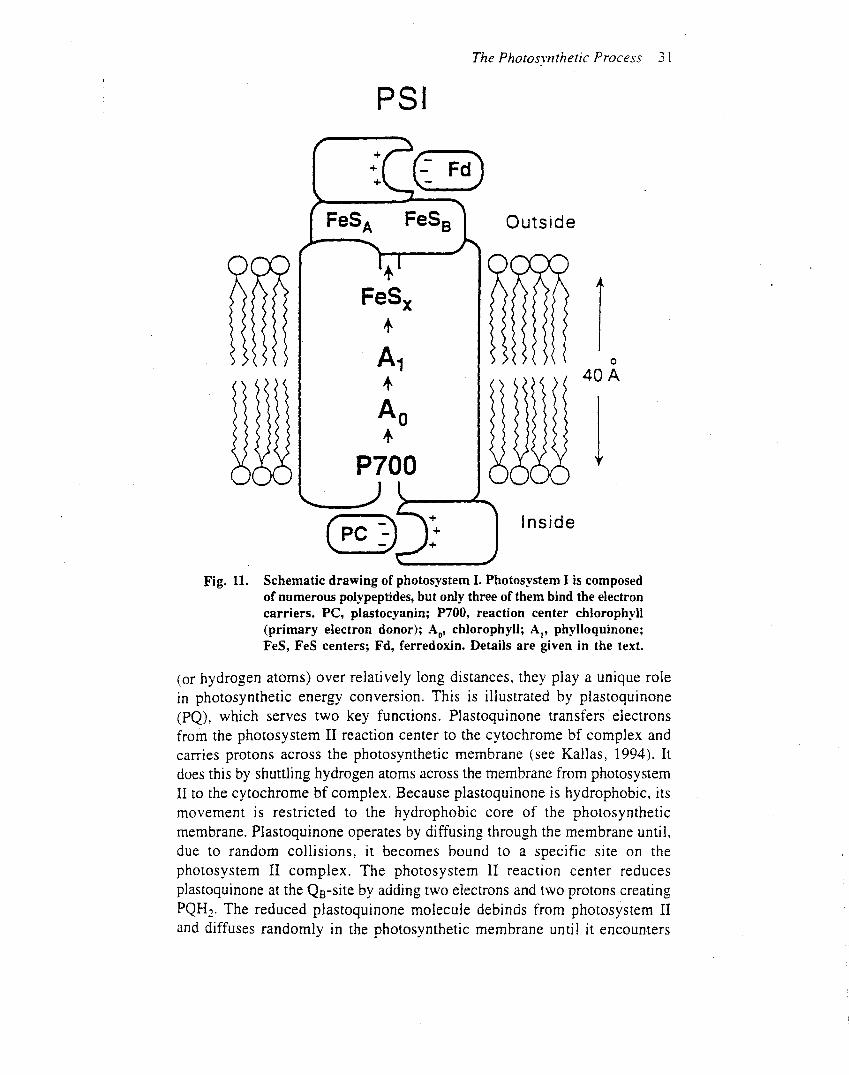
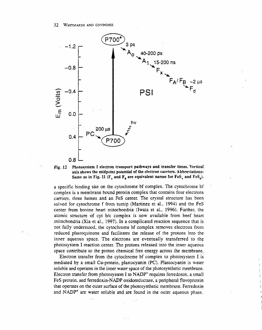
Oxygenic photosynthesis depends on two reactioncentre protein complexes, photosystem II and photosys-tem I, which are linked by the cytochrome bf complex andsmall mobile electron carriers (Whitmarsh and Govindjee,1999) (Figure 1). Photosystem II, the cytochrome bfcomplex and photosystem I are embedded in the thylakoidmembrane and operate in series to transfer electrons fromwater to nicotinamide–adenine dinucleotide phosphate(NADP1 ). The energy necessary to move electrons fromwater toNADP1 is providedby light,which is captured bythe photosystem II and photosystem I antenna systems. Inplants and algae the thylakoid membranes are locatedinside chloroplasts, which are subcellular organelles. Inoxygenic bacteria the thylakoids are located inside theplasma membrane.
Chloroplasts originated from photosynthetic bacteriainvading a nonphotosynthetic cell. In both chloroplastsand bacteria, thylakoid membranes form vesicles thatdefine an inner and outer aqueous space. Light-drivenelectron transfer through the photosystem II and photo-system I reaction centres provides the energy for creating aproton electrochemical potential across the thylakoidmembrane. The energy stored in the proton electrochemi-cal gradient drives a membrane-bound adenosine tripho-sphate (ATP) synthase that produces ATP. Overall, thelight-driven electron transport reactions, occurringthrough the thylakoid membrane, provide NADPH andATP for the productionof carbohydrates, the final productof oxygenic photosynthesis.Photosystem II uses light energy to drive two chemical
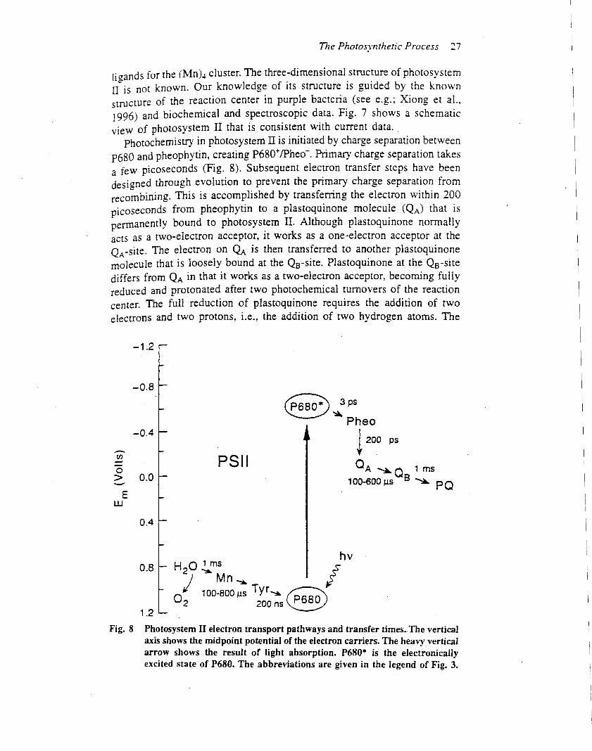
reactions: the oxidation of water and the reduction ofplastoquinone (Govindjee and Coleman, 1990; Nugent,2001). The primary photochemical reaction of photosys-tem II results in separating a positive and a negative chargewithin the reaction centre and is governed byEinstein’s lawof photochemistry: one absorbed photon drives thetransfer of one electron. This means that four photo-chemical reactions are needed to remove four electronsfrom water, which results in the release of one molecule ofdioxygen and four protons, and in the reduction of twomolecules of plastoquinone:
2H2O1 2PQ1 4H1 1 (4hn)!O21 2PQH21 4H1
This chapter focuses on the composition, structure andoperation of photosystem II. For brevity we describe whatwe know without explaining the experimental results thatunderlie our knowledge. The references given at the end of
Article Contents
Secondary article
. Introduction
. Organization, Composition and Structure
. Light Capture: The Antenna System
. Primary Photochemistry: The Reaction Centre
. Oxidation of Water: The Source of Molecular Oxygen
. Reduction of Plastoquinone: The Two-electron Gate
. Photosystem II Contributes to a Proton
Electrochemical Potential that Drives ATPase
. Downregulation: Energy is Diverted Away from the
Reaction Centre when there is Excess Light
. Secondary Electron Transfer Reactions in Photosystem
II Protect Against Photodamage
. Inactive Photosystem II: A Significant Proportion of
Reaction Centres do not Work In Vivo
. Fluorescence: Monitoring Photosystem II Activity In
Vivo
. Summary
1ENCYCLOPEDIA OF LIFE SCIENCES / & 2002 Macmillan Publishers Ltd, Nature Publishing Group / www.els.net
the article are an entryway into the vast literature onphotosystem II that extends backmore than half a century.
Organization, Composition andStructure
Photosystem II is embedded in the thylakoid membrane,with the oxygen-evolving site near the inner aqueous phaseand the plastoquinone reduction site near the outeraqueous phase (Figure 2), an orientation that enables theoxidation–reduction chemistry of the reaction centre tocontribute to the proton electrochemical gradient acrossthe thylakoidmembrane. In chloroplasts, the photosystemII and photosystem I complexes are distributed in differentregions of the thylakoid membrane. Most of the photo-system II complexes are located in the stacked membranes(grana), whereas the photosystem I complexes are locatedin the stomal membranes. It is not clear why two reactioncentres that operate in series are spatially separated ineukaryotic organisms. In prokaryotes, the thylakoidmembranes do not form grana and the photosystem IIand I complexes appear to be intermixed. In eukaryotes thephotosystem II complex is densely packed in the thylakoidmembrane, with average centre to centre distances of a fewhundred angstroms.One square centimetre of a typical leafcontains about 30 trillion photosystem II complexes.
Although photosystem II is found in prokaryoticcyanobacteria (e.g. Synechocystis spp.) and prochloro-phytes (e.g. Prochloron spp.), in eukaryotic green algae(e.g. Chlamydomonas spp.), red algae (e.g. Porphyridiumspp.), yellow-green algae (e.g.Vaucheria) and brown algae
(e.g. Fucus spp.) and all higher plants (e.g. Arabidopsisspp.), extensive research indicates that its structure andfunction are remarkably similar in these diverse organisms.
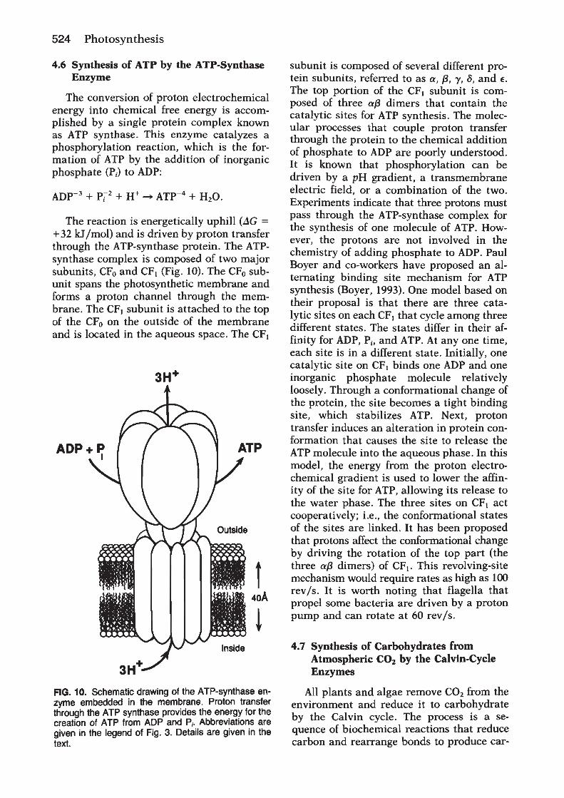
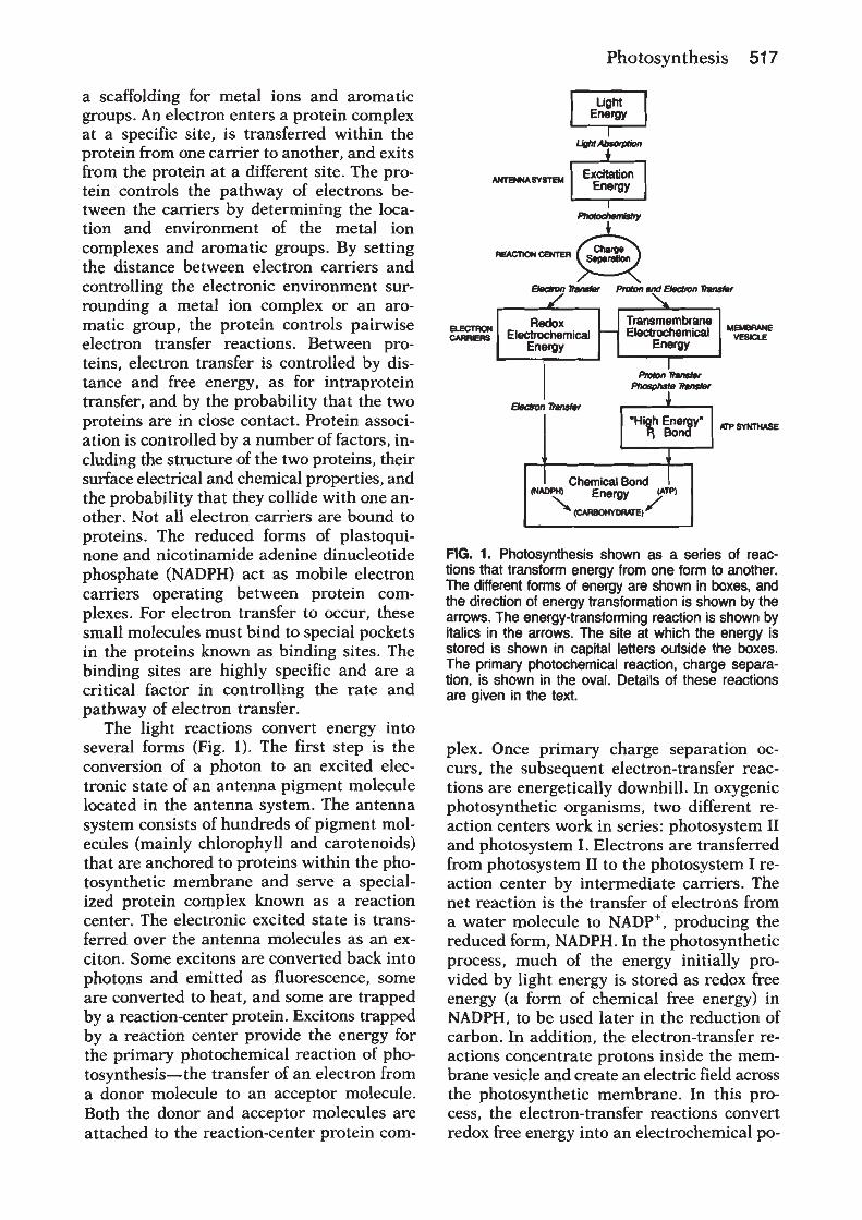
Light
40–200 ps
1–3 ps
15–200 ns200–500 ns
0.5–20 µs
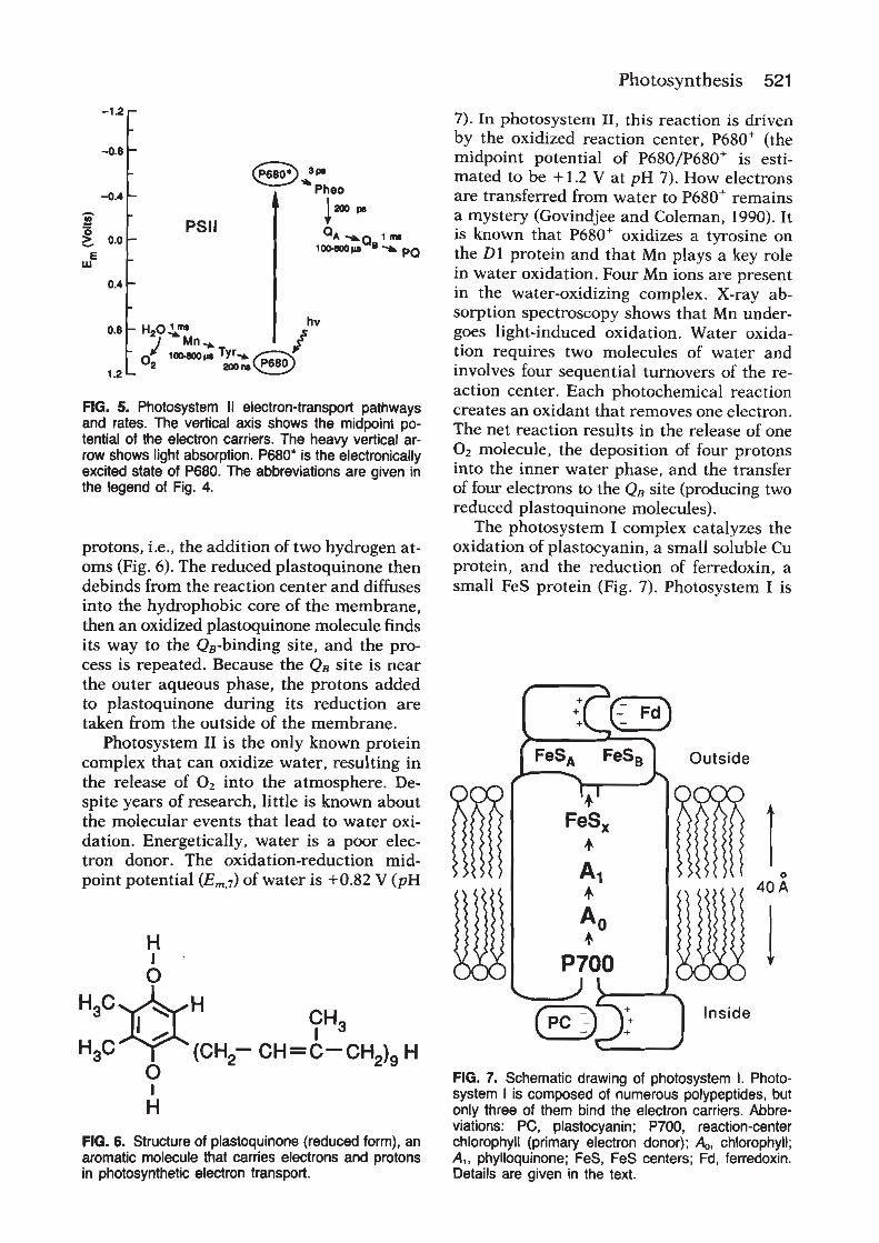
0.4
1.2
E m (
volts
)
–1.2
0
–0.4
O2 + 4H+
2H2O50 µs–1.5 ms
Light
200–800 µs
20 ns–200 µs
100–200 ps
3–20 ps
200–600 µs1 ms
5 ms50–100 µs
20–200 µs
2H+
PQPQPQPQ
Tyr4Mn
P680
Photosystem II
P680*Pheo
QAQB
PQCyt b6Cyt b6
FeS/Cyt fPC
P700
Photosystem I
P700*
AoA1
Fx
FA, FBFD
FNRNADPH
NADP+
2H+
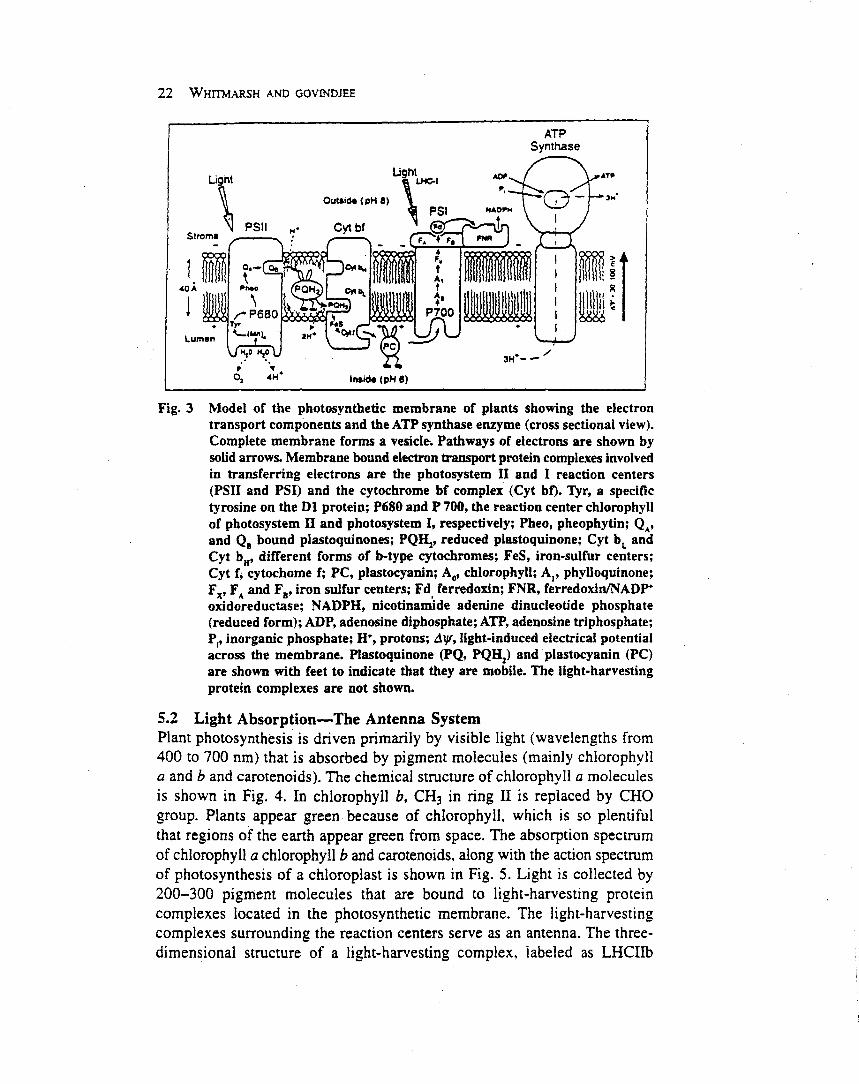
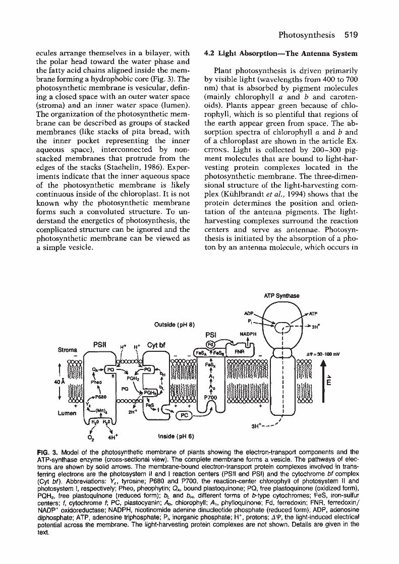
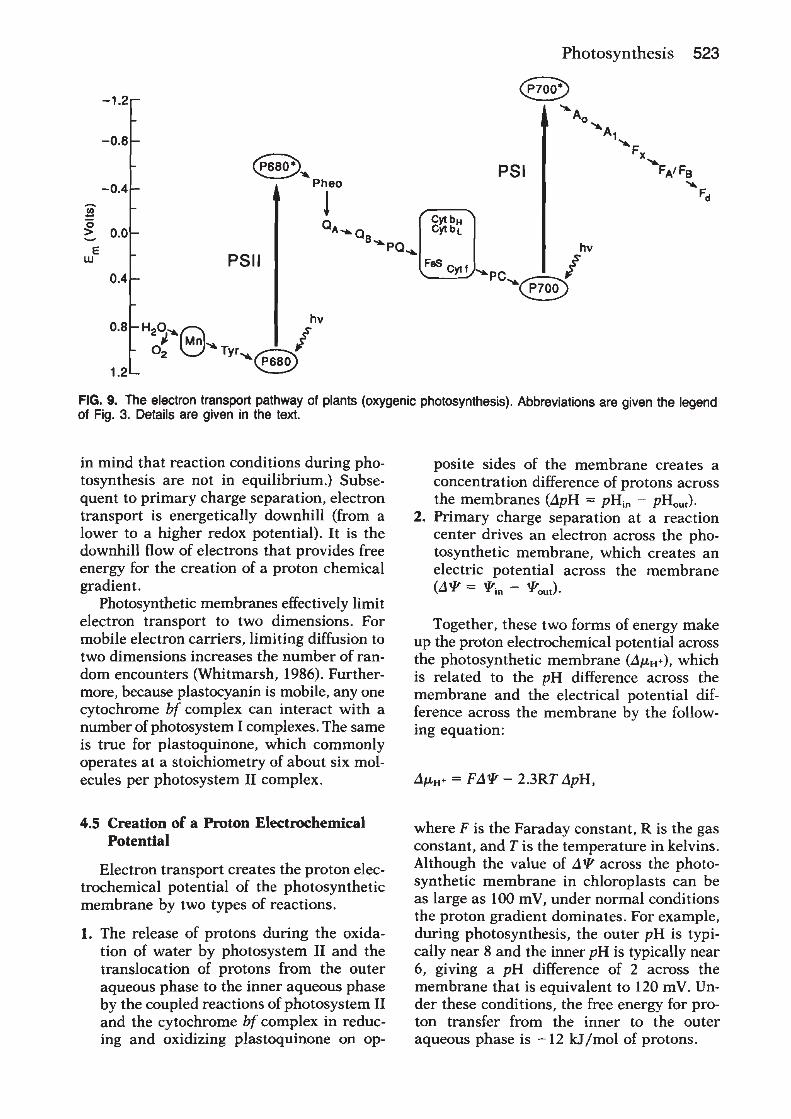
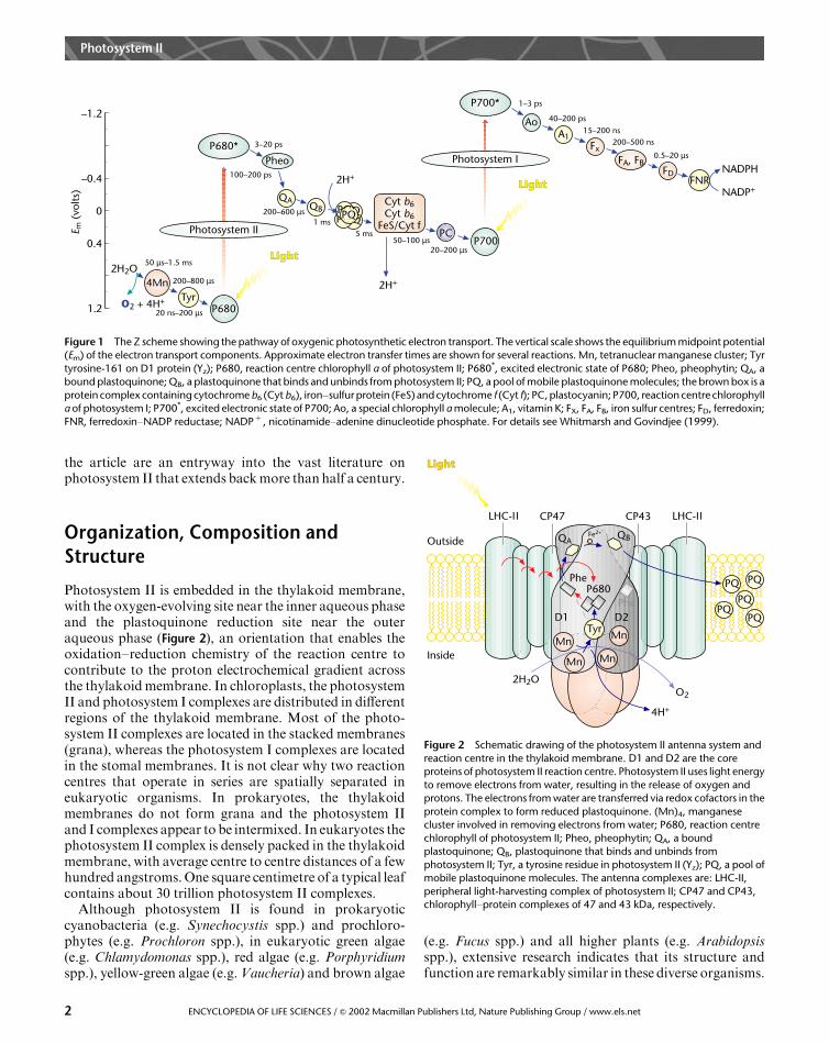
Figure 1 The Z scheme showing the pathway of oxygenic photosynthetic electron transport. The vertical scale shows the equilibrium midpoint potential(Em) of the electron transport components. Approximate electron transfer times are shown for several reactions. Mn, tetranuclear manganese cluster; Tyr
tyrosine-161 on D1 protein (Yz); P680, reaction centre chlorophyll a of photosystem II; P680*, excited electronic state of P680; Pheo, pheophytin; QA, abound plastoquinone; QB, a plastoquinone that binds and unbinds from photosystem II; PQ, a pool of mobile plastoquinone molecules; the brown box is a
protein complex containing cytochrome b6 (Cyt b6), iron–sulfur protein (FeS) and cytochrome f (Cyt f); PC, plastocyanin; P700, reaction centre chlorophylla of photosystem I; P700*, excited electronic state of P700; Ao, a special chlorophyll a molecule; A1, vitamin K; FX, FA, FB, iron sulfur centres; FD, ferredoxin;
FNR, ferredoxin–NADP reductase; NADP1 , nicotinamide–adenine dinucleotide phosphate. For details see Whitmarsh and Govindjee (1999).
Mn
Tyr
4H+
2H2O
Inside
Outside
Mn
Mn
Mn
O2
PQ
PQ
PQPQ
PQ
PheP680
PheD2
QAQB
D1
Fe2+
Light
LHC-ΙΙ CP47 LHC-ΙΙCP43
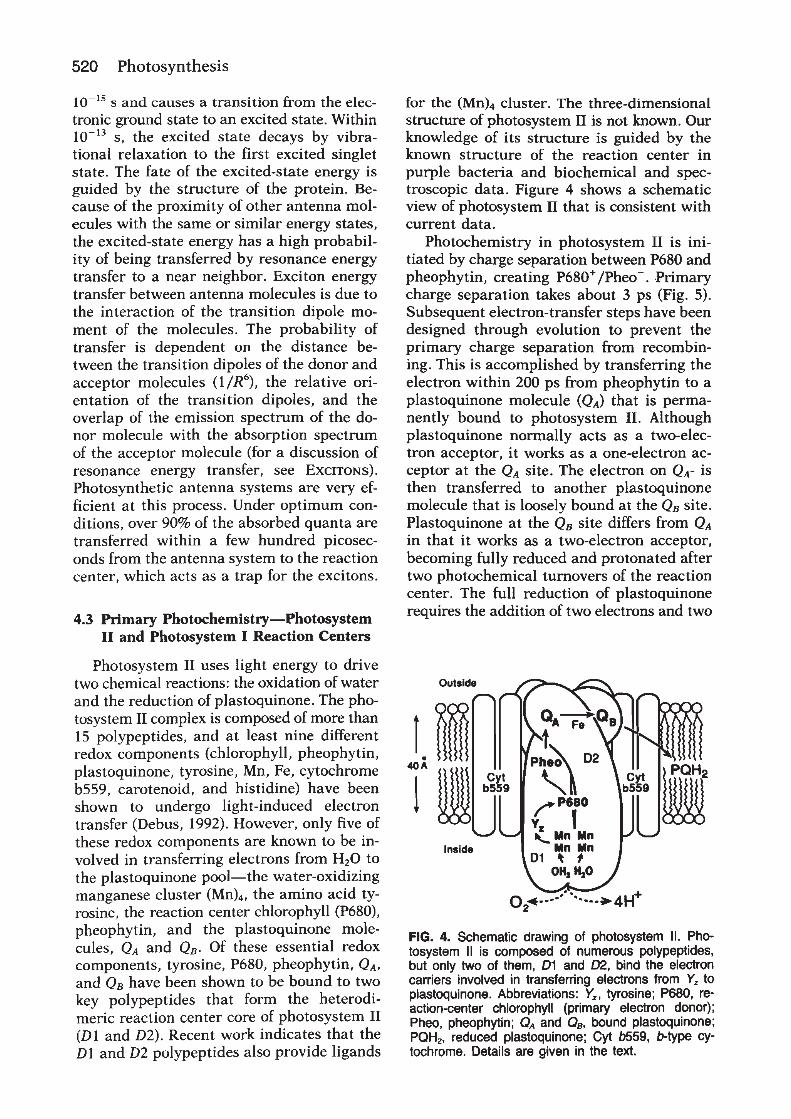
Figure 2 Schematic drawing of the photosystem II antenna system and
reaction centre in the thylakoid membrane. D1 and D2 are the coreproteins of photosystem II reaction centre. Photosystem II uses light energy
to remove electrons from water, resulting in the release of oxygen andprotons. The electrons from water are transferred via redox cofactors in the
protein complex to form reduced plastoquinone. (Mn)4, manganesecluster involved in removing electrons from water; P680, reaction centre
chlorophyll of photosystem II; Pheo, pheophytin; QA, a boundplastoquinone; QB, plastoquinone that binds and unbinds from
photosystem II; Tyr, a tyrosine residue in photosystem II (Yz); PQ, a pool ofmobile plastoquinone molecules. The antenna complexes are: LHC-II,
peripheral light-harvesting complex of photosystem II; CP47 and CP43,chlorophyll–protein complexes of 47 and 43 kDa, respectively.
Photosystem II
2 ENCYCLOPEDIA OF LIFE SCIENCES / & 2002 Macmillan Publishers Ltd, Nature Publishing Group / www.els.net
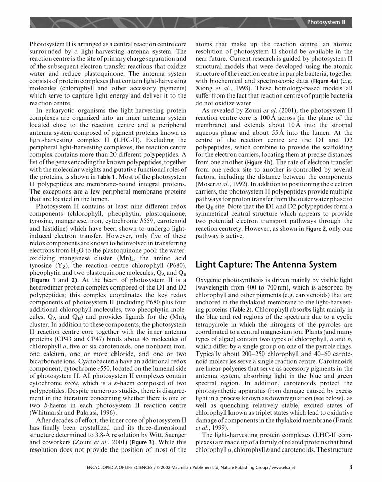
Photosystem II is arranged as a central reaction centre coresurrounded by a light-harvesting antenna system. Thereaction centre is the site of primary charge separation andof the subsequent electron transfer reactions that oxidizewater and reduce plastoquinone. The antenna systemconsists of protein complexes that contain light-harvestingmolecules (chlorophyll and other accessory pigments)which serve to capture light energy and deliver it to thereaction centre.
In eukaryotic organisms the light-harvesting proteincomplexes are organized into an inner antenna systemlocated close to the reaction centre and a peripheralantenna system composed of pigment proteins known aslight-harvesting complex II (LHC-II). Excluding theperipheral light-harvesting complexes, the reaction centrecomplex contains more than 20 different polypeptides. Alist of the genes encoding the knownpolypeptides, togetherwith themolecular weights and putative functional roles ofthe proteins, is shown in Table 1. Most of the photosystemII polypeptides are membrane-bound integral proteins.The exceptions are a few peripheral membrane proteinsthat are located in the lumen.
Photosystem II contains at least nine different redoxcomponents (chlorophyll, pheophytin, plastoquinone,tyrosine, manganese, iron, cytochrome b559, carotenoidand histidine) which have been shown to undergo light-induced electron transfer. However, only five of theseredox components are known tobe involved in transferringelectrons from H2O to the plastoquinone pool: the water-oxidizing manganese cluster (Mn)4, the amino acidtyrosine (YZ), the reaction centre chlorophyll (P680),pheophytin and two plastoquinone molecules, QA and QB
(Figures 1 and 2). At the heart of photosystem II is aheterodimer protein complex composed of the D1 and D2polypeptides; this complex coordinates the key redoxcomponents of photosystem II (including P680 plus fouradditional chlorophyll molecules, two pheophytin mole-cules, QA and QB) and provides ligands for the (Mn)4cluster. In addition to these components, the photosystemII reaction centre core together with the inner antennaproteins (CP43 and CP47) binds about 45 molecules ofchlorophyll a, five or six carotenoids, one nonhaem iron,one calcium, one or more chloride, and one or twobicarbonate ions. Cyanobacteria have an additional redoxcomponent, cytochrome c550, located on the lumenal sideof photosystem II. All photosystem II complexes containcytochrome b559, which is a b-haem composed of twopolypeptides. Despite numerous studies, there is disagree-ment in the literature concerning whether there is one ortwo b-haems in each photosystem II reaction centre(Whitmarsh and Pakrasi, 1996).
After decades of effort, the inner core of photosystem IIhas finally been crystallized and its three-dimensionalstructure determined to 3.8-A resolution by Witt, Saengerand coworkers (Zouni et al., 2001) (Figure 3). While thisresolution does not provide the position of most of the
atoms that make up the reaction centre, an atomicresolution of photosystem II should be available in thenear future. Current research is guided by photosystem IIstructural models that were developed using the atomicstructure of the reaction centre in purple bacteria, togetherwith biochemical and spectroscopic data (Figure 4a) (e.g.Xiong et al., 1998). These homology-based models allsuffer from the fact that reaction centres of purple bacteriado not oxidize water.As revealed by Zouni et al. (2001), the photosystem II
reaction centre core is 100 A across (in the plane of themembrane) and extends about 10 A into the stromalaqueous phase and about 55 A into the lumen. At thecentre of the reaction centre are the D1 and D2polypeptides, which combine to provide the scaffoldingfor the electron carriers, locating them at precise distancesfrom one another (Figure 4b). The rate of electron transferfrom one redox site to another is controlled by severalfactors, including the distance between the components(Moser et al., 1992). In addition to positioning the electroncarriers, the photosystem II polypeptides provide multiplepathways for proton transfer from the outerwater phase tothe QB site. Note that the D1 and D2 polypeptides form asymmetrical central structure which appears to providetwo potential electron transport pathways through thereaction centrety. However, as shown in Figure 2, only onepathway is active.
Light Capture: The Antenna System
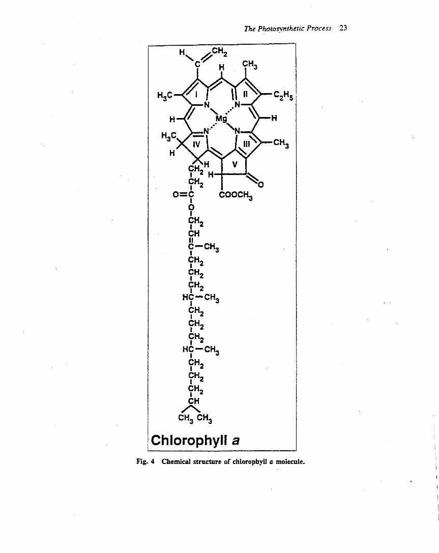
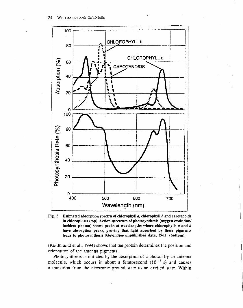
Oxygenic photosynthesis is driven mainly by visible light(wavelength from 400 to 700 nm), which is absorbed bychlorophyll and other pigments (e.g. carotenoids) that areanchored in the thylakoid membrane to the light-harvest-ing proteins (Table 2). Chlorophyll absorbs light mainly inthe blue and red regions of the spectrum due to a cyclictetrapyrrole in which the nitrogens of the pyrroles arecoordinated to a central magnesium ion. Plants (andmanytypes of algae) contain two types of chlorophyll, a and b,which differ by a single group on one of the pyrrole rings.Typically about 200–250 chlorophyll and 40–60 carote-noid molecules serve a single reaction centre. Carotenoidsare linear polyenes that serve as accessory pigments in theantenna system, absorbing light in the blue and greenspectral region. In addition, carotenoids protect thephotosynthetic apparatus from damage caused by excesslight in a process known as downregulation (see below), aswell as quenching relatively stable, excited states ofchlorophyll known as triplet states which lead to oxidativedamage of components in the thylakoidmembrane (Franket al., 1999).The light-harvesting protein complexes (LHC-II com-
plexes) aremade upof a family of relatedproteins that bindchlorophyll a, chlorophyll band carotenoids.The structure
Photosystem II
3ENCYCLOPEDIA OF LIFE SCIENCES / & 2002 Macmillan Publishers Ltd, Nature Publishing Group / www.els.net
of one of the LHC-II complexes has been determinedby electron crystallography (Kuhlbrandt et al., 1994).The complex forms a trimer in the membranes, witheach subunit binding seven molecules of chlorophylla, five molecules of chlorophyll b and two carotenemolecules.
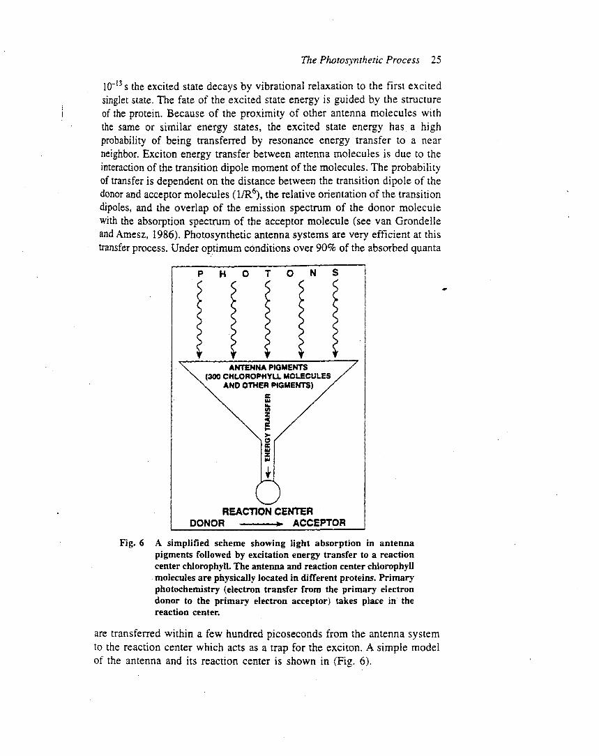
Photosynthesis starts with the absorption of a photon byan antenna molecule, which causes a rapid (102 15 s)
transition from the electronic ground state to an excitedstate.Within 102 13 s, the electronic excited state decays byvibrational relaxation to the first excited singlet state.These excited electron states are short lived, and the fate ofthe excitation energy in the antenna system is guided by thestructure of the protein–pigment complexes. Because ofthe proximity of other antenna molecules with the same orsimilar electron energy levels, the excited state energy has a
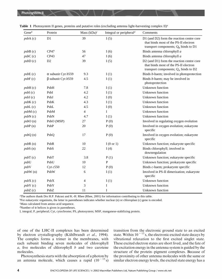
Table 1 Photosystem II genes, proteins and putative roles (excluding antenna light-harvesting complex II)a
aThe authors thank Drs H.P. Pakrasi and K.-H. Rhee (Rhee, 2001) for information contributing to this table.bFor eukaryotic organisms, the letter in parentheses indicates whether nuclear (n) or chloroplast (c) gene is encoded.cMass calculated from amino acid sequence.dNumber of α helices is given in parentheses.I, integral; P, peripheral; Cyt, cytochrome; PS, photosystem; MSP, manganese-stabilizing protein.
Geneb Protein Mass (kDa)c Integral or peripherald Comments
psbA (c) D1 39 I (5) D1 (and D2) form the reaction centre core that binds most of the PS-II electron transport components; QB binds to D1
psbB (c) CP47 56 I (6) Binds antenna chlorophyll a
psbC (c) CP43 47 I (6) Binds antenna chlorophyll a
psbD (c) D2 39 I (5) D2 (and D1) form the reaction centre core that binds most of the PS-II electron transport components; QA binds to D2
psbE (c) α subunit Cyt b559 9.3 I (1) Binds b-haem; involved in photoprotection
psbF (c) β subunit Cyt b559 4.5 I (1) Binds b-haem; may be involved inphotoprotection
psbH (c) PsbH 7.8 I (1) Unknown function
psbI (c) PsbI 4.2 I (1) Unknown function
psbJ (c) PsbJ 4.2 I (0) Unknown function
psbK (c) PsbK 4.3 I (1) Unknown function
psbL (c) PsbL 4.5 I (0) Unknown function
psbM (c) PsbM 4 I Unknown function
psbN (c) PsbN 4.7 I (1) Unknown function
psbO (n) PsbO (MSP) 27 P (0) Involved in regulating oxygen evolution
psbP (n) PsbP 20 P (0) Involved in oxygen evolution; eukaryote specific
psbQ (n) PsbQ 17 P (0) Involved in oxygen evolution; eukaryote specific
psbR (n) PsbR 10 I (0 or 1) Unknown function; eukaryote specific
psbS (n) PsbS 22 I (4) Binds chlorophyll; involved indownregulation
psbT (c) PsbT 3.8 P (1) Unknown function; eukaryote specific
psbU PsbU 10 P Unknown function; prokaryote specific
psbV Cyt c550 12 P (0) Binds c-haem; prokaryote specific
psbW (n) PsbW 6 I (1) Involved in PS-II dimerization; eukaryote specific
psbX (c) PsbX 4 I (1) Unknown function
psbY (c) PsbY 3 I Unknown function
psbZ (c) PsbZ 11 I Unknown function
Photosystem II
4 ENCYCLOPEDIA OF LIFE SCIENCES / & 2002 Macmillan Publishers Ltd, Nature Publishing Group / www.els.net

Figure 3 Organization of Synechococcus elongatus photosystem II a helices determined by X-ray crystallography. (a) Viewed from above the thylakoid
membrane. (b) Viewed in the plane of the membrane. Abbreviations are as in Figure 1 and Table 1. From Zouni et al. (2001); reproduced with permissionfrom H. T. Witt.
Photosystem II
5ENCYCLOPEDIA OF LIFE SCIENCES / & 2002 Macmillan Publishers Ltd, Nature Publishing Group / www.els.net

Figure 4 Organization of photosystem II cofactors. (a) Determined by modelling. Abbreviations are as in Figure 1, except D1-Y161 is Tyr (YZ) and D2-Y160 is a tyrosine in D2 (YD). From Xiong et al. (1998). (b) Organization of Synechococcus elongatus photosystem II cofactors determined by X-ray
crystallography. The numbers represent centre to centre distances in angstroms. From Zouni et al. (2001); reproduced with permission from H. T. Witt.
Photosystem II
6 ENCYCLOPEDIA OF LIFE SCIENCES / & 2002 Macmillan Publishers Ltd, Nature Publishing Group / www.els.net
high probability of being transferred to a neighbouringmolecule by a process known as Forster resonance energytransfer (Lakowicz, 1999).
The transfer of excitation energy between chlorophyllmolecules is due to interaction between the transitiondipole moments of the donor and acceptor molecules. Theprobability of transfer falls off quickly as the distancebetween the pigments increases (the rate is proportional toR2 6, where R is the distance between the transitiondipoles), and depends strongly on the overlap of theemission spectrum of the donor molecule and the absorp-tion spectrum of the acceptor molecule, as well as therelative orientation of the donor and acceptor chromo-phores.
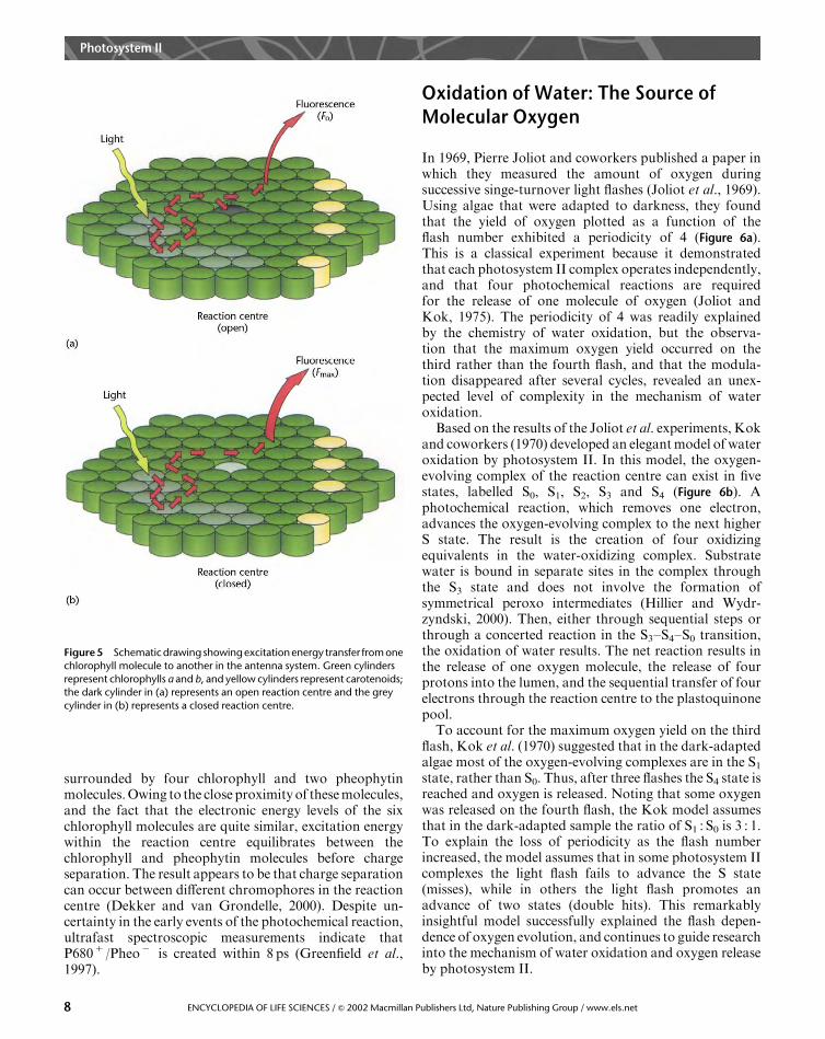
As shown in Figure 5, resonance energy transfer enablesexcitation energy to migrate over the antenna system.Because the first excited singlet state of chlorophyll a islower than that of chlorophyll b or the carotenoids,excitation energy is rapidly localized on the chlorophyll amolecules. As a consequence, the energy that escapes theantenna system as fluorescence comes almost entirely fromchlorophyll a.
During the migration process, the excitation energy iseither trapped by a reaction centre, converted into heat orreleased as photons. Photosynthetic antenna systems aredesigned to be very efficient at getting the excited stateenergy to a reaction centre. Measurements of photosynth-esis at low light intensities show that over 90%of absorbedphotons can be trapped by a reaction centre and promoteprimary charge separation. However, environmental con-ditions may impose limitations on photosynthesis thatsignificantly slow the rate of electron transport, whichgreatly increases the fraction of absorbed light energy thatgoes into heat and fluorescence. Measurements of chlor-ophyll fluorescence provide a noninvasive method formonitoring photosynthetic performance in vivo (seebelow).
Primary Photochemistry: The ReactionCentre
The primary photochemical reaction of photosystem IIdepends on electron transfer from P680 to pheophytin(Pheo), creating the charge separated state: P6801 /Pheo2
(Dekker and van Grondelle, 2000). This primary photo-chemical reaction differs from subsequent electron transferreactions in that the equilibrium redox midpoint potentialof the primary donor (P680) is lower in energy than that ofthe primary electron acceptor (Pheo). As a consequence,electron transfer from P680 to Pheo can occur only if P680is in an excited electronic state (denoted by P680*), which iscreated either by excitation energy from the antennasystem or by direct absorption of a photon by the reactioncentre.Subsequent electron transfer steps prevent the primary
charge separation from recombining by transferringthe electron within 200 ps from Pheo2 to QA (Figures 1,2 and 4). From QA
2 , the electron is transferred toanother plastoquinone molecule bound at the QB site.After two photochemical turnovers, QB becomes fullyreduced (PQH2), after which it unbinds from photo-system II and is released into the thylakoid membrane.While the electron removed fromP680 is rapidly sent away,an electron from a tyrosine residue (YZ) on the D1polypeptide reduces P6801 . Electrons for the reduction ofYZ are extracted from the water-oxidizing complex, whichincludes the (Mn)4 cluster. The rate of electron transferfrom YZ to P6801 ranges from 20 ns to 200ms, dependingon the redox state of components involved in wateroxidation.Although photochemistry in photosystem II leads to
charge separation between P680 and Pheo, the stepsleading to this relatively stable state are complicated andnot well understood. As shown in Figure 4b, P680 is
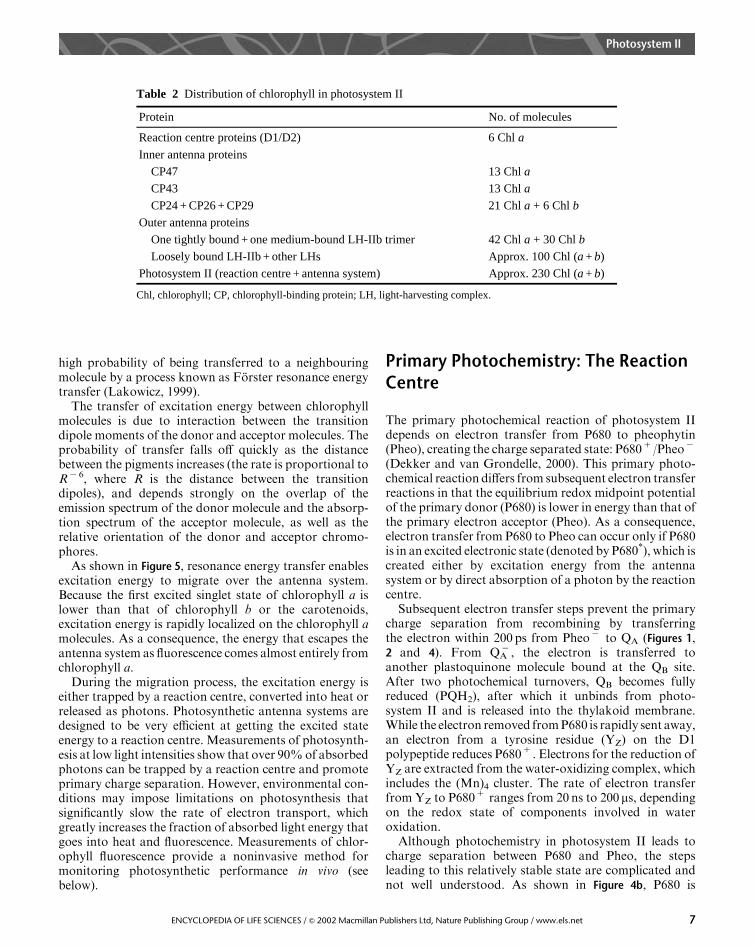
Table 2 Distribution of chlorophyll in photosystem II
Chl, chlorophyll; CP, chlorophyll-binding protein; LH, light-harvesting complex.
Protein No. of molecules
Reaction centre proteins (D1/D2) 6 Chl a
Inner antenna proteins
CP47 13 Chl a
CP43 13 Chl a
CP24 + CP26 + CP29 21 Chl a + 6 Chl b
Outer antenna proteins
One tightly bound + one medium-bound LH-IIb trimer 42 Chl a + 30 Chl b
Loosely bound LH-IIb + other LHs Approx. 100 Chl (a + b)
Photosystem II (reaction centre + antenna system) Approx. 230 Chl (a + b)
Photosystem II
7ENCYCLOPEDIA OF LIFE SCIENCES / & 2002 Macmillan Publishers Ltd, Nature Publishing Group / www.els.net
surrounded by four chlorophyll and two pheophytinmolecules.Owing to the close proximityof thesemolecules,and the fact that the electronic energy levels of the sixchlorophyll molecules are quite similar, excitation energywithin the reaction centre equilibrates between thechlorophyll and pheophytin molecules before chargeseparation. The result appears to be that charge separationcan occur between different chromophores in the reactioncentre (Dekker and van Grondelle, 2000). Despite un-certainty in the early events of the photochemical reaction,ultrafast spectroscopic measurements indicate thatP6801 /Pheo2 is created within 8 ps (Greenfield et al.,1997).
Oxidation of Water: The Source ofMolecular Oxygen
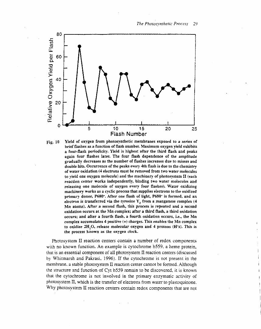
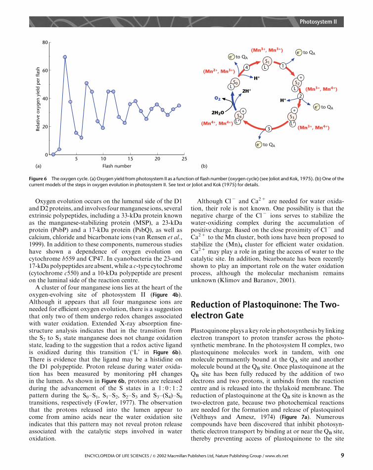
In 1969, Pierre Joliot and coworkers published a paper inwhich they measured the amount of oxygen duringsuccessive singe-turnover light flashes (Joliot et al., 1969).Using algae that were adapted to darkness, they foundthat the yield of oxygen plotted as a function of theflash number exhibited a periodicity of 4 (Figure 6a).This is a classical experiment because it demonstratedthat each photosystem II complex operates independently,and that four photochemical reactions are requiredfor the release of one molecule of oxygen (Joliot andKok, 1975). The periodicity of 4 was readily explainedby the chemistry of water oxidation, but the observa-tion that the maximum oxygen yield occurred on thethird rather than the fourth flash, and that the modula-tion disappeared after several cycles, revealed an unex-pected level of complexity in the mechanism of wateroxidation.Based on the results of the Joliot et al. experiments, Kok
and coworkers (1970) developed an elegantmodel of wateroxidation by photosystem II. In this model, the oxygen-evolving complex of the reaction centre can exist in fivestates, labelled S0, S1, S2, S3 and S4 (Figure 6b). Aphotochemical reaction, which removes one electron,advances the oxygen-evolving complex to the next higherS state. The result is the creation of four oxidizingequivalents in the water-oxidizing complex. Substratewater is bound in separate sites in the complex throughthe S3 state and does not involve the formation ofsymmetrical peroxo intermediates (Hillier and Wydr-zyndski, 2000). Then, either through sequential steps orthrough a concerted reaction in the S3–S4–S0 transition,the oxidation of water results. The net reaction results inthe release of one oxygen molecule, the release of fourprotons into the lumen, and the sequential transfer of fourelectrons through the reaction centre to the plastoquinonepool.To account for the maximum oxygen yield on the third
flash, Kok et al. (1970) suggested that in the dark-adaptedalgae most of the oxygen-evolving complexes are in the S1state, rather than S0. Thus, after three flashes the S4 state isreached and oxygen is released. Noting that some oxygenwas released on the fourth flash, the Kok model assumesthat in the dark-adapted sample the ratio of S1 : S0 is 3 : 1.To explain the loss of periodicity as the flash numberincreased, the model assumes that in some photosystem IIcomplexes the light flash fails to advance the S state(misses), while in others the light flash promotes anadvance of two states (double hits). This remarkablyinsightful model successfully explained the flash depen-dence of oxygen evolution, and continues to guide researchinto the mechanism of water oxidation and oxygen releaseby photosystem II.
Figure 5 Schematicdrawing showingexcitationenergy transfer from onechlorophyll molecule to another in the antenna system. Green cylinders
represent chlorophylls a and b, and yellow cylinders represent carotenoids;the dark cylinder in (a) represents an open reaction centre and the grey
cylinder in (b) represents a closed reaction centre.
Photosystem II
8 ENCYCLOPEDIA OF LIFE SCIENCES / & 2002 Macmillan Publishers Ltd, Nature Publishing Group / www.els.net
Oxygen evolution occurs on the lumenal side of the D1andD2proteins, and involves fourmanganese ions, severalextrinsic polypeptides, including a 33-kDa protein knownas the manganese-stabilizing protein (MSP), a 23-kDaprotein (PsbP) and a 17-kDa protein (PsbQ), as well ascalcium, chloride and bicarbonate ions (van Rensen et al.,1999). In addition to these components, numerous studieshave shown a dependence of oxygen evolution oncytochrome b559 and CP47. In cyanobacteria the 23-and17-kDapolypeptides are absent,while a c-type cytochrome(cytochrome c550) and a 10-kDa polypeptide are presenton the luminal side of the reaction centre.
A cluster of four manganese ions lies at the heart of theoxygen-evolving site of photosystem II (Figure 4b).Although it appears that all four manganese ions areneeded for efficient oxygen evolution, there is a suggestionthat only two of them undergo redox changes associatedwith water oxidation. Extended X-ray absorption fine-structure analysis indicates that in the transition fromthe S2 to S3 state manganese does not change oxidationstate, leading to the suggestion that a redox active ligandis oxidized during this transition (‘L’ in Figure 6b).There is evidence that the ligand may be a histidine onthe D1 polypeptide. Proton release during water oxida-tion has been measured by monitoring pH changesin the lumen. As shown in Figure 6b, protons are releasedduring the advancement of the S states in a 1 : 0 : 1 : 2pattern during the S0–S1, S1–S2, S2–S3 and S3–(S4)–S0transitions, respectively (Fowler, 1977). The observationthat the protons released into the lumen appear tocome from amino acids near the water oxidation siteindicates that this pattern may not reveal proton releaseassociated with the catalytic steps involved in wateroxidation.
Although Cl2 and Ca21 are needed for water oxida-tion, their role is not known. One possibility is that thenegative charge of the Cl2 ions serves to stabilize thewater-oxidizing complex during the accumulation ofpositive charge. Based on the close proximity of Cl2 andCa21 to the Mn cluster, both ions have been proposed tostabilize the (Mn)4 cluster for efficient water oxidation.Ca21 may play a role in gating the access of water to thecatalytic site. In addition, bicarbonate has been recentlyshown to play an important role on the water oxidationprocess, although the molecular mechanism remainsunknown (Klimov and Baranov, 2001).
Reduction of Plastoquinone: The Two-electron Gate
Plastoquinone plays a key role in photosynthesis by linkingelectron transport to proton transfer across the photo-synthetic membrane. In the photosystem II complex, twoplastoquinone molecules work in tandem, with onemolecule permanently bound at the QA site and anothermolecule bound at the QB site. Once plastoquinone at theQB site has been fully reduced by the addition of twoelectrons and two protons, it unbinds from the reactioncentre and is released into the thylakoid membrane. Thereduction of plastoquinone at the QB site is known as thetwo-electron gate, because two photochemical reactionsare needed for the formation and release of plastoquinol(Velthuys and Amesz, 1974) (Figure 7a). Numerouscompounds have been discovered that inhibit photosyn-thetic electron transport by binding at or near the QB site,thereby preventing access of plastoquinone to the site
02510
Rela
tive
oxyg
en y
ield
per
flas
h
Flash number
80
5 15 20
60
40
20
S1L
S2L
S0L
+
e–
e–
S3
L+
+S4
L+
+
2
14
3
(Mn3+, Mn3+) to QA
to QA
e– to QA
(Mn4+, Mn4+)
(Mn2+, Mn3+)
e– to QA
(Mn3+, Mn4+)
(Mn3+, Mn4+)
H+
H+
2H+
O2
2H2O
(a) (b)
Figure 6 The oxygen cycle. (a) Oxygen yield from photosystem II as a function of flash number (oxygen cycle) (see Joliot and Kok, 1975). (b) One of the
current models of the steps in oxygen evolution in photosystem II. See text or Joliot and Kok (1975) for details.
Photosystem II
9ENCYCLOPEDIA OF LIFE SCIENCES / & 2002 Macmillan Publishers Ltd, Nature Publishing Group / www.els.net

(Wraight, 1981). A few of these compounds (e.g. atrazine)are used as commercial herbicides.
The pathway of electrons from P680 to QB is shown inFigures 1 and 2. The reduction of plastoquinone requirestwo photochemical reactions. In the first reaction anelectron is transferred from QA
2 to QB within 100–200ms,producing the state QA/QB
2 (Figure 7b). In the secondreaction an electron is transferred from QA
2 to QB2 within
400–600ms, producing the state QA/QB2 2 , which rapidly
takes up protons, producing PQH2. Protons involved inthe reduction of plastoquinone come from the outer waterphase and are delivered by branched pathways through theprotein that include amino acids near the QB site. There isevidence that bicarbonate ions also play a role by bindingnear the QB site. Reduced plastoquinone unbinds from theQB site and enters the hydrophobic core of the membrane,which allows an oxidized plastoquinone molecule to bindto the site so that the cycle can be repeated.
Photosystem II Contributes to a ProtonElectrochemical Potential that DrivesATPase
The production of ATP in photosynthesis depends on theconversion of redox free energy into a proton electricalchemical potential, which is made up of a pH difference(DpH)andan electrical potential difference (DC) across thethylakoid membrane, and is given by the followingequation:
DmH1 5FDC2 2.3RTDpH
where F is the Faraday constant,R is the gas constant, andT is the temperature in kelvin. Photosystem II contributesto this protonpotential energy by: (1) the release of protonsduring the oxidation of water by photosystem II into thelumen; (2) the uptake of protons from the stromal phaseassociated with the reduction of plastoquinone at the QB
site – this reaction is the first half of a proton-transportingmechanism that is completed by the oxidation of plasto-quinol by the cytochrome bf complex, which releases theprotons initially taken up by photosystem II into thelumen; and (3) the creation of an electrical potential acrossthe membrane due to electron transfer through thephotosystem II reaction centre from water to plastoqui-none.
Downregulation: Energy is DivertedAway from the Reaction Centre whenthere is Excess Light
Although photosynthesis can be very efficient, environ-mental conditions typically impose severe limitations onboth rate and efficiency. One of the most common stresssituations for a photosynthetic organism is the absorptionof more light than they can use for carbon reduction.Excess light can drive inopportune electron transferreactions, which may cause both long-term and short-term damage to photosystem II, impairing photosyntheticproductivity.Photosynthetic organisms have evolved different strate-
gies to avoid injury due to excess light.Oneof the dominant
H+
H+
PQQAQBH2
QAQB2–
(H+) QA–QB
–(H+)
QAQB–(H+)
QA–QB(H+)QAQB
PQH2
QAQB(H+)
300–600 µs
100–200 µs
1
2
0 104
Chl
orop
hyll
a flu
ores
cenc
e, ∆
F (r
elat
ive
units
)
Number of preilluminating flashes
1.0
2 6 8
0.6
0.4
0.2
31 5 7 9
0.8
(a) (b)
Figure 7 The two-electron gate. (a) Chlorophyll a fluorescence from photosystem II as a function of flash number showing the two-flash dependence. (b)
Steps in the two-electron reduction of plastoquinone at the QB site of photosystem II. See text for details.
Photosystem II
10 ENCYCLOPEDIA OF LIFE SCIENCES / & 2002 Macmillan Publishers Ltd, Nature Publishing Group / www.els.net

protective mechanisms in plants and algae is known asdownregulation or nonphotochemical quenching: this is adynamic regulation of excitation energy transfer pathwayswithin the antenna system that diverts excitation energyinto heat before it reaches the reaction centre (Demmig-Adams et al., 1996). It is not unusual for half of theabsorbed quanta to be directed from the antenna array andconverted into heat. Under such conditions the efficiencyof photosynthetic light energy conversion can be reducedby more than 50%. It is not known why plants respond toexcess light by downregulating light capture rather thanincreasing photosynthetic capacity.
Secondary Electron Transfer Reactionsin Photosystem II Protect AgainstPhotodamage
Photosystem II is susceptible to damage by excess light.This is not surprising in viewof the fact that photosystem IImust switch between various high-energy states thatinvolve powerful oxidants required for the oxidation ofwater, and strong reductants for the reduction ofplastoquinone. In saturating light, a single reaction centrecan have an energy throughput of 600 eV s2 1, which isequivalent to 60 000 kW per mole of photosystem II. Toavoid damage, photosystem II contains redox componentsthat appear to serve as safety valves by accepting ordonating electrons at opportune times. For example, it hasbeen proposed that cytochrome b559 serves to deactivate ararely formed, but highly damaging, redox state ofphotosystem II, by accepting an electron from pheophytinwhen forward electron transfer is overloaded (Whitmarshand Pakrasi, 1996).
Inactive Photosystem II: A SignificantProportion of Reaction Centres do notWork In Vivo
Although most photosystem II reaction complexes workefficiently to oxidize water and reduce plastoquinone, anumber of in vivo assays have shown that a significantproportion are unable to transfer electrons to theplastoquinone pool at physiologically significant rates.Experiments using higher plants, algae and cyanobacteriaindicate that photosynthetically inactive photosystem IIcomplexes are a common feature of oxygenic organisms.For example, in healthy spinach leaves, in vivo measure-ments show that 30% of the photosystem II complexes areinactive (Chylla and Whitmarsh, 1989). Inactive photo-system II centres are impaired at the QA site, which isreoxidized approximately 1000 times slower than in active
centres. In addition, the antenna system serving inactivephotosystem II complexes is approximately half the size ofthat serving each active complex.Membrane fractionationstudies indicate that stromal membranes are enriched ininactive photosystem II centres, but they are also present ingranalmembranes. These differences, between the antennasize and membrane distribution of active and inactivecentres, are shared by photosystems IIa and IIb, which aredefined by their relative antenna size. It is not known whyplants contain photosystem II reaction centre complexesthat do not contribute to energy transduction, but it hasbeen estimated that inactive centres could reduce thequantum efficiency of photosynthesis by as much as 10%.
Fluorescence: Monitoring PhotosystemII Activity In Vivo
Measurements of chlorophyll fluorescence provide anoninvasive technique for monitoring photosyntheticprocesses in plants, algae and cyanobacteria. One of theprimary applications is determining the activity of photo-system II reaction centres in vivo. The technique relies onthe observation that the yield of chlorophyll fluorescencedepends in large part on the capacity of photosystem II tocarry out a stable charge separation between P680, theprimary donor, and QA, the primary quinone acceptor ofthe reaction centre. When QA is oxidized, the reactioncentre is able to utilize the light energy harvested by theantenna system for charge separation and the fraction ofexcitation lost to fluorescence is low, giving rise to lowfluorescence yields (Figure 5). In contrast, when QA isreduced, the reaction centre is unable to undergo stablecharge separation and the fraction of excitation lost tofluorescence is high, giving rise to the maximum fluores-cence yield.Measurements of the chlorophyll fluorescence emission
from leaves provide data for calculating photochemicalyields under physiological conditions (Schreiber et al.,1998). The recent introduction of highly sensitive charge-coupled device cameras has enabled instrumentation thatimages chlorophyll fluorescence in cells, leaves and plants(Nedbal et al., 2000; Holub et al., 2000). The next stage indevelopment is to use remote sensing to measure chlor-ophyll fluorescence dynamics for crops, forests, grasslandsand aquatic photosynthesis.
Summary
Photosystem II is amultiunit chlorophyll–protein complexthat uses light energy to transfer electrons from water toplastoquinone. It is located in the thylakoidmembrane andis made up of an antenna system, which captures light, and
Photosystem II
11ENCYCLOPEDIA OF LIFE SCIENCES / & 2002 Macmillan Publishers Ltd, Nature Publishing Group / www.els.net

a reaction centre core, which uses the light energy to driveelectron and proton transfer. The antenna system iscomposed of protein complexes that contain pigmentmolecules, chlorophyll a and b plus accessory molecules,which capture light by converting photon energy toexcitation energy. The reaction centre contains carriersthat create a pathway for electrons from water toplastoquinone. These carriers include a cluster of fourmanganese ions, a tyrosine (YZ) residue, a specialized pairof chlorophyll molecules (P680), pheophytin, a perma-nently bound plastoquinone (QA) and a plastoquinonethat binds reversibly to photosystem II at the QB site.
The primary photochemical reaction leads to theformation of a charge-separated state in which P680, aspecialized pair of chlorophyll molecules, is oxidized and apheophytin molecule is reduced. Oxidized P680 serves toremove electrons from water, while reduced pheophytinprovides the electrons for the reduction of plastoquinone.Four consecutive photochemical reactions lead to theoxidation of two water molecules, which results in therelease of one dioxygen molecule.
The structure of the photosystem II reaction centre isjust becoming available, which means that the long-timegoal of understanding the molecular mechanism of wateroxidation is finally within reach.
References
Chylla RA and Whitmarsh J (1989) Inactive photosystem II complexes
in leaves: turnover rate and quantitation. Plant Physiology 90: 765–
772.
Dekker JP and van Grondelle R (2000) Primary charge separation in
photosystem II. Photosynthesis Research 63: 195–208.
Demmig-Adams B, Gilmore A and Adams WW III (1996) In vivo
functions of carotenoids in plants. FASEB Journal 10: 403–412.
Des Marais DJ (2000) When did photosynthesis emerge on earth?
Science 289: 1703–1704.
Fowler CF (1977) Proton evolution from photosystem II. Stoichiometry
and mechanistic considerations. Biochimica et Biophysica Acta 462:
414–421.
Frank HA, Young AJ, Britton G and Cogdell RJ (eds) (1999) The
photochemistry of carotenoids, vol. 8. In: Govindjee (series ed.)
Advances in Photosynthesis. Dordrecht: Kluwer Academic.
Govindjee and Coleman W (1990) How does photosynthesis make
oxygen? Scientific American 262: 50–58.
Greenfield SR, SeibertM,Govindjee andWasielewskiMR (1997)Direct
measurement of the effective rate constant for primary charge
separation in isolated photosystem II reaction centers. Journal of
Physical Chemistry B101: 2251–2255.
Hillier W and Wydrzyndki T (2000) The affinities for the two substrate
water binding sites in theO2 evolving complex of Photosystem III vary
independently during the S-state turnover. Biochemistry 39: 4399–
4405.
Holub O, Seufferheld MJ, Gohlke C, Govindgee and Clegg RM (2000)
Fluorescence lifetime imaging (FLI) in real time – a new technique in
photosynthesis research. Photosynthica 38: 581–599.
Joliot P and Kok B (1975) Oxygen evolution in photosynthesis. In:
Govindjee (ed.) Bioenergetics in Photosynthesis, pp. 387–412. New
York: Academic Press.
Joliot P, Barbieri G and Chabaud R (1969) Un nouveau modele des
centres photochimiques du systeme II. Photochemistry and Photo-
biology 10: 309–329.
Klimov VV and Baranov SV (2001) Bicarbonate requirement for the
water-oxidizing complex of photosystem II. Biochimica et Biophysica
Acta 1503: 187–196.
Kok B, Forbush B and McGloin M (1970) Cooperation of charges in
photosynthetic oxygen evolution. Photochemistry and Photobiology
11: 457–475.
Kuhlbrandt W, Wang DN and Fujiyoshi Y (1994) Atomic model of
plant light harvesting complex by electron crystallography. Nature
367: 614–621.
Lakowicz JR (1999) Principles of Fluorescence Spectroscopy, 2nd edn.
New York: Kluwer Academic–Plenum.
Moser CC, Keske JM, Warncke K, Farid RS and Dutton PL (1992)
Nature of biological electron transfer. Nature 355: 796–802.
Nedbal L, Soukupova J, Whitmarsh J and Trilele M (2000) Postharvest
imaging of chlorophyll fluorescence from lemons can be used to
predict fruit quality. Photosynthetica 38: 571–579.
Nugent J (ed.) (2001) Photosynthetic water oxidation. Biochimica et
Biophysica Acta 1503: 1–259.
Ort D and Whitmarsh J (2001) Photosynthesis. Encyclopedia of Life
Sciences. London: Macmillan.
RheeK-H (2001) Photosystem II: the solid structural era.Annual Review
of Biophysical and Biomolecular Structure 30: 307–328.
Schreiber U, BilgerW,HormannH andNeubauer C (1998) Chlorophyll
fluorescence as a diagnostic tool: basics and some aspects of practical
relevance. In: RaghavendraAS (ed.)Photosynthesis: AComprehensive
Guide, pp. 320–336. Cambridge.
van Rensen JJS, Xu C and Govindjee (1999) Role of bicarbonate in
photosystem II, the water-plastoquinone oxido-reductase of plant
photosynthesis. Physiol. Plantarum 105: 585–592.
Velthuys BR and Amesz J (1974) Charge accumulation at the reducing
side of system 2 of photosynthesis. Biochimica et Biophysica Acta 325:
277–281.
Whitmarsh J and Pakrasi H (1996) Form and function of cytochrome
b559. In: Ort DR and Yocum CF (eds) Oxygenic Photosynthesis: The
Light Reactions, pp. 249–264. Dordrecht: Kluwer Academic.
Whitmarsh J and Govindjee (1999) The photosynthetic process. In:
Singhal GS, Renger G, Sopory SK, Irrgang K-D and Govindjee (eds)
Concepts in Photobiology: Photosynthesis and Photomorphogenesis,
pp. 11–51. Dordrecht: Kluwer Academic.
Wraight C (1981) Oxidation–reduction physical chemistry of the
acceptor complex in bacterial photosynthetic reaction centers:
evidence for a new model of herbicide activity. Israel Journal of
Chemistry 21: 348–354.
Xiong J, Subramaniam S and Govindjee (1998) A knowledge-based
three dimensional model of the photosystem II reaction center of
Chlamydomonas reinhardtii. Photosynthesis Research 56: 229–254.
Zouni A, Witt HT, Kern J et al. (2001) Crystal structure of photosystem
II from Synechococcus elongatus at 3.8Angstrom resolution. Nature
409: 739–743.
Further Reading
Blankenship RE (2002) Mechanisms of Photosynthesis. Oxford: Black-
well Science.
Falkowski PG and Raven JA (1997) Aquatic Photosynthesis. Oxford:
Blackwell Science.
Govindjee (1999) Milestones in photosynthesis research. In: Yunus M,
Pathre U and Mohanty P (eds) Probing Photosynthesis: Mechanisms,
Regulation and Adaptation, pp. 9–39. London: Taylor and Francis.
Photosystem II
12 ENCYCLOPEDIA OF LIFE SCIENCES / & 2002 Macmillan Publishers Ltd, Nature Publishing Group / www.els.net

Ke B (2001) Photosynthesis: Photobiochemistry and Photobiophysics.
Dordrecht: Kluwer Academic.
Ort D and Yocum CF (eds) (1996) Oxygenic photosynthesis: the light
reactions. In: Govindjee (ed.) Advances in Photosynthesis. Dordrecht:
Kluwer Academic.
RaghavendraAS (ed.) (1998)Photosynthesis: AComprehensive Treatise.
Cambridge: Cambridge University Press.
van Amerongen H, Valkunas L and van Grondelle R (2000)
Photosynthetic Excitons. Singapore: World Scientific.
Photosystem II
13ENCYCLOPEDIA OF LIFE SCIENCES / & 2002 Macmillan Publishers Ltd, Nature Publishing Group / www.els.net