18 report circular polarization vision in a stomatopod ... pdfs/chiou_etal_2008... · report...
TRANSCRIPT

Current Biology 18, 429–434, March 25, 2008 ª2008 Elsevier Ltd All rights reserved DOI 10.1016/j.cub.2008.02.066
ReportCircular Polarization Visionin a Stomatopod Crustacean
Tsyr-Huei Chiou,1,4 Sonja Kleinlogel,2,4,5 Tom Cronin,1
Roy Caldwell,3 Birte Loeffler,2,6 Afsheen Siddiqi,1
Alan Goldizen,2 and Justin Marshall2,*1Department of Biological SciencesUniversity of Maryland Baltimore County1000 Hilltop CircleBaltimore, Maryland 212502Sensory Neurobiology GroupVision Touch and Hearing Research CentreSchool of Biomedical Sciences and Queensland Brain InstituteThe University of QueenslandBrisbane, Queensland 4072Australia3Department of Integrative BiologyUniversity of California, Berkeley3060 Valley Life Sciences Building #3140Berkeley, California 94720-3140
Summary
We describe the addition of a fourth visual modality in theanimal kingdom, the perception of circular polarized light.
Animals are sensitive to various characteristics of light,such as intensity, color, and linear polarization [1, 2]. This
latter capability can be used for object identification, con-trast enhancement, navigation, and communication through
polarizing reflections [2–4]. Circularly polarized reflections
from a few animal species have also been known for sometime [5, 6]. Although optically interesting [7, 8], their signal
function or use (if any) was obscure because no visualsystem was known to detect circularly polarized light.
Here, in stomatopod crustaceans, we describe for the firsttime a visual system capable of detecting and analyzing cir-
cularly polarized light. Four lines of evidence—behavior,electrophysiology, optical anatomy, and details of signal
design—are presented to describe this new visual function.We suggest that this remarkable ability mediates sexual
signaling and mate choice, although other potential func-tions of circular polarization vision, such as enhanced con-
trast in turbid environments, are also possible [7, 8]. Theability to differentiate the handedness of circularly polarized
light, a visual feat never expected in the animal kingdom, isdemonstrated behaviorally here for the first time.
Results and Discussion
Stomatopod Visual Systems
Stomatopod crustaceans possess an unusual visual system,unlike that of any other animal yet described [9]. Their eyes
*Correspondence: [email protected] authors contributed equally to this work.5Present address: Max-Planck-Institut fur Biophysik, Max-von-Laue-
Strasse 3, D-60438 Frankfurt/M, Germany.6Present address: University of Applied Sciences Bremen, Dept. for Biomi-
metics, Neustadtswall 30, 28199 Bremen, Germany.
possess extraordinary capabilities such as tunable, eight-channel color vision [10–12], complex linear polarization vision[13, 14], potential monocular stereopsis or range-finding [15],and luminance and form vision. Here, we add another visualcapability to the repertoire of their astonishing eyes, circularpolarization vision, a previously unrecognized visual modality.To detect these many channels of information, stomatopodsemploy a basic retinal design element consisting of eight cells(called the rhabdom) common to many crustaceans. Eachrhabdom receives light through an individual set of optics:hexagonal lens elements and spacers called crystalline cones.The whole unit of photoreceptive rhabdom and optics com-bined is known as an ommatidium (Figure 1). Stomatopod om-matidia are highly modified and arranged into spatially dis-crete subsections within the eye [10, 13]. The six-rowmidband region of the eye (Figure 1) contains most of the un-usual retinal specializations [10, 11, 13]. We now demonstratethat two ommatidial rows within this midband are specializedfor circular polarization vision in some stomatopod species(Figures 1 and 2).
Circular Polarized Light: The Physical Basis
Before describing the sensory basis for circular polarizationvision in stomatopods, we first revise the physics of linearand circular polarization of light. Although humans are nor-mally unaware of any aspect of polarized light, we use linearpolarizing filters in sunglasses or camera filters to reduce theinherently polarized glare from reflective surfaces such aswater or glass [16]. Circular polarizing filters are also used,especially in photography, for related reasons [16]. Light thatis linearly polarized has its electric vector (e-vector) confinedto one orientation, e.g. vertical, and this vector is the resultof having both x and y vibrational e-vector components inphase [17]. When these components are not in phase, the re-sultant e-vector projects an ellipse as the ray of polarized lighttravels through space. When the phase difference is 6 90�, thee-vector describes a circle. This is circularly polarized light. Ifthe e-vector of circularly polarized light rotates in a counter-clockwise direction as seen from the direction of the sensor,it is called right-handed circularly polarized light (R-CPL). If itrotates in a clockwise direction, it is left-handed circularlypolarized light (L-CPL) (Figure 2; for a good description ofthis, see Chapter 33 in the Feynman Lectures on Physics[17]). One further detail of the physics needs mentioninghere. If circularly polarized light is passed through transparentmaterial that is birefringent (having nonisotropic refractiveproperties) with a thickness and refractive index such that lightis slowed down (retarded) by ¼ of its wavelength in one e-vec-tor orientation (this is called a ¼ wave plate or retarder), thenthe circularly polarized light becomes linearly polarized.Here, the 90� phase difference between x and y vectors incircularly polarized light is brought back into phase, and there-fore linearly polarized light results [16, 17]. Importantly for thestomatopod story, the angle of the resultant e-vector forleft-handed circularly polarized light is exactly orthogonal(at 90�) to that for right-handed circularly polarized light(Figure 2).

Current Biology Vol 18 No 6430
Detection of Circularly Polarized Light:The Structural Basics
Photoreceptors, in both vertebrates and invertebrates, may bepreferentially sensitive to a specific e-vector direction [1, 2]. Ininvertebrates such as stomatopods, this requires precisealignments of microvilli, the basic elements that constructthe photoreceptive rhabdom. Maximum sensitivity to linearlypolarized light in such a receptor occurs when the e-vector isparallel to the microvillar axis [18, 19]. Many insects, includingdesert ants, crickets, and bees, use receptors with specificallyaligned microvilli to detect the natural e-vector variation in thesky, using this information for orientation and navigation [1, 2,20]. Underwater, some crustaceans and cephalopods mayemploy linear-polarization-sensitive systems based on recep-tors with orthogonal microvilli. In crustaceans, this frequentlyresults in square- or diamond-shaped rhabdom profiles intransverse section (Figure 2) [9]. This organization is seen invarious areas of stomatopod compound eyes, most notablyin the main rhabdoms of midband rows five and six, theR1–7 cells (see Figures 1 and 2 and [13] for cell nomenclature).
Figure 1. Circular Polarizing Signals and General
Eye Anatomy in Stomatopods
(A) The stomatopod crustacean Odontodactylus
cultrifer (male). The scale bar represents 1 cm.
(Photograph by Chrissy Huffard.)
(B) Detail of telson keel (inset in [A]) photo-
graphed through a left-handed circular polarizing
filter.
(C) As (B) except photographed through a right-
handed circular polarizing filter. Note the striking
contrast difference compared to (B).
(D) The eye of Odontodactylus scyllarus, a close
relative of O. cultrifer, seen from the front. The
vertical line is section direction and extent in
(E). The following abbreviations are used: mid-
band (MB), dorsal hemisphere (DH), and ventral
hemisphere (VH). The scale bar represents
800 mm.
(E) Diagrammatic representation of a sagittal sec-
tion (line in [D]) of rows five and six of the mid-
band of the eye of a generalized gonodactyloid
stomatopod (for full details, see [10, 13, 26]).
The dotted line is approximate section level in
Figure 2A.
As occurs in many other crustaceans, insome stomatopods [14, 21, 22], theseand other photoreceptors are capableof sensing the linear polarization of light.
In common with most malacostracancrustaceans [9], stomatopods have ineach ommatidium a single retinular celldesignated R8 with its centrally posi-tioned rhabdom sitting on top of andoptically coupled to seven retinular cells(R1–7) that between them make a singlelonger rhabdom (Figure 2). Cells arenumbered by convention on the basisof their specific, asymmetrical positionaround the rhabdom (Figure 2) [13]. Cells1, 4, and 5 make microvilli orthogonallyoriented to those of cells 2, 3, 6, and 7,and these two cell populations normallyprovide opponent channels for linearpolarized light discrimination [22–24].
This basic system, also present in the peripheral or hemi-spheric region outside the midband of stomatopod eyes(Figure 1) [10, 13], is modified in rows five and six of the mid-band in species of Odontodactylus for circular polarizationvision, where the two cell populations (1, 4, and 5 versus 2,3, 6, and 7) swap linear polarization discrimination for circular.
The R1–7 cells of Odontodactylus sp. rows five and sixremain intrinsically linearly polarized light detectors, but onlyafter light has been converted to this state from circularlypolarized light by specialized overlying structures, formed bythe rhabdomeres of the R8 photoreceptor cells (Figure 2).These photoreceptors are anatomically distinct from otherR8s in the eye [10, 13] and act as ¼ wave plates with a fastaxis parallel to their microvilli [13, 17]. This optical activity ofthe R8 rhabdom can be seen using polarization microscopy(Figure 3). Because of the specific orientation of the R8 rhab-domeres over the R1–7 cells (their microvilli are at 45� to thoseof R1–7 in both rows, Figure 2) the linear polarization directionsresulting from conversion are orthogonal for L-CPL (+45�) andR-CPL (245�, Figure 2) and are thus oriented precisely for the

Circular Polarization Vision in Mantis Shrimp431
most efficient stimulation of the underlying orthogonal R1–7photoreceptor set. The overall result is an R-CPL- andL-CPL-detecting system in each ommatidium. There are actu-ally two ommatidial rows containing units of this system,midband rows five and six; note that row six has all elementsrotated 90� relative to row five (Figure 2) [10, 13].
Row Five and Six R8 Cells Are ¼ Wave RetardersTypical R8 cells in stomatopods and other crustaceans havemicrovilli that are either bidirectional or random in orientation[9, 13]. However, the R8 cells of rows five and six of the
Figure 2. The Anatomy of ¼ Wave Retardation and Polarization Sensitivity
(A) Semithin toluidine-blue-stained (2 mm) transverse section through the
rhabdoms of midband rows five and six at a level (indicated by dotted line
in Figure 1E) to include both R8 and R1–7 rhabdoms. Cell numbering
according to anatomical position is as previously published [13]. White
arrows indicate both the microvillar direction and the resulting direction of
linear polarization sensitivity. The scale bar represents 10 mm.
(B) Diagrammatic representations of transverse sections through the R8 and
R1–7 rhabdomeres of rows five and six in (A) [13, 26]. Note the 90� twist
between rows. The hatching shows microvillar direction in R8 (unidirec-
tional) and R1–7 (orthogonal) rhabdoms.
(C) Three dimensional diagrammatic representation of row 6 R8 and R1–7
rhabdom (left) and how circular polarized light is changed and detected
by this system (right). Arrows indicate three things: the direction of orthog-
onal microvillar layers in R1–7 cells, the cells producing these microvilli (1, 4,
and 5 and 2, 3, 6, and 7), and the circular-polarization-sensitivity handed-
ness that the R1–7 cells end up with as a result of the ¼ wave retardation
of the overlying R8 cell.
midband of stomatopods are structurally unusual, being ovoidin transverse section, packed with very precisely aligned,unidirectional microvilli, and having substantially longer rhab-domeres than other R8 cells (Figures 1 and 2) [10, 13]. It isapparently this ultrastructural modification of the R8 cellsthat results in ¼ wave retardation of light passing throughit’s rhabdom in Odontodactylus species (Figure 3). Althoughall other gonodactyloid and lysiosquilloid stomatopods alsopossess R8 rhabdoms similar to this in structure [13], it isnot known whether they have the same optical properties.
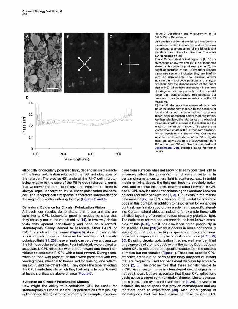
To estimate the R8 retardance, we measured the phase shiftinduced by the sections of the rhabdom with a polarizationmicroscope. Here, the sample, in this case the frozen sectioncontaining the R8 rhabdom viewed on axis (that is, lookingdown the length of the photoreceptor), is placed betweenthe two crossed polarizers of the polarization microscope.Light is polarized by the first polarizer and passes throughthe sample, and if the sample is birefringent, it becomes ellip-tical. The phase shift introduced by the sample that producesthis elipticity can be obtained by analysis of its polarizationstate (e.g., elipticity) with measured rotations of the secondpolarizer in the light path. We then calculated the retardanceon the basis of the approximate thickness of the section,around 10 mm, and the length of the whole rhabdom measuredby tracking the number of sections one can get from an R8 cell.Figure 3D shows the phase shift (f) of a whole length of the R8rhabdom as a function of wavelength (see [25] for full method).Our results indicate that the retardance of the R8 is slightlylower but fairly close to ¼ of a wavelength from 400 nm tonear 700 nm.
Microvilli in these R8 cells are oriented at 45� compared tothe orthogonal microvilli in the R1–7 cells rhabdom below(Figures 2 and 3 see also [13]), and it was this angle, the exactconformation required for conversion of CPL, that firstsuggested the possibility for circular polarization sensitivityin this eye. Three lines of evidence supporting this opticallyand anatomically driven hypothesis are now presented: elec-trophysiological, behavioral, and signal design.
Electrophysiological Evidence for CircularPolarization Vision
If it is true that the structural design of row five and six omma-tidia makes them sensitive to circularly polarized light, thenintracellular electrophysiological recordings from the R1–7cells should have the following properties. First, cells fromthe two orthogonal populations (1, 4, and 5 versus 2, 3, 6,and 7) should be sensitive only to either left- or right-handedcircularly polarized light. Results shown in Figures 4A and 4Cconfirm this prediction. Because of the 90� rotation betweenrows five and six, the same receptor cell populations in rowsfive and six are sensitive to the same handed CPL; i.e., recep-tor cells 1, 4, and 5 in both rows five and six are sensitive toL-CPL but not to R-CPL, and receptor cells 2, 3, 6, and 7 inboth rows five and six are sensitive to R-CPL but not toL-CPL. The identity of the cells was confirmed with intracellularstaining after recordings were made (Figure 4D). In addition tothis, all row five and six R1–7 receptor cells should be insensi-tive to the e-vector orientation of incoming linearly polarizedlight. That is, a light stimulus transmitted through a rotatinglinear polarizer should produce a constant response in thesecells, independent of the angle of rotation (and the resultante-vector angle) that they are exposed to. As shown inFigure 4B, this is what was found. The R8 rhabdomeres, actingas ¼ wave retarders, convert linearly polarized light into
Current Biology Vol 18 No 6432
Figure 3. Description and Measurement of R8
Cell ¼ Wave Retardance
(A) Semithin section of the R8 cell rhabdoms in
transverse section in rows five and six to show
the orthogonal arrangement of the R8 cells and
therefore their microvillar direction. The scale
bar represents 10 mm.
(B and C) Equivalent retinal region to (A), 10 mm
cryosection of row five and six R8 cell rhabdoms
viewed with a polarizing microscope. In (B), the
bright appearance of the R8 rhabdom eliptical
transverse sections indicates they are birefrin-
gent or depolarizing. The crossed arrows
indicate the microscope polarizer and analyzer
direction, and the disappearance of the bright
elipses in (C) when these are rotated 45� confirms
birefringence as the property of the material
rather than depolarization. This suggests but
does not prove ¼ wave retardance in the R8
rhabdoms.
(D) The R8 retardance was measured by record-
ing of the phase shift induced by the sections of
the rhabdom with a polarization microscope
in dark-field, or crossed-polarizer, configuration.
We then calculated the retardance on the basis of
the approximate thickness of the section and the
length of the whole rhabdom. The phase shift
(f) of a whole length of the R8 rhabdom as a func-
tion of wavelength is shown here. Our results
indicate that the retardance of the R8 is slightly
lower but fairly close to ¼ of a wavelength from
400 nm to near 700 nm. See the main text and
Supplemental Data available online for further
details.
elliptically or circularly polarized light, depending on the angleof the linear polarization relative to the fast and slow axes ofthe retarder. The precise 45� angle of the R1–7 cell microtu-bules relative to the axes of the R8 ¼ wave retarder ensuresthat whatever the state of polarization transmitted, there isalways equal absorption by a linear-polarization-sensitivecell. The receptor cell’s response is therefore independent ofthe angle of e-vector entering the eye (Figures 2 and 3).
Behavioral Evidence for Circular Polarization Vision
Although our results demonstrate that these animals aresensitive to CPL, behavioral proof is needed to show thatthey actually make use of this ability [14]. In two-way choicetests with operant conditioning and food as a reward,stomatopods clearly learned to associate either L-CPL orR-CPL stimuli with the reward (Figure 5). As with their abilityto distinguish colors or the e-vector orientation of linearlypolarized light [14, 26] these animals can perceive and analyzethe light’s circular polarization. Four individuals were trained toassociate L-CPL reflection with a food reward and three indi-viduals to associate R-CPL with a food reward. During tests,when no food was present, animals were presented with twofeeding tubes, identical to those used for training, one reflect-ing L-CPL and the other R-CPL. They chose the tube reflectingthe CPL handedness to which they had originally been trainedat levels significantly above chance (Figure 5).
Evidence for Circular Polarization SignalsHow might the ability to discriminate CPL be useful forstomatopods? Humans use circular polarization filters (usuallyright-handed filters) in front of cameras, for example, to reduce
glare from surfaces while not allowing linearly polarized light toadversely affect the camera’s internal sensor systems. Incertain circumstances where light is scattered, e.g., in turbidmedia or living tissue, the light can become circularly polar-ized, and in these instances, discriminating between R-CPLand L-CPL may be useful for enhancing the contrast betweenobjects and their background [7, 8]. CPL exists in the marineenvironment [27], so CPL vision could be useful for stomato-pods in this context. In addition to its potential for enhancingcontrast, such vision could play a role in intraspecific signal-ing. Certain natural objects, including for example those witha helical layering of proteins, reflect circularly polarized light.The cuticles of scarab beetles provide the best known exam-ples of this [5, 6], but it has also been noted previously incrustacean tissue [28] (where it occurs in areas not normallyvisible). Stomatopods use highly specialized color and linearpolarization signals for complex social interactions [4, 26, 29,30]. By using circular polarization imaging, we have identifiedthree species of stomatopods within the genus Odontodactuswhere CPL is reflected from specific locations on the cuticlesof males but not females (Figure 1). These sex-specific CPL-reflective areas are on parts of the body (uropods or telson)that are frequently used for behavioral displays by stomato-pods [2, 8]. The precise role that these signals, visible toa CPL visual system, play in stomatopod sexual signaling isnot yet known, but we speculate that these CPL reflectionscould act as a secret communication channel. Linear polariza-tion signals, used by marine invertebrates [4, 30], are visible toanimals like cephalopods that prey on stomatopods and aretherefore open to exploitation [30]. Also, other genera ofstomatopods that we have examined have variable CPL

Circular Polarization Vision in Mantis Shrimp433
Figure 4. Electrophysiology of Circular-Polarization-Sensitive Photoreceptors
(A) Intensity-response curves (R-logI, normalized voltage response versus log intensity) of an R2 cell in row five to right- and left-handed circularly polarized
white light. The cell is stimulated with increasing light intensity through a right-handed (triangles) and left-handed (squares) providing circular polarizing
filter. Circular polarization sensitivity (CPS) is defined by the intensity shift (DS in log units—see arrow) between the two R-logI curves such that CPS =
10DS. This cell is sensitive to right-handed circular polarized light with a CPS of 10.2.
(B) Similar recording as (A) but this shows the cells response to linear polarized light. A linear polarization filter was arranged with its axis at 6 45�, the angles
that would theoretically elicit maximum and minimum response in row 5 R2 cell if it was sensitive to linear polarized light. Significantly, the cell shows very
low (1.38) linear polarization sensitivity because of conversion of the incoming linear polarized light to circular polarized light that can not be detected by
rhabdomeric photoreceptors.
(C) Average (10.4) of six intensity-response curves with 6 standard deviation (SD) to left- and right-handed circular polarization flashes of increasing inten-
sity. Symbols are as in (A).
(D) Dye-filled cells shown in 7 mm transverse sections of row five and six photoreceptors in the left eye (for orientation with such sections, see [13]). In row
five, cell R1 is filled with Lucifer yellow, and in row six, cell R5 is filled with ethidium bromide. Each cell showed CPS similar to that in (A), although both cells
here showed left-handed circular polarization sensitivity. The scale bar represents 10 mm.
sensitivity and may be unable to view the sexual displays ofOdontodactylus species, making this a private channel ofcommunication, unavailable to both predators and potentialstomatopod competitors.
Whatever the use of CPL signals and CPL vision to stomato-pods, comparing design features of their CPL reflectors andsensors to those of man-made systems will be interesting.Humans use CPL filters and imaging in everyday photography,medical photography, and object-detection systems in turbidenvironments [7, 8]. The reefs and waters that many stomato-pods inhabit are often turbid, and it is no surprise that, perhapsas long as 400 million years ago (when stomatopod crusta-ceans first appeared), nature got there first.
Supplemental Data
Experimental Procedures are available at http://www.current-biology.com/
cgi/content/full/18/6/429/DC1/.
A
W
f
v
l
o
R
t
e
J
B
w
b
c
o
R
R
A
P
cknowledgments
e would like to thank Horace Barlow for seeding the initial idea, Mike Land
or continual inspiration, and Jack Pettigrew for enthusiasm and advice. Da-
id Vaney and Andrew White provided help with optical and electrophysio-
ogical equipment. This work was supported by grants from the Asian Office
f Aerospace Research and Development, the Air Force Office of Scientific
esearch, the Australian Research Council, the National Science Founda-
ion, and the Swiss National Foundation. T-H.C. and S.K. are responsible
qually for the majority of the work described here, under the guidance of
.M and T.C., and are viewed as shared first author. Work by S.K. and
.L. was all performed at The University of Queensland. Behavioral tests
ere performed both in the USA by T.C., R.C., and A.S. and in Australia
y J.M., A.G., and B.L. Animal signals were identified, photographed, and
haracterized by R.C. All authors discussed the results and commented
n the manuscript.
eceived: January 20, 2008
evised: February 13, 2008
ccepted: February 13, 2008
ublished online: March 20, 2008

Current Biology Vol 18 No 6434
References
1. Wehner, R. (1983). The perception of polarised light. In The Biology of
Photoreception, D.J. Cosens and D. Vince-Prue, eds. (Cambridge, Lon-
don: Cambridge University Press), pp. 331–369.
2. Horvath, G., and Varju, D. (2004). Polarised Light in Animal Vision (New
York: Springer).
3. Wehner, R., and Lanfranconi, B. (1981). What do ants know about the
rotation of the sky? Nature 293, 731–733.
Figure 5. Behavioral Results, Discriminating Left- from Right-Circular-
Polarization-Reflecting Feeding Tubes
(A) Diagrammatic representation of the construction of feeding tubes. The
end of each tube is made to reflect either left-handed (L) or right-handed
(R) circular polarized light. This is achieved by gluing of a laminate of two fil-
ters, a linear polarizing filter, and a ¼ wave retarder on top of a diffuse white
reflective background disk (shown here as light gray for diagram clarity) that
is in turn glued on one end of the feeding tube, blocking this end completely.
During training, food is placed in the other, open end of the tube. In training
and test situations, two tubes are presented to the animals, circular-polar-
izing-reflector-side first. Animals rapidly learn to pick up and feed from
tubes and learn to discriminate L from R feeding tubes (see the Supplemen-
tal Data for more detailed description and [14] for similar experimental pro-
cedure).
(B) Photographs of the L- and R-circular-polarized-light-reflecting ends of
feeding tubes: left, as seen through no filter; middle, through a right-handed
circular polarizing filter analyzer; and right, through a left-handed circular
polarizing filter analyzer. The tube on the left reflects left-handed circular po-
larizing light, and the one on the right reflects right-handed circular polariz-
ing light. This ‘‘intensity difference’’ between tubes is therefore only seen by
a system capable of circular polarization sensitivity.
(C) Operant condition training tests where individual stomatopods (O. scyl-
larus) were required to learn that food was present in either feeding tubes.
Black bars are correct choices, and gray bars are incorrect choices. The
chance level is half way up each combined bar. In a Fisher’s exact test
(one tailed), * indicates choices are made significantly more often than by
chance; p values as follows: Individual 1, 0.045; 2, 0.034; 3, 0.072; 4, 0.040;
5, 0.049; 6, 0.072; and 7, 0.013.
4. Cronin, T.W., Shashar, N., Caldwell, R.L., Marshall, N., and Chiou, T.H.
(2003). Polarization vision and its role in biological signaling. Integr.
Comp. Biol. 43, 549–558, Cheroske AG.
5. Neville, A.C., and Caveney, S. (1969). Scarabid beetle exocuticle as an
optical analogue of cholesteric liquid crystals. Biol. Rev. Camb. Philos.
Soc. 44, 531–562.
6. Brink, D.J., van der Berg, N.J., Prinsloo, L.C., and Hodgkinson, I.C.
(2007). Unusual colouration in scarabid beetles. J. Phys. D Appl. Phys.
40, 2189–2196.
7. Yao, G., and Wang, L.V. (1999). Two-dimensional depth-resolved
Mueller matrix characterization of biological tissue by optical coherence
tomography. Opt. Lett. 24, 537–539.
8. Rakovic, M.J., Kattawar, G.W., Mehrubeoglu, M., Cameron, B.D., Wang,
L.V., Rastegar, S., and Cote, G.L. (1999). Light backscattering polariza-
tion patterns from turbid media: Theory and experiments. Appl. Opt. 38,
3399–3408.
9. Marshall, N.J., Kent, J., and Cronin, T.W. (1999). Visual adaptations in
crustaceans. In Adaptive Mechanisms in the Ecology of Vision, S.
Archer, M. Djamgoz, E. Lowe, J.C. Partridge, and S. Vallerga, eds. (Lon-
don: Kluwer), pp. 285–328.
10. Marshall, N.J. (1988). A unique colour and polarisation vision system in
mantis shrimps. Nature 333, 557–560.
11. Cronin, T.W., and Marshall, J. (1989). A retina with at least ten spectral
types of photoreceptors in a mantis shrimp. Nature 339, 137–140.
12. Cronin, T.W., Caldwell, R.L., and Marshall, N.J. (2001). Tunable colour
vision in a mantis shrimp. Nature 411, 547–548.
13. Marshall, N.J., Land, M.F., King, C.A., and Cronin, T.W. (1991). The
compound eyes of mantis shrimps (Crustacea, Hoplocarida, Stomato-
poda). I. Compound eye structure: The detection of polarised light.
Philos. Trans. R. Soc. Lond. B Biol. Sci. 334, 33–56.
14. Marshall, J., Cronin, T.W., Shashar, N., and Land, M. (1999). Behavioural
evidence for polarisation vision in stomatopods reveals a potential
channel for communication. Curr. Biol. 9, 755–758.
15. Schiff, H., Abbott, B.C., and Manning, R.B. (1985). Possible monocular
range-finding mechanisms in stomatopods from different environmen-
tal light conditions. Comp. Biochem. Physiol. A 80, 271–280.
16. Goldstein, D. (2003). Polarised Light (New York: Marcel Dekker).
17. Feynman, R.P., Leighton, R.B., and Sands, M. (1971). Lectures on
Physics (London: Addison-Westley).
18. Kirschfeld, K., and Snyder, A.W. (1975). Waveguide mode effects.
birefringence and dichroism in fly photoreceptors. In Photoreceptor Op-
tics, A.W. Snyder and R. Menzel, eds. (New York: Springer), pp. 56–77.
19. Snyder, A.W. (1973). Polarisation sensitivity of individual retinula cells.
J. Comp. Physiol. [A] 83, 331–360.
20. Labhart, T. (1999). How polarisation-sensitive interneurons of crickets
see the polarisation pattern of the sky: A field study with an optoelec-
tronic model neuron. J. Exp. Biol. 202, 757–770.
21. Cronin, T.W., Marshall, N.J., Quinn, C.A., and King, C.A. (1994). Ultravi-
olet photoreception in mantis shrimp. Vision Res. 34, 1443–1452.
22. Kleinlogel, S., and Marshall, J.N. (2006). Electrophysiological evidence
for linear polarisation sensitivity in the compound eyes of the stomato-
pod crustacean Gonodactylus chiragra. J. Exp. Biol. 209, 4262–4272.
23. Sabra, R., and Glantz, R.M. (1985). Polarisation sensitivity of crayfish
photoreceptors is correlated with their termination sites in the lamina
ganglionaris. J. Comp. Physiol. [A] 156, 315–318.
24. Kleinlogel, S., Marshall, N.J., Horwood, L.M., and Land, M.F. (2003).
Neuroarchitecture of the color and polarization vision system of the
stomatopod Haptosquilla. J. Comp. Neurol. 467, 326–342.
25. Huard, S. (1997). Polarization of Light (Paris: John Wiley and Sons).
26. Marshall, N.J., Cronin, T.W., and Kleinlogel, S. (2007). Stomatopod eye
structure and function: A review. Arthropod Struct. Dev. 4, 420–448.
27. Ivanoff, A., and Waterman, T.H. (1958). Elliptical polarisation of
submarine illumination. J. Mar. Res. 16, 255–282.
28. Neville, A.C., and Luke, B.M. (1971). Form optical activity in crustacean
cuticle. J. Insect Physiol. 17, 519–526.
29. Caldwell, R.L. (1991). Variation in reproductive behavior in stomatopod
crustacea. In Crustacean Sexual Biology, R.T. Bauer and J.W. Martin,
eds. (New York: Columbia University Press), pp. 67–90.
30. Chiou, T.-H., Cronin, T.W., Caldwell, R.L., and Marshall, N.J. (2005).
Biological polarized light reflectors in stomatopod crustaceans. SPIE
5888, 1–9.