17.occlusal schemes anatomic and semiamatomic occlusion
TRANSCRIPT

17. Occlusal Schemes - Anatomic and 17. Occlusal Schemes - Anatomic and Semi-anatomicSemi-anatomic
John Beumer III, DDS, MSJohn Beumer III, DDS, MSandand
Michael Hamada DDSMichael Hamada DDSDivision of Advanced Prosthodontics, Division of Advanced Prosthodontics, Biomaterials and Hospital DentistryBiomaterials and Hospital Dentistry
UCLA School of DentistryUCLA School of Dentistry
This program of instruction is protected by copyright ©. No portion of This program of instruction is protected by copyright ©. No portion of this program of instruction may be reproduced, recorded or transferred this program of instruction may be reproduced, recorded or transferred by any means electronic, digital, photographic, mechanical etc., or by by any means electronic, digital, photographic, mechanical etc., or by any information storage or retrieval system, without prior permission.any information storage or retrieval system, without prior permission.

Semi-anatomic Denture TeethSemi-anatomic Denture Teeth
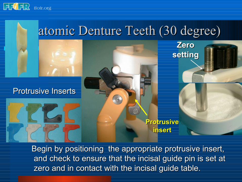
Begin by positioning the appropriate protrusive insert, Begin by positioning the appropriate protrusive insert, and check to ensure that the incisal guide pin is set at and check to ensure that the incisal guide pin is set at zero and in contact with the incisal guide table. zero and in contact with the incisal guide table.
Protrusive Protrusive insertinsert
Protrusive Inserts Protrusive Inserts
Zero Zero settingsetting

Semi-anatomic Denture TeethSemi-anatomic Denture Teeth
Background - Balanced occlusionBackground - Balanced occlusionOur objective in setting anatomic or semi-anatomic posterior teeth is to create a balanced occlusion. We wish to insure that all the posterior teeth as well as the anterior teeth maintain contact in lateral excursions.
To ensure bilateral balance we place an anterior-posterior curve in the arch, called a compensating curve, which is analogous to the curve of Spee in natural dentition.
In addition , we place a curve from side to side, the so called curve of Wilson.

Mark the casts indicating midline, crest of the ridge, and the retromolar pad. These landmarks will be used to check your denture setup.
MaxillaMidlineAnterior land
MandibleRidgeRetromolar pad
Cast LandmarksCast Landmarks

Anterior land
Cast Landmarks - MaxillaCast Landmarks - Maxilla
Midline
Incisive papilla

Lines indicating the crest of the ridge
Cast Landmarks -MandibleCast Landmarks -MandibleMidpoint of
retromolar pad
Land Mark on land indicating the midpoint of the retromolar pad

As previously mentioned (13c, 1a), the wax rim is ideally contoured on As previously mentioned (13c, 1a), the wax rim is ideally contoured on the patient and used to mount the upper cast with a facebow transfer the patient and used to mount the upper cast with a facebow transfer record. When the lower cast is mounted on the articulator with a centric record. When the lower cast is mounted on the articulator with a centric relation record the plane of occlusion is readily seen.relation record the plane of occlusion is readily seen.
The three landmarks used to identify the plane of occlusion are:The midpoint of the retromolar pads bilaterally as previously marked on the mandibular cast.The incisal edge of the maxillary central incisors
Setting the Maxillary Anterior TeethSetting the Maxillary Anterior Teeth

To set the remaining maxillary anterior teeth a clear glass or plastic slab is positioned on the mandibular record base to represent the plane of occlusion. When setting anatomic posterior teeth we recommend setting the maxillary posterior teeth before the mandibular posterior teeth. To aid in positioning the maxillary teeth, a line is inscribed on the slab indicating the crest of the mandibular ridge.
Setting the Maxillary Anterior TeethSetting the Maxillary Anterior Teeth
Mark indicating midpoint Mark indicating midpoint of the retromolar padof the retromolar pad

Setting the Maxillary Anterior TeethSetting the Maxillary Anterior TeethThese two lines, inscribed on the plastic plane, indicate the crest of the alveolar ridge. These lines will be used to position the maxillary posterior denture teeth to insure that the mandibular posterior teeth are centered over the ridge.
The lingual cusp tips of the posterior maxillary teeth should contact these lines.Lines indicating the Lines indicating the
crest of the ridgecrest of the ridge

Setting the Maxillary Anterior TeethSetting the Maxillary Anterior Teeth
Soften some baseplate wax and attach some to the ridge lap portion of the other maxillary central incisor and attach it to the record base as shown. Set the lateral incisors and cuspids as shown previously (Section 13c, 1a Lingualized occlusion).

Setting the Maxillary Anterior TeethSetting the Maxillary Anterior Teeth
Note the angulations of the anterior teeth in relation to the occlusal plane when viewed in profile.
Occlusal Occlusal planeplane

Setting the Maxillary Anterior TeethSetting the Maxillary Anterior Teeth
““Toed-in” PositionToed-in” Position
Note how the cervical and incisal edges of the cuspid are aligned vertically (yellow line). The facial surface of the cuspid however, is canted inward and appears “toed in” (red line) due to the prominence of the cervical area of the tooth (yellow arrow). The centrals and laterals are inclined slightly towards the distal.

The long axis of the premolars should be perpendicular to the occlusal plane and the buccal and lingual cusp tips should touch the occlusal plane. Arranging the premolars in this way insures that the adjacent marginal ridges will be on the same level. This is an important factor when setting the opposing premolars.
Setting the Maxillary Posterior TeethSetting the Maxillary Posterior Teeth
Occlusal Occlusal planeplane

Setting the Maxillary Posterior TeethSetting the Maxillary Posterior Teeth
Both the buccal and lingual cusp tips of the maxillary premolars should contact the plane of occlusion. The lingual cusp tips should also contact the line inscribed on the plastic plane indicating the crest of the mandibular ridge. This will ensure that when the opposing mandibular denture teeth are properly positioned and in occlusion, their central fossae will be centered over the mandibular ridge.
The Maxillary Premolars

Setting the Maxillary Posterior TeethSetting the Maxillary Posterior Teeth
The curve of Wilson and the curve of Spee begin in the molar region. The mesial lingual cusp tip of the 1st molar contacts the occlusal plane but the buccal cusp tips and the distal lingual cusp are elevated about .5mm off the
occlusal plane (yellow line).
The Maxillary 1st Molar

Setting the Maxillary Posterior TeethSetting the Maxillary Posterior Teeth
The Maxillary 2nd Molar
The set up viewed in profile. Note that the mesial lingual cusp tip touches the plane of occlusion along with the buccal and lingual cusps of the premolars. The curve of Spee begins at the 1st molar.

Setting the Maxillary Posterior TeethSetting the Maxillary Posterior Teeth
The Maxillary 2nd Molar
The curve of Spee is continued by elevating the 2nd molar off the plane of occlusion as shown. The 2nd molar is elevated to an even greater degree than the 1st molar, about 15 degrees in the average patient. When viewed from the distal it is set, like the 1st molar, with a slight curve of Wilson.
Occlusal Occlusal planeplane

Setting the Mandibular Posterior TeethSetting the Mandibular Posterior Teeth
Note that with this particular posterior tooth form, the mandibular Note that with this particular posterior tooth form, the mandibular cusps tips are designed to engage the embrasures of the opposing cusps tips are designed to engage the embrasures of the opposing maxillary teeth. This true of almost all anatomic tooth forms maxillary teeth. This true of almost all anatomic tooth forms designed for bilateral balance except the Ivoclar Ortholingual.designed for bilateral balance except the Ivoclar Ortholingual.
Completed set up
CentricCentric

Setting the Mandibular Posterior TeethSetting the Mandibular Posterior Teeth
The Mandibular 1st Molar
Begin by positioning the mandibular 1st molar. The mesial buccal cusp tip should engage the embrasure between the 1st molar and 2nd premolar. Therefore adjacent marginal ridges of the maxillary premolar and molar must be at the same level for the lower molar to properly engage them.

Setting the Mandibular Posterior TeethSetting the Mandibular Posterior Teeth
The Mandibular 1st Molar
Check the relationship from the lingual side. Make sure that the maxillary lingual cusp tips engage the the central fossa of the mandibular molar.
Setting the Mandibular Posterior TeethSetting the Mandibular Posterior Teeth
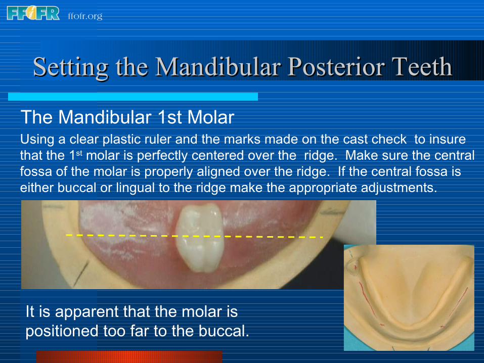
The Mandibular 1st MolarUsing a clear plastic ruler and the marks made on the cast check to insure that the 1st molar is perfectly centered over the ridge. Make sure the central fossa of the molar is properly aligned over the ridge. If the central fossa is either buccal or lingual to the ridge make the appropriate adjustments.
It is apparent that the molar is positioned too far to the buccal.
Setting the Mandibular Posterior TeethSetting the Mandibular Posterior Teeth
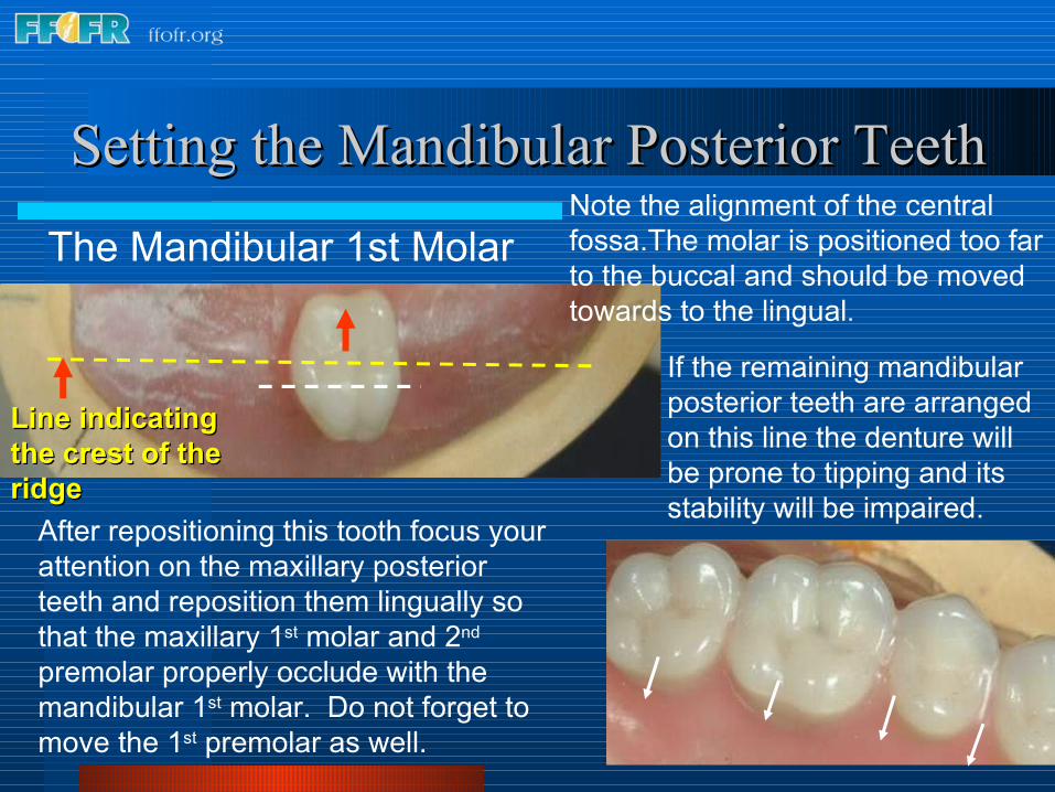
The Mandibular 1st MolarNote the alignment of the central fossa.The molar is positioned too far to the buccal and should be moved towards to the lingual.
If the remaining mandibular posterior teeth are arranged on this line the denture will be prone to tipping and its stability will be impaired.
Line indicating Line indicating the crest of the the crest of the ridgeridge
After repositioning this tooth focus your attention on the maxillary posterior teeth and reposition them lingually so that the maxillary 1st molar and 2nd premolar properly occlude with the mandibular 1st molar. Do not forget to move the 1st premolar as well.

Setting the Mandibular Posterior TeethSetting the Mandibular Posterior Teeth
The Mandibular 1st Molar
The molar is twisted and positioned too far to the lingual. If the remaining posterior mandibular teeth are arranged along this line the space for the tongue will likely be insufficient. The molar must be moved slightly to the buccal and twisted clockwise so that the central fossa is parallel to the ridge line.
After repositioning this tooth the posterior maxillary teeth should be moved to the buccal so that they properly occlude with the mandibular molar and permit positioning the remaining mandibular posterior teeth over the ridge.
Line Line indicating indicating the crest of the crest of the ridgethe ridge

Setting the Mandibular Posterior TeethSetting the Mandibular Posterior Teeth
The Mandibular Premolars - Centric
Position the mandibular premolars. The buccal cusp of the 2nd premolar should occlude with the the adjacent marginal ridges of the maxillary 1st and 2nd premolars.

Setting the Mandibular Posterior TeethSetting the Mandibular Posterior Teeth
The Mandibular Premolars - Centric
The buccal cusp of the mandibular 1st premolar should engage the mesial marginal ridge of the opposing 1st premolar. There should be a space between the 1st premolar and the cuspid.

Setting the Mandibular Posterior TeethSetting the Mandibular Posterior Teeth
The Mandibular Premolars - Centric
Verify that the premolars are in solid centric occlusion. If lingual centric contacts are lacking, contacts in balancing position will be lacking.
Setting the Mandibular Posterior TeethSetting the Mandibular Posterior Teeth
The Mandibular Premolars - Working
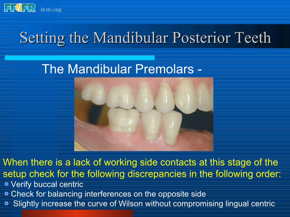
When there is a lack of working side contacts at this stage of the setup check for the following discrepancies in the following order:
Verify buccal centric Check for balancing interferences on the opposite side Slightly increase the curve of Wilson without compromising lingual centric

Complete Denture OcclusionComplete Denture Occlusion
The Mandibular Premolars - Working
During the lateral excursion into working, with this cusp form, when the teeth are properly arranged, there should be no lingual cusp contacts between the maxillary and mandibular teeth.

Setting the Mandibular Posterior TeethSetting the Mandibular Posterior Teeth
The Mandibular Molars – Balancing
When you lack balancing side contacts at this stage of the setup, check for the following discrepancies in the following order.
Verify lingual centric Check for working interferences on the opposite side
Setting the Posterior Mandibular TeethSetting the Posterior Mandibular Teeth
The Mandibular 2nd Molar - Centric
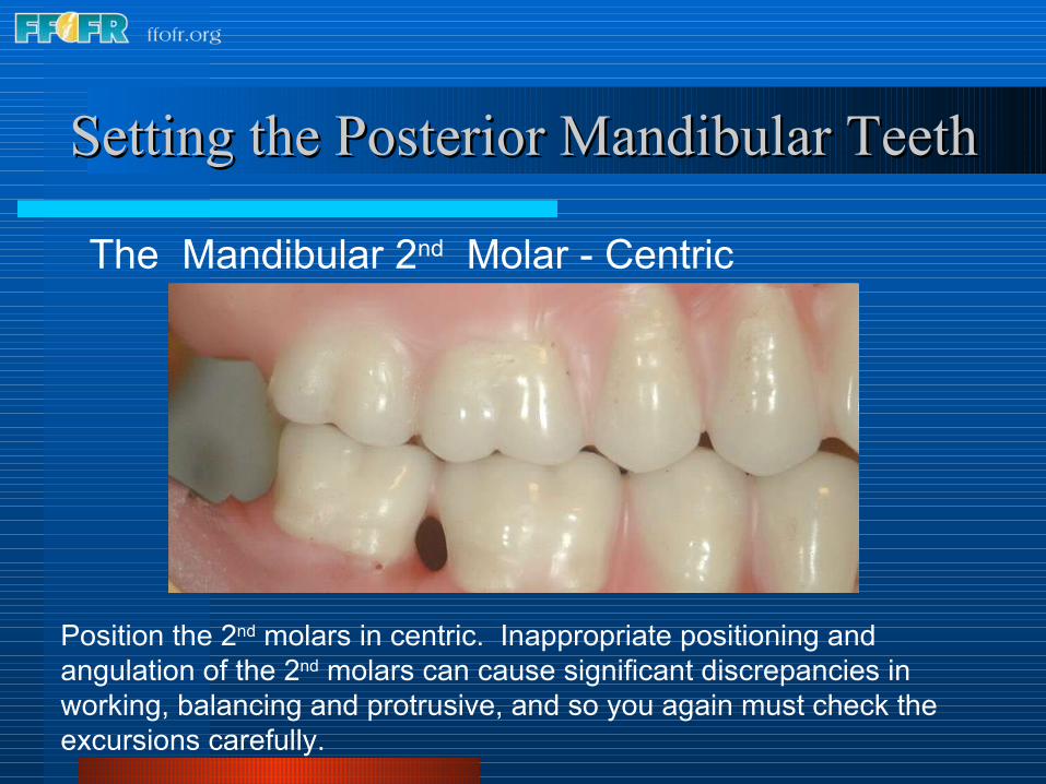
Position the 2nd molars in centric. Inappropriate positioning and angulation of the 2nd molars can cause significant discrepancies in working, balancing and protrusive, and so you again must check the excursions carefully.

Setting the Mandibular Posterior TeethSetting the Mandibular Posterior Teeth
The Mandibular 2nd Molar - Centric
Check lingual centric. Make sure that the lingual cusps of the maxillary 2nd molar properly occludes with the central fossa of the mandibular 2nd molar as shown.

Setting the Mandibular Posterior TeethSetting the Mandibular Posterior Teeth
Verify contacts in working excursions
Lack of working side contacts may be the result of: Poor buccal centric Insufficient curve of Wilson Working interferences in the 2nd molar region Balancing interferences on the opposite side **Check these in Check these in
the order citedthe order cited..

Setting the Mandibular Posterior TeethSetting the Mandibular Posterior Teeth
Verify contacts in during balancing excursion
Lack of balancing side contacts may be the result of: Poor lingual centric Working interferences on the opposite side, particularly in the 2nd
molar region

Setting the Mandibular Anterior TeethSetting the Mandibular Anterior Teeth
The Central Incisors
Begin by setting the central incisors. The mesial surfaces should be on the midline.

Setting the Mandibular Central IncisorsSetting the Mandibular Central Incisors
In most patients the labial surface of the mandibular incisors should be roughly perpendicular to the occlusal plane.
Occlusal Occlusal planeplane
The Central IncisorsThe Central Incisors

Setting the Mandibular Anterior TeethSetting the Mandibular Anterior Teeth
Determining the Amount of Vertical and Horizontal Overlap
The articulator is placed in the working and protrusive position, and the position of the central incisors adjusted to permit passive contact during lateral excursions.
WorkingWorking ProtrusiveProtrusive

Setting the Mandibular Anterior Teeth Setting the Mandibular Anterior Teeth
Horizontal Horizontal overlap overlap
This practice will idealize the amount of horizontal and vertical overlap This practice will idealize the amount of horizontal and vertical overlap and ensure that anterior guidance is not introduced into the setup. and ensure that anterior guidance is not introduced into the setup.
Horizontal Horizontal overlapoverlap
Vertical Vertical overlapoverlap

Setting the Mandibular Anterior TeethSetting the Mandibular Anterior Teeth
The Lateral Incisors and the Cuspids
Position the lateral incisors and cuspids in the same fashion as the central incisors. Take care not to introduce anterior guidance into the set up. The anterior teeth should be in only passive contact during lateral excursions.
WorkingWorking ProtrusiveProtrusive

Setting the Mandibular Anterior TeethSetting the Mandibular Anterior Teeth
The Lateral Incisors and the Cuspids
Upon completion, the amount of vertical and horizontal overlap will be idealized and anterior guidance has not been introduced into the setup. Bilateral balance has been maintained.

Semi-anatomic Denture TeethSemi-anatomic Denture Teeth Check to ensure you have Check to ensure you have
retained appropriate contacts in retained appropriate contacts in working, balancing andworking, balancing and protrusive.protrusive.
BalancingBalancing WorkingWorking
ProtrusiveProtrusive
Completed set up
CentricCentric

Anatomic Denture Teeth (30 degree)Anatomic Denture Teeth (30 degree)

Anatomic Denture Teeth (30 degree)Anatomic Denture Teeth (30 degree)
These teeth are arranged in the same fashion as the semi-anatomic teeth shown previously.Use the same sequence of steps as we have just shown.
Anatomic Denture Teeth (30 degree)Anatomic Denture Teeth (30 degree)
Begin by positioning the appropriate protrusive insert, Begin by positioning the appropriate protrusive insert, and check to ensure that the incisal guide pin is set at and check to ensure that the incisal guide pin is set at zero and in contact with the incisal guide table. zero and in contact with the incisal guide table.
Protrusive Protrusive insertinsert
Protrusive Inserts Protrusive Inserts
Zero Zero settingsetting

Setting Anatomic Teeth (30 degree)Setting Anatomic Teeth (30 degree)
When you are finished check to see that the posterior When you are finished check to see that the posterior teeth are on plane and the posterior teeth centered over teeth are on plane and the posterior teeth centered over the mandibular ridge. Make corrections as necessary.the mandibular ridge. Make corrections as necessary.
Setting Anatomic Teeth (30 degree)Setting Anatomic Teeth (30 degree)
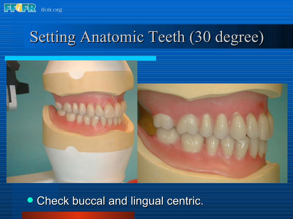
Check buccal and lingual centric.Check buccal and lingual centric.

Setting Anatomic Teeth (30 degree)Setting Anatomic Teeth (30 degree)
Check to ensure you have retained appropriate Check to ensure you have retained appropriate contacts in working, balancing andcontacts in working, balancing and protrusive.protrusive.
Balancing Protrusive
Working

Anatomic and Semi-anatomic Denture TeethAnatomic and Semi-anatomic Denture Teeth
Trouble shooting
Lack of working side contacts
Verify buccal centric. Restore centric contacts as necessary.
Increase the curve of Wilson Eliminate any anterior guidance present on the
working side. Check for balancing interferences on the opposite
side and make adjustments as necessary.

Anatomic and Semi-anatomic Denture TeethAnatomic and Semi-anatomic Denture Teeth
Trouble shooting
Lack of balancing side contacts
Verify lingual centric. Restore missing contacts as necessary.
Eliminate any anterior guidance present on the opposite or working side.
Check for working interferences on the opposite side particularly in the 2nd molar region.