14. muscle fiber plasticity - animal...
TRANSCRIPT

1
ANSC/FSTC 607 Biochemistry and Physiology of Muscle as a Food
MYOFIBRIL SYNTHESIS AND MUSCLE FIBER PLASTICITY
I. Conversion of myotubes to myofibers
A. Fusion of myoblasts à myotubes
1. Large increase in transcription,
translation of myofibrillar proteins
2. Later migration of myofibrillar
proteins (e.g., desmin) to Z-lines
4. Cytoplasm and nuclei in core of
myotube.
B. Formation of myofibrils
1. Aggregation of Z-line material (α-
actinin) around filaments
2. Synthesis of myofilaments, no
apparent development of sarcomeres
3. Synthesis of sarcomeres and
appearance of striations
2
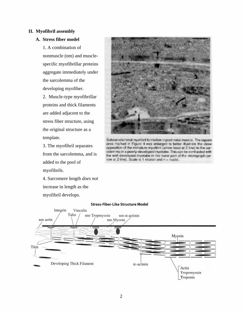
II. Myofibril assembly
A. Stress fiber model
1. A combination of
nonmuscle (nm) and muscle-
specific myofibrillar proteins
aggregate immediately under
the sarcolemma of the
developing myofiber.
2. Muscle-type myofibrillar
proteins and thick filaments
are added adjacent to the
stress fiber structure, using
the original structure as a
template.
3. The myofibril separates
from the sarcolemma, and is
added to the pool of
myofibrils.
4. Sarcomere length does not
increase in length as the
myofibril develops.

3
B. Premyofibril model
1. A premyofibril containing only nonmuscle proteins forms under the sarcolemma.
2. This detaches from sarcolemma and serves as the template for the developing
myofibril.
3. Nonmuscle proteins are gradually replaced with muscle-type myofibrillar proteins.
4. There is a lengthening of the sarcomere, which does not occur in the stress fiber
model.

4
III. Changes in fiber type distributions during growth
A. Type I myofibers
1. Even at birth, type I myofibers
(lightly stained) make up only a small
proportion of total myfibers.
2. Change in muscle mass is caused by
an increase in myofiber diameter.
B. Type II myofibers
1. Most myofibers in the neonate are
type II.
2. The proportion of type IIB increases
and IIA decreases.

5
IV. Conversion of type II
myofibers to type I
myofibers in early postnatal
muscle
A. In adult muscle, there is
a clear distinction
between type I and type
II myofibers.
1. Type I myofibers
stain strongly for acid-
stable ATPase.
2. Type I myofibers
stain strongly for
mitochondrial
activities. 2. SM = strong for acid-stable ATPase, medium for alkali-stable ATPase.
3. Type II myofibers
stain strongly for
alkali-stable ATPase.
Histochemical profiles of myofiber types of the serratus ventralis thoracis muscle of adult sheep. Myosin ATPase activity after preincubation at pH 4.3 (1a), 4.4 (1b), and 10.3 (1c). NADH-TR, G-3-P, and ß-HBD (1d – 1f).

6
B. At birth, a small population
of myofibers stains both
for acid- and alkali-stable
ATPase.
1. SS = strongly staining
for both acid- and alkali-
stable ATPase.
2. SM = strong for acid-
stable ATPase, medium for
alkali-stable ATPase. 2. SM = strong for acid-stable ATPase, medium for alkali-stable ATPase.
3. MS = medium for acid-
stable ATPase, strong for
alkali-stable ATPase.
C. These “intermediate” types
convert to type I myofibers
within weeks after birth.
1. All “intermediate” types
disappear.
2. Type I myofibers
increase in proportion.
Changes in proportions of the number (solid line) and cross-sectional area (dashed line) in myofibers. The decline in type II myofibers is due to a sharp decline if type IIA (fast-twitch oxidative) to type IIB (fast-twitch glycolytic). Type IIB are completely absent at birth in this muscle.
Histochemical profiles of myofiber types of the serratus ventralis thoracis muscle 0-week-old sheep. Myosin ATPase activity after preincubation at pH 4.3 (1a), 4.4 (1b), and 10.3 (1c). NADH-TR, G-3-P, and ß-HBD (1d – 1f). Myofiber types are labeled I, II, SM, SS, and MS.

7
V. Abnormal muscle growth A. Double-muscle cattle
1. Semitendinosus and
longissimus muscles contain 3.36
and 3.77 x 10⁶ myofibers.
2. This is twice as many as in
normal cattle.
3. The myostatin gene, which
halts myotube formation, is
mutated.
B. Callipyge lambs
1. Larger than normal myofibers
2. Same number of myofibers as normal sheep.
3. Defect is not due to myostatin.