1.3 cells and transport across them
DESCRIPTION
pTRANSCRIPT

Section 3.1 – Investigating the structure of cells Microscopy Lenses work more effectively if they are in a compound light microscope. Light waves a have a relatively long wavelength; therefore, they can only distinguish between objects that are at least 0.2 micrometers apart. Beams of electrons have shorter wavelengths and are therefore able to distinguish between objects as close as 0.1 nm apart. Magnification When viewed under a microscope, the material seen in called an image. Magnification tells you how many times bigger the image is in relation to the actual size of the object. It can be found using the following formula: Magnification = size of image/size of object The previous formula can also be rearranged to find the size of an object. Size of object = size of image/magnification Resolution The resolving power of a microscope is the minimum distance two objects can be apart in order for them to appear as separate items. The greater the resolution, the greater the clarity of the image that is produced. Cell fractionation Cell fractionation is the process where cells are broken up and the different organelles they contain are separated out. Before fractionation begins, the cells are put in a solution that is: Cold – to reduce enzyme activity that might break down the organelles. Isotonic – to prevent organelles bursting or shrinking as a result of osmotic gain or loss of water. An isotonic solution is one that has the same water potential as the original tissue. Buffered – to maintain a constant pH. Homogenation Cells are broken up by a homogeniser that releases the organelles. The fluid is called a homogenate. It is then filtered to remove complete cells and large pieces of debris.

Ultracentrifugation Ultracentrifugation is the process by which the homogenate is separated in a machine called a centrifuge. This spins tubes of the homogenate, creating a centrifugal force that makes the mixture separate.
• The tube of filtrate is placed in the ultracentrifuge and spun at a slow speed. • The heaviest organelles such as the nucleus are forced to the bottom where
they form a thin sediment. • The fluid at the top, called the supernatant is removed, leaving just the
sediment of nuclei at the bottom. • The supernatant is then put in another tube where it is spun at an even higher
speed than before. • The next heaviest organelles (mitochondria) are forced to the bottom and the
process continues until all the organelles are separated.

Section 3.2 – The electron microscope Electrons have a shorter wavelength than light and so they have a greater resolving power. As electrons are negatively charged, the beam can be focused using an electromagnet. Because electrons are absorbed by molecules in the air, a near vacuum must be created within the chamber of an electron microscope for it to work effectively.
There are two types of electron microscope: - Transmission electron microscope and scanning electron microscope. Transmission electron microscope The TEM consists of a gun that fires electrons which are focused onto the specimen by an electromagnet. Some of the electrons are absorbed by the specimen and appear dark on the image; other parts allow the electrons through and so appear light. This produces an image of the specimen.
The image that appears on screen is called a photomicrograph. Because the process takes place in a vacuum, living specimens cannot be observed. A complex staining process is required and even then the image is only in B&W. The specimen must be extremely thin. Artefacts (structure not present in the organism when it was alive) may appear on the image, these appear as a result of the way the specimen is prepared. Scanning electron microscope All the limitations of the TEM apply to the SEM but the specimen does not have to be extremely thin as the electrons do not penetrate. The beam of electrons is directed over the surface of the specimen in a regular pattern. The electrons bounce on the contours of the specimen and are scattered. The scattering of the electrons can be analysed and from this an image can be produced using a computer. The SEM has a lower resolving power than the TEM (20nm) but is still ten times better than a light microscope.

Section 3.3 – Structure of epithelial cells Epithelial cells are eukaryotic cells. Eukaryotic cells have a distinct nucleus and a membrane that surrounds each organelle. The function of an epithelial cells is to absorb and secrete. The nucleus
The nucleus controls the cells activities and contains hereditary material.
• The Nuclear envelope is a double membrane that surrounds the nucleus. Its outer membrane is continuous with the endoplasmic reticulum and often has ribosomes on its surface. It can control the substances entering and leaving
the nucleus.
• Nuclear pores allow the passage of large materials into and out of the nucleus.
• Nucleoplasm is granular jelly like material that makes up the bulk of the nucleus.
• Chromatin is the DNA found within the nucleoplasm This is the diffuse form
chromosomes take up when the cells is not dividing.
• The nucleolus is small spherical body within the nucleoplasm. It manufactures ribosomal RNA and assembles ribosomes.
The mitochondria
• A double membrane surrounds the
organelle, the outer one controlling the entry and exit of material. The inner membrane inner membrane is folded to form extensions known as cristae.
• Cristae are shelf like extensions of the
inner membrane. These provide a large surface area for the attachment of enzymes during respiration.

• The matrix makes up the remainder of the mitochondria. It is a semi-rigid material that contains proteins, lipids and traces of DNA that allows the mitochondria to control the production of its own proteins. The enzymes involved in respiration are found in the matrix.
Mitochondria are responsible for the production of the energy-carrier molecule ATP. Because of this, high numbers of mitochondria are found in cells where there is a high level of metabolic activity. Endoplasmic Reticulum
Rough endoplasmic reticulum – has ribosomes present on the outer surface of the membranes. Its functions are to: a) provide a large surface area for the synthesis of proteins and glycoproteins, b) provide a pathway for the transport of materials, especially proteins throughout the cell. Smooth endoplasmic reticulum - lacks ribosomes on its surface and is often more tubular in appearance.
Its functions are to: a) synthesise, store and transport lipids, b) synthesise store and transport carbohydrates. Golgi Apparatus
The Golgi apparatus is similar to the SER in structure but is more compact. It consists of a stack of membranes that form flattened sacks, or cisternae with small rounded hollow structures called vesicles. The proteins and lipids produced in the ER are passed through the Golgi apparatus in strict sequence. The Golgi apparatus modifies these proteins often by adding non-protein structures to them such as carbohydrates. It is also labels them so they can be sorted
and sent to their correct destination. Once sorted and modified, proteins are transported in vesicles which are regularly removed from the edge of the Golgi cisternae.

These vesicles move to the cell membrane where they fuse and release their contents to the outside (exocytosis). Lysosomes Lysosomes are formed when a vesicle contains enzymes. Lysosomes isolate potentially dangerous enzymes from the rest of the cell before releasing them outside of the cell or into phagocytic vesicles within the cell. Lysosomes digest worn out organelles so that the useful chemicals they are made of can be reused. They can completely break down cells after they have died. (Apoptosis) Ribosomes
Ribosomes occur in either the cytoplasm or the RER. There are two types depending on which cell they are found in: 80S Type – found in eukaryotic cells, is around 25nm in diameter. 70S Type – found in prokaryotic cells, is slightly smaller.
Microvilli Microvilli are finger like projections of the epithelial cells. Their function is to increase the surface area for diffusion.

Section 3.4 - Lipids
• Lips contain carbon, hydrogen and oxygen. • The proportion of oxygen to carbon and hydrogen is smaller than in
carbohydrates. • They are insoluble in water. • They are soluble in organic solvents such as alcohol and acetone.
Roles of lipids Phospholipids contribute to the flexibility of membranes and the transfer of lipid-soluble substances across them. In addition to this, lipids can be used as: An energy source. Lipids can provide more than twice the energy of carbohydrate. Waterproofing. Lipids are insoluble in water and are therefore suitable for waterproofing. Insulation. Fats are slow conductors of heat, kept under skin to retain heat in the body. Protection. Often stored around delicate organs. Triglycerides are so called because they have three fatty acids (tri-) combined with glycerol (-glyceride). Each fatty acids combines with glycerol in a condensation reaction. CH2OH + HOOC CH2OOC + H2O (Glycerol) + (fatty acid) Phospholipids Phospholipids are similar to lipids except that the fatty acid is replaced with a phosphate molecule. Fatty acid molecules repel water whereas phosphate molecules are attracted to water. Test for lipids
1. Take a dry, grease free test tube.
2. Take 2cm3 of the sample being tested and add 5cm3 of ethanol.
3. Shake the test tube and dissolve the lipids.
4. Add 5cm3 of water and shake gently.
5. A cloudy white colour indicates the presence of a lipid.
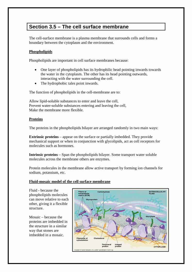
Section 3.5 – The cell surface membrane The cell-surface membrane is a plasma membrane that surrounds cells and forms a boundary between the cytoplasm and the environment. Phospholipids Phospholipids are important in cell surface membranes because:
• One layer of phospholipids has its hydrophilic head pointing inwards towards the water in the cytoplasm. The other has its head pointing outwards, interacting with the water surrounding the cell.
• The hydrophobic tales point inwards. The function of phospholipids in the cell-membrane are to: Allow lipid-soluble substances to enter and leave the cell, Prevent water-soluble substances entering and leaving the cell, Make the membrane more flexible. Proteins The proteins in the phospholipids bilayer are arranged randomly in two main ways: Extrinsic proteins – appear on the surface or partially imbedded. They provide mechanical support or when in conjunction with glycolipids, act as cell receptors for molecules such as hormones. Intrinsic proteins – Span the phospholipids bilayer. Some transport water soluble molecules across the membrane others are enzymes. Protein molecules in the membrane allow active transport by forming ion channels for sodium, potassium, etc. Fluid-mosaic model of the cell surface membrane Fluid - because the phospholipids molecules can move relative to each other, giving it a flexible structure. Mosaic – because the proteins are imbedded in the structure in a similar way that stones are imbedded in a mosaic.

Section 3.6 - Diffusion Diffusion is defined as the net movement of molecules or ions from a region where they are more highly concentrated to one where their concentration is lower. All particles are constantly in motion due to the kinetic energy that they posses. The motion is random and there is no set pattern to the way they move. Rate of diffusion
• The greater the difference in concentration, the greater the rate of diffusion
• The larger the area of an exchange surface, the greater the rate of diffusion.
• The thinner the exchange surface, the faster the rate of diffusion.
• The nature of the plasma membrane; its composition and the number of pores.
• The size and nature of the diffusing molecule. For example smaller molecules diffuse faster than big ones.
Diffusion is proportional to: surface area x difference in concentration Length of diffusion path Facilitated diffusion Facilitated is a passive process as it only relies on the kinetic motion of particles. Facilitated diffusion can only occur at specific point along the plasma membrane where there are special protein molecules. The proteins for special water filled channels. The channels only open for specific molecules. This allows water soluble ions and molecules to pass through. Such molecules such as glucose and amino acids would take much longer to diffuse through the phospholipids bilayer. When a molecule that is specific to the carrier protein is present, the carrier protein changes shape, causing it to release the molecule on the other side of the plasma membrane.

Section 3.7 – Osmosis Osmosis is defined as the passage of water from a region where it has a higher water potential to a region where it has a lower water potential through a partially permeable membrane. Water potential is measured in Pascal’s. Under standard conditions of temperature (25oC), pure water is said to have a water potential of 0. Water with a solute dissolved in it will have a water potential that is less than 0. Water molecules move from one side where the water potential is higher (less negative) across a partially permeable membrane to another side where the water potential is lower (more negative). The water moves along a water potential gradient. At the point where the water potentials on either side of a partially permeable membrane are equal, a dynamic equilibrium is established and there is no net movement of water. Osmosis in animal cells If a red blood cell is place in pure water it will absorb water by osmosis because it has a lower water potential. The cell-surface membrane will eventually burst if too much water enters the cells. To prevent cells bursting due to too much water entering the cells, cells are often bathed in solutions where the water potential outside the cell is the same as the water potential inside the cell. This is called an isotonic solution. A hypotonic solution is one where the concentration outside is greater than the concentration inside. A hypertonic solution is one where the water potential outside the cell is lower than
the water potential inside the cell.

Section 3.8 – Active transport Active transport allows cells to exchange molecules against a concentration gradient. Metabolic energy is required for this process. Active transport is the movement of molecules or ions into or out of a cell from a region of lower concentration to a region on higher concentration using energy and carrier molecules. Metabolic energy is needed in the form ATP. Carrier molecules which act as “pumps” are involved.
• Active transport uses ATP in two main ways: by using ATP to directly move molecules.
• By using a concentration gradient that has already been set up by direct active
transport. This is known as co-transport.
• The carrier molecules accept molecules or ions to be transport on one side of it.
• The molecules of the ions bind to the receptors on the channels of the carrier protein.
• On the other side of the membrane ATP bind to the carrier protein causing it
to split into ADP and a phosphate molecule. This as a result, causes the carrier protein to change shape, releasing the molecule onto the other side.
• The phosphate molecule then recombines with the ADP to form ATP again,
which causes the carrier protein to revert back to its original shape. Occasionally, the molecule or ion is moved into the cell at the same time as a different one is being removed from it. One example of this is the sodium-potassium pump Sodium ions are actively taken in by the cell whilst potassium ones are actively removed from the cell.

Section 3.9 – Absorption in the small intestine Villi and Microvilli Villi have walls lined with epithelial cells. Villi are situated at the interface between the lumen of the intestines and the blood and other tissues inside the body. Their properties increase the efficiency of absorption because:
• They increase the surface area for diffusion
• They are very thin walled, thus reducing the distance over which diffusion takes place.
• They are able to move and so maintain a concentration gradient
• They are well supplied with blood vessels so that the blood can carry away
absorbed molecules and hence maintain a diffusion gradient. The epithelial cells possess Microvilli which further increase the surface area for diffusion. They are situated on the cell surface membrane. Villi contain muscles which move the food ensuring the glucose is absorbed from the food adjacent to the villi, new glucose rich food replaces it, thus maintains a concentration gradient for diffusion. Role of active transport in absorption The way in which most glucose is absorbed from small intestine is an example of co-transport.
1. Sodium ions are actively transported out of the epithelial cells by the sodium potassium pump.
2. There is now a much higher concentration of sodium ions in the lumen than in
the cells.
3. The sodium ions diffuse into the cells down a concentration gradient. As they flood back into the cells, they are coupled with glucose molecules which are drawn in with them.
4. The glucose diffuses into the blood through a carrier protein.
It is the sodium ion concentration, rather than the ATP directly, that powers the movement of glucose into the cell.

Section 3.10 – Cholera Structure of a bacteria cell The bacterium that causes cholera is called Vibrio Cholerae.
• All bacteria possess a cells wall that is made up of peptidoglycan. This is a mixture of polysaccharides and peptides.
• Many bacteria also protect themselves by producing a capsule of mucilaginous slime around this wall.
• Flagella occur at certain types of bacteria.
• Inside the cell-surface membrane is
the cytoplasm that contains ribosomes that are smaller than the ones found in eukaryotic cells. (70s type)
• Bacteria store food as glycogen granules and oil droplets.
• The genetic material of a bacterium is found in the form of a circular strand of DNA.
• Separate from this are smaller circular pieces of DNA, called plasmids. How the cholera bacterium causes disease Almost all Vibrio cholerae bacteria ingested by humans are killed by the low pH in the stomach but many can still survive, especially if the pH is above 4.5. When the bacteria enter the lumen of the small intestine they use their flagella to propel themselves through the mucus lining of the intestinal wall. They then start to produce a toxic protein. The protein has two parts: one part binds to the carbohydrate receptors of the intestinal epithelial cells, whereas the other part enters the epithelial cells. The causes the ion channels of the cell-surface membrane to open, that the chloride ions that are normally contained within the epithelial cells flood into the lumen of the intestine. The loss of chloride ions from cells increases the water potential in the cell, but lowers the water potential outside the cells. This causes water to move into the small intestine. The loss of ions from the cells establishes a concentration gradient. Ions move by diffusion into the epithelial cells. This establishes a water potential gradient that causes water to move by osmosis from the blood and other tissues into the small intestine.

Section 3.11- Oral rehydration therapy What causes diarrhoea?
• Damage to the epithelial cells in the lining of the small intestine
• Loss of Microvilli due to toxins
• Excessive secretion of water due to toxins What is oral rehydration therapy? Drinking water to treat diarrhoea is ineffective because: Water is not being absorbed by the intestine. Indeed, as in the case of cholera, water is actually being lose from cells. The drinking water does not replace electrolytes that are being lost from cells of the intestine. As sodium ions are being absorbed, the water potential falls and water enters the cells by osmosis. A rehydration solution should therefore contain:
• Water – to rehydrate tissues
• Sodium – to replace the ions lost from the epithelium of the small intestine and to make optimum use of the sodium-glucose carrier proteins.
• Glucose – to stimulate the uptake of sodium ions from the intestine and to
provide energy
• Potassium – to replace lost potassium ions and to stimulate appetite
• Other electrolytes – such as chloride and citrate ions, to help prevent electrolyte imbalance
The solution must be given regularly and in large amounts whilst the person has the illness.