128-slice dual source coronary cta: defining optimal arterial enhancement levels
TRANSCRIPT
ORIGINAL ARTICLE
128-Slice dual source coronary CTA: defining optimal arterialenhancement levels
Ashkan A. Malayeri & Stefan L. Zimmerman &
Spencer T. Lake & Elliot K. Fishman & Pamela T. Johnson
Received: 25 November 2013 /Accepted: 13 March 2014# Am Soc Emergency Radiol 2014
Abstract This study aims to correlate coronary artery en-hancement levels with quality of vessel visualization andcalcified plaque visualization using a 128-slice dual-sourceCT (DSCT) scanner. Coronary CT angiography exams from52 patients, mean age of 55 years (range, 22–90) and meanweight of 184 lbs (range, 120–320 lbs), were reviewed retro-spectively. Contrast infusion rates ranged from 4.5 to 7 mL/s(mean, 5.8 mL/s). Postcontrast density of the largest calcifiedplaque and postcontrast density of the left main (LM) andright coronary arteries (RCA) were recorded. Enhancementquality was graded as 1=suboptimal, 2=adequate for diagno-sis, and 3=excellent. Pre- and postcontrast acquisitions werecompared for calcified plaque conspicuity. The largest calci-fied plaque density was a mean of 862 HU (range, 376 to1,384 HU) on the postcontrast scan. The mean LM and RCAcoronary artery enhancement levels for studies of excellentenhancement quality (N=43) were 468 and 457 HU, respec-tively, higher than mean enhancement levels of 320 and322 HU for adequate enhancement quality (N=8) (p<0.0001and 0.009). One study was graded as a nondiagnostic en-hancement quality. Twenty-five subjects had calcified plaque,3/8 with adequate and 22/43 with excellent enhancementquality. At least one calcified plaque measuring <2 mm wasisodense to contrast enhancement on axial images in 5/25; all
five were in the highest enhancement quality group. Highcoronary artery enhancement quality using 128-DSCT is as-sociated with mean proximal coronary artery enhancementlevels over 400 HU. High levels of enhancement may obscuresmall, calcified plaques.
Keywords Coronary . CTA . Calcium score . Enhancementlevel . Coronary artery
Introduction
With the advent of faster scanners, the accuracy of coronaryCT angiography (CTA) for the detection of coronary arterydisease has dramatically improved [1–3]. In a recent study of75 subjects by Achenbach et al., the sensitivity and negativepredictive value of dual-source 128-slice CTA for detection ofpatients with at least one coronary artery stenosis were 100 %[4]. Owing to the unprecedented speed of scanners that ac-quire more than 64 slices and the very narrow window of dataacquisition, an understanding of protocol optimization is keyto generating high-quality examinations. Consideration ofcontrast resolution, spatial resolution, and temporal resolutionall factor into image quality and affect diagnostic accuracy.
The purpose of this study was to focus on contrast enhance-ment, and, in particular, to identify the optimal arterial en-hancement levels for high-quality coronary CTA. Prior re-search on four-slice scanners has defined the minimum ac-ceptable enhancement levels for coronary artery CTA of250 HU, with a caveat that high enhancement could obscurecalcified plaque [5]. Despite this potential pitfall, more recentresearch has demonstrated that higher enhancement levels,measuring more than 320 HU, correlated with higher diag-nostic accuracy [6]. Implementation of scanners that acquiremore than 64 slices and routine use of infusion levels above5 mL/s generates even higher arterial enhancement levels. Todetermine whether high levels of enhancement obscuring
A. A. Malayeri : S. L. Zimmerman : E. K. Fishman :P. T. Johnson (*)Russell H. Morgan Department of Radiology, Johns HopkinsHospital, Baltimore, MD, USAe-mail: [email protected]
S. T. LakeDepartment of Radiology, UCSF Medical Center,San Francisco, CA, USA
P. T. JohnsonThe Russell H. Morgan Department of Radiology and RadiologicScience, Johns Hopkins School of Medicine, 601N, Caroline Street,Room 3140D, Baltimore, MD 21287, USA
Emerg RadiolDOI 10.1007/s10140-014-1214-4

calcification are a valid concern, our study correlated thecoronary artery enhancement levels with the quality of vesselvisualization and the calcified plaque visualization using a128-slice dual-source CT (DSCT) scanner.
Methods
The institutional review board approved the study proto-col with a waiver of informed consent. A search of theradiology information system identified 52 consecutiveadult patients (29 men and 23 women) who had under-gone coronary CT angiography on a 128-slice DSCTscanner (Somatom Definition Flash CT scanner, SiemensMedical Solutions, Malvern, PA, USA) between March2010 and April 2012. The mean age of the patient popu-lation was 55 years (range, 22–90) and the mean weightwas 184 lbs (range, 110–320 lbs.).
CT technique
Following a noncontrast acquisition for calcium scoring, arte-rial phase images were performed with 100- or 120-kVp tube
voltage depending on patient size. CARE dose is used todictate the milliamperes-second (mAs). Beta blockers areroutinely administered to obtain a target heart rate of 60 beatsper minute. Retrospective gating and ECG dose modulationtechniques were used, with minimal tube current (5 % ofmaximum mAs) delivered outside of the optimal diastolicimaging window (60–80 % of the R-R interval). Scans areacquired with 0.6-mm detector thickness, and the multiphase(0–100 %) reconstructions performed at 0.75×0.5 mm. Infu-sion protocol generally includes administration of iodixanol320 (320 mg/mL, GE Healthcare, Princeton, NJ, USA)followed by a 30-mL saline flush. The target infusion ratesrange from 5 to 7 mL/s, with the higher rates used for largerpatients who have adequate intravenous (IV) access with an18-gauge IV catheter. The CT technologist selected infusionrate after testing the patients’ IV catheter with saline flush,taking into account the quality of IV access and the patient’ssize. Contrast infusion parameters were available retrospec-tively in 31 of the 52 patients. Average IV contrast (iodixanol,320 mgI/mL) volume was 105 ml (range, 80–120 mL). Con-trast was administered at a mean rate of 5.9 mL/s (range, 4.5–7 mL/s). The CT images and contrast infusion parameterswere retrieved retrospectively from the PACS.
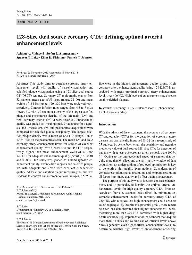
Fig. 1 A 57-year-old man withhigh-quality coronary arteryenhancement on CTA. a, bCoronal oblique volumerendering of the heart (a) andcurved planar reconstruction ofthe LAD and RCA (b) show highlevel of enhancement andexcellent visualization of thecoronary arteries, as well asdistinction between calcifiedplaque and intraluminal contrastenhancement. c, d Multiplanarreconstructions cross-sections ofproximal right coronary artery (c)and left main coronary artery (d)demonstrating how enhancementlevels were measured
Emerg Radiol

Quantitative enhancement assessment
The coronary artery enhancement levels were measured by abody fellowship-trained CT attending with 7 years of experi-ence. To determine the postcontrast enhancement level, short-axis images of the left main (LM) and the right coronaryarteries (RCA) were reconstructed using interactivemultiplanar reconstructions (Fig. 1). For each coronary artery,the proximal portion with maximum diameter and homoge-neity in short axis was chosen. A circular region of interestwas centered in the artery to measure the postcontrast densityof the blood pool in Hounsfield units, covering at least half ofthe intraluminal contrast pool, while avoiding volume averag-ing near the vessel wall. From these, the mean enhancementlevel was derived, by averaging the RCA and LM postcontrastattenuation levels. Enhancement levels across subgroups wascompared using unpaired T tests, with p values of <0.05considered significant.
Qualitative enhancement assessment
The image quality with respect to coronary arterial enhance-ment was assessed by a separate cardiovascular fellowshiptrained cardiac radiologist (3 years of attending experience incardiac imaging), who graded enhancement level on a scale of
1 to 3, with 1 being suboptimal, 2 being adequate, and 3 beingexcellent enhancement.
Calcified plaque assessment
The Agatston scores for the entire heart were recorded. Foreach case with coronary calcification, the density of the largestcalcified plaque was measured on postcontrast acquisitions byan upper level radiology resident. Pre- and postcontrast acqui-sitions were then compared for calcified plaque conspicuity bythe same attending who measured coronary artery attenuationat an interval of greater than 1 month from the time that theenhancement levels were measured, using axial images andmultiplanar reconstructions (MPRs). The precontrast imagesacquired for calcium scoring were first reviewed to delineatethe calcified plaques and then compared to the postcontrastimages. This enabled identification of calcified plaques thatwere not visualized on the postcontrast study.
Results
The mean enhancement level for studies of excellent enhance-ment quality (N=43) was 462 HU, significantly higher thanthe mean enhancement level for adequate enhancement qual-ity studies (N=8) of 321 HU (p=0.001; Table 1). The contrastenhancement level of one study, with mean coronary arteryenhancement level of 299 HU, was deemed subjectivelysuboptimal.
Most patients were imaged with 120 kVp. The subjectswho were imaged with 100 kVp had significantly higherenhancement levels despite use of similar contrast volumesand infusion rates (Table 2).
Agatston scores were available in 48 patients and measured>0 in 26 of these. The mean score was 123, ranging between 0and 1,495. The density of the largest calcified plaque wasmeasured in 25 subjects with calcified plaque density, withmean of 862 HU (range, 376 to 1,384 HU) on the postcontrastscan, which was much higher than the mean enhancementlevel of the corresponding coronary artery (Table 3).
Table 2 Comparison of en-hancement levels in Hounsfieldunit to peak kilovoltage used fordata acquisition. Use of lowerpeak kilovoltage was associatedwith significantly higher contrastenhancement levels, despite ab-sence of significant differencesbetween contrast volume and in-fusion rate in these two groups
a Statistically significantdifferences
100 kVp (n=10) 120 kVp (n=42) p values
Mean (range) age 49 years (22 to 67) 57 years (28 to 90) 0.17
Mean (range) weight 150 lbs (120 to 180) 192 lbs (145 to 320) <0.001a
Mean (range) contrast volume 104 mL (80 to 120) 105 mL (90 to 120) 0.79
Mean (range) contrast infusion rate 6.1 mL/s (6 to 6.5) 5.8 mL/s (4.5 to 7) 0.07
Mean (range) left main coronary artery (LM) 542 HU (265 to 687) 419 HU (260 to 568) 0.02a
Mean (range) right coronary artery (RCA) 529 HU (252 to 671) 409 HU (189 to 578) 0.03a
Mean (range) of LM and RCA HU 535 HU (259 to 679) 414 HU (225 to 564) 0.03a
Number of subjects with calcified plaque obscured 1/10 (10 %) 5/42 (12 %) n/a
Table 1 Mean (and range) HU enhancement level by coronary artery,correlated with subjective assessment of enhancement quality
LM HU RCA HU Mean HU
Excellent quality(n=43)
468 (316 to 687) 456 (313 to 671) 462 (335 to 679)
Adequate quality(n=8)
320 (260 to 422) 322 (189 to 545) 321 (225 to 484)
Suboptimalquality (n=1)
320 278 299
p Values <0.0001a 0.009a 0.001a
LM left main coronary artery, RCA right coronary artery,mean average ofright and left coronary arteriesa Statistically significant difference between high and moderate qualityenhancement levels
Emerg Radiol

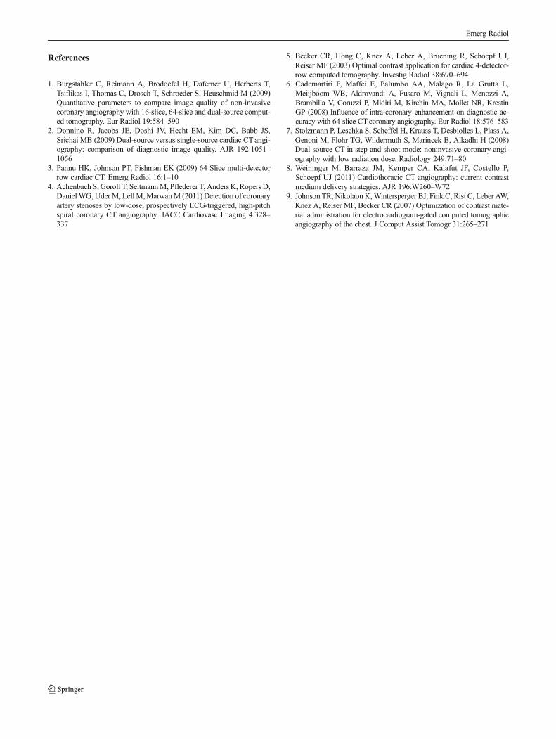
The 25 patients with calcified plaque included 38 % (3/8)of the subjects with adequate enhancement and 22 of the 43subjects (51 %) with excellent enhancement quality. At leastone small, calcified plaque was isodense to contrast enhance-ment on axial images in 5 of these 25 subjects (Fig. 2); all 5were in the excellent enhancement quality group. The maxi-mum diameter of largest obscured plaque ranged from 1.4 to1.8 mm, with a mean of 1.6 mm. Comparison of meanenhancement of the 22 excellent enhancement level subjectswith and without obscured plaque showed significantly highermean enhancement in those with obscured plaque (540 HU)than those without (434 HU; p=0.04, Table 4).
Discussion
Imaging of coronary arteries has dramatically improved asfaster scanners enable higher spatial and temporal resolution.As demonstrated in our study, current protocols with DSCTcan produce coronary CTA studies with excellent qualitycoronary artery enhancement, as 83 % of cases showed ex-cellent enhancement quality for evaluation of the coronaryarteries and 98 % had acceptable enhancement quality fordiagnosis. This concurs with published data reporting thatdiagnostic image quality can be achieved in more than 95 %cases when imaged on DSCT [7].
The faster acquisition times contributing to improvementsin image quality afforded by of state-of-the-art multidetectorCT scanners have precipitated use of contrast infusion rates of>5 mL/s, resulting in higher enhancement levels [8]. Studieshave shown that significantly higher enhancement levels with-in the coronary artery lumen can be achieved with DSCTcompared to 16- and 64-slice single-source CT scanners. Aninvestigation by Burgstahler et al. [1] that included 256 coro-nary CTstudies conducted on 16- and 64-slice DSCTscannersreported that quantitative level of enhancement and signal-to-noise ratio on DSCTwas significantly higher than the 16- and64-slice scanners single-source scanners. The mean level ofenhancement of the four coronary arteries in that study rangedbetween 310 and 362 HU.
Prior research has confirmed that higher enhancementlevels translate into excellent image quality and improveddiagnostic accuracy [4]. However, whether very high levelsof enhancement interfere with visualization of calcified plaquehas been a matter of debate. Becker et al. [5] evaluated 60
patients with four-slice MDCT and defined a minimum ac-ceptable enhancement levels for coronary artery CTA of
Fig. 2 A 50-year-old female with two small adjacent foci of calcificationin the midportion of RCA (a). Following injection of intravenous con-trast, proximal RCA enhancement level measured 509 HU, and theintraluminal attenuation adjacent to the plaque increased to 471 HU (b),with obscuration of the small calcified plaque. Plaque density was124 HU precontrast and 417 HU postcontrast, with the postcontrastincrease presumably related to beam hardening artifact. The left-sidedplaque was selected during calcium scoring (c)
Table 3 Attenuation of the largest plaque compare to proximal luminalenhancement in the same coronary artery
Density HU
Density of the largest calcified plaque (n=25) 862 (376 to 1384)
Density of the coronary artery (n=25) 415 (173 to 662)
Emerg Radiol

250 HU with a caveat that enhance values more than 350 HUcould obscure calcified plaque. A more recent study by John-son et al. [9] using a 64-slice scanner in 30 patients demon-strated inadequate visualization of only four coronary arteries,and, for each of these, the proximal coronary artery enhance-ment level was less than 200 HU. These authors recommend-ed target enhancement of 300 HU as optimum for evaluationof the coronary arteries. They also raised concern that en-hancement values of more than 350 HU adjacent calcifiedplaque obscure calcified plaque and result in an underestima-tion of coronary artery stenosis. These studies, however, didnot perform direct assessment of calcium visualization on pre-and postcontrast images.
In the current study, we demonstrate that higher enhance-ment level within the coronary arteries is associated withsubjectively higher image quality when assessed by a cardio-vascular radiologist. The proximal coronary artery contrastenhancement level in the high-enhancement quality groupranged from 335 to 679 HU. In comparison, adequate imagequality was associated with a contrast enhancement levelranging from 225 to 484 HU. Balanced with image qualityis the requirement to discriminate calcified plaque from vas-cular enhancement. In this series, high contrast enhancementlevels did not impede visualization of any calcifications on thecontrast-enhanced scan in the majority of patients. In a minor-ity of patients, small, calcified plaques were obscured whenthe contrast enhancement level was very high. The plaquesthat were obscured were <2mm inmaximum diameter; so thatit is unlikely that a significant coronary artery stenosis wouldbe missed. The downside of not visualizing a small plaquewould be potentially mischaracterizing a patient’s cardiac risk.Specifically, if a precontrast study is not performed, a patientwith obscured calcification could be misdiagnosed as beingentirely free from atherosclerotic coronary artery disease. Thiscould impact patient management by incorrectly stratifyingtheir risk for future significant coronary artery disease. Ourcomparison of patients with all plaque visualized to those withobscured plaque suggests that the target enhancement level toavoid this pitfall is in the range of 400–450 HU.
Limitations of this study include the small sample size andthe retrospective nature, which resulted in incomplete infusionparameters for all subjects, inability to determine BMI. The
small sample size and limited contrast infusion informationprecluded a determination of how iodine load and iodinedelivery rate affect contrast enhancement level. Accordingly,the data analyzed does not provide information to guideinfusion protocols in terms of volume, iodine load, and rate.These parameters are ideally evaluated by prospective collec-tion of contrast infusion data correlated with patient BMI.Furthermore, different concentrations of contrast, saline flushvolume, and timing methods are used in other institutions,necessitating individualized protocol optimization to achievethe target enhancement levels defined here. As demonstratedin this study, the use of lower peak kilovoltage is associatedwith higher coronary artery enhancement levels, indicatingthat infusion parameters and voltage should be analyzed withmultivariate analysis in a larger set of subjects to delineate theoptimal infusion parameters. An additional limitation is thatwe did not evaluate the utility of curved planar MPRs andmaximum intensity projections (MIP) renderings for visuali-zation of isodense plaque when enhancement levels werehigh. Experienced cardiac imagers recognize that MIP render-ings enable distinction of calcified plaque from highintraluminal contrast levels, owing to differences in perceiveddensity, mural configuration of calcified plaque, and bloomingartifact of calcification.
In conclusion, high levels of coronary artery enhance-ment achieved with current generation DSCT are associat-ed with better visualization of the coronary arteries. Fromour data, target enhancement level is in the range of 400–450 HU, higher than previous estimates. Following con-trast infusion, small (<2 mm), calcified plaques can beobscured when the luminal density approximates that ofthe small plaque. While this is not likely to result in cardiacimagers missing significant coronary stenosis, if anoncontrast study is not performed in conjunction withCTA, patients could potentially be misdiagnosed as havingno coronary atherosclerotic disease. When interpreting coro-nary CTA, radiologists should be cognizant that smallplaques may be subtle or obscured if contrast enhancementlevels are high.
Conflict of Interest The authors declare that they have no conflict ofinterest.
Table 4 Mean (and range) post-IV contrast coronary artery HU in 22 patients with excellent image quality, divided according to those who had calcifiedplaque visualized or obscured
LM HU RCA HU Mean HU
All calcified plaques visualized (n=17) 442 (340 to 570) 426 (326 to 587) 434 (338 to 579)
At least 1 calcified plaque obscured (n=5) 544 (438 to 681) 535 (435 to 614) 540 (437 to 648)
p Values 0.07 0.02a 0.04a
LM left main coronary artery, RCA right coronary artery, mean average of right and left coronary arteriesa Statistically significant differences
Emerg Radiol
References
1. Burgstahler C, Reimann A, Brodoefel H, Daferner U, Herberts T,Tsiflikas I, Thomas C, Drosch T, Schroeder S, Heuschmid M (2009)Quantitative parameters to compare image quality of non-invasivecoronary angiography with 16-slice, 64-slice and dual-source comput-ed tomography. Eur Radiol 19:584–590
2. Donnino R, Jacobs JE, Doshi JV, Hecht EM, Kim DC, Babb JS,Srichai MB (2009) Dual-source versus single-source cardiac CT angi-ography: comparison of diagnostic image quality. AJR 192:1051–1056
3. Pannu HK, Johnson PT, Fishman EK (2009) 64 Slice multi-detectorrow cardiac CT. Emerg Radiol 16:1–10
4. Achenbach S, Goroll T, SeltmannM, Pflederer T, Anders K, Ropers D,DanielWG, UderM, Lell M,MarwanM (2011) Detection of coronaryartery stenoses by low-dose, prospectively ECG-triggered, high-pitchspiral coronary CT angiography. JACC Cardiovasc Imaging 4:328–337
5. Becker CR, Hong C, Knez A, Leber A, Bruening R, Schoepf UJ,Reiser MF (2003) Optimal contrast application for cardiac 4-detector-row computed tomography. Investig Radiol 38:690–694
6. Cademartiri F, Maffei E, Palumbo AA, Malago R, La Grutta L,Meiijboom WB, Aldrovandi A, Fusaro M, Vignali L, Menozzi A,Brambilla V, Coruzzi P, Midiri M, Kirchin MA, Mollet NR, KrestinGP (2008) Influence of intra-coronary enhancement on diagnostic ac-curacy with 64-slice CT coronary angiography. Eur Radiol 18:576–583
7. Stolzmann P, Leschka S, Scheffel H, Krauss T, Desbiolles L, Plass A,Genoni M, Flohr TG, Wildermuth S, Marincek B, Alkadhi H (2008)Dual-source CT in step-and-shoot mode: noninvasive coronary angi-ography with low radiation dose. Radiology 249:71–80
8. Weininger M, Barraza JM, Kemper CA, Kalafut JF, Costello P,Schoepf UJ (2011) Cardiothoracic CT angiography: current contrastmedium delivery strategies. AJR 196:W260–W72
9. Johnson TR, Nikolaou K,Wintersperger BJ, Fink C, Rist C, Leber AW,Knez A, Reiser MF, Becker CR (2007) Optimization of contrast mate-rial administration for electrocardiogram-gated computed tomographicangiography of the chest. J Comput Assist Tomogr 31:265–271
Emerg Radiol