12/7/2011 - iap-ad.org sneige-issues in... · cap: single microsopic section from each ln block, no...
TRANSCRIPT

12/7/2011
1
Kamal Ishak Memorial Lecture

12/7/2011
2
Issues in Assessment of Axillary
Sentinel Lymph Nodes
Nour Sneige, M.D.
MD Anderson Cancer Center
Houston, Texas
Axillary Lymph Nodes Status
Most significant prognostic factor
Goals of ALN dissection:
Accurate staging and guiding
treatment selection
---potential complications---
First node to receive
lymphatic drainage
from the area of the
primary tumor
Lymphatic mapping
with SLN biopsy for
breast cancer
introduced in 1990
Sentinel Lymph Node (SLN)

12/7/2011
3
• Accurate (negative SLNs accurately predicts
absence of metastasis in the remaining axillary
nodes (95% to 100%)
• Safe (significant reduction in surgical morbidity)
• More sensitive to detect small metastases than
ALN dissection – small number of LNs removed
can be subjected to a more detailed pathologic
evaluation.—standard of care---
Advantages
Sentinel Lymph Node
- Objectives -
Definition of micrometastases
Prognostic Significance
Identification/ Intraoperative
evaluation (H&E, IHC and mol.)
Conclusions
12/7/2011
4
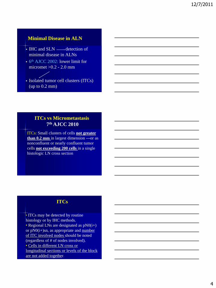
Minimal Disease in ALN
IHC and SLN detection of
minimal disease in ALNs
6th AJCC 2002: lower limit for
micromet >0.2 - 2.0 mm
Isolated tumor cell clusters (ITCs)
(up to 0.2 mm)
ITCs vs Micrometastasis
7th AJCC 2010
ITCs: Small clusters of cells not greater
than 0.2 mm in largest dimension ---or as
nonconfluent or nearly confluent tumor
cells not exceeding 200 cells in a single
histologic LN cross section
ITCs
• ITCs may be detected by routine
histology or by IHC methods.
• Regional LNs are designated as pN0(i+)
or pN0(i+)sn, as appropriate and number
of ITC involved nodes should be noted
(regardless of # of nodes involved).
• Cells in different LN cross or
longitudinal sections or levels of the block
are not added together.

12/7/2011
5
ITCs
ITCs
Single cluster
(<0.1 mm) of
tumor
pN0 (i+)
ITC

12/7/2011
6
Micrometastases
Tumor deposits greater than 0.2 mm but not
larger than 2 mm in largest dimension or
> 200 tumor cells identified as single
dispersed or as a nearly confluent in a
single slide (even if <0.2 mm).
PN1mi (sn), as appropriate, and the number
of involved nodes should be noted.
Largest dimension of any group of cells
that are touching one another (confluent
or contiguous tumor cells) regardless of
whether the deposit is confined to the
LN, extends outside the node
(extranodal or extracapsular extension),
or is totally present outside the LN and
invading adipose tissue.
Size of a Tumor Deposit
2.0 mm micrometastasis

12/7/2011
7
Desmoplastic stromal reaction present:
the combined contiguous dimension of
tumor cells and fibrosis determines
size of the metastases.
ITC s or Micrometastasis?
Micrometastasis

12/7/2011
8
Prognostic Significance of Micrometastases in
Axillary Lymph Nodes
In studies that have evaluated complete axillary
dissection: • Occult mets not associated with poor prognosis
Khan et.al. 2006
Chen et al. 2007
• Associated with poorer outcome than N0 :
Sakorafas et.al. 2004
Maibenco et.al. 2006
Kuijt et al. 2005 (if no adjuvant chemo given)
Prognostic Significance of Micrometastases
and ITCs in Axillary Nodes
A. Incidence of further metastases in
non-sentinel axillary lymph nodes
B. Survival differences
SLNs

12/7/2011
9
Incidence of further metastases in NSLNs
(#1228 pts):
SLN NSLN mets
9.5 % ITCs 15%
26% micromets 21%
SLN stratified as to size of micromets 1mm
vs 1-2 mm: ITCs and micromets up to 1 mm
had a significantly lower risk of additional
mets (Viale et al.)
Prognostic Significance of
Micrometastases and ITCs in SLNs
Prognostic Significance of
Micrometastases and ITCs in SLN
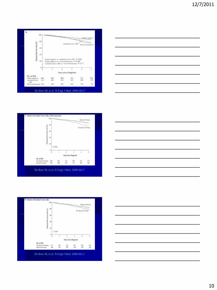
Survival differences: Controversial
Associated with a significantly shorter
disease-free interval than was SLN
negativity (Reed et al.2009, de Boer et
al.2009, Truong et.al 2010)
Not significant (majority adjuvant)
Hansen et al, 2009; Weaver et al. 2011
(78%); Giuliano et al. (Z0010, 83%)
De Boer M, et al. N Engl J Med. 2009:361;7
12/7/2011
10
De Boer M, et al. N Engl J Med. 2009:361;7
De Boer M, et al. N Engl J Med. 2009:361;7
De Boer M, et al. N Engl J Med. 2009:361;7

12/7/2011
11
De Boer M, et al. N Engl J Med. 2009:361;7
Effect of Occult Metastases on Survival
in Node-Negative Breast Cancer
NSABP-B32 occult mets (neg SLN at initial exam but
pos. at 2 widely spaced additional H&E levels and IHC)
were detected in 15.9% of 3887 patients.
Occult metastases were an independent prognostic
variable (with respect to overall survival p=0.03, DFS
p=0.02, and D- D-FS p=0.04), however the magnitude
of the difference in outcome at 5 years was small 1.2
percentage points) (5 yr estimates of OS among pts
were 94.6% and 95.8% respectively).
Weaver et al, N Eng J Med 2011
NSABP Trial B-32
12/7/2011
12
Sentinel Lymph Node
- Objectives -
Definition of micrometastases
Prognostic Significance
Identification/ Intraoperative
evaluation (H&E, IHC and mol.)
Conclusions
Pathologic Evaluation of SLNs
Goals:
Identify all macrometastases
(larger than 2 mm, pN1) and
most micrometastases (pN1mi)
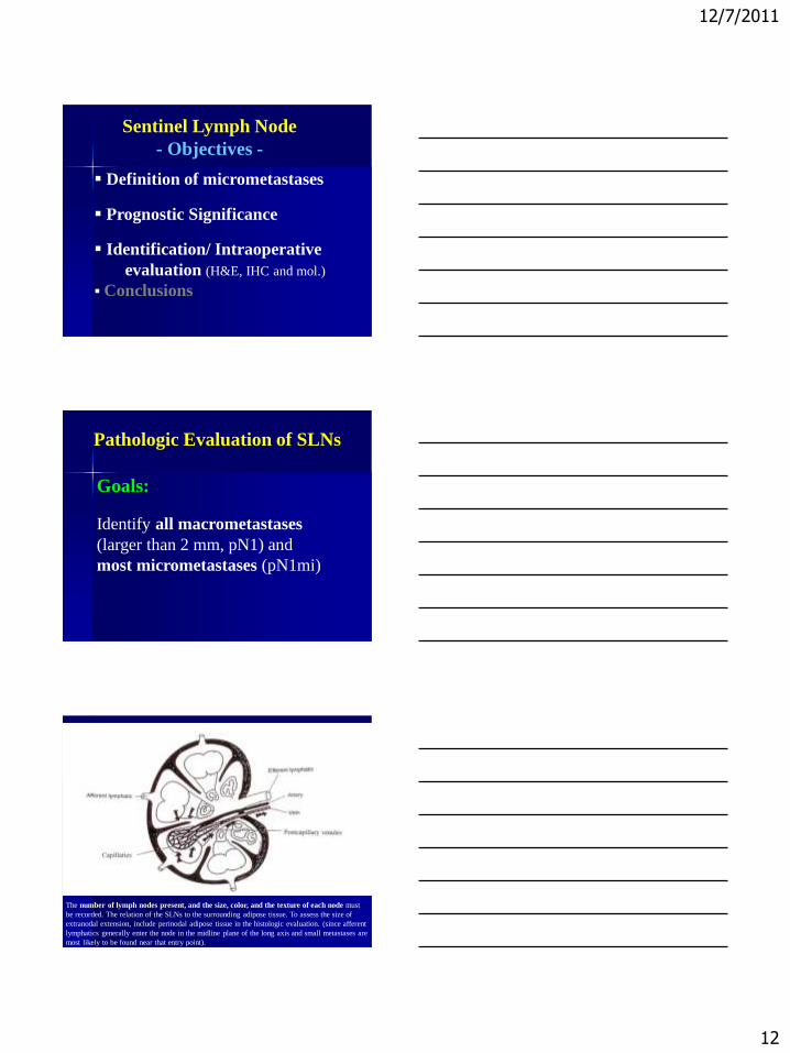
The number of lymph nodes present, and the size, color, and the texture of each node must
be recorded. The relation of the SLNs to the surrounding adipose tissue. To assess the size of
extranodal extension, include perinodal adipose tissue in the histologic evaluation. (since afferent
lymphatics generally enter the node in the midline plane of the long axis and small metastases are
most likely to be found near that entry point).

12/7/2011
13
Gross Handling of LNs
Serial sections at 1-to-2 mm intervals through
and parallel to the longest axis.
Entire LN should be submitted for evaluation.
Each LN is submitted in a separate cassette or
identified by colored ink to permit accurate
assessment of the total number of LN and
number of involved nodes.
-- short half-life and limited penetration of
technetium, health risks to those handling SLNs
are negligible (ASCO Guidelines 2005).
Intraoperative Examination of the
Sentinel Node
Allows immediate axillary dissection when
metastasis is found in the SLN.
For every 100 patients who have SLNs
evaluation intraoperatively, 16 to 17 will have
positive nodes and 8 to 9 will have false-negative
results.
Each institution must have a policy on
intraoperative assessment or deferral to
permanent sections.

12/7/2011
14
Intraoperative Examination of the
Sentinel Node
• Standard techniques:
Imprint cytology or evaluation of cells scraped
from the cut surface of the node
Frozen section (carry the risk of significant
destruction of potentially diagnostic tissue).
Suspicious findings should be reported as not
diagnostic for tumor and deferred to paraffin
section
• Molecular techniques
Permanent
Frozen section
12/7/2011
15
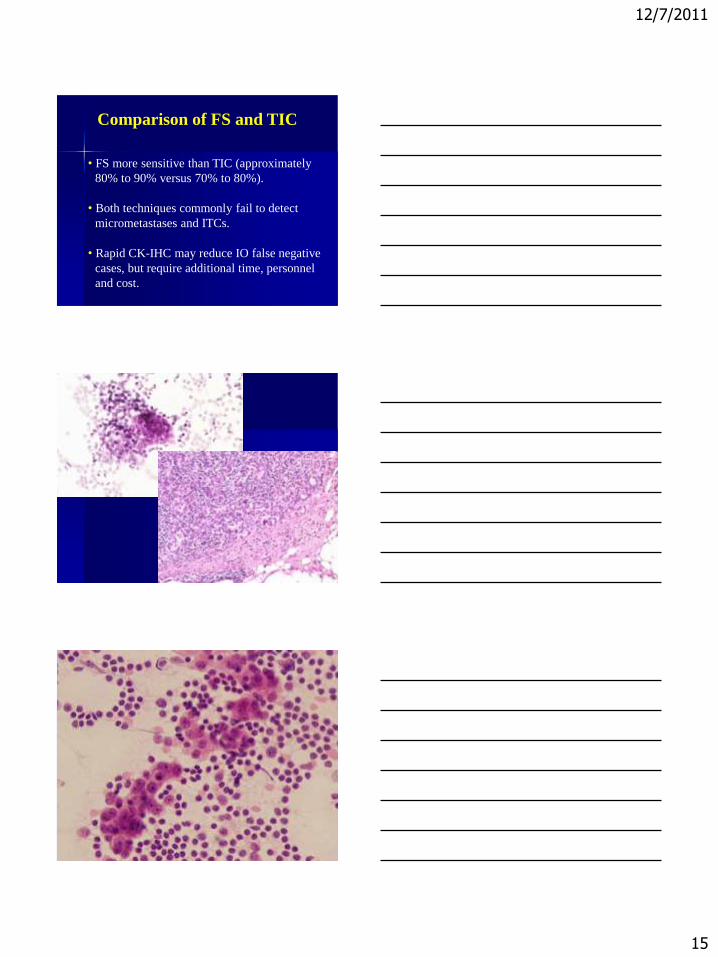
Comparison of FS and TIC
• FS more sensitive than TIC (approximately
80% to 90% versus 70% to 80%).
• Both techniques commonly fail to detect
micrometastases and ITCs.
• Rapid CK-IHC may reduce IO false negative
cases, but require additional time, personnel
and cost.

12/7/2011
16
Potential Sources of Trouble
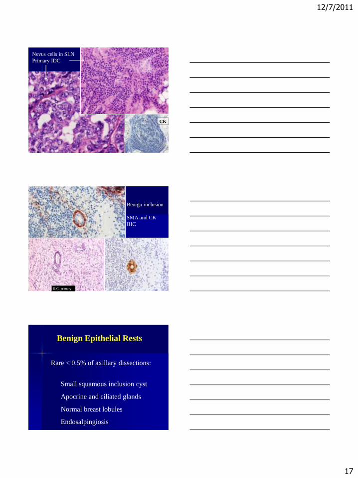
1. Nevus Cell aggregates
2. Benign glandular inclusions
3. Displacement of benign epithelium
4. Extramedullary hematopoesis
5. Sinus histiocytosis
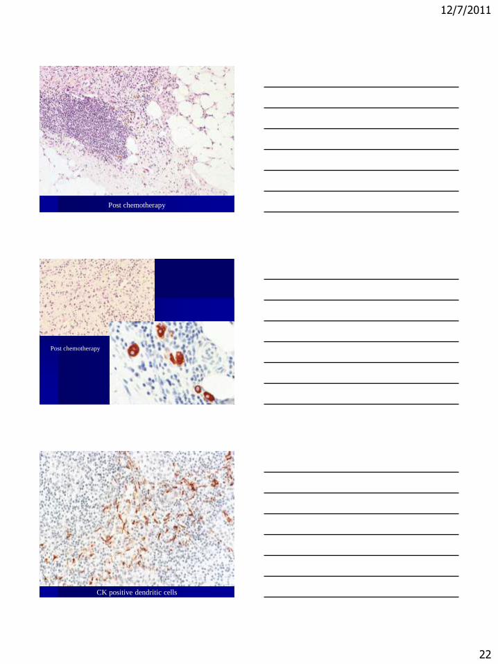
6. IHC CK positive dendritic cells
12/7/2011
17
Nevus cells in SLN
Primary IDC
CK
Benign inclusion
SMA and CK
IHC
ILC, primary
Benign Epithelial Rests
Rare < 0.5% of axillary dissections:
Small squamous inclusion cyst
Apocrine and ciliated glands
Normal breast lobules
Endosalpingiosis
12/7/2011
18
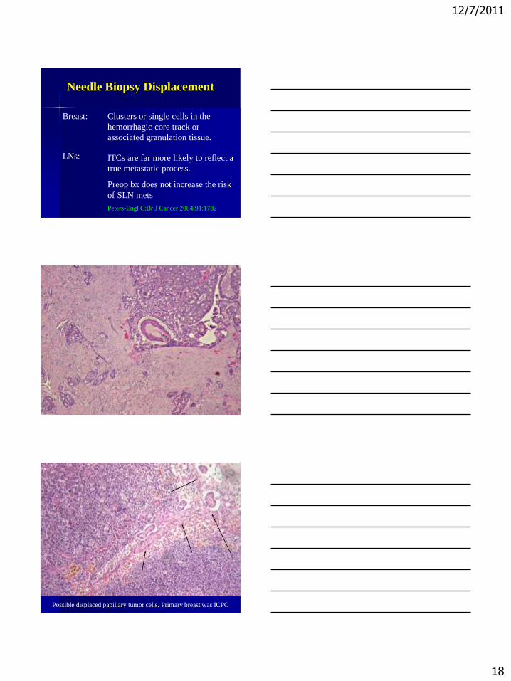
Needle Biopsy Displacement
Breast:
Clusters or single cells in the
hemorrhagic core track or
associated granulation tissue.
LNs: ITCs are far more likely to reflect a
true metastatic process.
Preop bx does not increase the risk
of SLN mets
Peters-Engl C:Br J Cancer 2004;91:1782
Possible displaced papillary tumor cells. Primary breast was ICPC

12/7/2011
19
Endothelial cells
Imprint cytology.
Blood vessel
Imprint Cytology: Histiocytes mimicking metastatic
lobular carcinoma (CK neg), primary is IDC, grade 3
12/7/2011
20
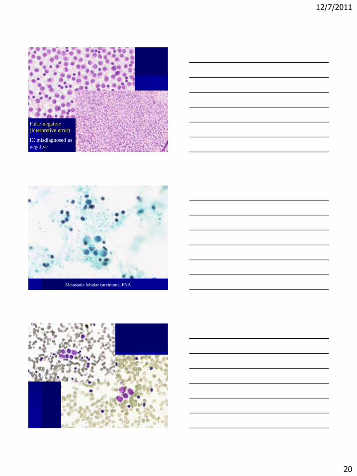
False-negative
(interpretive error)
IC misdiagnosed as
negative
Metastatic lobular carcinoma, FNA

12/7/2011
21
Imprint Cytology:
Clusters of giant cells
Both giant cells and single tumor cells present
12/7/2011
22
Post chemotherapy
Post chemotherapy
CK positive dendritic cells

12/7/2011
23
Dots like CK-positive cells in germinal center
Dot like Pan-CK positive, but CK 7, MOC31, and ER - . Morphology c/w
GC cells not with ILC, classic.
Sampling SLNs/Paraffin Section
Levels-Current Practice-
No consensus among pathologists regarding how many
H&E and CK IHC levels should be examined
CAP: single microsopic section from each LN block, no IHC.
ADASP ―several‖ microscopic sections from each block and also
do not routinely recommend IHC
Limited step sections: (top level plus 1 or 2 sections cut at 200-
to-500 micrometer intervals into the block)
Comprehensive analysis: The European Institute of Oncology
group performs FS analysis of the entire sentinel node in
multiple step sections separated by 50 µm
12/7/2011
24
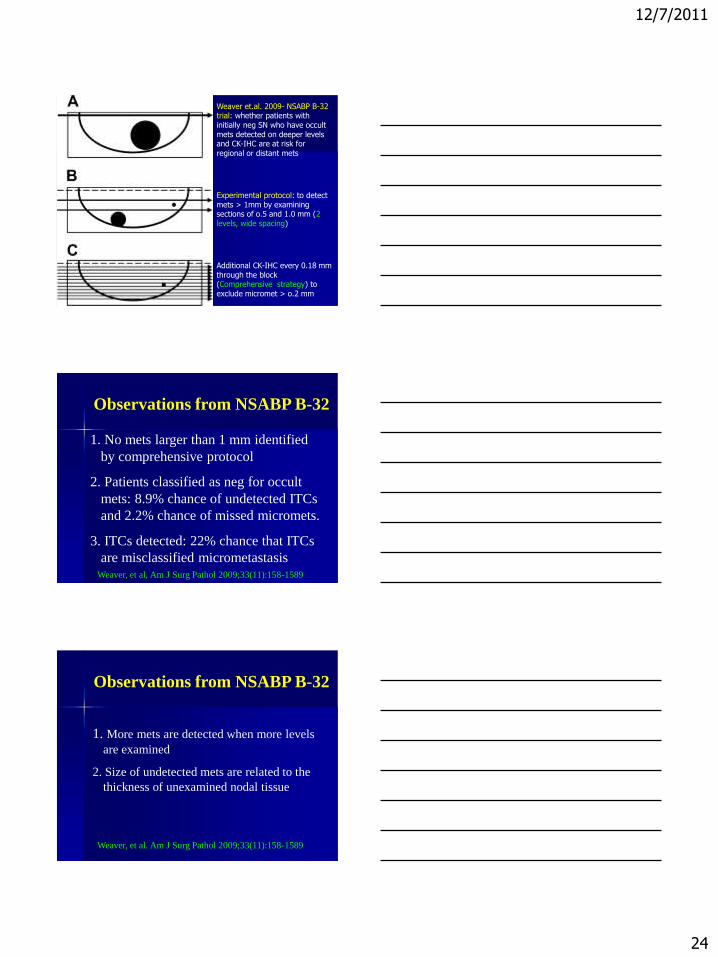
Weaver et.al. 2009- NSABP B-32 trial: whether patients with initially neg SN who have occult mets detected on deeper levels and CK-IHC are at risk for regional or distant mets
Experimental protocol: to detect mets > 1mm by examining sections of o.5 and 1.0 mm (2 levels, wide spacing)
Additional CK-IHC every 0.18 mm through the block (Comprehensive strategy) to exclude micromet > o.2 mm
Observations from NSABP B-32
1. No mets larger than 1 mm identified
by comprehensive protocol
2. Patients classified as neg for occult
mets: 8.9% chance of undetected ITCs
and 2.2% chance of missed micromets.
3. ITCs detected: 22% chance that ITCs
are misclassified micrometastasis Weaver, et al, Am J Surg Pathol 2009;33(11):158-1589
Observations from NSABP B-32
1. More mets are detected when more levels
are examined
2. Size of undetected mets are related to the
thickness of unexamined nodal tissue
Weaver, et al. Am J Surg Pathol 2009;33(11):158-1589

12/7/2011
25
Sampling SLNs/Paraffin Section
Levels and Use of IHC
NSBPB-32 clinical trial conclusion:
Observed difference in 5 yrs survival 1.2
percentage points between patients in whom
occult mets were detected and patients in whom
occult mets were not detected was not significant
Raised the argument against analysis of
additional tissue levels or routine IHC-CK for
SLN evaluation. .
Use of IHC Analysis?
Survey by Syed Hoda on the continued use of
CK on sentinel nodes, there were 30
respondents. Responses equally divided
among 3 groups:
1. Advocates the use of IHC.
2. Never did it.
3. Contemplating discontinuation of IHC. -driven by the oncologists/surgeons ---uncertainty
about the significance of micromets or ITCs-
Our practice
- Serial sections of LN at 1-2 mm
- 1 H&E and 3 unstained
(if H&E negative, stain one IHC-CK)
To facilitate Dx of small-volume mets.
Detection of each possible ITC should not
be the goal of routine histologic evaluation

12/7/2011
26
Molecular Techniques
Require carefully controlled assay
conditions to avoid false positive results.
Test manufacturers have set cut off
levels at the lower limit of
micrometastasis, 0.2 mm with generally
favorable results.
Molecular Study
Purpose: to increase the likelihood of detecting breast
cancer metastases.
Veridex Gene Search ™ Breast Lymph Node (BLN),
FDA approved 2007 for in vitro intra-operative diagnosis
of metastasis greater than 0.2 mm in SLN to guide decision
making on CLA at the time of sentinel node surgery.
One Step Nuclei Acid Amplification (OSNA).
Molecular Study
Viale et al. (2008), sensitivity 98.1% (> 2mm), 94.7% (>1
mm), 77.8% > 0.2 mm), overall concordance 90.8%.
Taffe et.al. (2009), sampled the outer node tissue (two 1 mm
slices): Sensitivity 88.9%, specificity 93.5%.
Another study from Japan (half of the node was submitted):
sensitivity 95.7% for macrometastases, 60% for
micrometastases, and 55.6% for isolated tumor cells
Veys et.al. (2009), overall concordance 93%, with 87%
sensitivity, 94% specificity and 75% positive and 97%
negative predictive values.

12/7/2011
27
SLN are serially sectioned along the long axis into equal size and numbers of
sections. Alternate sections (50% of the node) are used for intraoperative PCR to
detect the presence of cytokeratin 19 and mamoglobin mRNA. The remaining
sections are used for histology.
Node sampling and sharing method. Note that this pattern was continued until the node was exhausted.
Viale G, et.al., Surg 2008;247: 136-142
Correlation between Histopathologic
Examination of the SLN and BLN Assay
Results
BLN Assay
Histology Positive Negative
Metastases >2 mm 51 1 52
Micrometastases 0.2-2 mm 5 15 20
ITC 0 10 10
Negative 11 200 211
Total 67 226 293
Viale G, et. al., Ann Surg 2008;247:136-142
12/7/2011
28
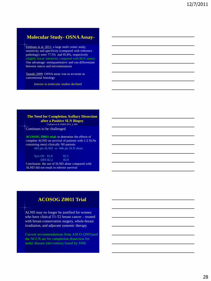
Molecular Study- OSNA Assay-
Feldman et al. 2011: a large multi center study,
sensitivity and specificity (compared with reference
pathology) were 77.5% and 95.8%, respectively
(slightly lower sensitivity compared with BLN assay).
One advantage: semiquantitative and can differentiate
between macro and micrometastasis
Tamaki 2009: OSNA assay was as accurate as
conventional histology
Interest in molecular studies declined
The Need for Completion Axillary Dissection
after a Positive SLN Biopsy Giuliano et al JAMA 2011, p 569
Continues to be challenged
ACOSOG Z0011 trial: to determine the effects of
complete ALND on survival of patients with 1-2 SLNs
containing mets) clinically N0 patients
445 pts ALND vs 446 pts SLN alone
5yrs OS 91.8 92.5
DFS 82.2 83.9
Conclusion: the use of SLND alone compared with
ALND did not result in inferior survival
ACOSOG Z0011 Trial
ALND may no longer be justified for women
who have clinical T1-T2 breast cancer – treated
with breast-conservation surgery, whole-breast
irradiation, and adjuvant systemic therapy.
Current recommendations from ASCO (2005)and
the NCCN are for completion dissection for
nodal disease (micromets) found by SNB.

12/7/2011
29
Survey of ASCO Members on Management of Sentinel
Node Micrometastases in Breast Cancer:
Wasif N, et al. Ann Surg Oncol. 2009;16:2442-49
382 med
onc, 100
surgeon,
55 rad onc
Wasif N, et al. Ann Surg Oncol. 2009;16:2442-49
Wasif N, et al. Ann Surg Oncol. 2009;16:2442-49
12/7/2011
30
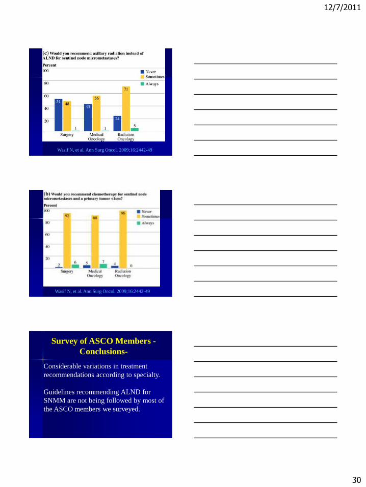
Wasif N, et al. Ann Surg Oncol. 2009;16:2442-49
Wasif N, et al. Ann Surg Oncol. 2009;16:2442-49
Survey of ASCO Members -
Conclusions-
Considerable variations in treatment
recommendations according to specialty.
Guidelines recommending ALND for
SNMM are not being followed by most of
the ASCO members we surveyed.
12/7/2011
31
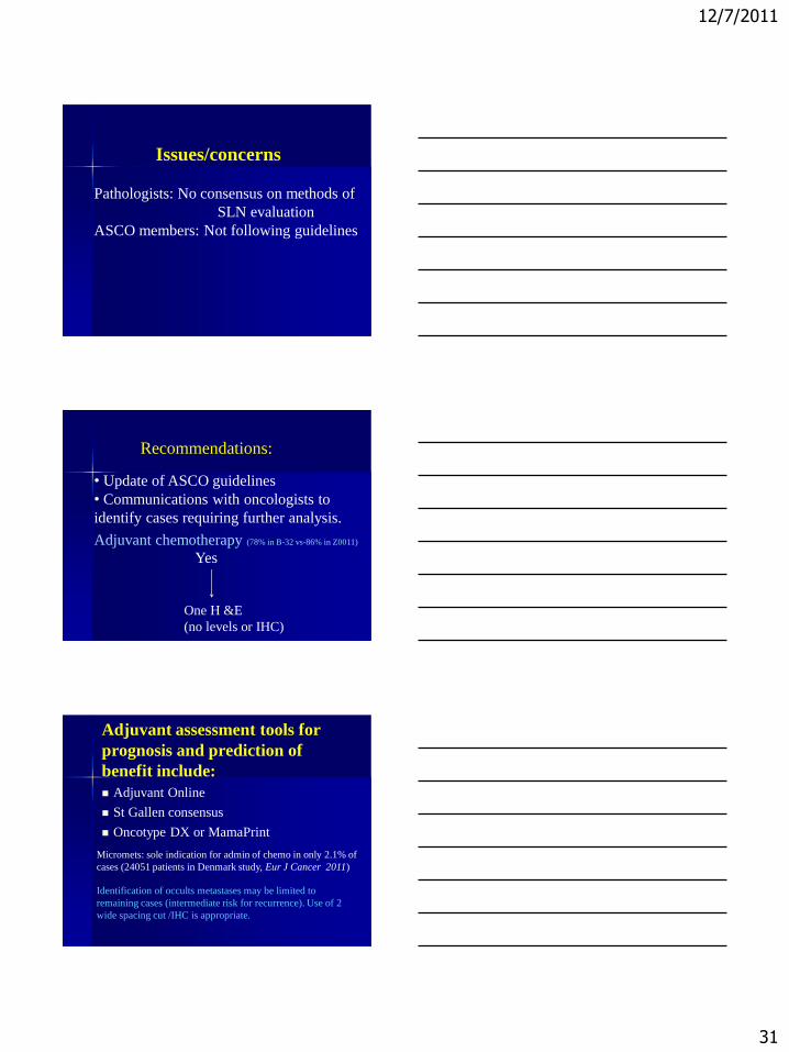
Issues/concerns
Pathologists: No consensus on methods of
SLN evaluation
ASCO members: Not following guidelines
Recommendations:
• Update of ASCO guidelines
• Communications with oncologists to
identify cases requiring further analysis.
Yes
One H &E
(no levels or IHC)
Adjuvant chemotherapy (78% in B-32 vs-86% in Z0011)
Adjuvant assessment tools for
prognosis and prediction of
benefit include:
Adjuvant Online
St Gallen consensus
Oncotype DX or MamaPrint
Micromets: sole indication for admin of chemo in only 2.1% of
cases (24051 patients in Denmark study, Eur J Cancer 2011)
Identification of occults metastases may be limited to
remaining cases (intermediate risk for recurrence). Use of 2
wide spacing cut /IHC is appropriate.

12/7/2011
32
Pathology reporting
Provide sufficient information in the
pathology reports --documentation of the
tumor burden in the nodes and number of
nodes involved--to facilitate accurate
staging using the criteria of the current
ASCO/UICC system Future targeted therapy and targetable
signaling pathways, rather than molecularly defined subgroups, may ultimately drive
treatment decisions.
.

12/7/2011
33
Rare well diff
clusters on cytology
best called
suspicious
From: Turner et al
JCO 2008;26:258-263
Dx: Micrometastasis? Non confluent cluster
>0.2mm & <2.0mm
But >200 cells
SLN + % Non SLN Involvement
ITC 0-12
Micrometastases
<1 mm
1-2 mm
15-30 15-17
20-30
Macrometastases 40-50

12/7/2011
34
Wasif N, et al. Ann Surg Oncol. 2009;16:2442-49