11th international meeting on low frequency noise and
TRANSCRIPT

11th International Meeting on
Low Frequency Noise and Vibration and its Control Maastricht The Netherlands 30 August to 1 September 2004
VIBROACOUSTIC DISEASE – THE RESPONSE OF BIOLOGICAL TISSUE TO LOW FREQUENCY NOISE
Mariana Alves-Pereira1, João Joanaz de Melo1 Maria Cristina Marques2, Nuno A. A.
Castelo Branco3
1New University of Lisbon, DCEA-FCT, 2825 Caparica ([email protected]) 2Dept. Physiology, School of Pharmacology, University of Lisbon, Portugal
3Center for Human Performance, 2615 Alverca, Portugal ([email protected])
Summary BACKGROUND: Vibroacoustic disease (VAD) is a systemic pathology caused by excessive exposure to low frequency noise (LFN). Until 1987, it was thought that the pathological effects of excessive LFN exposure were limited to the realm of cognitive and neurological disturbances. After the autopsy findings in a deceased VAD patient, it became clear that LFN impinges on the entire body, particularly the cardio-respiratory systems. In 1992, rodents were exposed to LFN, and the respiratory tract was studied through scanning and transmission electron microscopy. Pericardial, tracheal and lung fragments, removed with informed consent from VAD patients, have also been studied with light and electron microscopy. This report summarizes what is known to date on the tissue and cellular response to LFN exposure. TUBULIN-BASED STRUCTURES: Cilia are tubulin-based and exist in normal pericardia as well as in the respiratory tract. In VAD patients, pericardial cilia cease to exist, while tracheal and bronchial cilia are distributed in abnormal arrangements. In LFN-exposed rodents, respiratory tract cilia appear sheared, clipped or shaggy. ACTIN-BASED STRUCTURES: Cochlear cilia are actin-based structures, as are brush-cell microvilli that protrude into the respiratory tract airway. In LFN-exposed rodents, both structures appear fused. Actin filaments are also a fundamental element of the cellular cytoskeleton. In VAD patients’ pericardia, cytoskeletal deformations may be a consequence of LFN-induced changes of the actin filaments. BIOTENSEGRITY HYPOTHESIS: One of the most consistent findings in almost all human and rodent tissue fragments is the abnormal proliferation of collagen and elastin. It is hypothesized that the principles of biotensegrity structures may contribute to the explanation of tissue and cellular responses to LFN exposure. Introduction For the past 24 years, the effects of low frequency noise (LFN) (<500 Hz, including infrasound) exposure have been the object of intense scientific inquiry. Vibroacoustic

disease (VAD) is a whole-body pathology caused by excessive exposure to LFN (1-3), either due to occupational sources (4-6) or environmental sources (7-9). The response of biological tissue to LFN has drawn great interest, particularly given the significant structural, or morphological, changes of the exposed organs, tissues and cells. In 1987, an autopsy was performed on a deceased VAD patient, as specifically bequeathed by the patient in his will (10). Until then, it was thought that LFN-induced pathology was restricted to the realm of neuropathophysiology (11-13). Autopsy findings disclosed, among several other extraordinary features, widespread thickening of blood vessel walls, and abnormally thickened cardiac structures, namely valves and pericardium. Fibrosis (collagen proliferation) was also identified in the lungs (10). In 1992, Wistar rats began to be used as animal models for VAD. Rodents were exposed to LFN, and fragments of different sections of the respiratory tract were studied with electron microscopy (14). In 1996, the first pericardial fragments were taken from fully informed VAD patients who were undergoing cardiac surgery (for other reasons) (15). Since then, 12 VAD patients have provided pericardial fragments for our study (16-19). Similarly, several other VAD patients have provided fragments of respiratory tract tissue (epithelia) through biopsy (conducted for other reasons). All these tissue samples were examined with electron microscopy. The goal of this report is to contribute to the characterisation of the biomechanical response of tissue to the presence of excessive LFN, drawing upon the data collected from the microscopy studies. SOURCES OF DATA Autopsy In 1987, the first autopsy was performed on a VAD patient employed as an aircraft technician (10). Until then, the observed pathology in this professional group was mostly related to neuro-physiological and –psychological parameters: delays in nerve conduction times (20), deteriorated cognitive function (21), and brain lesions, as visualized through magnetic resonance imaging (20). In autopsy, this 58-year-old VAD patient disclosed two silent tumours, in the brain and kidney. The cause of death was a silent cardiac infarct. The myocardium exhibited several small scars from previous, silent events. Blood vessel walls were thickened with fibrosis (composed by collagen and elastic fibers). Fibrosis (focal and interstitial) was also identified in the lungs. The pericardium (a thin sac that surrounds the heart) and the cardiac valves were very thickened, also with abnormal amounts of fibrosis, i.e., abnormal amounts of collagen. It should be noted that the thickening observed in the blood vessel walls is not related to artherosclerotic plaques, which are located in lumps along the inner lumen of the vessel. In VAD, vessel walls thicken due to a continuous lining of fibrosis, which develops in one of the tissue layers that constitutes vessel walls – the intima. Pericardial Fragments The pericardium is a fibrous sac that encases the heart, with the purpose of maintaining it in its normal position. External forces, due to respiration or changes in body posture, are absorbed by the pericardium so as to keep the heart and its cardiac rhythm intact (22).

Consisting of three tissue layers – mesothelium, fibrosa and epipericardium – the pericardium is a highly organized mass of connective tissue, with a predominance of collagen fibers arranged in accordion-like bundles. Elastic fibers, much less numerous than collagen fibers, intersect the collagen bundles at right angles. This anatomical arrangement taken together with the viscoelastic properties of both collagen and elastin, provide the pericardium with the mechanical capability of protecting the integrity of the cardiac cycle. The thickness of the normal parietal leaflet of the pericardium is approximately 0.5mm (22). In VAD patients, the pericardial sac is greatly thickened, measuring 1.0 to 2.3 mm. Microscopy studies of VAD patients’ pericardial fragments have uncovered the origin of this excessive thickness. In VAD patients, the pericardium exhibits 5 distinct layers, intead of the normal 3 layers: the fibrosa layer splits into two thickened layers and, in between, sandwiches a newly formed layer of loose tissue (containing blood vessels, fatty tissue and nerves) (5,16-19). The normal pericardium also features strands of cilia. In the all samples of each of the 12 pericardial fragments studied to date, no cilium strand has ever been observed (5,16-19). Respiratory Tract Fragments Tracheal fragments were removed through endoscopic biopsy from 2 VAD patients. One was 62-year old smoker, employed as an aircraft technician, diagnosed with lung cancer (squamous cell carcinoma of the upper right lobe), and since deceased (23). The other is a 58-year-old non-smoker female, retired military parachutist (23). Pleural and lung fragments were also removed from the 62-year-old male, with his informed consent, during lung surgery for the carcinoma (24). Abnormally increased amounts of collagen and elastin were present in all observed fragments. Ciliated cells featured unusual distributions and arrangements. More recently, bronchoscopy has disclosed lesions that appear to be specific to VAD patients. Biopsies of these lesions, as well as from the non-lesioned areas, have been studied with light microscopy in 9 VAD patients. In the lesioned areas, massive amounts of collagen were identified, in which vascular neo-formations were embedded. In the non-lesioned areas, collagen was also more abundant than normal, but less so than in the lesioned areas. Animal Studies Extensive electron microscopy studies have been performed on Wistar rat respiratory epithelia (the respiratory epithelium is the tissue layer that lines the respiratory tract and that is open to the airway). Over the years, different animal groups have been exposed to LFN on different exposure schedules. To date, the following exposures have been conducted: occupationally-simulated (8 hours/day, 5 days/week, weekends in silence); 48-hour of continuous LFN exposure; 2160 hours of continuous LFN exposure; gestated and born in occupationally-simulated LFN and subsequently kept in silence for 1 year; and born and gestated in occupationally-simulated LFN, and subsequently exposed to additional LFN (14,25-31). Other rat organs that have briefly been studied are the kidney (32) and the cochlea (33). The most remarkable and consistent feature in all samples that were studied is the abnormal growth of collagen, seen in blood vessel walls, tracheal wall, pleural sac,
stomach wall, and kidney glomeruli. The cilia that line the respiratory tract are severely damaged, as are other cell populations, such as brush cells and secretory cells. THE BUILDING BLOCKS In order to fully understand the implications of the data collected to date, it is necessary to regard the human body as a set of mechanical (viscoelastic) elements. Inside the cell, organelles are not floating about. Instead, there is an intricate network of fibres that criss-crosses the entire intra-cellular space, and that provides both mechanical strength as well as a method of cellular transportation (among other functions). This network is called the cytoskeleton. Two of the major constituents of cytoskeletal fibers are actin and tubulin. Actin and tubulin are proteins that can be found in numerous mammalian tissues and cellular structures. Microtubules (MT) are polar structures, consisting of 13 linear filaments, aligned in parallel and forming long hollow cylinders, each composed of alternating α- and β-tubulin molecules. With an outer diameter of 25 nm, MT are the primary organizers of the cytoskeleton. Actin protein forms filaments of 2-stranded helical polymers, 5-9 nm in diameter. Actin filaments (AF) can, in turn, form linear bundles, 2D networks and 3D gels. Within the cytoskeleton, AF are highly concentrated underneath the plasma membrane, at the cell cortex. They are more rigid than MT, and are highly polarized structures. AF restructuring of the cell cortex can be triggered by extracellular signalling, and re-organization of the actin cortex can influence the plasma membrane above. Microvilli, are finger-like, cell-surface projections that increase the active surface area of cells. At the core of each microvillus is a rigid bundle of parallel AF, cross-linked by actin-binding proteins. Laterally, the AF core is connected to the plasma membrane through motor proteins (myosin). Auditory hair cells in the cochlea, or stereocilia, are also formed by actin bundles. At the tissue level, each cell can be thought of as an individual element, connected to adjacent cells by “ropes” (or ligaments) that, in turn, interconnect with each cell’s cytoskeleton. The spaces between the cells are collectively called the extra-cellular matrix. Collagen and elastin are the major constituents of the extra-cellular matrix. Ciliary strands, or axonemes, with an outer diameter of 25 µm, are composed of 9 MT doublets, organized in a ring centered on an additional pair of MT. Axoneme MT are associated with several proteins that form cross-links maintaining MT bundles together, and generating the driving force for cilliary bending motion. MT also form mechanically-driven relay systems that control motion. Cilia allow for the movement of fluid over a surface, for the propelling of cells through a fluid, and are also secretory structures (spp-controls). Cilia distribution in the human respiratory epithelia: 109/cm2. In the pericardium, the mesothelium is in direct contact with the pericardial sac, and is formed by a one-layer thick sheet of mesothelial (cuboidal) cells (MC). Anchoring junctions (AJ) interconnect MC among themselves, through their cytoskeletal fibers. AJ that interconnect MC cytoskeletons through intermediate filaments are called desmosomes. Intermediate filament architecture consists of fibrous subunits associated side-by-side in overlapping arrays, forming a meshwork that extends throughout the cytoplasm, providing mechanical strength to the cell. Intermediate filaments do not rupture when stretched, and

can withstand larger stresses and strains than actin filaments. Intermediate filaments are crucial to maintain cellular integrity. THE BUILDING BLOCKS WITH LOW FREQUENCY NOISE EXPOSURE Thickened Blood Vessels
Scanning electron microscopy of rat alveolar structure exposed to 2160 hours of continuous LFN. Amplification of small artery with an adjacent vein, both with thickened walls. Intima is thickened. Internal and external elastic laminae are also thickened and exhibit disruptions. This response is also observed in humans, as seen through autopsy. Note that this thickening is not similar to artherosclerotic plaques since it does not occur in clumps along the lumen.
Light microscopy of the lung parenchyma of a 62-year-old, smoker, VAD patient. A functional unit consisting of a very thickened artery (encircled with dashed lines), and a bronquiole filled with macrophages containing brown pigment. (x200)
Transmission electron microscopy of a VAD patient’s parietal pericardium. Loose tissue layer exhibiting a blood vessel with thickened walls (dashed lines), two myofibroblasts, and numerous cytoplasmic extensions surrounding bundles of collagen. (x2800)
Thickening of blood vessel walls is an intriguing finding. It was seen in all sources of data: autopsy, pericardial fragments, respiratory tract fragments, and in animal model repiratory tract images. Carotid artery thickening has also been documented in VAD patients (34,35). Considering the blood vessel as a flexible hose, the thickening of the walls of such a hose could, in theory, increase its rigidity. Consider, also, that the impact of LFN upon viscoelastic tissue can produce acoustically-induced vibration that can propagate within the organism. Could this thickening, somehow, be the response to propagating, LFN-induced vibration within the organism? Perhaps, the desired end result is not rigidity of vessel walls, but an increased compliance.

Thickening of Alveoli Walls The lungs can be viewed as two dangling lobes, in certain areas composed of a very fine mesh of connective tissue, such as the alveoli. Alveoli walls also thicken with LFN exposure.
Scanning electron microscopy of non-exposed rat alveolar structure Alveolar walls are thin (see arrows) and wall structure is visible.
Scanning electron microscopy of the rat alveolar structure, exposed to 2160 hours of continuous LFN, followed by 8 days in silence. No edema is present. Alveolar walls are greatly thickened (see arrows), and wall structure appears effaced.
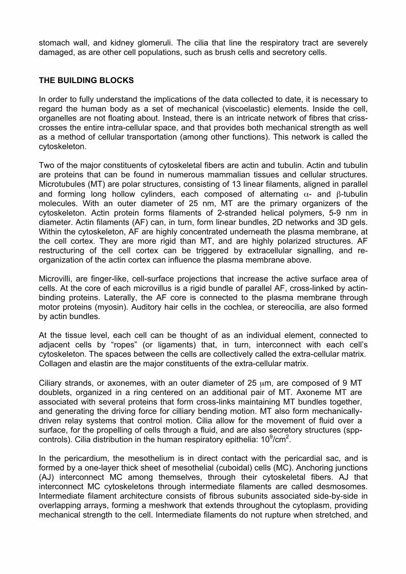
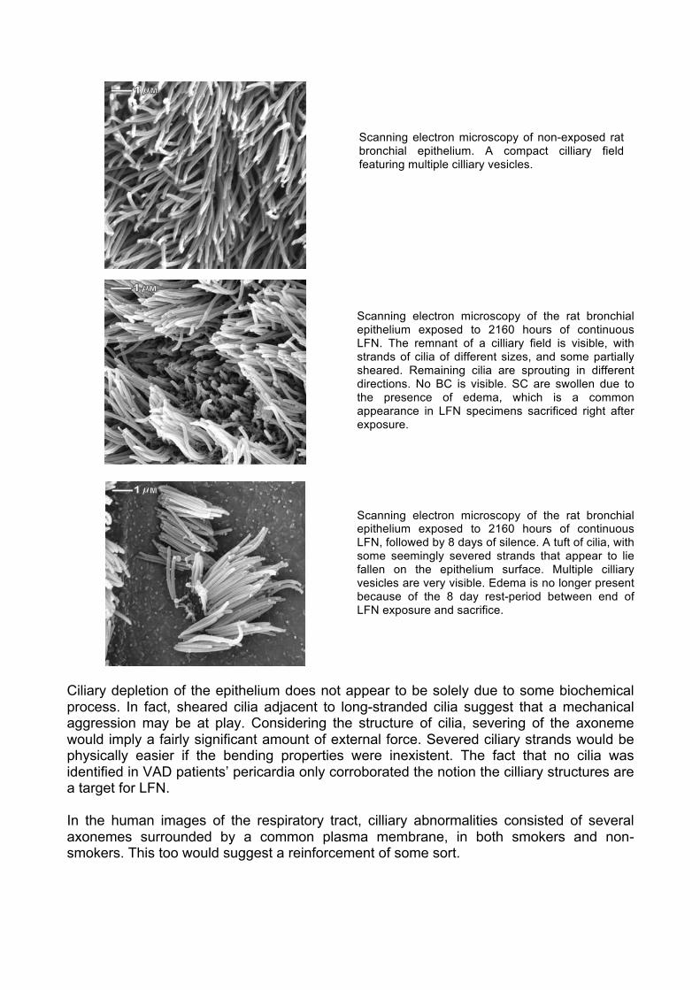
If it is true that LFN exposure induces the propagation of vibration throughout the different viscoelastic tissues of the organism, then it would seem that structures that are not prepared to assimilate this LFN-induced vibration would respond in a manner that would “ward off” the deleterious effects of undesired (and unpredicted) vibration. Perhaps, by increasing their thickness, the “flapping” motion induced by the propagating vibration, is minimized. Cilia Damage In LFN-exposed rodents, ciliary fields become depleted. A distinct pattern of destruction, though, is not identifiable. Within the same image, some areas had sheared cilia while other adjacent, ciliated cells exhibited longer, non-sheared strands, albeit shaggy. Shaggy cilia were seen in many micrographs although axoneme structures seemed intact. Sheared cilia seemed as though they had been clipped, and strands of seemingly clipped cilia were observed lying on the epithelial surface. This is in stark contrast to control rats, where the respiratory epithelium possesses many dense cilliary fields.
Scanning electron microscopy of non-exposed rat bronchial epithelium. A compact cilliary field featuring multiple cilliary vesicles.
Scanning electron microscopy of the rat bronchial epithelium exposed to 2160 hours of continuous LFN. The remnant of a cilliary field is visible, with strands of cilia of different sizes, and some partially sheared. Remaining cilia are sprouting in different directions. No BC is visible. SC are swollen due to the presence of edema, which is a common appearance in LFN specimens sacrificed right after exposure.
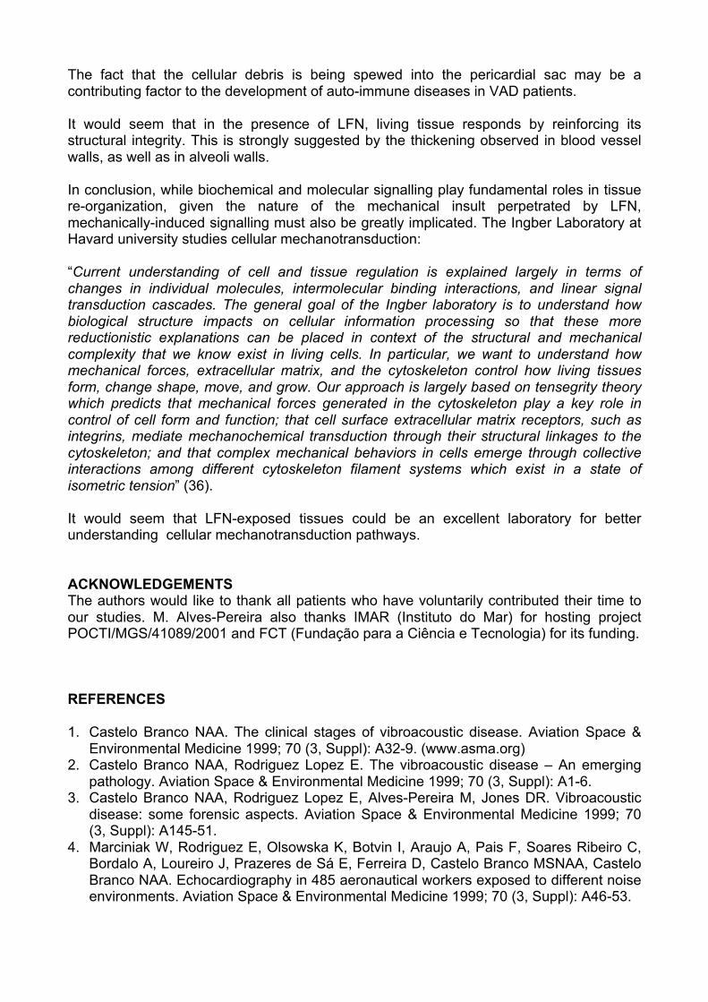
Scanning electron microscopy of the rat bronchial epithelium exposed to 2160 hours of continuous LFN, followed by 8 days of silence. A tuft of cilia, with some seemingly severed strands that appear to lie fallen on the epithelium surface. Multiple cilliary vesicles are very visible. Edema is no longer present because of the 8 day rest-period between end of LFN exposure and sacrifice.
Ciliary depletion of the epithelium does not appear to be solely due to some biochemical process. In fact, sheared cilia adjacent to long-stranded cilia suggest that a mechanical aggression may be at play. Considering the structure of cilia, severing of the axoneme would imply a fairly significant amount of external force. Severed ciliary strands would be physically easier if the bending properties were inexistent. The fact that no cilia was identified in VAD patients’ pericardia only corroborated the notion the cilliary structures are a target for LFN. In the human images of the respiratory tract, cilliary abnormalities consisted of several axonemes surrounded by a common plasma membrane, in both smokers and non-smokers. This too would suggest a reinforcement of some sort.

Fusion of Actin Structures Brush cells (BC) possess microvilli uniformly distributed over the apical surface that is open to the airway. In LFN-exposed rodents, microvilli clustered together, and with increasing exposure time, became fused. Secretory cell microvilli in exposed specimens appeared stunted. Cochlear stereocilia also appeared fused in LFN-exposed rats, both among themselves as well as with the upper tectorial membrane. Why BC microvilli respond to prolonged LFN stress by fusing is unknown. However, the fact that AF can form both rigid (but flexible) bundles as well as gel-like networks, taken together with the fact that motor proteins connect the AF core to the plasma membrane, microvilli fusion does not seem to be such a remote possibility, given the right triggering events. Fusion of cochlear stereocilia AF has also been observed, and corroborates this notion.
Scanning electron microscopy of non-exposed rat tracheal epithelum. Two brush cell (BC) (arrows), at different stages of development, are surrounded by secretory cells (SC) in rosetta-shaped structures. Cilia are long, uniform, and exuberant. BC microvilli are uniformly distributed. Rosetta-forming SC have exuberant microvilli at different lengths indicating that these SC are at different stages of life cycles.
Scanning electron microscopy of the rat tracheal epithelium exposed to 1864 hours of occupationally-simulated LFN. Edema is uniformly distributed. Cell borders are well defined as valleys. The rosetta centered on the BC (arrow) is conserved. BC microvilli are clustering into groups losing their uniform appearance. SC microvilli are still in clearly different stages of growth, but are visibly shorter than in controls.
Scanning electron microscopy of rat tracheal epithelium exposed to 4399 hours of occupationally-simulated LFN. Amplification of a tracheal BC in an exposed rat. Microvilli are clearly grouped together and, in some locations, appear almost fused. Surrounding SC microvilli are uniformly short and stubby.
Scanning electron microscopy of rat tracheal epithelium exposed to 4399 hours of occupationally-simulated LFN. BC (arrow) with fused microvilli, seemingly spreading outward from the center, forming a type of indentation. Around the edges, individual microvilli are still identifiable. Rosetta structures are difficult to identify. Cilia of different lengths are slightly shaggy, and SC microvilli are short and disorganized. A few vesicles budding from cilia are visible.

Scanning electron microscopy of rat tracheal epithelium exposed to 5304 hours of occupationally-simulated LFN. A BC in destruction (arrow). BC microvilli are variable in shape and size, and some are fused. Part of the BC seems to be sinking. Ciliated cells have a variable number of cilia, some very long, others very short, and some of which are shaggy.
Scanning electron microscopy of non-exposed rat bronchial epithelium. The BC, in the center of the image, exhibits a tuft of microvilli that are individually identifiable, uniformly distributed, and sprouting upward into the airway. Surrounding the BC are SC with microvilli of different sizes. Tufts of cilia featuring vesicles are also visible. No sheared, shaggy or wilted cilia are visible. No edema is present.
Scanning electron microscopy of rat bronchial epithelium exposed to 2160 hours of continuous LFN. A BC is in the center of the image. Its microvilli are not sprouting upward, and instead have formed an central indentation of fused microvilli that seem to be spreading outward. The prominent SC that surround the BC are swollen due to edema that causes the deep valleys at the intercellular junctions. SC microvilli are very irregular. Cilliary vesicles are visible. c
Scanning electron microscopy of rat cochlear stereocilia exposed to 4399 hours of occupationally-simulated LFN. Cochlear stereocilia are fused with the upper tectonic membrane.
Scanning electron microscopy of the rat cochlear stereocilia exposed to 4399 hours of occupationally-simulated LFN. Cochlear stereocilia after removal of the tectonic membrane, portions of which remain fused to the stereocilia forming bridges between adjacent cells.
The Pericardium
Instead of 3 layers of tissue, as expected in normal percardia, five distinct layers were identifiable: the fibrosa layer had thickened considerably, split in two, and now sandwiched a newly formed layer of loose tissue, i.e., containing blood vessels, fatty and nerve tissues. Both the internal (closest to the mesothelium) and the external (closest to the epipericardium) exhibited the classical arrangement of accordion-like wave-forms of collagen bundles, intertwined with elastic fibers at seemingly regular intervals. No cilia were identified in any samples. Cytoplasmic extensions, emanating from fibroblast cells also accompany the wavy collagen bundles. Cellular death was observed in all layers, although more so in the internal fibrosa. This was not the classical, apoptotic death.

Light microscopy of a VAD patient pericardium, with pericardial sac on left. Five layers are identifiable: (from left) mesothelial, internal fibrosa, loose tissue, external fibrosa, and epipericardium. In both fibrous layers, wavy collagen bundles are visible. The loose tissue is rich in vessels. No inflammatory cellularity was identified in any of the five layers. (x200)
Transmission electron microscopy of a VAD patient’s parietal pericardium. Mesothelial cell with ruptured membrane and partial loss of cytoplasm. Burst cell in submesothelial layer with large amount of small debris. (x4000)
Transmission electron microscopy of the loose tissue layer. Empty cell membrane in the vicinity of three fragments of nuclear material, embedded within multiple collagen bundles with different orientations. (x5300)
Transmission electron microscopy of VAD patient’s parietal pericardium. Deep within the internal fibrosa layer: a burst myofibroblast near a small elastic fiber (arrow) surrounded by abnormally abundant collagen bundles. (x4000)
Transmission electron microscopy of a VAD patient’s parietal pericardium. Collagen bundles with different orientations, cytoplasmic extensions of a myofibroblast, and an elastic fiber (black arrow) deep in one of the halves of the fibrosa layer. (x10000)
Instead, cellular debris, consisting of seemingly live organelles, were scattered throughout all layers, usually in the vicinity of elastic fibers, and frequently in the neighborhood of burst cytoplasmic extensions. In the mesothelium layer, herniations of mesothelial cell

plasma membranes into the pericardial sac were frequently observed, some of which contained cellular organelles. No nuclear material was observed within these cytoplasmic extensions. Individual mesothelial cells were seen protruding into the pericardial sac, in a process of surface extrusion of that cell. Mesothelial cells with ruptured surfaces in direct contact with the pericardial sac were also observed. Anchoring junctions between mesothelial cells were very strong, sometimes formed by more than two, closely packed, desmosomes, and mostly located in the upper junctional area, closer to the pericardial sac. Unusual gaps were seen in the lower section of the mesothelial layer, where desmosomes were less frequent. These gaps possess great plasticity, and are reversible occurrences. DISCUSSION LFN induces tissue reorganization and neo-formation. One of the underlying purposes may be the need to maintain structural integrity in a viscoelastic environment undergoing LFN-induced vibratory propagation. Actin-based structures seem to have a tendency to fuse. Indeed, microvilli fusion (as seen in the brush cell) will alter the kinetic properties of the structure. Cochlear cilia, for example, are supposed to vibrate freely against the upper tectorial membrane, when an acoustical pressure wave is transduced along the basal membrane. This movement is what relays the acoustical signal to the brain. However, in LFN-exposed rats, cilia are fused together, as well as with the upper tectorial membrane. Hence, when the basal membrane attempts to transduce the acoustical signal, instead of freely vibrating, cochlear cilia, now a non-vibrating structure, will be pulled. If something similar occurs in the cochlear cilia of LFN-exposed humans, then perhaps discomfort might be felt. Discomfort that may be closely associated with the concept of annoyance. The destruction of cilliary fields is dramatic, and might be related to its structural specificities. The cillium is anchored to the cellular cortex through an actin-based network located within the cytoskeleton directly under the plasma membrane. Given the response of other actin-based structures, namely brush cell microvilli as well as cochlear stereocilia, it is not unreasonable to hypothesize that perhaps the actin filaments that compose the cytoskeleton might also be reacting to LFN exposure. Corroborating this notion are transmission electron microscopy images showing intact internal cilliary structures (14). Yet, strands of apparently sheared cilia appear lying horizontally on the epithelial surface, and cilliary fields are depleted. The response of the pericardium to LFN certainly appears to be an adaptation response. This does not exclude the loss of functional capabilities, for example, not a single cilium was found in mesothelial cells. Despite the dramatic alterations of the pericardia, heart function is normal and no diastolic dysfunction exists in VAD patients. It would seem that this newly formed loose tissue layer, rich in vessels and adipose tissue, with numerous elastic components, plays a very important role, possibly of a pneumatic and logistic nature, in maintaining normal function of the heart in these patients. The ruptured cellular membranes seen in the pericardial mesothelial layer are very unusual. Cellular debris is seen in all layers of ther pericardium. This sort of cellular death is not related to the normal, programmed, or apoptotic, cellular death. In VAD patients’ pericardia, cellular death seems to be associated with mechanical processes and stresses.
The fact that the cellular debris is being spewed into the pericardial sac may be a contributing factor to the development of auto-immune diseases in VAD patients. It would seem that in the presence of LFN, living tissue responds by reinforcing its structural integrity. This is strongly suggested by the thickening observed in blood vessel walls, as well as in alveoli walls. In conclusion, while biochemical and molecular signalling play fundamental roles in tissue re-organization, given the nature of the mechanical insult perpetrated by LFN, mechanically-induced signalling must also be greatly implicated. The Ingber Laboratory at Havard university studies cellular mechanotransduction: “Current understanding of cell and tissue regulation is explained largely in terms of changes in individual molecules, intermolecular binding interactions, and linear signal transduction cascades. The general goal of the Ingber laboratory is to understand how biological structure impacts on cellular information processing so that these more reductionistic explanations can be placed in context of the structural and mechanical complexity that we know exist in living cells. In particular, we want to understand how mechanical forces, extracellular matrix, and the cytoskeleton control how living tissues form, change shape, move, and grow. Our approach is largely based on tensegrity theory which predicts that mechanical forces generated in the cytoskeleton play a key role in control of cell form and function; that cell surface extracellular matrix receptors, such as integrins, mediate mechanochemical transduction through their structural linkages to the cytoskeleton; and that complex mechanical behaviors in cells emerge through collective interactions among different cytoskeleton filament systems which exist in a state of isometric tension” (36). It would seem that LFN-exposed tissues could be an excellent laboratory for better understanding cellular mechanotransduction pathways. ACKNOWLEDGEMENTS The authors would like to thank all patients who have voluntarily contributed their time to our studies. M. Alves-Pereira also thanks IMAR (Instituto do Mar) for hosting project POCTI/MGS/41089/2001 and FCT (Fundação para a Ciência e Tecnologia) for its funding. REFERENCES 1. Castelo Branco NAA. The clinical stages of vibroacoustic disease. Aviation Space &
Environmental Medicine 1999; 70 (3, Suppl): A32-9. (www.asma.org) 2. Castelo Branco NAA, Rodriguez Lopez E. The vibroacoustic disease – An emerging
pathology. Aviation Space & Environmental Medicine 1999; 70 (3, Suppl): A1-6. 3. Castelo Branco NAA, Rodriguez Lopez E, Alves-Pereira M, Jones DR. Vibroacoustic
disease: some forensic aspects. Aviation Space & Environmental Medicine 1999; 70 (3, Suppl): A145-51.
4. Marciniak W, Rodriguez E, Olsowska K, Botvin I, Araujo A, Pais F, Soares Ribeiro C, Bordalo A, Loureiro J, Prazeres de Sá E, Ferreira D, Castelo Branco MSNAA, Castelo Branco NAA. Echocardiography in 485 aeronautical workers exposed to different noise environments. Aviation Space & Environmental Medicine 1999; 70 (3, Suppl): A46-53.

5. Castelo Branco NAA, Monteiro E, Alves-Pereira M, Águas AP, Sousa Pereira A, Grande NR. Morphological changes in the pericardia of military helicopter pilots. Proc. Microscopy Barcelona 2001, 4-7 Sep 2001: 318-19.
6. Araujo A, Pais F, Lopo Tuna JMC, Alves-Pereira M, Castelo Branco NAA. Echocardiography in noise-exposed flight crew. Internoise 2001, The Hague, Holland 2001: 1007-10.
7. Torres R, Tirado G, Roman A, Ramirez R, Colon H, Araujo A, Pais F, Marciniak W, Nóbrega J, Bordalo e Sá A, Lopo Tuna JMC, Castelo Branco MSNAA, Alves-Pereira M, Castelo Branco NAA. Vibroacoustic disease induced by long-term exposure to sonic booms. Internoise 2001, The Hague, Holland; 2001: 1095-98.
8. Araujo A, Alves-Pereira M, Joanaz de Melo J, Castelo Branco NAA. Environmentally-induced vibroacoustic disease in a suburban family. 11th Intern Conf Sound & Vibration, St. Petersburg, Russia, 2004: 1767-1774
9. Castelo Branco NAA, Reis Ferreira J, Marques MC, Alves-Pereira M. Vibroacoustic disease: the impact of low frequency noise on quality of life. ECQual-Journal Eur. Comm. Quality of Life (In press) 2004.
10. Castelo Branco NAA. A unique case of vibroacoustic disease. A tribute to an extraordinary patient. Aviation Space & Environmental Medicine 1999; 70 (3, Suppl): A27-3.
11. GIMOGMA. [Epilepsy of vascular etiology, a clinical picture of vibration disease?] Rev Port Med Mil 1984; 32: 5-9. (In Portuguese)
12. GIMOGMA. [Evoked potential study in a population exposed to occupational vibration.] Rev Port Med Mil 1984; 32: 10-6. (In Portuguese)
13. GIMOGMA. [Vibration and noise as the cause of acoustic hypo and hypersensibility in an industrial population]. Rev Port Med Mil 1984; 32: 17-20.(In Portuguese)
14. Castelo Branco NAA, Alves-Pereira M, Martins dos Santos J, Monteiro E. SEM and TEM study of rat respiratory epithelia exposed to low frequency noise. In Science and Technology Education in Microscopy: An Overview, A. Mendez-Vilas (ed.), Formatex: Badajoz, Spain, 2003; Vol. II: 505-33.
15. Castelo Branco NAA, Águas AP, Sousa Pereira A, Monteiro E, Fragata JIG, Grande NR. The pericardium in the vibroacoustic syndrome. J Cardiovas Diag & Prog 1996; 13(4): 284. (Abstract)
16. Castelo Branco NAA, Águas AP, Sousa Pereira A, Monteiro E, Fragata JIG, Tavares F, Grande NR. The human pericardium in vibroacoustic disease. Aviation Space & Environmental Medicine 1999; 70 (3, Suppl): A54-62.
17. Castelo Branco NAA, Fragata JI, Monteiro E, Alves-Pereira M. Pericardial features in vibroacoustic disease patients. Proc. 8th Intern. Conf. Noise as Public Health Problem (ICBEN), Rotterdam, Holland, 2003: 380-381.
18. Castelo Branco NAA, Fragata JI, Martins AP, Monteiro E, Alves-Pereira M. Pericardial cellular death in vibroacoustic disease. Proc. 8th Intern. Conf. Noise as Public Health Problem (ICBEN), Rotterdam, Holland, 2003: 376-377.
19. Castelo Branco NAA, Fragata JI, Martins AP, Monteiro E, Alves-Pereira M. Pericardial cellular death in vibroacoustic disease patients. Proc. 11th Intern Conf Sound & Vibration, St. Petersburg, Russia, 2004: 1753-1760.
20. Pimenta MG, Martinho Pimenta AJF, Castelo Branco MSN, Castelo Branco NAA. ERP P300 and brain magnetic resonance imaging in patients with vibroacoustic disease. Aviation Space & Environmental Medicine 1999; 70 (3, Suppl): A107-14.
21. Gomes L, Martinho Pimenta AJF, Castelo Branco NAA. Effects of occupational exposure to low frequency noise on cognition. Aviation Space & Environmental Medicine 1999; 70 (3, Suppl): A115-8.

22. Shabetai R. Diseases of the pericardium. In: Hurst’s The Heart, Arteries and Veins. Schlant RC, Wayne Alexander R (eds.) (8th ed). McGraw-Hill, Inc.: New York; 1994: 1647-9.
23. Reis Ferreira J, Mendes CP, Castelo Branco NAA, Monteiro E, Alves-Pereira M. The human trachea in vibroacoustic disease. Proc. 8th Intern. Conf. Noise as Public Health Problem (ICBEN), Rotterdam, Holland, 2003: 388-389.
24. Reis Ferreira J, Mendes CP, Castelo Branco NAA, Monteiro E & Alves-Pereira M. The human lung and pleura in vibroacoustic disease. Proc. 8th Intern. Conf. Noise as Public Health Problem (ICBEN), Rotterdam, Holland, 2003: 386-387.
25. Oliveira MJR, Sousa Pereira A, Águas AP, Monteiro E, Grande NR, Castelo Branco NAA. Effects of low frequency noise upon the reaction of pleural milky spots to mycobacterial infection. Aviat Space Environ Med 1999; 70 (March, Suppl): A137-40.
26. Grande N, Águas AP, Sousa Pereira A, Monteiro E, Castelo Branco NAA. Morphological changes in the rat lung parenchyma exposed to low frequency noise. Aviat Space Environ Med 1999; 70 (3, Suppl): A70-7.
27. Sousa Pereira A, Águas A, Grande NR, Castelo Branco NAA. The effect of low frequency noise on rat tracheal epithelium. Aviat Space Environ Med 1999; 70 (3, Suppl): A86-90.
28. Castelo Branco NAA, Monteiro E, Costa e Silva A, Reis Ferreira J, Alves-Pereira M. Respiratory epithelia in Wistar rats. Rev. Port. Pneumol. 2003; IX: 381-88.
29. Castelo Branco NAA, Monteiro E, Costa e Silva A, Reis Ferreira J, Alves-Pereira M. Respiratory epithelia in Wistar rats born in low frequency noise plus varying amount of additional exposure. Rev. Port. Pneumol. 2003; IX (6): 481-492.
30. Castelo Branco NAA, Gomes-Ferreira P, Monteiro E, Costa e Silva A, Reis Ferreira J, Alves-Pereira M. Respiratory epithelia in Wistar rats after 48 hours of continuous exposure to low frequency noise. Rev. Port. Pneumol. 2003; IX (6): 474-79.
31. Castelo Branco NAA, Monteiro E, Costa e Silva A, Martins dos Santos J, Reis Ferreira J, Alves-Pereira M. The lung parenchyma in low frequency noise exposed Wistar rats. Rev. Port. Pneumol. 2004; X (1): 77-85.
32. 23. Castelo Branco NAA, Monteiro E, Martins dos Santos J & Alves-Pereira M. Low frequency noise and intra-cellular edema. Proc. 8th Intern. Conf. Noise as Public Health Problem (ICBEN), Rotterdam, Holland, 2003: 378-379.
33. Lousã N, Monteiro E, Alves-Pereira M & Castelo Branco NAA. Rat cochlea exposed to low frequency noise. Proc. 8th Intern. Conf. Noise as Public Health Problem (ICBEN), Rotterdam, Holland, 2003: 43-45.
34. Albuquerque e Sousa J, Martinho Pimenta AJF, Castelo Branco NAA. Three Cases of the Vibroacoustic Syndrome with Significant Carotid Stenosis. Aviation Space Environmental Medicine 1995; 66: 494 (Abstract)
35. Albuquerque e Sousa J, Dinis da Gama A, Macedo MV, Cassio I, et al. Carotid angiodynographic studies in individuals occupationally exposed to noise and vibration. Aviation Space Environmental Medicine 1991; 62: 134 (Abstract).
36. Donald E. Ingber, MD, PhD, Professor of Pathology, Harvard Medical School, [email protected]