114 molecular and serotyping characterization of …
TRANSCRIPT

114
Fayoum J. Agric. Res. & Dev., Vol. 33, No.1, January, 2019
MOLECULAR AND SEROTYPING CHARACTERIZATION OF NON-
SHIGA TOXIGENIC ESCHERICHIA COLI ASSOCIATED WITH
FOOD COLLECTED FROM THE LOCAL MARKET IN FAYOUM
GOVERNORATE, EGYPT
Amr E. M. Mahmoud1, Ghada O. El-demerdash
2, Mohamed H. H. Roby
3
and Sahar R. Mohamed2
1Biochemistry Department, Faculty of Agriculture, Fayoum University,
Fayoum, Egypt. 2Animal Health Research Institute, Dokki, Giza, Egypt.
3 Food Science and Technology Department, Faculty of Agriculture, Fayoum
University, Fayoum, Egypt.
ABSTRACT:
Escherichia coli is considered as one of the bacteria that causing
diarrhea outbreaks all over the world, and it is responsible for diseases
for human and animals as well
A total number of 50 raw milk and 50 raw beef meat samples were
collected from the local market in Fayoum Governorate. These samples
were subjected for bacteriological, serological and molecular
investigations. E. coli was isolated from raw milk and raw beef samples
with an isolation rate of 58% and 14%, respectively.
Serogrouping of the E. coli isolates from the raw milk samples
revealed presence of O142, O55, O111, O27, and O26 with percentage of
20.69%, 20.69%, 17.24%, 17.24%, and 3.45%, respectively. However,
the serogrouping of the E. coli isolates from raw beef meat samples
revealed presence of O111, O27, O142, O55, and O127 with percentage of
28.56%, 14.28%, 14.28%, 14.28%, and 14.28%, respectively.
Multiplex PCR was applied for the detection of virulence genes
including shiga-toxin genes (stx1 and stx2), and the intimin gene (eae)
which detected in E. coli. All the isolates were negative to both stx1 and
stx2 genes. Meanwhile, the raw milk isolates of O142, O55, O111, and O27
were positive to eae gene. However, O26 isolate was negative to this
gene. Also, the raw beef meat isolates of O142, O55, O111, and O27 were
positive to eae gene. But, O127 isolate was negative to this gene.
KEY WORDS: E. coli, Virulence genes, Raw milk and Raw meat.
INTRODUCTION
Diarrhea is the second cause of death after pneumonia in children aged
between 1 to 59 months, with mortality number of 0.509 million per year
worldwide. Developing countries had the majority of this mentioned mortality
cases of children by diarrhea (Liu et al., 2016 and WHO, 2017). Diarrhea
outbreaks are happening all over the world which has the attention as an

Amr E. M. Mahmoud1, et al., 115
Fayoum J. Agric. Res. & Dev., Vol. 33, No.1, January, 2019
important public health problem that caused by Escherichia coli bacteria
causing diseases for human and animals as well (Buchholz et al., 2011;
Watson et al., 2017 and Valilis et al., 2018). This Gram-negative bacteria
exists as part of the normal flora of animals and humans' gastrointestinal tract
and responsible for infection between both of them (Karmali et al., 2010 and
Lim et al., 2010). The Majority of its strains are harmless. Unfortunately,
there are many virulence factors such as the mobile genetic elements
including; bacteriophages, plasmids….etc and pathogenicity islands that may
be acquired by these strains resulting in turning them to the pathogenic state
(Kaper et al., 2004). Moreover, most of E. coli carrying hosts are apparently
healthy and asymptomatic (Hussein and Bollinger 2005, and Bogitsh et al.,
2018). And, ruminants such as sheep, goats and especially cattle, are counted
as the main reservoir of E. coli bacteria (Kaper et al., 2004). E. coli
transmission route starts when the bacteria could pass into the food chain via
any contaminated food, drinks and water with feces (Suardana et al., 2017).
So, E. coli transmission may occur via the consumption of any contaminated
type of uncooked meat, fruits, vegetables, unpasteurized milk and its products
(Karmali et al., 2010 and Tzschoppe et al., 2012). Consequently, E. coli
infection may lead to many food-borne diseases in human including, diarrhea,
renal failure, brain failure and hemolytic uremic syndrome which considered
as life-threatening disease (Karmali et al., 2010 and Lim et al., 2010). There
are six pathotypes of this bacterium, enterotoxigenic E. coli {ETEC},
enteropathogenic E. coli {EPEC}, enteroaggregative E. coli {EAEC},
diffusely adherent E. coli {DAEC} enterohemorrhagic E. coli {EHEC}, and
enteroinvasive E. coli {EIEC} (Nataro and Kaper 1998, and Lei et al.,
2018). Also, there are many strains of E. coli that produce toxins called "shiga
toxins" which cause illness in the vertebrates. These strains are called "shiga-
toxin producing" E. coli (STEC) or verocytotoxic E. coli which are the
pathotype group of enterohemorrhagic E. coli {EHEC}(Nguyen and
Sperandio 2012; Lacher et al., 2016, and Valilis et al., 2018). Also, it is
classified by its serotypes which include more than 700 serotypes according to
their O and H antigen (Lacher et al., 2016 and Bai et al., 2018). E. coli
O157:H7 is the major serotype that was associated with human illness world
widely. This strain was classified as the most common strain responsible of the
E. coli outbreaks in the USA, German, Northern Ireland, South Korea, Japan,
England, Scotland and many other countries (Tarr et al., 2005; Money et al.,
2010; Buchholz et al., 2011; Dallman et al., 2012; Park et al., 2014;
Watahiki et al., 2014; Launders et al., 2016; Saeedi et al., 2017 and Yang
et al., 2017). However, in the last twenty years the non-O157 stains were
classified as responsible serotypes for 20 to 50% of the E. coli associated
illness' outbreaks world widely, these serotypes are including the O26, O45,
O103, O111, O121 and O145 (Wasilenko et al., 2012; Gould et al., 2013;

MOLECULAR AND SEROTYPING CHARACTERIZATION OF…….. 116
Fayoum J. Agric. Res. & Dev., Vol. 33, No.1, January, 2019
Albonico et al., 2017 and Balamurugan et al., 2017). Chapman et al.,
(2006), found that there were more than 50 virulence factors of E. coli
participated in its pathogenicity. E. coli bacteria produce many factors
associated with human illness such as Shiga toxins (stx1 and stx2), besides the
other virulence factors that responsible for the attachment of the bacteria to the
hosts' intestinal epithelial cells. This attaching lesions caused by the intimin
protein that encoded by the eae gene (Chapman et al., 2006; Farfan and
Torres 2012; and Gharieb et al., 2015). So, the main aim of this work is to
detect the virulence genes stx1, stx2 and eae of Escherichia coli bacteria
growing in raw milk and beef meat collected from the local market in Fayoum
Governorate. Besides, characterizing the serotypes of the Escherichia coli
isolates.
MATERIALS AND METHODS
Sample collection
A total number of 50 raw milk and 50 raw beef meat samples were
collected from the local market in Fayoum Governorate. Samples were
collected in sterile marked container then inoculated in Carry and Blair
transport medium. The last was kept in ice box for the laboratory bacterial
culturing and identification.
Bacteriological examination
The collected samples were cultured using MacConkey agar. The
plates were aerobically incubated up to two days at 37 ᵒC. Then the suspected
colonies were picked up and tested for Gram's reaction. The positive colonies
were identified biochemically by using Vitek2 compact system (bioMérieux,
Durham, NC, USA), according to the manufacturer's instructions
(Chatzigeorgiou et al., 2011 and Quinn et al., 2011), using the Gram-
Negative (GN) card which is a complete system for routine identification
testing of most clinically significant Gram-Negative organisms. Colonies were
transferred to the 0.45 % saline to prepare the organism suspension with a
density equivalent to a 0.50 to 0.63 McFarland using a calibrated VITEK® 2
DensiCHEK™ Plus. Then, the last suspension used to fill the test cards for
Vitek2 instrument.
Serological identification
Escherichia coli isolates were serologically identified using the rapid
diagnostic E. coli antisera set (Denka sieken comp. LTD) according to
Edwards and Ewing (1972).
Molecular examination
DNA extraction
Escherichia coli isolates' DNA extraction was done using the
QIAamp DNA Mini kit (Qiagen, Germany, GmbH) with modifications
from the manufacturer’s recommendations and according to (Sambrook et

Amr E. M. Mahmoud1, et al., 117
Fayoum J. Agric. Res. & Dev., Vol. 33, No.1, January, 2019
al., 1989). In brief, a 200 µl of the each sample suspension was added to
the proteinase K solution (10 µl), 200 µl of the lysis buffer and incubated
at 56˚C for 10 min. Then, 200 µl of 100% ethyl alcohol was added to the
lysate. After washing and centrifuging the sample, 100 µl of elution buffer
that provided by the kit was used to elute the nucleic acid.
PCR amplification
PCR amplification of the E. coli isolates' DNA of the virulent genes
was carried out using the primers that revealed to (stx1, stx2, and eae) genes as
indicated in (Table1). This PCR amplification was applied on 10 random
isolates (one of each serotype) of E. coli, 5 of each raw milk and beef meat
samples for the detection of the virulence genes. The PCR amplification of
these primers were utilized in a 25 µl reaction containing 12.5 µl of PCR
Master Mix (Takara, Japan), 1 µl of each primer of 20 pmol concentration, 6
µl of DNA template and 4.5 µl of nuclease-free water. The reaction was
performed in an (Applied Biosystem Thermal Cycler). Cycling conditions
were used as recommended by the manufacturer as follow: primary
denaturation: 94˚C/5 min., secondary denaturation: 94˚C/30 sec., annealing:
55˚C/45 sec., extension: 72˚C/45 sec., no. of cycles: 35 and final extension:
72˚C/10 min.
Analysis of the PCR Products
1.5 % agarose gel (Applichem, Germany, GmbH) was used to separate the
PCR products by electrophoresis in 1x TBE buffer at room temperature using
gradients of 5V/cm. For gel analysis, 20 µl of the PCR products was loaded in
each gel well. The fragments sizes were determined using a gelpilot 100bp
plus DNA Ladders (Qiagen, Germany, GmbH). The gel was photographed by
a gel documentation system (Alpha Innotech, Biometra).
Table (1): Primers used for the detection of virulent genes of E. coli,
F: Forward and R: Reverse. Target
Genes Primers sequences
Amplified
Segment (bp) Reference
eae F: ATG CTT AGT GCT GGT TTA GG
248 Bisi-Johnson et al., 2011 R: GCC TTC ATC ATT TCG CTT TC
stx1 F:ACACTGGATGATCTCAGTGG
614 Shetty et al., 2012 R:CTGAATCCCCCTCCATTATG
stx2 F:CCATGACAACGGACAGCAGTT
779 Shetty et al., 2012 R:CCTGTCAACTGAGCAGCACTTTG

MOLECULAR AND SEROTYPING CHARACTERIZATION OF…….. 118
Fayoum J. Agric. Res. & Dev., Vol. 33, No.1, January, 2019
RESULTS
The results of bacteriological examination
Out of the 100 raw milk and meat samples collected from the local
market in Fayoum Governorate, Egypt, the E. coli was isolated as (58%) of the
raw milk samples followed by (14%) of the raw beef meat samples as shown
in table (2). Also, the biochemical identification of the positive E. coli isolates
by Vitek2 system is shown in table (3).
Table (2): Prevalence of E. coli bacteria isolated from collected samples.
No. of examined
Samples
Escherichia coli
No. of +ve samples % of +ve samples
50 raw milk samples 29 58
50 raw beef meat samples 7 14
No.: Number of positive isolates of E. coli and %: Percentage in relation to No.
of examined samples (50).
Table (3): Biochemical details of Escherichia coli using Vitek 2 compact
system.
**Vitek2 Gram-negative card well contents according to BioMerieux,
manufacturer manual are indicted in Appendix 1.
The results of serotyping of E. coli positive isolates
Serogrouping of the 29 E. coli isolates from raw milk samples revealed
presence of O142, O55, O111, O27, and O26 with percentage of 20.69%, 20.69%,
17.24%, 17.24%, and 3.45%, respectively. Also, there were 6 isolates untyped
as shown in table (4). However, the serogrouping of the 7 E. coli isolates from
raw beef meat samples revealed presence of O111, O27, O142, O55, and O127 with
percentage of 28.56%, 14.28%, 14.28%, 14.28%, and 14.28%, respectively.
Besides, there was one isolate untyped as shown in table (4).
Table 3: Biochemical details of Escherichia coli
2 APPA - 3 ADO - 4 PyrA - 5 IARL - 7 dCEL - 9 BGAL +
10 H2S - 11 BNAG - 12 AGLTP - 13 dGLU + 14 GGT - 15 OFF +
17 BGLU - 18 dMAL + 19 dMAN + 20 dMNE + 21 BXYL - 22 BAlap -
23 proA - 26 LIP - 27 PLE - 29 TyrA - 31 URE - 32 dSOR +
33 SAC - 34 dTAG - 35 dTER + 36 CIT - 37 MNT - 39 5KG +
40 ILATK - 41 AGLU - 42 SUCT + 43 NAGA - 44 AGAL - 45 PHOS -
46 GlyA - 47 ODC + 48 LDC + 53 IHISa - 56 CMT + 57 BGUR -
58 O129R + 59 GGAA - 61 IMLTa - 62 ELLM - 64 ILATa -
Amr E. M. Mahmoud1, et al., 119
Fayoum J. Agric. Res. & Dev., Vol. 33, No.1, January, 2019
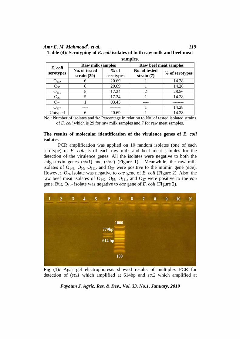
Table (4): Serotyping of E. coli isolates of both raw milk and beef meat
samples.
E. coli
serotypes
Raw milk samples Raw beef meat samples
No. of tested
strain (29)
% of
serotypes
No. of tested
strain (7) % of serotypes
O142 6 20.69 1 14.28
O55 6 20.69 1 14.28
O111 5 17.24 2 28.56
O27 5 17.24 1 14.28
O26 1 03.45 ---- -------
O127 ---- ------- 1 14.28
Untyped 6 20.69 1 14.28
No.: Number of isolates and %: Percentage in relation to No. of tested isolated strains
of E. coli which is 29 for raw milk samples and 7 for raw meat samples.
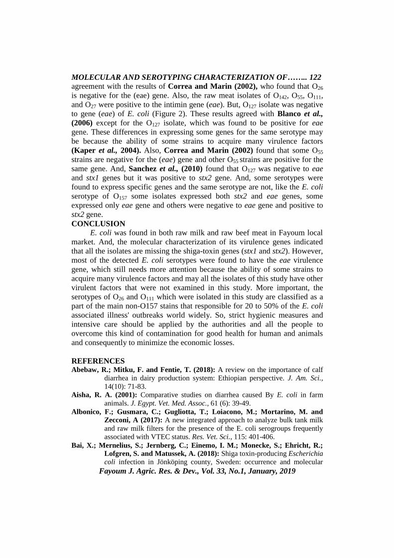
The results of molecular identification of the virulence genes of E. coli
isolates
PCR amplification was applied on 10 random isolates (one of each
serotype) of E. coli, 5 of each raw milk and beef meat samples for the
detection of the virulence genes. All the isolates were negative to both the
shiga-toxin genes (stx1) and (stx2) (Figure 1). Meanwhile, the raw milk
isolates of O142, O55, O111, and O27 were positive to the intimin gene (eae).
However, O26 isolate was negative to eae gene of E. coli (Figure 2). Also, the
raw beef meat isolates of O142, O55, O111, and O27 were positive to the eae
gene. But, O127 isolate was negative to eae gene of E. coli (Figure 2).
Fig (1): Agar gel electrophoresis showed results of multiplex PCR for
detection of (stx1 which amplified at 614bp and stx2 which amplified at
1 2 3 4 5 P L 6 7 8 9 10 N
1000
100
614 bp
779bp

MOLECULAR AND SEROTYPING CHARACTERIZATION OF…….. 120
Fayoum J. Agric. Res. & Dev., Vol. 33, No.1, January, 2019
779bp), L: represents the molecular size marker (100pb plus ladder), N:
Negative control, P: Positive control, Lanes from 1:5 represent the raw milk
isolates O142, O55, O111, O27, and O26 which are negative for both stx1 and stx2
genes, and Lanes from 6:10 represent the raw beef meat isolates O142, O55,
O111, O27, and O127 which are negative for both stx1 and stx2 genes.
Fig (2): Agarose gel electrophoresis showed results of Multiplex PCR for
detection of eae gene, L: represents the molecular size marker (100pb plus
ladder), N: Negative control, P: Positive control of eae gene (248bp), Lanes
from 1:5 represents the raw milk isolates; Lanes 1:4 are the isolates of O142,
O55, O111, and O27 which are positive for eae gene, Lane 5 is the O26 isolate
which is negative for the eae gene, Lanes from 6:10 represent the raw beef
meat isolates; Lanes 6:9 are the isolates of O142, O55, O111, and O27 which are
positive for eae gene, lane 10 is the O127 isolate which is negative for the eae
gene.
DISCUSSION
Escherichia coli considered as one of the bacteria that causing diarrhea
outbreaks all over the world, and it is responsible for diseases for human and
animals as well (Buchholz et al., 2011: Watson et al., 2017 and Valilis et al.,
2018). Unfortunately, the developing countries had the majority of mortality
cases of children aged from 1 to 59 months caused by diarrhea (Liu et al.,
2016 and WHO, 2017). Also, neonatal calf diarrhea is considered as of the
most important health problems in livestock causing high economic losses
worldwide either directly due to mortality and needs for treatment or indirectly
through poor growth (El-Seedy et al., 2016; Abebaw et al., 2018 and Bokma
et al., 2019). In present study, E. coli was isolated from raw milk samples with an
isolation rate of 58%. This result was lower than the isolation rate that
100
1 2 3 4 5 P L N6 7 8 9 10
600
248bp
100

Amr E. M. Mahmoud1, et al., 121
Fayoum J. Agric. Res. & Dev., Vol. 33, No.1, January, 2019
described by (Ombarak et al., (2016), who isolated E. coli with an incidence
of 76.4%. But, this result was higher than the isolation rate that described by
(Metwally and Ali (2015); Bedasa et al., 2018 and Singh et al., 2018), who
isolated E. coli with an incidence of 44%, 32% and 17.19% respectively.
However, this percentage was almost similar to the rate that obtained by El
Nahas et al., (2015), who isolated E. coli with an incidence of 55%. There
were only 7 E. coli isolates out of the 50 raw meat samples with an isolation
rate of 14%. This percentage was almost similar to the rate that obtained by
Bedasa et al., (2018). However, this result was higher than the rates described
by Rahimi et al., (2012) and Moawad et al., (2017), who isolated E. coli with
an incidence of 8.2% and 11.7%, respectively. This high rate may be
explained by that transmission of infection occurs during the milking process
by milkers' hands, contaminated equipments and milking machine Scherrer et
al., (2004). Also, this may be the same in case of meat rates which is more
likely as cause of poor hygienic measures and customs during slaughter,
handling, transportation and even during all stages of storage Rahimi et al.,
(2010). Also, contamination level may be varied due to the differences in
geographic or national region, processing environments, meat sources and the
methodologies which the samples were taken such as; the samples amount,
numbers and even the periods of which the samples were tested Kegode et al.,
(2008). Serogrouping of E. coli isolates from the raw milk samples revealed
presence of O142, O55, O111, O27, and O26 with percentage of 20.69%, 20.69%,
17.24%, 17.24%, and 3.45%, respectively. Also, the serogrouping of E. coli
isolates from raw meat samples revealed presence of O111, O27, O142, O55, and
O127 with percentage of 28.56%, 14.28%, 14.28%, 14.28%, and 14.28%,
respectively. The above mentioned results are in agreement with results of
Aisha (2001), who isolated O26, O127 and O27, Correa and Marin (2002),
who isolated O26, O55, O111, O127 and O142, Blanco et al., (2006), who isolated
O55, O111, O127 and O142, Lin et al., (2011) who isolated O26, O142 and O111,
Fadel et al., (2017), who isolated O26 ,O27,O55, O111, and O142,and Kalule et
al., (2018), who isolated O26, O55, and O111,
Molecular characterization of the E. coli isolates from both raw milk
and beef meat samples through applying different conditions of multiplex PCR
for detection of genes encoding virulence factors (stx1, stx2 and eae). All the
isolates were negative to both the shiga-toxin genes (stx1) and (stx2) (Figure
1). Meanwhile, the raw milk isolates of O27, O55, O111, and O142 were positive
to the intimin gene (eae). These results agreed with the results of Blanco et al.,
(2006) who found that O55, O111 and O142 are negative for both shiga-toxin
genes (stx1) and (stx2) and positive to the intimin gene (eae). However, O26
isolate was negative to gene (eae) of E. coli (Figure 2). This result is in
MOLECULAR AND SEROTYPING CHARACTERIZATION OF…….. 122
Fayoum J. Agric. Res. & Dev., Vol. 33, No.1, January, 2019
agreement with the results of Correa and Marin (2002), who found that O26
is negative for the (eae) gene. Also, the raw meat isolates of O142, O55, O111,
and O27 were positive to the intimin gene (eae). But, O127 isolate was negative
to gene (eae) of E. coli (Figure 2). These results agreed with Blanco et al.,
(2006) except for the O127 isolate, which was found to be positive for eae
gene. These differences in expressing some genes for the same serotype may
be because the ability of some strains to acquire many virulence factors
(Kaper et al., 2004). Also, Correa and Marin (2002) found that some O55
strains are negative for the (eae) gene and other O55 strains are positive for the
same gene. And, Sanchez et al., (2010) found that O127 was negative to eae
and stx1 genes but it was positive to stx2 gene. And, some serotypes were
found to express specific genes and the same serotype are not, like the E. coli
serotype of O157 some isolates expressed both stx2 and eae genes, some
expressed only eae gene and others were negative to eae gene and positive to
stx2 gene.
CONCLUSION E. coli was found in both raw milk and raw beef meat in Fayoum local
market. And, the molecular characterization of its virulence genes indicated
that all the isolates are missing the shiga-toxin genes (stx1 and stx2). However,
most of the detected E. coli serotypes were found to have the eae virulence
gene, which still needs more attention because the ability of some strains to
acquire many virulence factors and may all the isolates of this study have other
virulent factors that were not examined in this study. More important, the
serotypes of O26 and O111 which were isolated in this study are classified as a
part of the main non-O157 stains that responsible for 20 to 50% of the E. coli
associated illness' outbreaks world widely. So, strict hygienic measures and
intensive care should be applied by the authorities and all the people to
overcome this kind of contamination for good health for human and animals
and consequently to minimize the economic losses.
REFERENCES Abebaw, R.; Mitku, F. and Fentie, T. (2018): A review on the importance of calf
diarrhea in dairy production system: Ethiopian perspective. J. Am. Sci.,
14(10): 71-83.
Aisha, R. A. (2001): Comparative studies on diarrhea caused By E. coli in farm
animals. J. Egypt. Vet. Med. Assoc., 61 (6): 39-49.
Albonico, F.; Gusmara, C.; Gugliotta, T.; Loiacono, M.; Mortarino, M. and
Zecconi, A (2017): A new integrated approach to analyze bulk tank milk
and raw milk filters for the presence of the E. coli serogroups frequently
associated with VTEC status. Res. Vet. Sci., 115: 401-406.
Bai, X.; Mernelius, S.; Jernberg, C.; Einemo, I. M.; Monecke, S.; Ehricht, R.;
Lofgren, S. and Matussek, A. (2018): Shiga toxin-producing Escherichia
coli infection in Jönköping county, Sweden: occurrence and molecular

Amr E. M. Mahmoud1, et al., 123
Fayoum J. Agric. Res. & Dev., Vol. 33, No.1, January, 2019
characteristics in correlation with clinical symptoms and duration of stx
shedding. Front. Cell. Infect. Microbiol., 8:125. doi:
10.3389/fcimb.2018.00125.
Balamurugan, S.; Ahmed, R.; Gao, A. and Strange, P. (2017): Comparison of the
fate of the top six non-O157 shiga-toxin producing Escherichia coli
(STEC) and E. coli O157:H7 during the manufacture of dry fermented
sausages. Int. J. Food Microbiol., 259: 14-21.
Bedasa, S.; Shiferaw, D.; Abraha, A. and Moges, T. (2018): Occurrence and
antimicrobial susceptibility profile of Escherichia coli O157:H7 from food
of animal origin in Bishoftu town, Central Ethiopia. Int. J. Food Contam.,
5 (2): 1-8.
Bisi-Johnson, M. A.; Obi, C. L.; Vasaikar, S. D.; Baba, K. A. and Hattori, T.
(2011): Molecular basis of virulence in clinical isolates of Escherichia coli
and Salmonella species from a tertiary hospital in the Eastern Cape, South
Africa. Gut Pathogens, 3 (9): 1-8.
Blanco, M.; Blanco,J. Dahbi, G.; Mora,A.; Alonso, M. P.; Varela, G.; Gadea,M.
P.; Schelotto,F.; Gonza, E. A. and Blanco, J. (2006): Typing of intimin
(eae) genes from enteropathogenic Escherichia coli (EPEC) isolated from
children with diarrhoea in Montevideo, Uruguay: identification of two
novel intimin variants (mB and jR/b2B). J. Med. Microbiol., 55: 1165-
1174.
Bogitsh, B. J.; Carter, C. E. and Oeltmann, T. N. (2018): Human parasitology, 5th
ed.; Elsevier, London, United Kingdom.
Bokma, J.; Boone, R.; Deprez, P. and Pardon, B. (2019): Risk factors for
antimicrobial use in veal calves and the association with mortality. J.
Dairy Sci., 102: 607-618.
Buchholz, U.; Bernard, H.; Werber, D.; Böhmer, M.; Remschmidt, C.; Wilking,
H.; Deleré, Y.; Heiden, M.; Adlhoch, M.; Dreesman, J. and Ehlers, J.
(2011): German outbreak of Escherichia coli O104:H4 associated with
sprouts. N. Engl. J. Med., 365: 1763-1770.
Chapman, T. A.; Wu, X. Y.; Barchia, I.; Bettelheim, K. A.; Driesen, S.; Trott,
D.; Wilson, M. and Chin, J. J. (2006): Comparison of virulence gene
profiles of Escherichia coli strains isolated from healthy and diarrheic
swine. Appl. Environ. Microbiol., 72: 4782-4795.
Chatzigeorgiou, K. S.; Sergentanis, T. N.; Tsiodras, S.; Hamodrakas, S. J. and
Bagos, P.G., (2011): Phoenix 100 versus Vitek 2 in the identification of
gram-positive and gram-negative bacteria: a comprehensive meta-analysis.
J. clin. microbiol.,49: 3284-3329.
Correa, M. G. P. and Marin, J. M. (2002): O-serogroups, eae gene and EAF
plasmid in Escherichia coli isolates from cases of bovine mastitis in
Brazil. Vet. Microbiol., 85: 125-132.
Dallman, T.; Smith, G. P.; O'brien, B.; Chattaway, M. A.; Finlay, D., Grant, K.
A. and Jenkins, C. (2012): Characterization of a verocytotoxin -
producing enteroaggregative Escherichia coli serogroup O111:H21 strain
MOLECULAR AND SEROTYPING CHARACTERIZATION OF…….. 124
Fayoum J. Agric. Res. & Dev., Vol. 33, No.1, January, 2019
associated with a household outbreak in Northern Ireland. J. Clin.
Microbiol., 50: 4116-4119.
Edwards, P. R. and Ewing, W. H. (1972): Identification of Enterobacteriacae, 3rd
ed., Burgess Pub. Co., MN, USA.
El Nahas, A. W.; Mohamed, H. A.; El Barbary, H. A. and Mohamed, H. S.
(2015): Incidence of E. coli in raw milk and its products. Benha Vet. Med.
J., 29 (1): 112-117.
El-Seedy, F. R.; Abed, A. H.; Yanni, H. A. and Abd El-Rahman, S. A. A. (2016): Prevalence of E. coli and Salmonella in neonatal calves with diarrhea. J.
Basic and Applied Sci. Cell. Mol. Bio. 62: 21–28. .
Fadel, H. M.; Afifi, R. and Al-Qabili, D. M. (2017): Characterization and zoonotic
impact of Shiga toxin producing Escherichia coli in some wild bird
species. Vet. World, 10 (9): 1118-1128.
Farfan, M. J. and Torres, A. G., (2012): Molecular mechanisms that mediate
colonization of Shiga toxin-producing Escherichia coli strains. Infect.
Immun., 80: 903-913.
Gharieb, R. M.; Fawzi, E. M.; Attia, N. E. and Bayoumi, Y. H. (2015): Calf
diarrhea in Sharkia province, Egypt: diagnosis; prevalence, virulence
profiles and zoonotic potential of the causative bacterial agents. Int. J.
Agric. Sci. Vet. Med., 3 (2): 71-87.
Gould, L. H.; Mody, R. K.; Ong, K. L.; Clogher, P.; Cronquist, A. B.; Garman,
K. N.; Lathrop, S.; Medus, C.; Spina, N. L.; Webb, T. H.; White, P.
L.; Wymore, K.; Gierke, R. E.; Mahon, B. E.; Griffin, F.T. ( 2013): Increased recognition of non-O157 shiga toxin–producing Escherichia
coli infections in the United States during 2000-2010: epidemiologic
features and comparison with E. coli O157 infections. Foodborne Pathog.
Dis., 10; 453-460.
Hussein, H. S. and Bollinger, L. M. (2005): Prevalence of Shiga toxin-producing
Escherichia coli in beef cattle. J. Food Prot., 68: 2224-2241.
Kalule, J. B.; Keddy, K. H. and Nicol, M. P. (2018): Characterisation of STEC and
other diarrheic E. coli isolated on CHROM agar™ STEC at a tertiary
referral hospital, Cape Town. BMC Microbiol., 18 (55): 1-8.
Kaper, J. B.; Nataro, J. P. and Mobley, H. L. (2004): Pathogenic Escherichia coli.
Nat. Rev. Microbiol., 2: 123-140.
Karmali, M. A.; Gannon, V. and Sargeant, J. M. (2010): Verocytotoxin-
producing Escherichia coli (VTEC), Vet. Microbiol., 140 (3-4): 360-370.
Kegode, R. B.; Doetkott, D. K.; Khaitsa, M. L. and Wesley, I. V. (2008): Occurrence of Campylobacter species, Salmonella species and generic
Escherichia coli in meat products from retail outlets in the Fargo
metropolitan area. J. Food Safety, 28: 111-125.
Lacher, D. W.; Gangiredla, J.; Patel, I.; Elkins, C. A. and Feng, P. C.H. (2016): Use of the Escherichia coli identification microarray for characterizing the
health risks of Shiga toxin-producing E. coli isolated from foods. J. Food
Prot., 79: 1656-1662.

Amr E. M. Mahmoud1, et al., 125
Fayoum J. Agric. Res. & Dev., Vol. 33, No.1, January, 2019
Launders, N.; Locking, M. E.; Hanson, M.; Willshaw, G.; Charlett, A.; Salmon,
R.; Cowden, J. and Adak, G. K. (2016): A large Great Britainwide
outbreak of STEC O157 phage type 8 linked to handling of raw leeks and
potatoes. Epidemiol. Infect., 144: 171-181.
Lei, L.; Rehman, M. U.; Huang, S.; Zhang, L.; Wang, L.; Mehmood, K.; Zhang,
H.; Tong, X.; Wang, M. and Li, J. (2018): Antimicrobial resistance and
prevalence of diarrheagenic Escherichia coli (DEC), in diarrheic yaks of
Tibetan Plateau, China. Acta Trop., 182: 111-114.
Lim, J. Y.; Yoon, J. W. and Hovde, C. J. (2010): A brief overview of Escherichia
coli O157:H7 and its plasmid O157. J. Microbiol. Biotechnol., 20 (1): 5-
14.
Lin, A.; Nguyen, L.; Lee, T.; Clotilde, L. M.; Kase, J. A.; Son, I.; Carter, J. M.
and Lauzon, C. R. (2011): Rapid O serogrouping of the ten most
clinically relevant STECs by Luminexmicrobead-based suspension array.
J Microbiol. Methods, 87 (1): 105-110.
Liu, L.; Oza, S.; Hogan, D.; Chu, Y.; Perin, J.; Zhu, j.; Lawn, j. E.; Cousens, S.;
Mathers, C. and Black, R. E. (2016): Global, regional, and national
causes of under-5 mortality in 2000-15: an updated systematic analysis
with implications for the Sustainable Development Goals. Lancet, 388:
3027-3035.
Metwally, A. M. M. and Ali, F. H. M. (2015): Escherichia coli O157 in dairy
products from retailers and small dairy shops. J. Food and Dairy Sci.,
Mansoura Univ., 6 (5): 349-355.
Moawad, A. A.; Hotzel, H.; Awad, O.; Tomaso, H.; Neubauer, H.; Hafez, H. M.
and El-Adawy, H. (2017): Occurrence of Salmonella entericaand
Escherichia coli in raw chicken and beef meat in northern Egypt and
dissemination of their antibiotic resistance markers. Gut Path., 9 (57): 1-
13.
Money, P.; Kelly, A. F.; Gould, S. W. J.; Denholm-Price, J.; Threlfall, E. J. and
Fielder, M. D. (2010): Cattle, weather and water: mapping Escherichia
coli O157:H7 infections in humans in England and Scotland. Environ.
Microbiol., 12: 2633-2644.
Nataro, J. P. and Kaper, J. B. (1998): Diarrheagenic Escherichia coli. Clin.
Microbiol. Rev., 11: 132-201.
Nguyen, Y. and Sperandio, V. (2012): Enterohemorrhagic E. coli (EHEC)
pathogenesis. Front. Cell. Infect. Microbiol., 2:90.
doi: 10.3389/fcimb.2012.00090.
Ombarak, R. A.; Hinenoya, A.; Awasthi, S. P.; Iguchi, A.; Shima, A.; Elbagory,
R. M. and Yamasaki, S. (2016): Prevalence and pathogenic potential of
Escherichia coli isolates from raw milk and raw milk cheese in Egypt. Int.
J. Food Microbiol., 221: 69-76.
Park, J. H.; Oh, S. S.; Oh, K. H.; Shin, J.; Jang, E. J.; Jun, B. Y.; Youn, S. K.
and Cho, S. H. (2014): Diarrheal outbreak caused by atypical
enteropathogenic Escherichia coli O157:H45 in South Korea. Foodborne
Pathog. Dis., 11: 775-781.

MOLECULAR AND SEROTYPING CHARACTERIZATION OF…….. 126
Fayoum J. Agric. Res. & Dev., Vol. 33, No.1, January, 2019
Quinn, P. J.; Markey, B. K.; Leonard, F. C.; Hartigan, P.; Fanning, S. and
Fitzpatric, E. S. (2011): Veterinary microbiology and microbial
diseases.2nd
edition. Willy-Blackwell publisher, IN, USA.
Rahimi, E.; Ameri, M. and Kazemeini, H. R. (2010): Prevalence and antimicrobial
resistance of Campylobacter species isolated from raw camel, beef, lamb
and goat meat in Iran. Foodborne Path. Dis., 7; 443-447.
Rahimi, E.; Kazemeini, H. R. and Salajegheh, M. (2012): Escherichia coli
O157:H7/NM prevalence in raw beef, camel, sheep, goat, and water
buffalo meat in Fars and Khuzestan provinces, Iran. Vet. Res. Forum, 3
(1): 13-17.
Saeedi, P.; Yazdanparast, M.; Behzadi, E.; Salmanian, A.; Mousavi, S.;
Nazarian, S. and Amani, J. (2017): A review on strategies for
decreasing E. coli O157:H7 risk in animals. Microb. Pathog., 103: 186-
195.
Sambrook, J.; Fritscgh, E. F. and Mentiates (1989): Molecular coloning. A
laboratory manual, Cold spring Harbor Laboratotry press, NY. USA.
Sanchez, S.; Martınez, R.; Garcıa, A.; Vidal, D.; Blanco, J.; Blanco, M.; Blanco,
J. E.; Mora, A.; Herrera-Leon, S.; Echeita, A.; Alonso, J. M. and
Rey, J. (2010): Detection and characterisation of O157:H7 and non-O157
Shiga toxin-producing Escherichia coli in wild boars. Vet. Microbiol.,
143: 420-423.
Scherrer, D.; Coti, S.; Muehlberr, J. E.; Zweife, C. and Stephan, R. (2004): Phenotypic and genotypic characteristics of S. aureus isolates from raw
bulk-tank milk samples. Vet. Microbiol., 101:101-107.
Shetty, V. A.; Kumar, S. H.; Shetty, A. K.; Karunasagar, I. and Karunasagar, I.
(2012): Prevalence and Characterization of Diarrheagenic Escherichia
coli Isolated from Adults and Children in Mangalore, India. J. Lab. Phys.
4 (1): 24-29.
Singh, A.; Chhabra, D.; Sikrodia, R.; Shukla, S.; Sharda, R. and Audarya, S.
(2018): Isolation of E. coli from bovine mastitis and their antibiotic
sensitivity pattern. Int. J. Curr. Microbiol. App. Sci., 7 (10): 11-18.
Suardana, I. W.; Widiasih, D. A.; Nugroho, W. S.; Wibowo, M. H. and Suyasa, I.
N. (2017): Frequency and risk-factors analysis of Escherichia coli
O157:H7 in Bali-cattle. Acta Tropica, (172): 223-228.
Tarr, P. I.; Gordon, C. A. and Chandler, W. L. (2005): Shiga-toxin-producing
Escherichia coli and haemolytic uraemic syndrome. Lancet, 365: 1073-
1086.
Tzschoppe, M.; Martin, A. and Beutin, L. (2012): A rapid procedure for the
detection and isolation of enterohaemorrhagic Escherichia coli (EHEC)
serogroup O26, O103, O111, O118, O121, O145 and O157 strains and the
aggregative EHEC O104:H4 strain from ready-to-eat vegetables. Int. J.
Food Microbiol., 152; 19-30.
Valilis, E.; Ramsey, A.; Sidiq, S. and DuPont, H. L. (2018): Non-O157 Shiga
toxin-producing Escherichia coli-A poorly appreciated enteric pathogen:
Systematic review. Int. J. Infect. Dis., 76: 82-87.

Amr E. M. Mahmoud1, et al., 127
Fayoum J. Agric. Res. & Dev., Vol. 33, No.1, January, 2019
Wasilenko, J. L.; Fratamico, P. M.; Narang, N.; Tillman, G. E.; Ladely, S.;
Simmons, M. and Cray, W.C. (2012): Influence of primer sequences
and DNA extraction method on detection of non-O157 Shiga toxin-
producing Escherichia coli in ground beef by real time PCR targeting the
eae, stx, and serogroup-specific genes. J. Food Prot., 75: 1939-1950.
Watahiki, M.; Isobe, J.; Kimata, K.; Shima, T.; Kanatani, J.; Shimizu, M.;
Nagata, A.; Kawakami, K.; Yamada, M.; Izumiya, H.; Iyoda, S.;
Morita-Ishihara, T.; Mitobe, J.; Terajima, J.; Ohnishi, M. and Sata,
T. (2014): Characterization of enterohemorrhagic Escherichia coli O111
and O157 strains isolated from outbreak patients in Japan. J. Clin.
Microbiol., 52: 2757-2763.
Watson, V. E.; Jacob, M. E.; Flowers, J. R.; Strong, S. J.; DebRoy, C. and
Gookin, J. L. (2017): Association of atypical
enteropathogenic Escherichia coli with diarrhea and related mortality in
kittens. J. Clin. Microbiol., 55: 2719-2735.
Widiasih, D. A.; Nugroho, W. S.; Wibowo, M. H. and Suyasa, I. N. (2017): Frequency and risk-factors analysis of Escherichia coli O157:H7 in Bali-
cattle. Acta Trop., 172: 223-228.
World Health Organization (2017): Diarrhoeal disease Key facts, Available
at: https://www.who.int / news-room / factsheets / detail / diarrhoeal-
disease. Accessed 20 December 2018.
Yang, S. C.; Lin, C. H.; Aljuffali, I. A.; and Fang, J. Y. (2017): Current pathogenic
Escherichia coli foodborne outbreak cases and therapy development.
Arch. Microbiol., 199: 811-825.
MOLECULAR AND SEROTYPING CHARACTERIZATION OF…….. 128
Fayoum J. Agric. Res. & Dev., Vol. 33, No.1, January, 2019
Appendix (1): Vitek2 Gram-negative card well contents according to
BioMerieux, manufacturer manual
5KG: 5-Keto-D-Gluconate
ADO: Adonitol
AGAL: α-galactosidase
AGLTP: GlutamylArylamidase-
transferase
AGLU: α-glucosidase
APPA: Ala-Phe-Pro-Arylamidase
BAlap: β-Alanine arylamidasepNA
BGAL: β- Galactosidase
BGLU: β-Glucosidase
BGUR: β-glucuronidasE
BNAG: β-N-Acetyl-Glucoaminidase
BXYL: β-Xylosidase
CIT: Sodium Citrate
CMT: Coumerate
dCEL: D-cellobiose
dGLU: D-glucose
dMAL: D-maltose
dMAN: D-mannitol
dMNE: D-mannose
dSOR: D-Sorbitol
dTAG: D-Tagatose
dTER: D-Trehalose
ELLM: Ellman
GGT: γ-Gutamyl- Transferase
GlyA: Glycine arylamidase
H2S: H2S production
IARL: L-arbitol
IHISa: L-histidine assimilation
ILATa: L-Lactate assimilation
ILATK: L-Lactate assimilation
IMLTa: L-Malate assimilation
LDC: Lysine decarboxylase
LIP: Lipase
MNT: Malonate
NAGA: β-N-Acetyl-Galactosaminidase
O129R: 0/129 resistance (comp.vibrio)
ODC: Ornithine decarboxylase
OFF: Fermentation Glucose
PHOS: Phosphate
PLE: Palatinose
proA: L-ProlineArylamidase
PyrA: L-Pyrrolydonyl-Arylamidase
SAC: Saccharose/Sucrose
SUCT: Succinate alkalinization
TyrA: Tyrosine Arylamidase
URE: Urease
STAG: D-Tagatose

Amr E. M. Mahmoud1, et al., 129
Fayoum J. Agric. Res. & Dev., Vol. 33, No.1, January, 2019
O˛
O˛
O˛
O˛
O
O˛
O˛
O˛
O˛
O
(PCR)stx2- stx1 eae
stx1stx2
O˛
O˛
O˛
Oeae
OO˛
O˛
O˛
O
eaeO