11.3.4 analysis of rna-binding proteins - uni … · 11.3.4 analysis of rna-binding proteins 1. ......
TRANSCRIPT
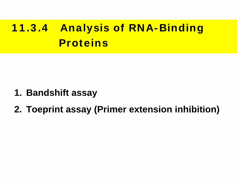
11.3.4 Analysis of RNA-Binding Proteins
1. Bandshift assay
2. Toeprint assay (Primer extension inhibition)
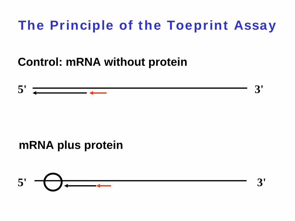
The Principle of the Toeprint Assay
5' 3'
5' 3'
Control: mRNA without protein
mRNA plus protein
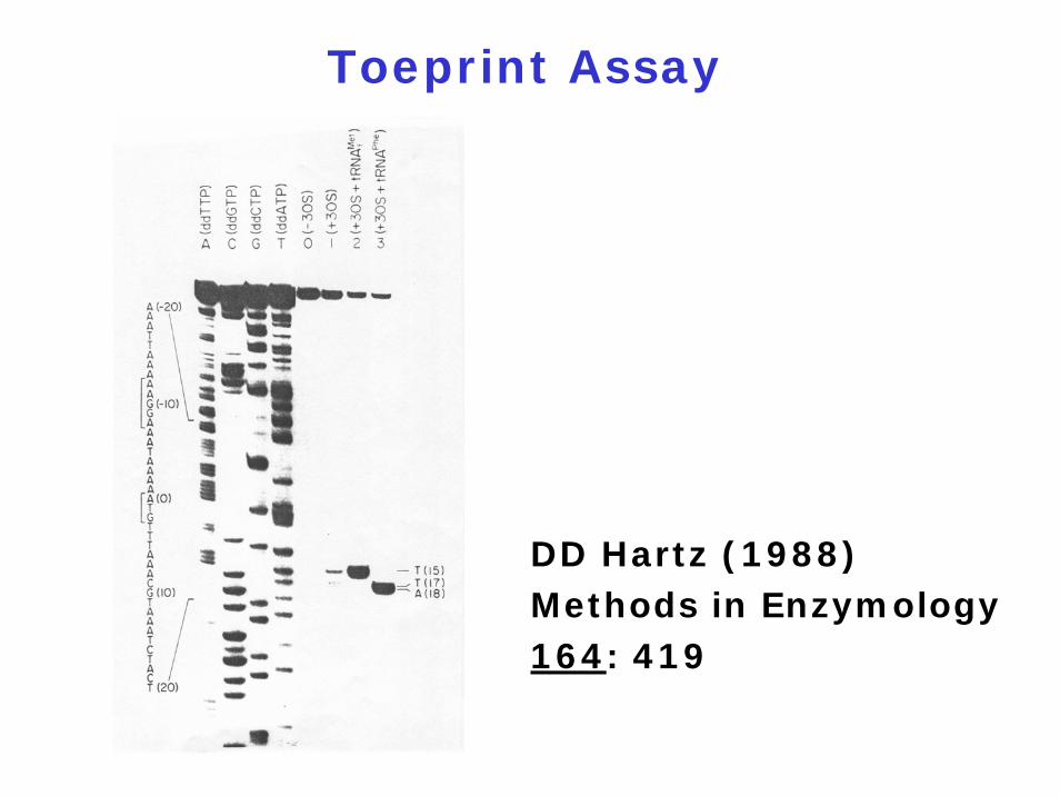
Toeprint Assay
DD Hartz (1988) Methods in Enzymology 164: 419
11.3.5 Analysis of Protein-Protein Interaction
How to study protein-protein interactions ?
Biochemical techniques
Molecular biological in vivo techniques
Genetic techniques
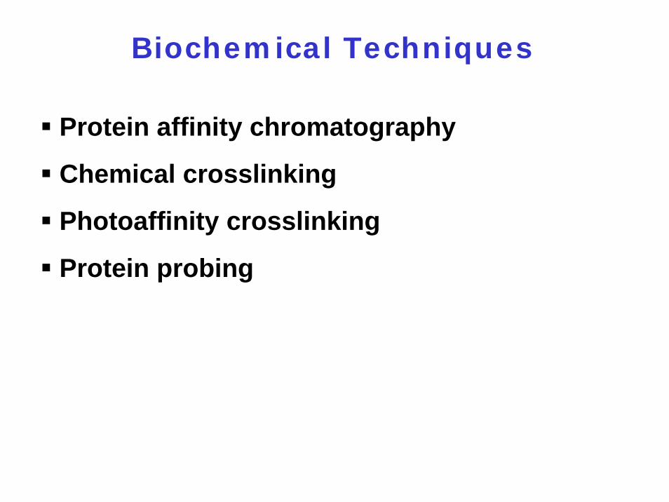
Biochemical Techniques
Protein affinity chromatography
Chemical crosslinking
Photoaffinity crosslinking
Protein probing
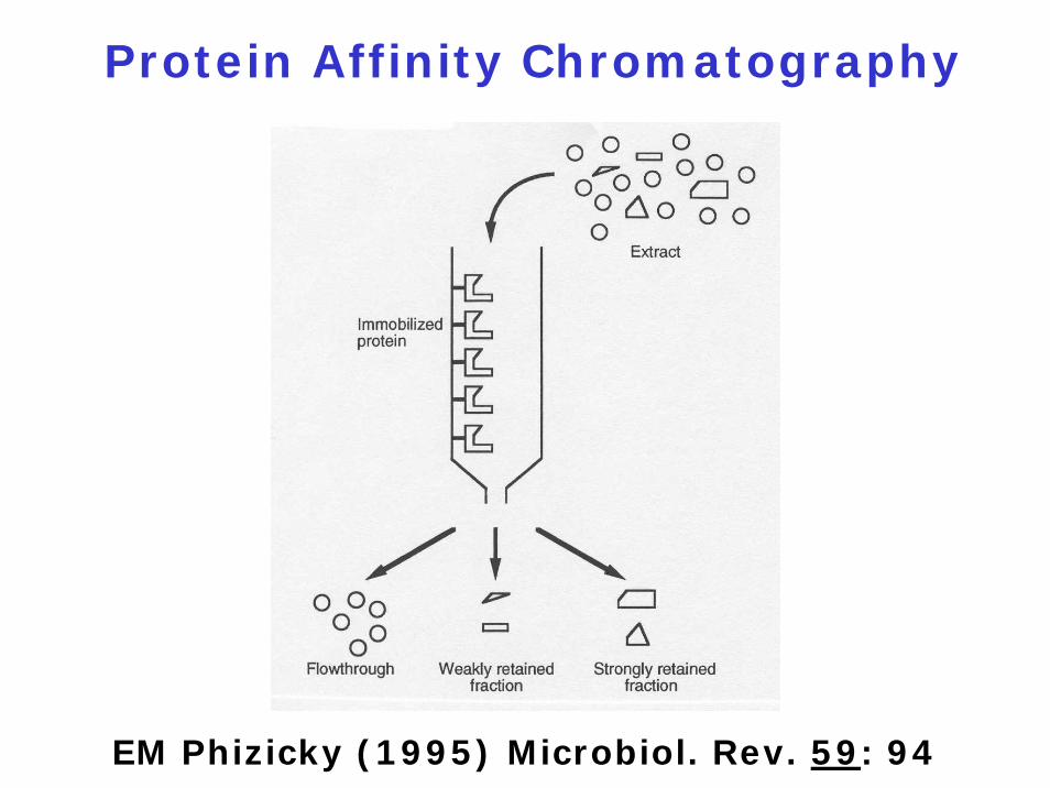
Protein Affinity Chromatography
EM Phizicky (1995) Microbiol. Rev. 59: 94
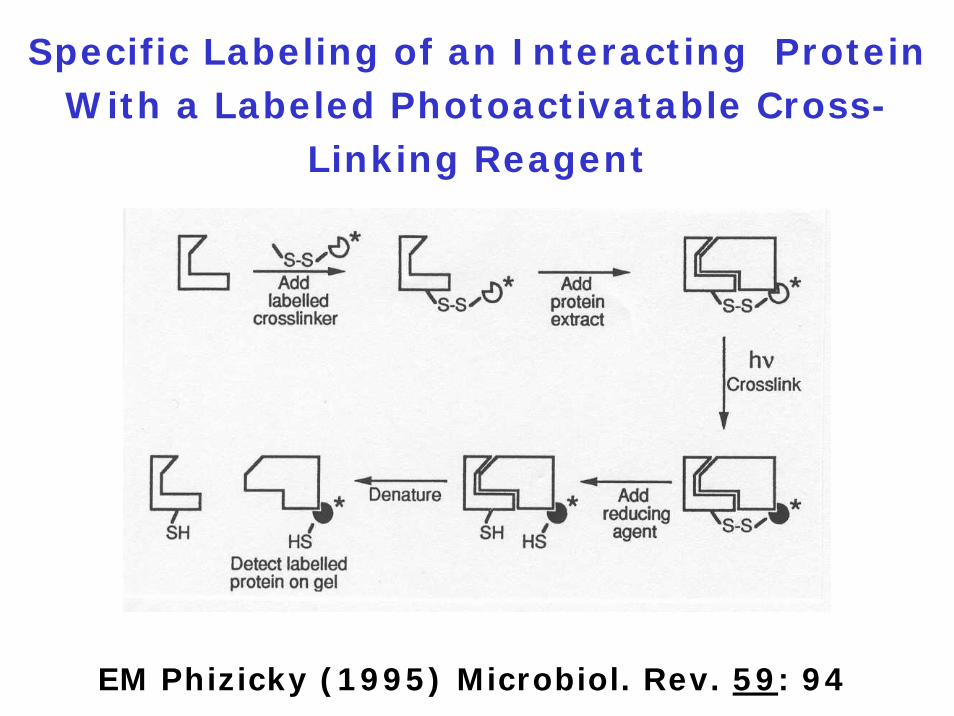
Specific Labeling of an Interacting Protein With a Labeled Photoactivatable Cross-
Linking Reagent
EM Phizicky (1995) Microbiol. Rev. 59: 94
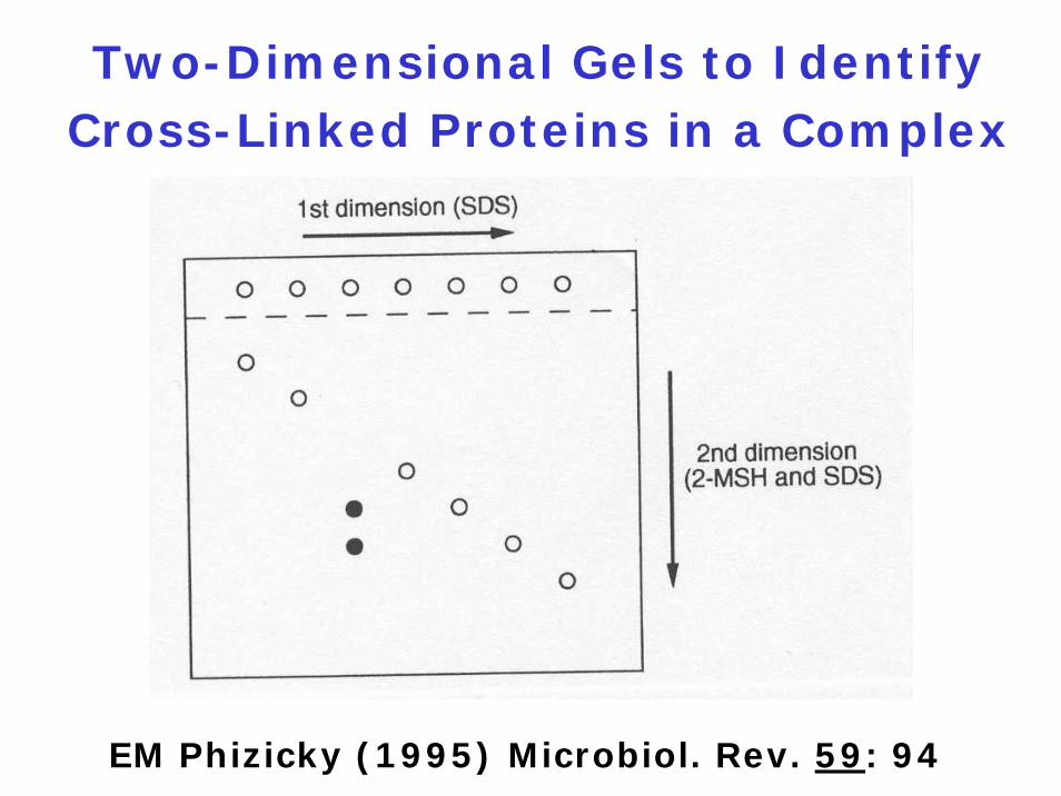
Two-Dimensional Gels to Identify Cross-Linked Proteins in a Complex
EM Phizicky (1995) Microbiol. Rev. 59: 94
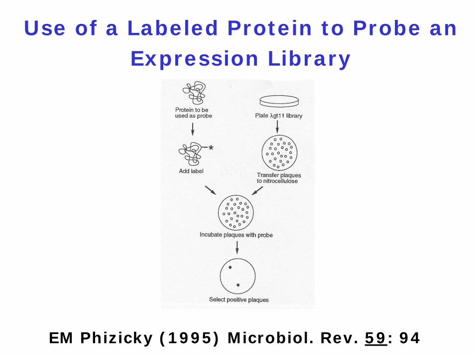
Use of a Labeled Protein to Probe an Expression Library
EM Phizicky (1995) Microbiol. Rev. 59: 94
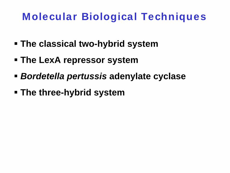
Molecular Biological Techniques
The classical two-hybrid system
The LexA repressor system
Bordetella pertussis adenylate cyclase
The three-hybrid system
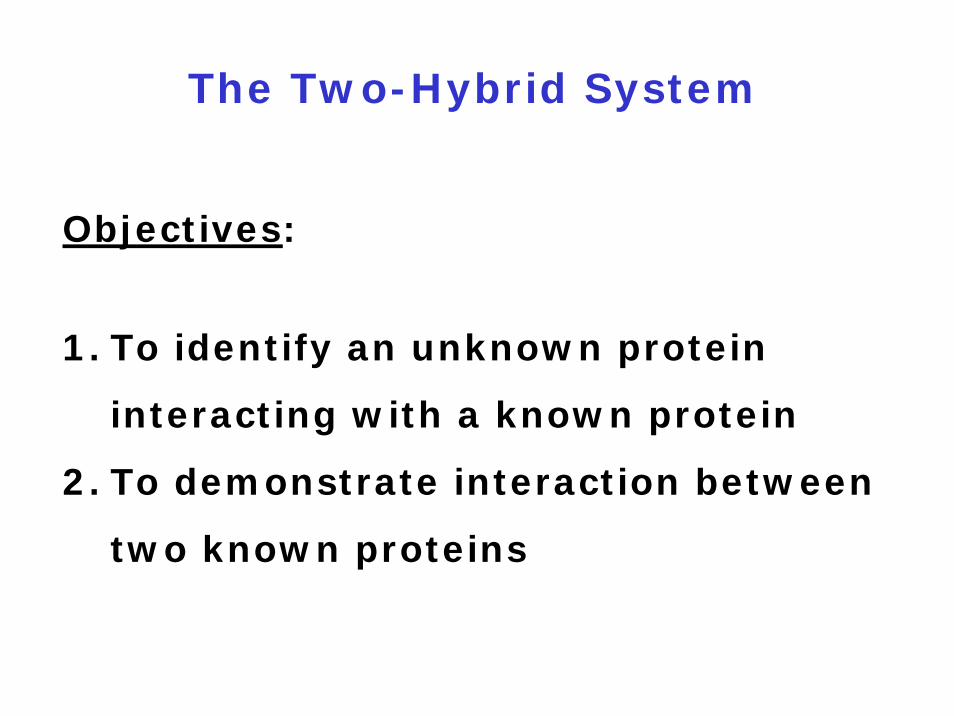
The Two-Hybrid System
Objectives:
1. To identify an unknown protein
interacting with a known protein
2. To demonstrate interaction between
two known proteins
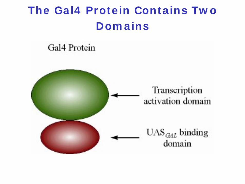
The Gal4 Protein Contains Two Domains
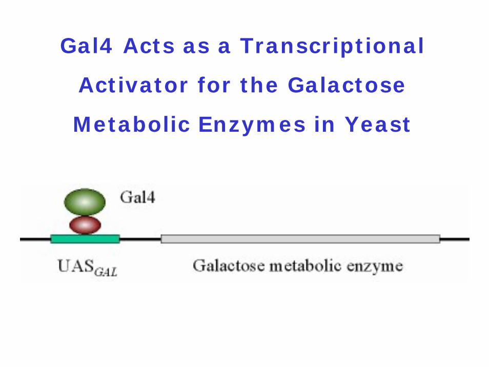
Gal4 Acts as a Transcriptional
Activator for the Galactose
Metabolic Enzymes in Yeast
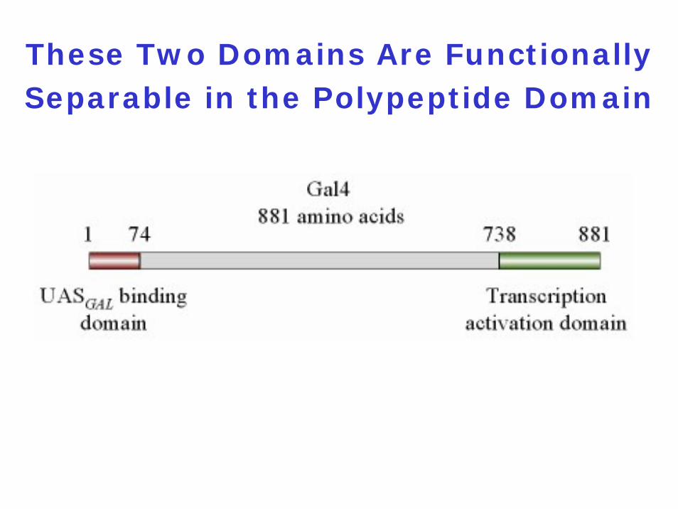
These Two Domains Are Functionally Separable in the Polypeptide Domain
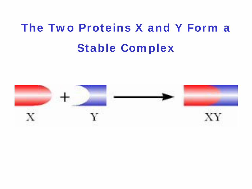
The Two Proteins X and Y Form a
Stable Complex
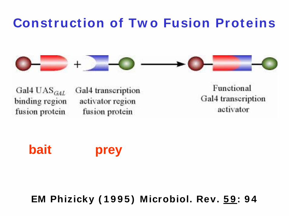
Construction of Two Fusion Proteins
bait prey
EM Phizicky (1995) Microbiol. Rev. 59: 94
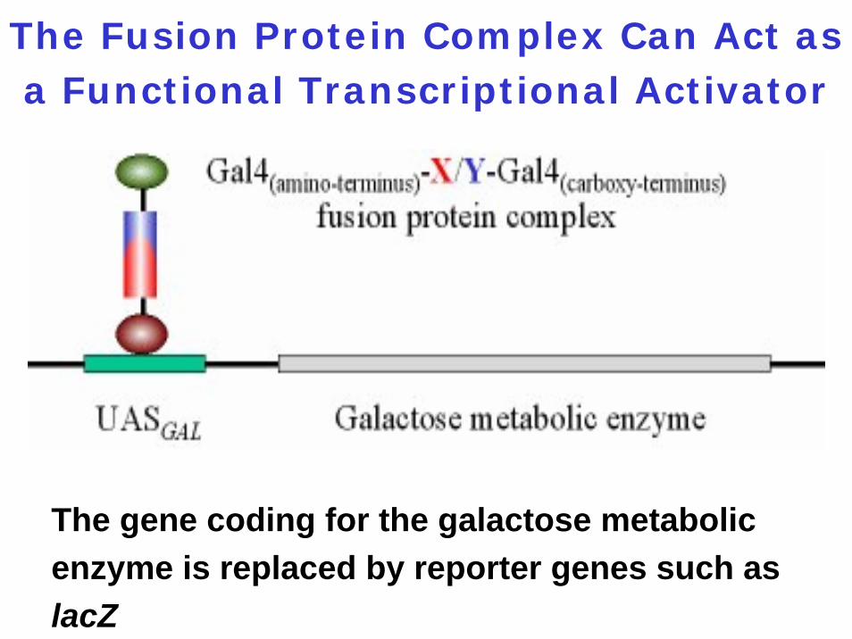
The Fusion Protein Complex Can Act as a Functional Transcriptional Activator
The gene coding for the galactose metabolic enzyme is replaced by reporter genes such as lacZ
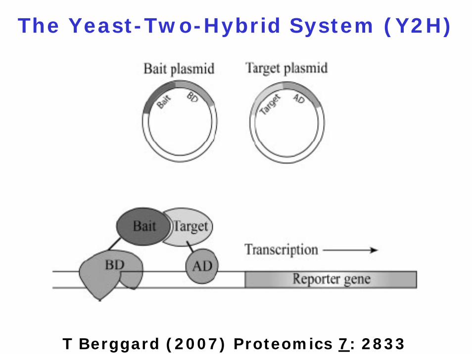
The Yeast-Two-Hybrid System (Y2H)
T Berggard (2007) Proteomics 7: 2833
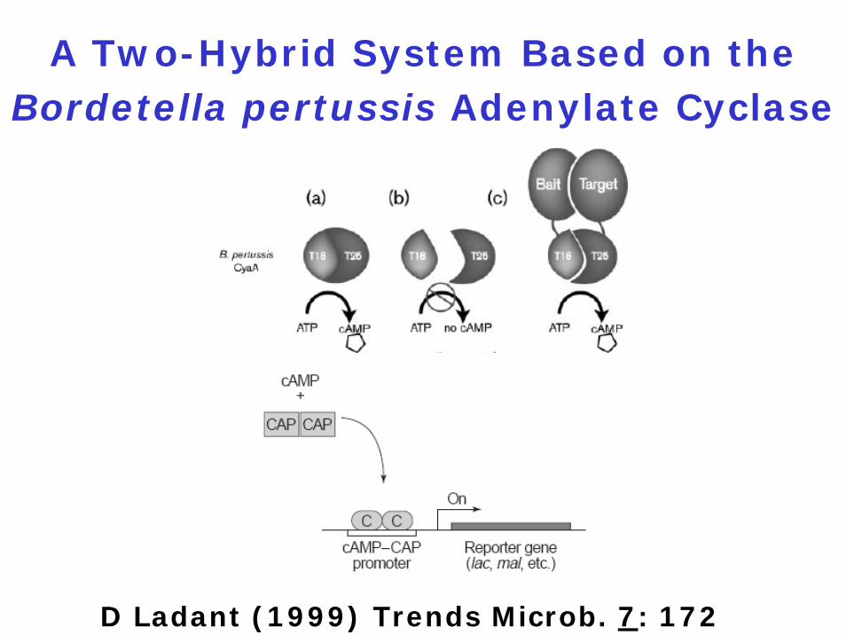
A Two-Hybrid System Based on the Bordetella pertussis Adenylate Cyclase
D Ladant (1999) Trends Microb. 7: 172
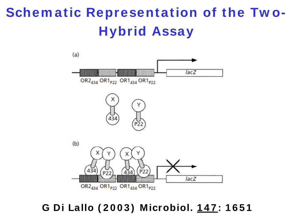
Schematic Representation of the Two-Hybrid Assay
G Di Lallo (2003) Microbiol. 147: 1651
The Three-Hybrid System
Objective:
To detect protein-RNA interactions
Principle:
Binding of a bifunctional RNA to each of two hybrid proteins activates transcription of a yeast reporter gene in vivo
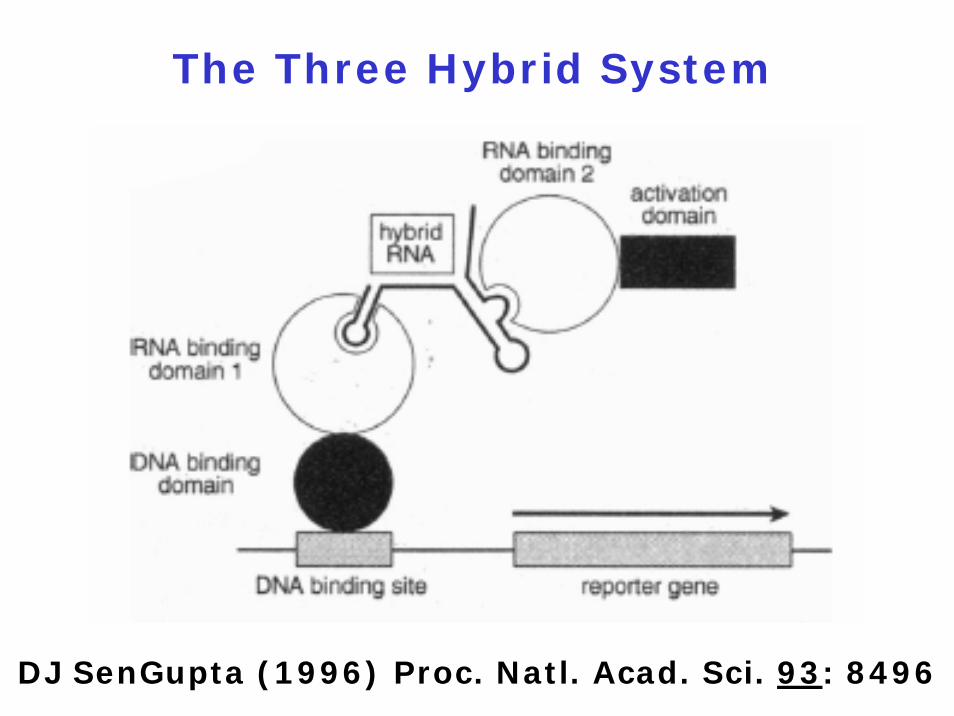
DJ SenGupta (1996) Proc. Natl. Acad. Sci. 93: 8496
The Three Hybrid System
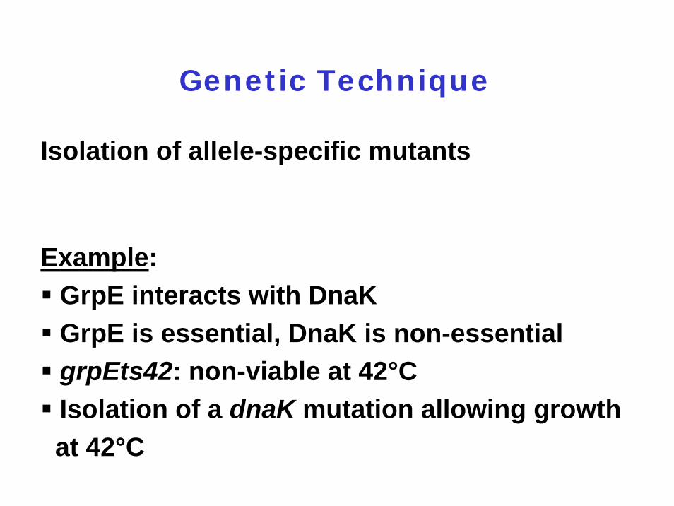
Genetic Technique
Isolation of allele-specific mutants
Example:GrpE interacts with DnaK GrpE is essential, DnaK is non-essentialgrpEts42: non-viable at 42°CIsolation of a dnaK mutation allowing growth at 42°C
11.3.6 Surface Display
Goal:
To identify ligands interacting with
a protein or peptide
1. in vivo systems
- prokaryotic
- eukaryotic
2. in vitro systems
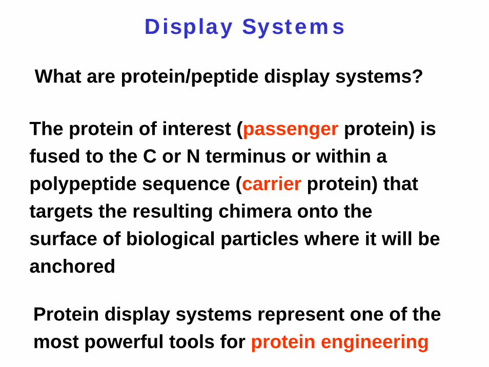
Display Systems
What are protein/peptide display systems?
The protein of interest (passenger protein) is fused to the C or N terminus or within a polypeptide sequence (carrier protein) that targets the resulting chimera onto the surface of biological particles where it will be anchored
Protein display systems represent one of the most powerful tools for protein engineering
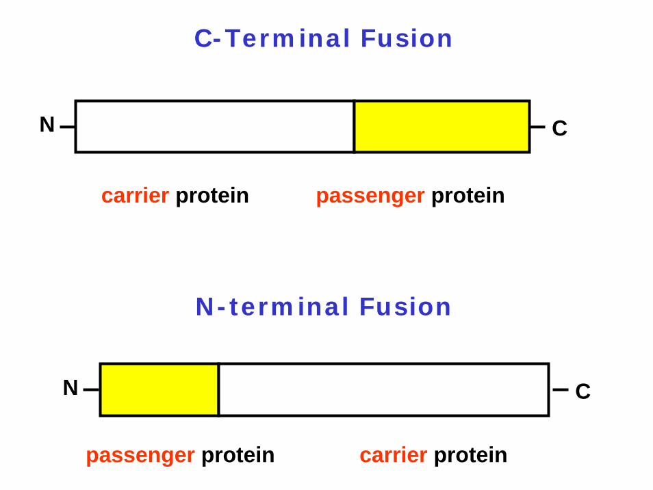
C-Terminal Fusion
N C
carrier protein passenger protein
N-terminal Fusion
N C
passenger protein carrier protein
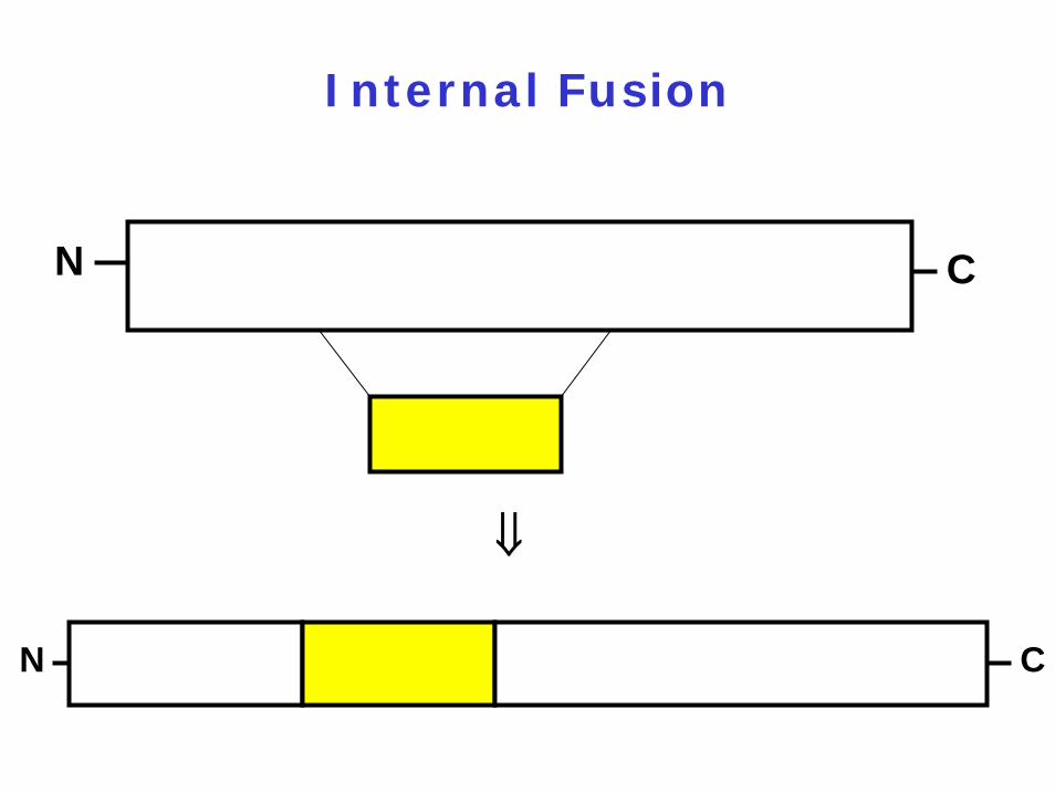
Internal Fusion
⇓
N C
N C
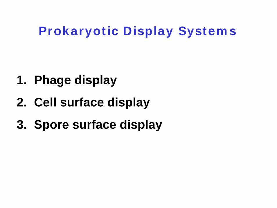
Prokaryotic Display Systems
1. Phage display
2. Cell surface display
3. Spore surface display
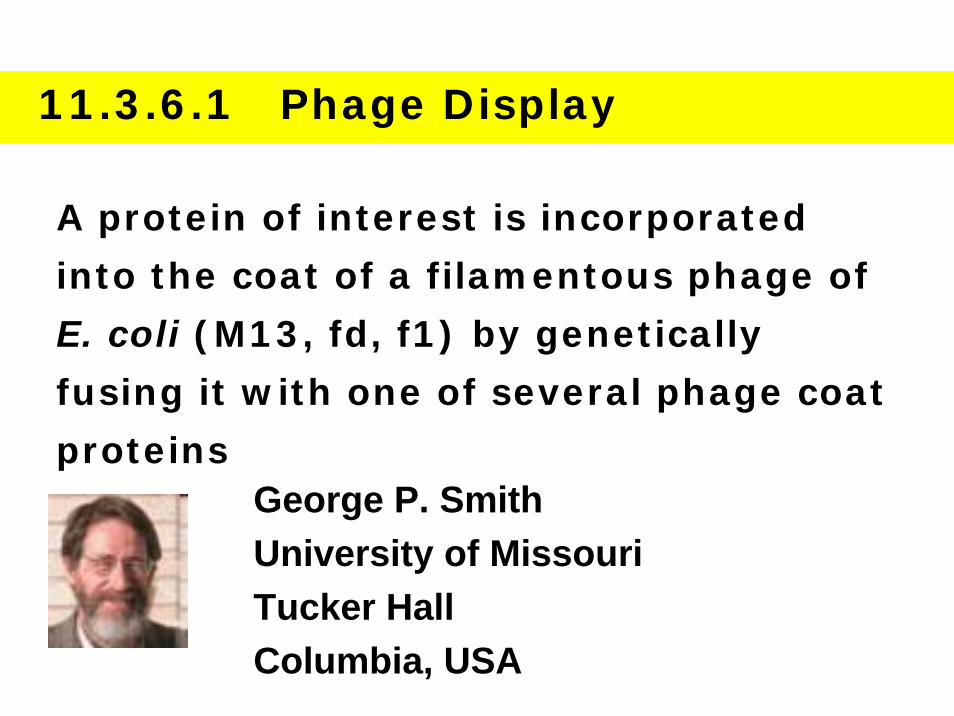
11.3.6.1 Phage Display
A protein of interest is incorporated
into the coat of a filamentous phage of
E. coli (M13, fd, f1) by genetically
fusing it with one of several phage coat
proteinsGeorge P. SmithUniversity of MissouriTucker HallColumbia, USA
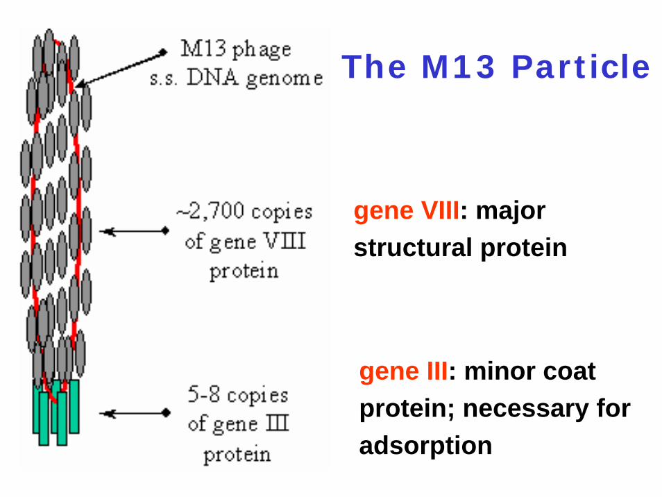
The M13 Particle
gene VIII: major structural protein
gene III: minor coat protein; necessary for adsorption
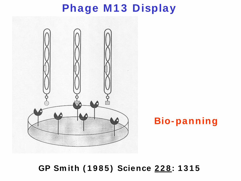
Phage M13 Display
Bio-panning
GP Smith (1985) Science 228: 1315
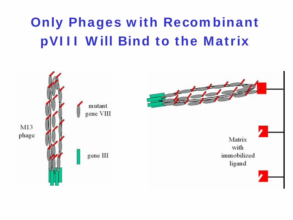
Only Phages with Recombinant pVIII Will Bind to the Matrix
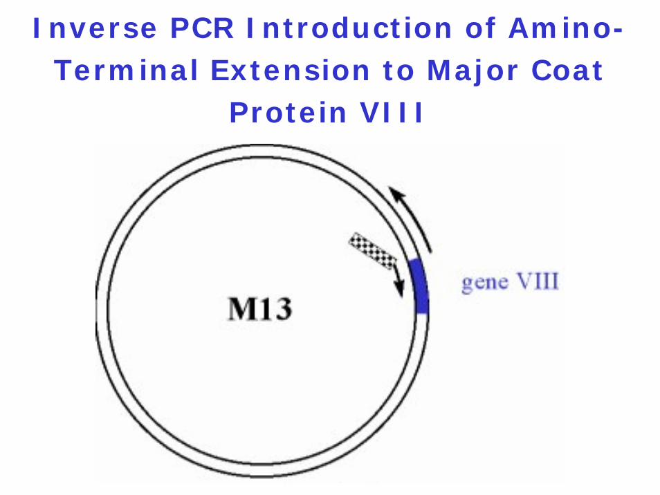
Inverse PCR Introduction of Amino-Terminal Extension to Major Coat
Protein VIII
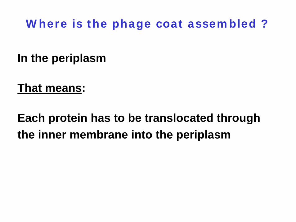
Where is the phage coat assembled ?
In the periplasm
That means:
Each protein has to be translocated through the inner membrane into the periplasm
Background:
Based on fusions of proteins or peptides to the capsid proteinspIIIpVIIITranslocated through the inner membrane via the Sec pathway, where they become incorporated into the phage particle
Phage display depends on its effective translocation
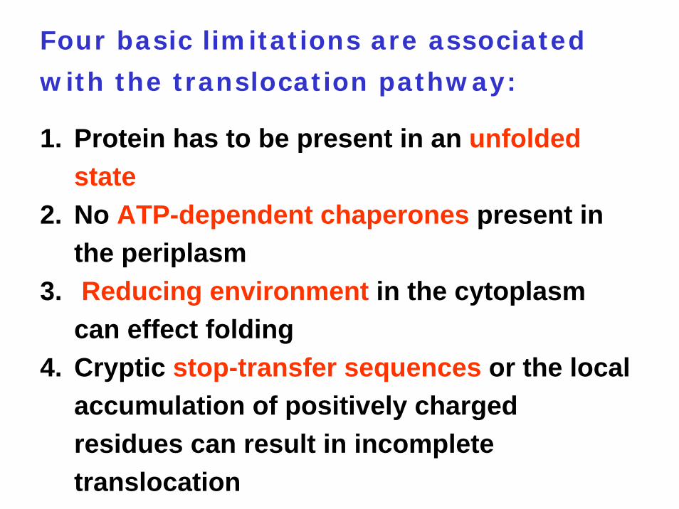
Four basic limitations are associated
with the translocation pathway:
1. Protein has to be present in an unfolded state
2. No ATP-dependent chaperones present in the periplasm
3. Reducing environment in the cytoplasm can effect folding
4. Cryptic stop-transfer sequences or the local accumulation of positively charged residues can result in incomplete translocation
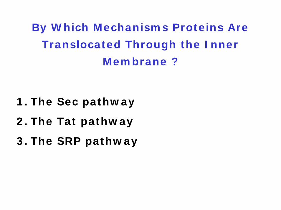
By Which Mechanisms Proteins Are
Translocated Through the Inner
Membrane ?
1. The Sec pathway
2. The Tat pathway
3. The SRP pathway

Components of the Sec Pathway
1. Proteins contain a cleavable N-terminal signal sequence
2. SecB binds to hydrophobic regions and prevents premature folding
3. SecA acts as a motor protein4. SecYEG heterotrimer forms the membrane-
embedded translocon
Important:Sec-dependent proteins are translocated in a translocation-competent form
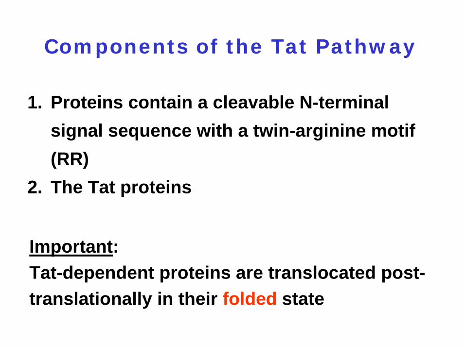
Components of the Tat Pathway
1. Proteins contain a cleavable N-terminal signal sequence with a twin-arginine motif (RR)
2. The Tat proteins
Important:Tat-dependent proteins are translocated post-translationally in their folded state
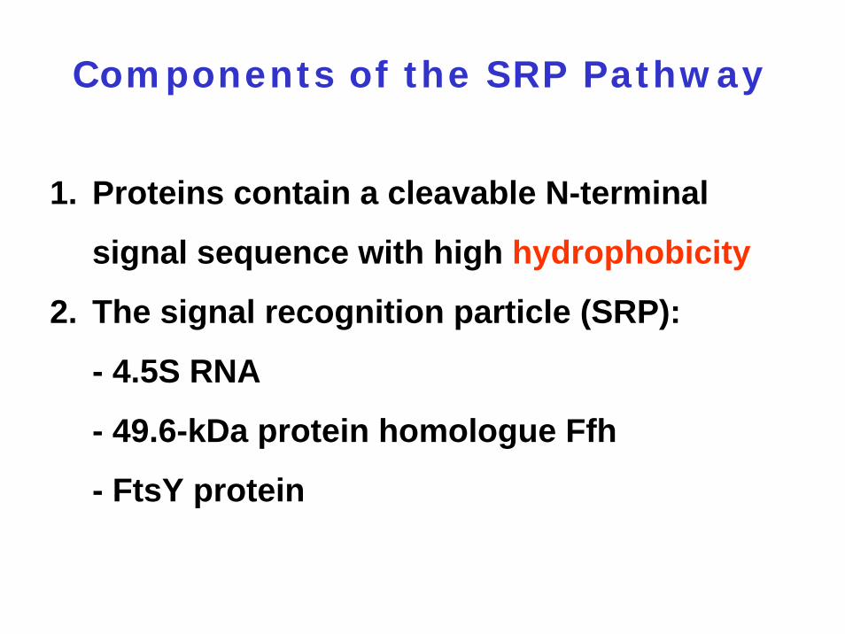
Components of the SRP Pathway
1. Proteins contain a cleavable N-terminal
signal sequence with high hydrophobicity
2. The signal recognition particle (SRP):
- 4.5S RNA
- 49.6-kDa protein homologue Ffh
- FtsY protein
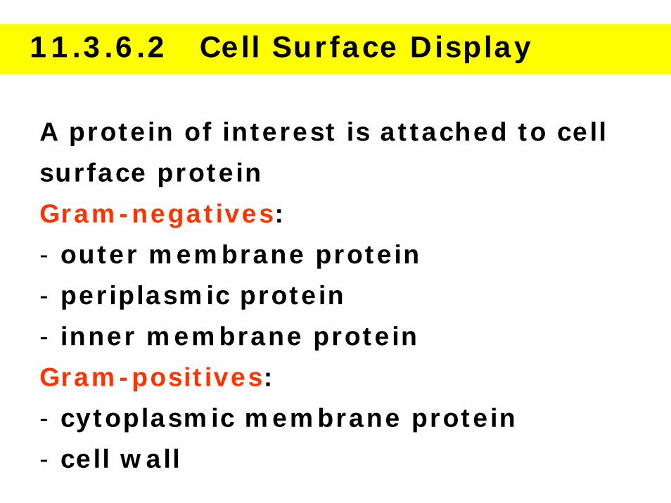
11.3.6.2 Cell Surface Display
A protein of interest is attached to cell
surface protein
Gram-negatives:
- outer membrane protein
- periplasmic protein
- inner membrane protein
Gram-positives:
- cytoplasmic membrane protein
- cell wall
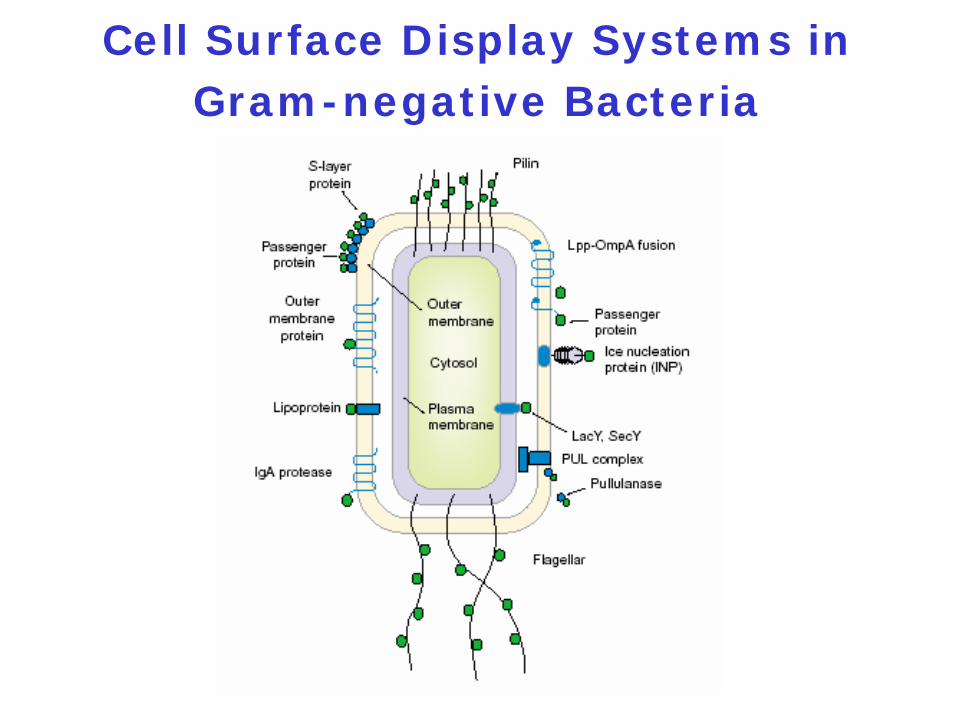
Cell Surface Display Systems in Gram-negative Bacteria
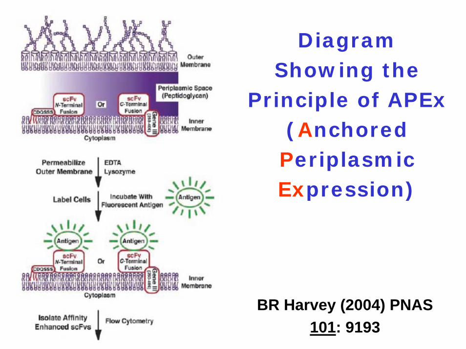
Diagram Showing the
Principle of APEx(Anchored Periplasmic Expression)
BR Harvey (2004) PNAS 101: 9193
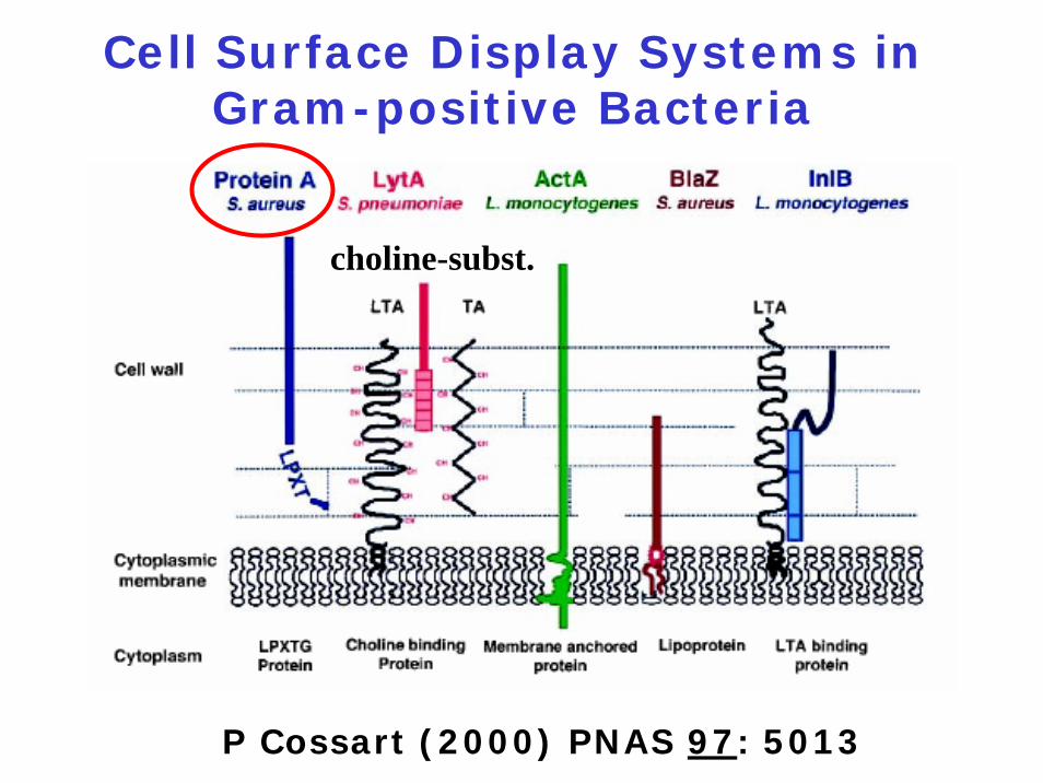
Cell Surface Display Systems in Gram-positive Bacteria
P Cossart (2000) PNAS 97: 5013
choline-subst.
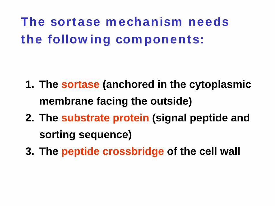
The sortase mechanism needs the following components:
1. The sortase (anchored in the cytoplasmic membrane facing the outside)
2. The substrate protein (signal peptide and sorting sequence)
3. The peptide crossbridge of the cell wall
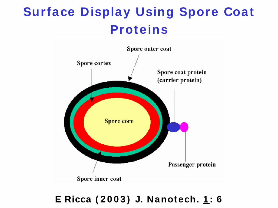
Surface Display Using Spore Coat Proteins
E Ricca (2003) J. Nanotech. 1: 6
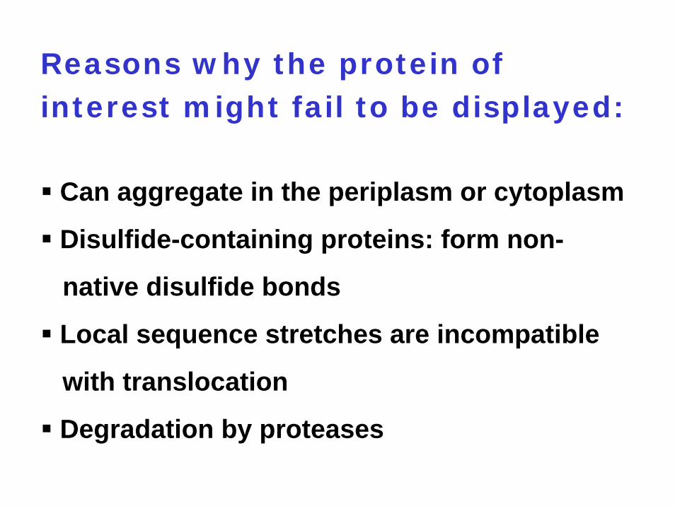
Reasons why the protein of interest might fail to be displayed:
Can aggregate in the periplasm or cytoplasm
Disulfide-containing proteins: form non-
native disulfide bonds
Local sequence stretches are incompatible
with translocation
Degradation by proteases
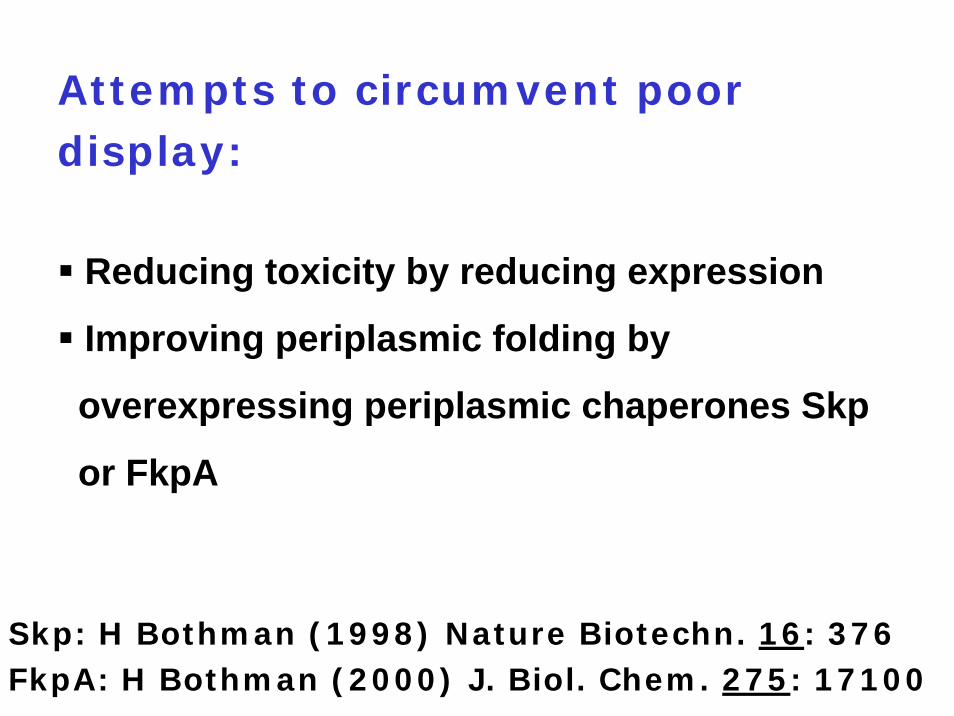
Attempts to circumvent poor display:
Reducing toxicity by reducing expression
Improving periplasmic folding by
overexpressing periplasmic chaperones Skp
or FkpA
Skp: H Bothman (1998) Nature Biotechn. 16: 376FkpA: H Bothman (2000) J. Biol. Chem. 275: 17100
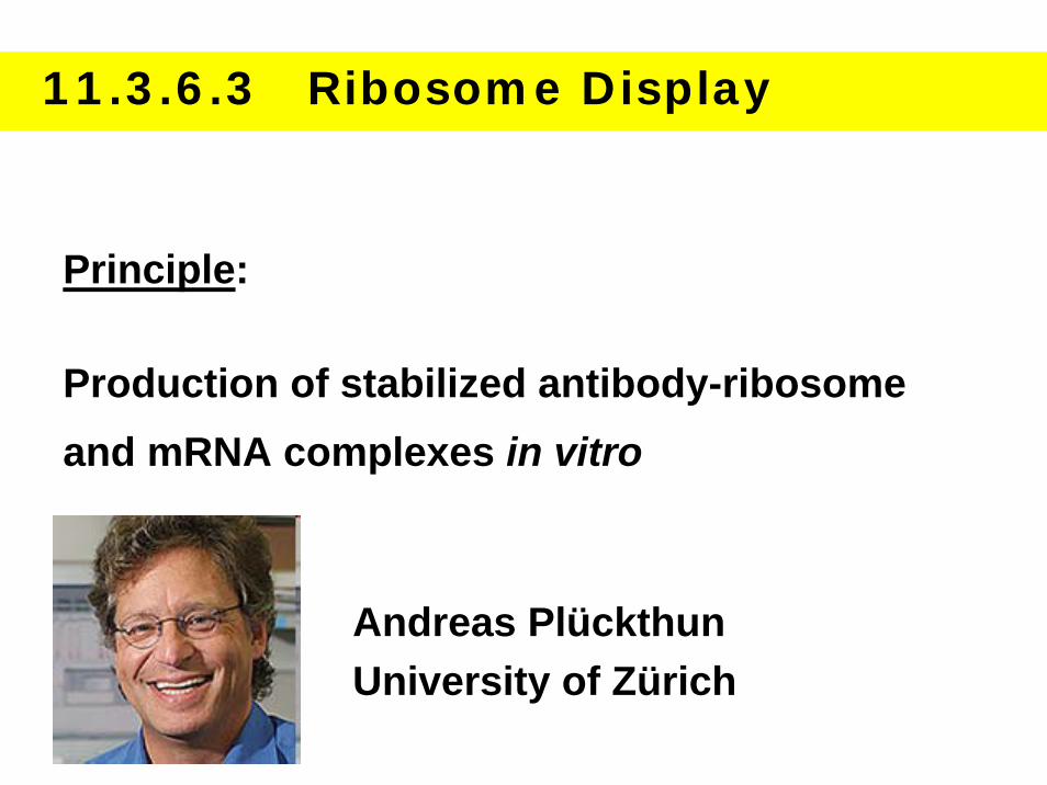
11.3.6.3 Ribosome Display
Principle:
Production of stabilized antibody-ribosome and mRNA complexes in vitro
Andreas PlückthunUniversity of Zürich

Principle of in vitro Ribosome Display for Screening Native Protein Libraries
for Ligand Binding
J. Hanes (1997) Proc. Natl. Acad. Sci. 94: 4937
mRNA without stop codon
Limitations:
Efficient in vitro translation and stalling can
be technically challenging
Concentrations of cellular factors required
for efficient scFv folding may differ from
concentrations found in vivo
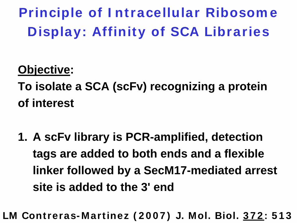
Principle of Intracellular Ribosome Display: Affinity of SCA Libraries
LM Contreras-Martinez (2007) J. Mol. Biol. 372: 513
Objective:To isolate a SCA (scFv) recognizing a protein of interest
1. A scFv library is PCR-amplified, detection tags are added to both ends and a flexible linker followed by a SecM17-mediated arrest site is added to the 3' end
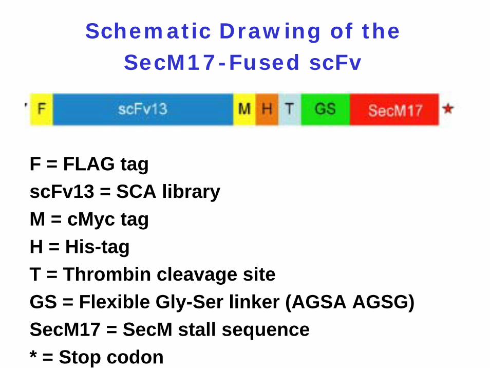
Schematic Drawing of the SecM17-Fused scFv
F = FLAG tagscFv13 = SCA libraryM = cMyc tag H = His-tag T = Thrombin cleavage site GS = Flexible Gly-Ser linker (AGSA AGSG)SecM17 = SecM stall sequence* = Stop codon
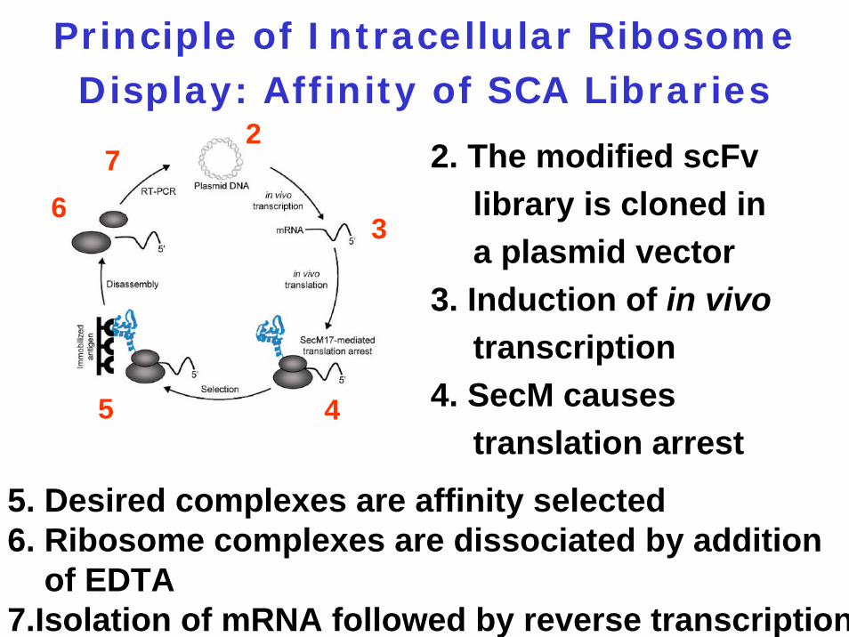
Principle of Intracellular Ribosome Display: Affinity of SCA Libraries
2. The modified scFv library is cloned in a plasmid vector
3. Induction of in vivotranscription
4. SecM causes translation arrest
5. Desired complexes are affinity selected 6. Ribosome complexes are dissociated by addition
of EDTA7.Isolation of mRNA followed by reverse transcription
2
3
45
6
7
11.3.7 Surface Plasmon Resonance (SPR)
Method to monitor the kinetics of a reaction in real timeMeasures the affinity and speed of binding between a capture agent and a probeYields apparent rate constants for the association and dissociation phases of the reaction
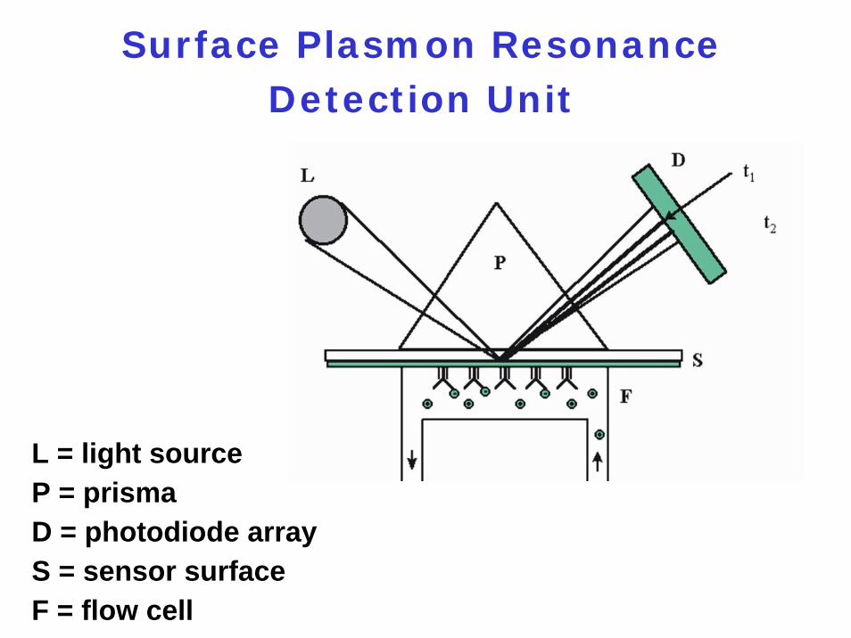
Surface Plasmon Resonance Detection Unit
L = light sourceP = prismaD = photodiode arrayS = sensor surface F = flow cell
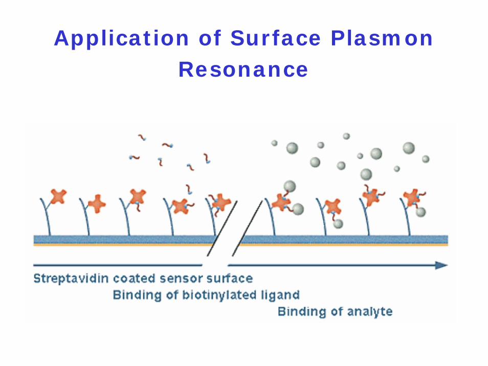
Application of Surface Plasmon Resonance
11.3.8 SELEX
Systematic Evolution of
Ligands by Exponential
Enrichment
T Tuerk and L Gold (1990) Science 249: 505
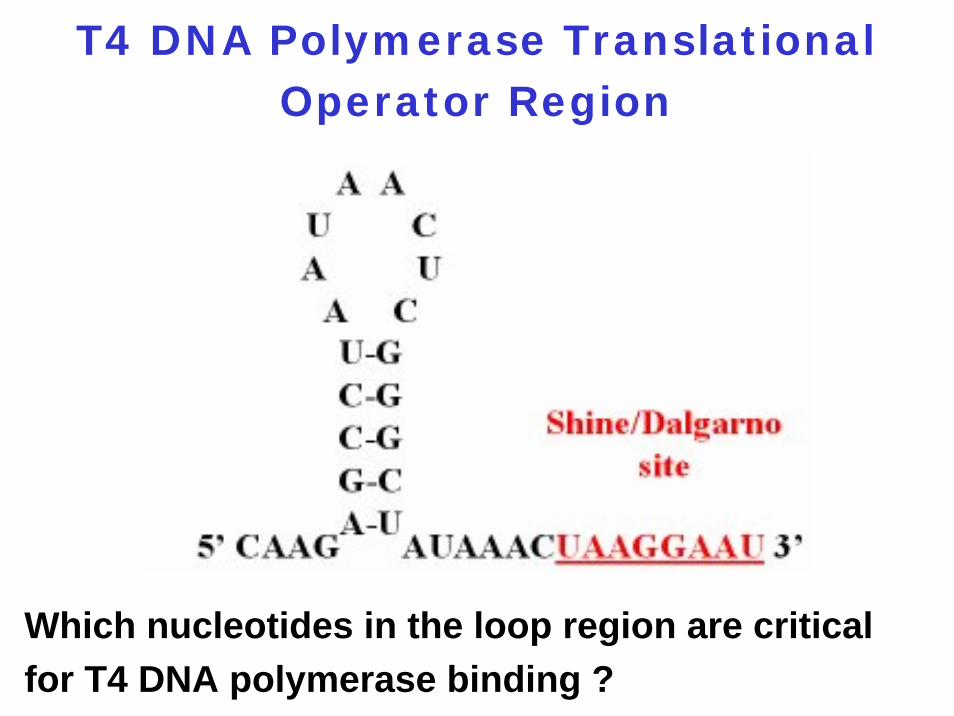
T4 DNA Polymerase Translational Operator Region
Which nucleotides in the loop region are critical for T4 DNA polymerase binding ?
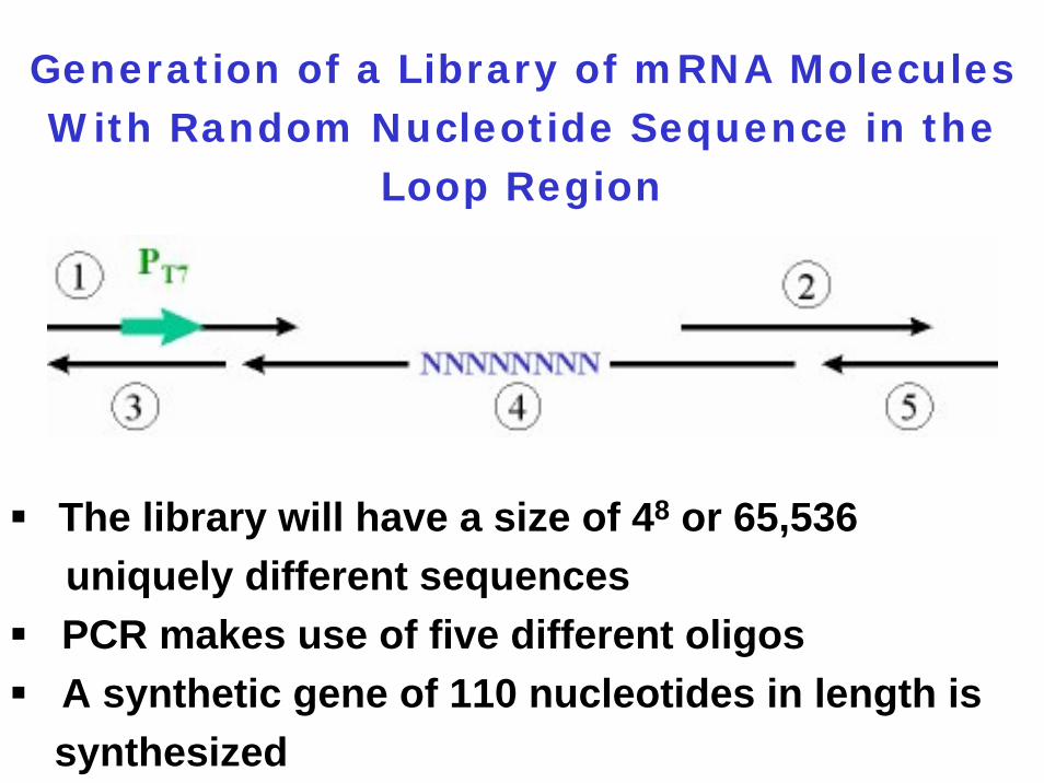
Generation of a Library of mRNA Molecules With Random Nucleotide Sequence in the
Loop Region
The library will have a size of 48 or 65,536 uniquely different sequencesPCR makes use of five different oligosA synthetic gene of 110 nucleotides in length is synthesized
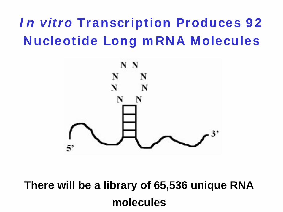
In vitro Transcription Produces 92 Nucleotide Long mRNA Molecules
There will be a library of 65,536 unique RNA molecules
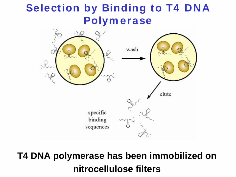
Selection by Binding to T4 DNA Polymerase
T4 DNA polymerase has been immobilized on nitrocellulose filters
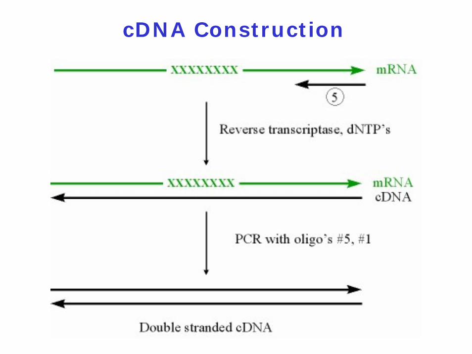
cDNA Construction
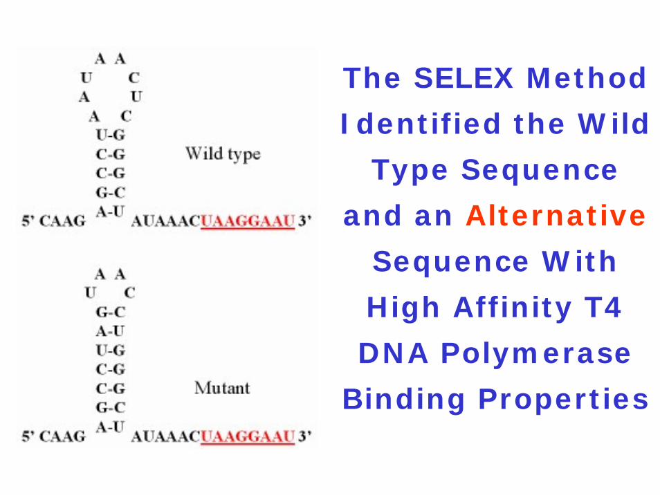
The SELEX Method
Identified the Wild
Type Sequence
and an Alternative
Sequence With
High Affinity T4
DNA Polymerase
Binding Properties
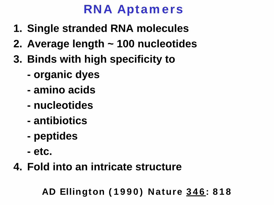
RNA Aptamers
1. Single stranded RNA molecules2. Average length ~ 100 nucleotides3. Binds with high specificity to
- organic dyes- amino acids- nucleotides- antibiotics- peptides- etc.
4. Fold into an intricate structure
AD Ellington (1990) Nature 346: 818
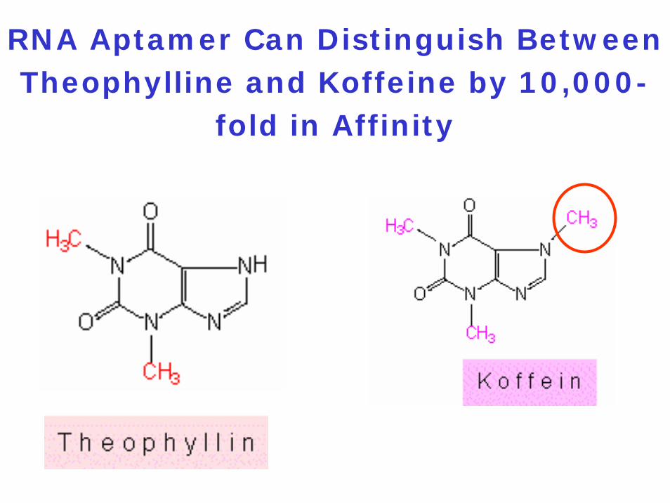
RNA Aptamer Can Distinguish Between Theophylline and Koffeine by 10,000-
fold in Affinity
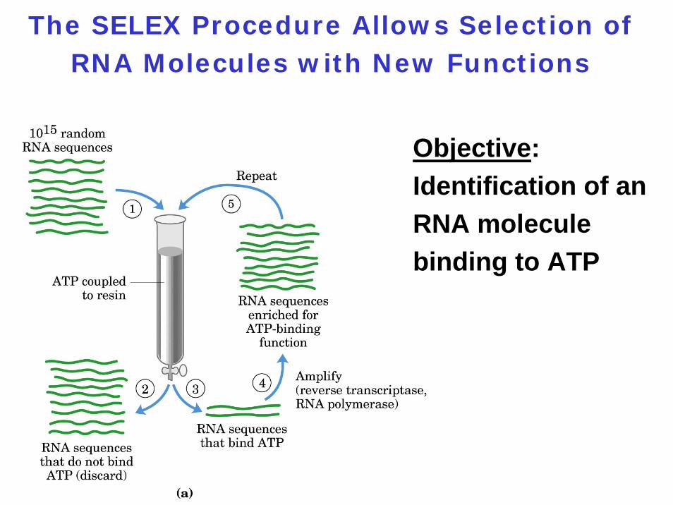
The SELEX Procedure Allows Selection of RNA Molecules with New Functions
Objective:Identification of an RNA molecule binding to ATP
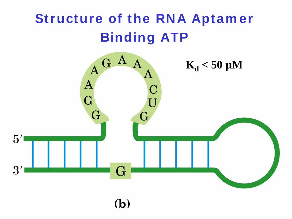
Structure of the RNA Aptamer Binding ATP
Kd < 50 µM
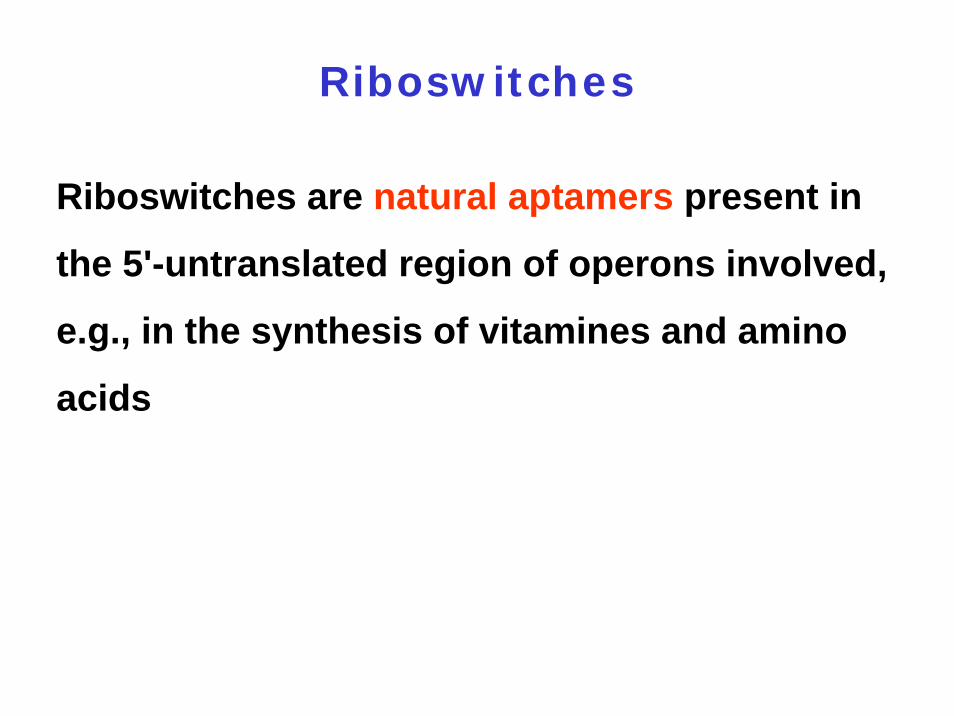
Riboswitches
Riboswitches are natural aptamers present in
the 5'-untranslated region of operons involved,
e.g., in the synthesis of vitamines and amino
acids
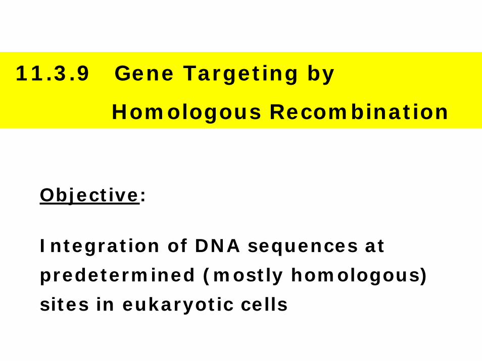
11.3.9 Gene Targeting by
Homologous Recombination
Objective:
Integration of DNA sequences at
predetermined (mostly homologous)
sites in eukaryotic cells
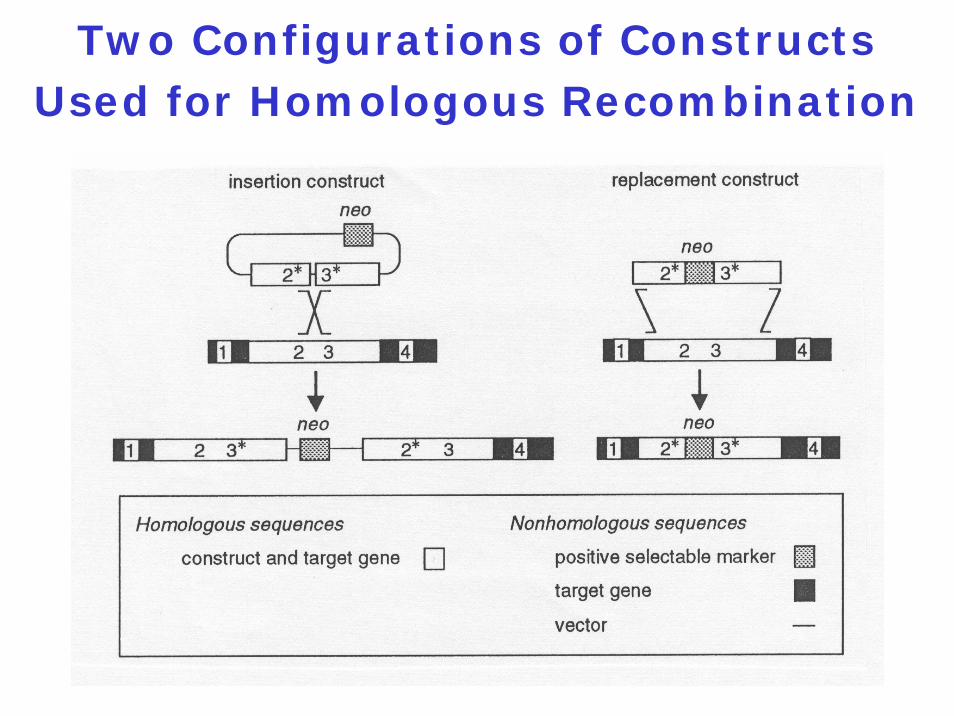
Two Configurations of ConstructsUsed for Homologous Recombination
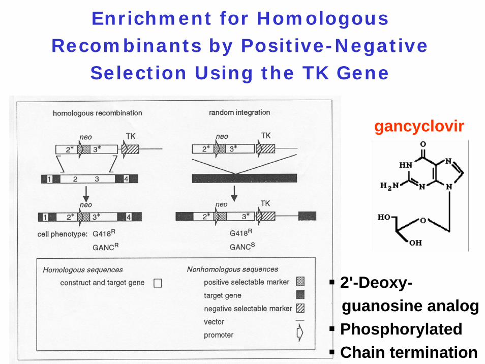
Enrichment for Homologous Recombinants by Positive-Negative
Selection Using the TK Gene
gancyclovir
2'-Deoxy-guanosine analogPhosphorylatedChain termination
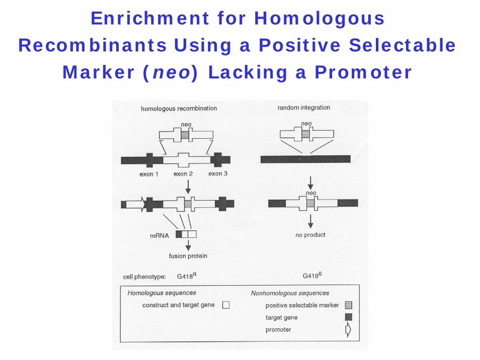
Enrichment for Homologous Recombinants Using a Positive Selectable
Marker (neo) Lacking a Promoter
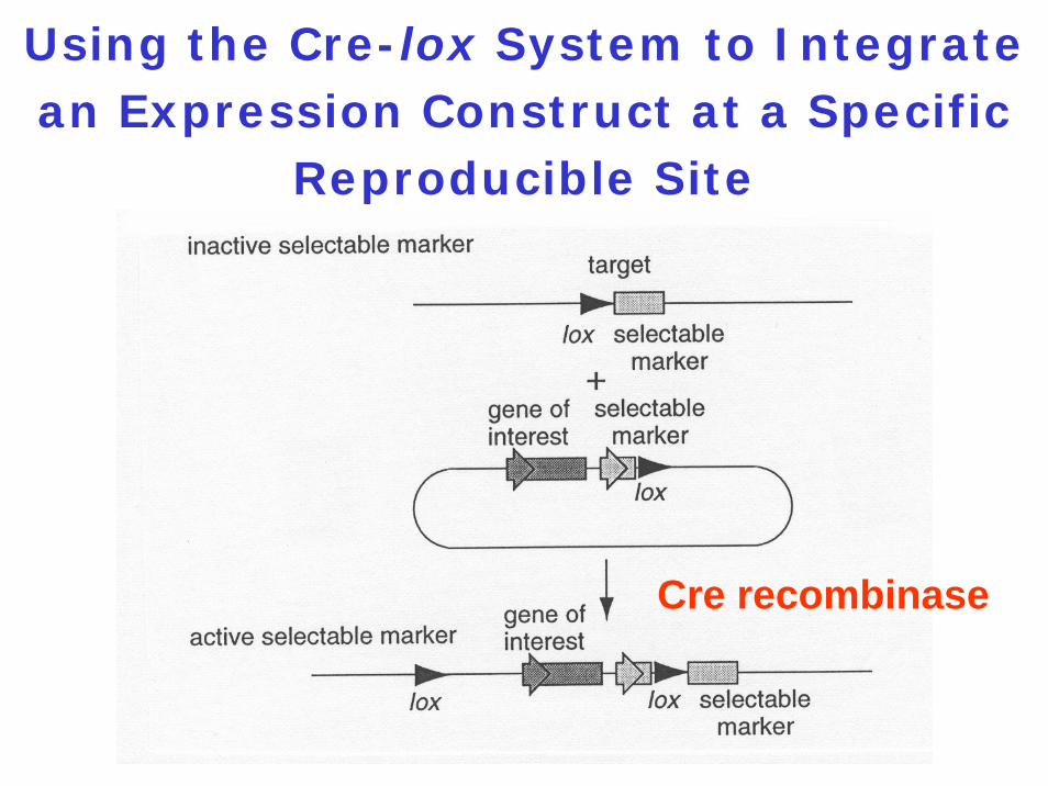
Using the Cre-lox System to Integrate an Expression Construct at a Specific
Reproducible Site
Cre recombinase
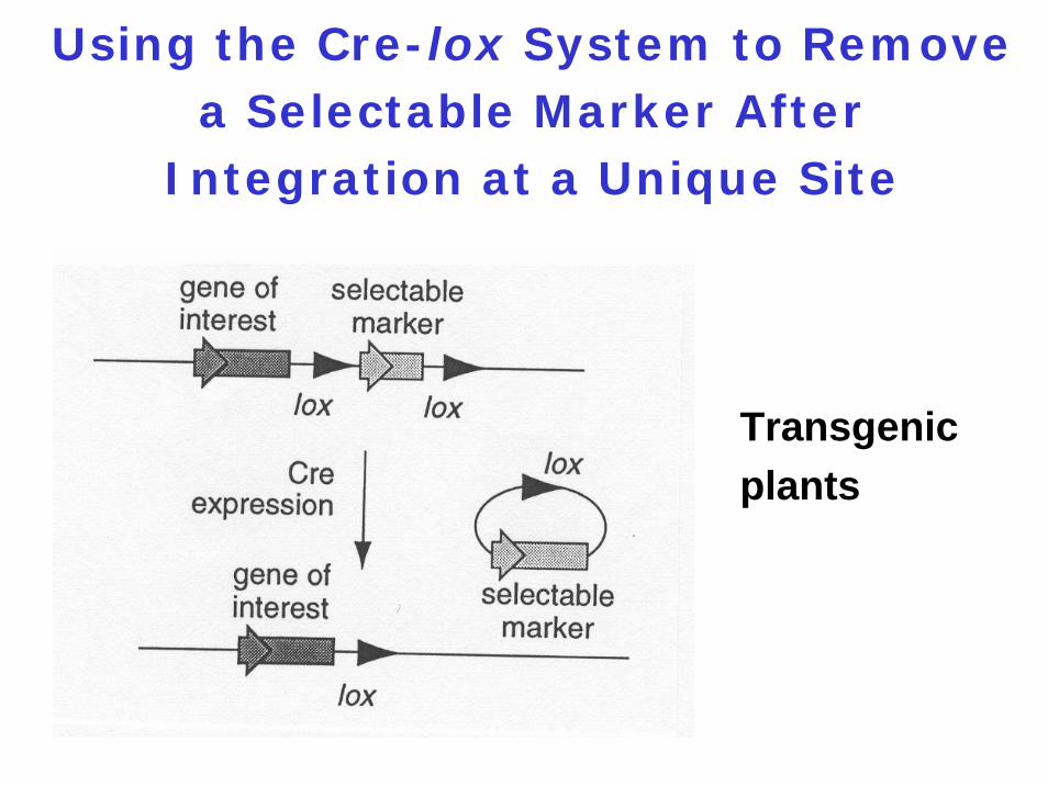
Using the Cre-lox System to Remove a Selectable Marker After
Integration at a Unique Site
Transgenic plants
11.3.10 Incorporation of non-
native amino acids into
proteins in E. coli cells
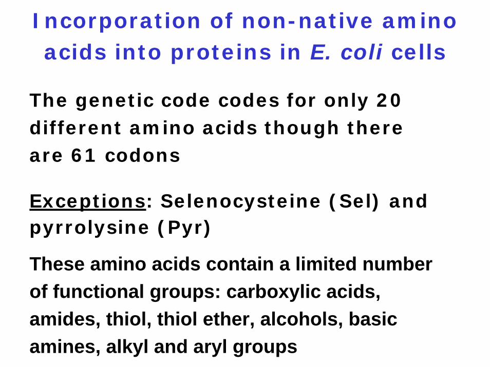
Incorporation of non-native amino acids into proteins in E. coli cells
The genetic code codes for only 20 different amino acids though there are 61 codons
Exceptions: Selenocysteine (Sel) and pyrrolysine (Pyr)
These amino acids contain a limited number of functional groups: carboxylic acids, amides, thiol, thiol ether, alcohols, basic amines, alkyl and aryl groups
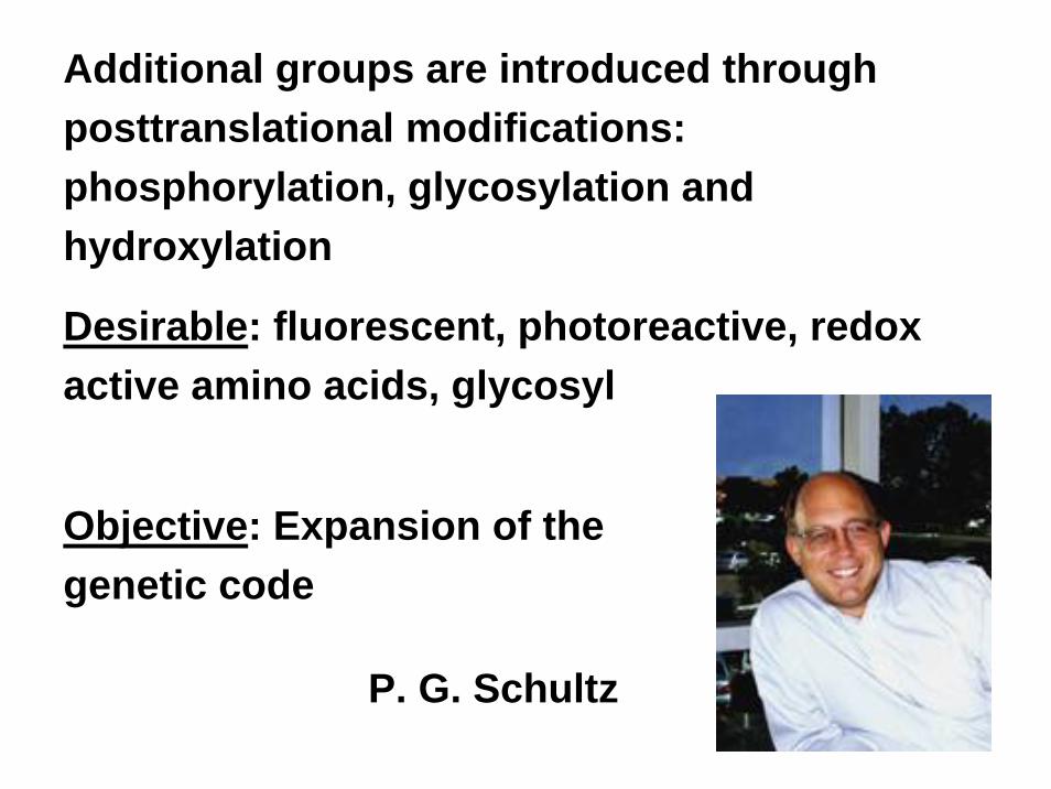
Additional groups are introduced through posttranslational modifications: phosphorylation, glycosylation and hydroxylation
Desirable: fluorescent, photoreactive, redox active amino acids, glycosyl
Objective: Expansion of the genetic code
P. G. Schultz
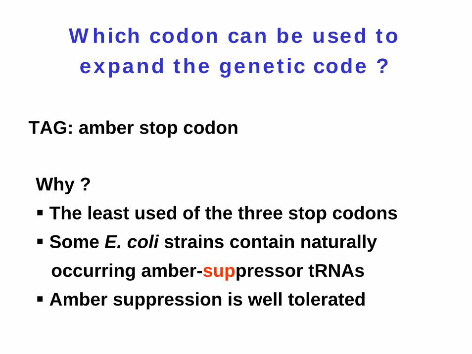
Which codon can be used to expand the genetic code ?
TAG: amber stop codon
Why ?The least used of the three stop codonsSome E. coli strains contain naturally occurring amber-suppressor tRNAsAmber suppression is well tolerated
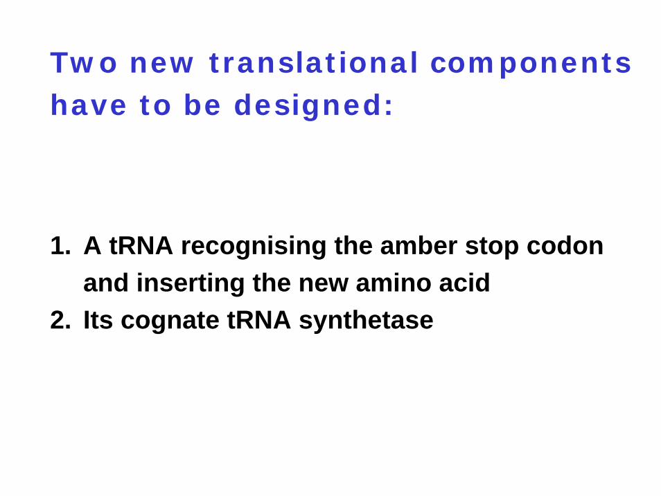
Two new translational components have to be designed:
1. A tRNA recognising the amber stop codon and inserting the new amino acid
2. Its cognate tRNA synthetase
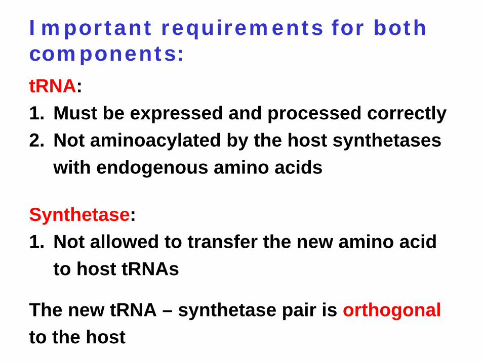
Important requirements for both components:tRNA:1. Must be expressed and processed correctly2. Not aminoacylated by the host synthetases
with endogenous amino acids
Synthetase:1. Not allowed to transfer the new amino acid
to host tRNAs
The new tRNA – synthetase pair is orthogonalto the host
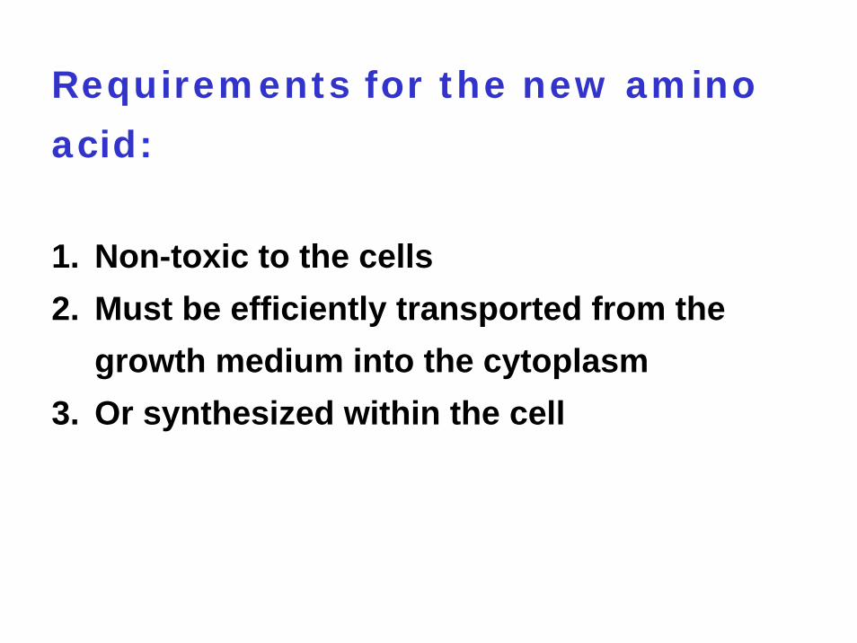
Requirements for the new amino
acid:
1. Non-toxic to the cells2. Must be efficiently transported from the
growth medium into the cytoplasm3. Or synthesized within the cell
First orthogonal pair:
tRNATyr and its cognate synthetase derived from Methanoccocus jannaschii
Inserts tyrosine instead of an amber codon
Problem:tRNA recognized to some degree by E. colisynthetases
Objective:tRNA should not be recognized by E. colisynthetases
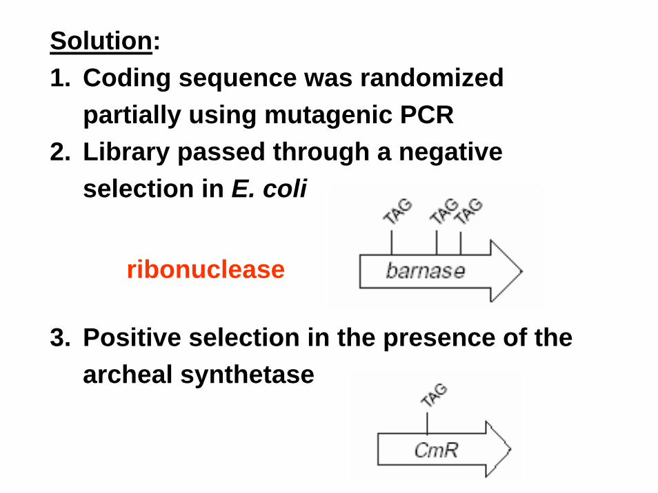
Solution: 1. Coding sequence was randomized
partially using mutagenic PCR2. Library passed through a negative
selection in E. coli
3. Positive selection in the presence of the archeal synthetase
ribonuclease
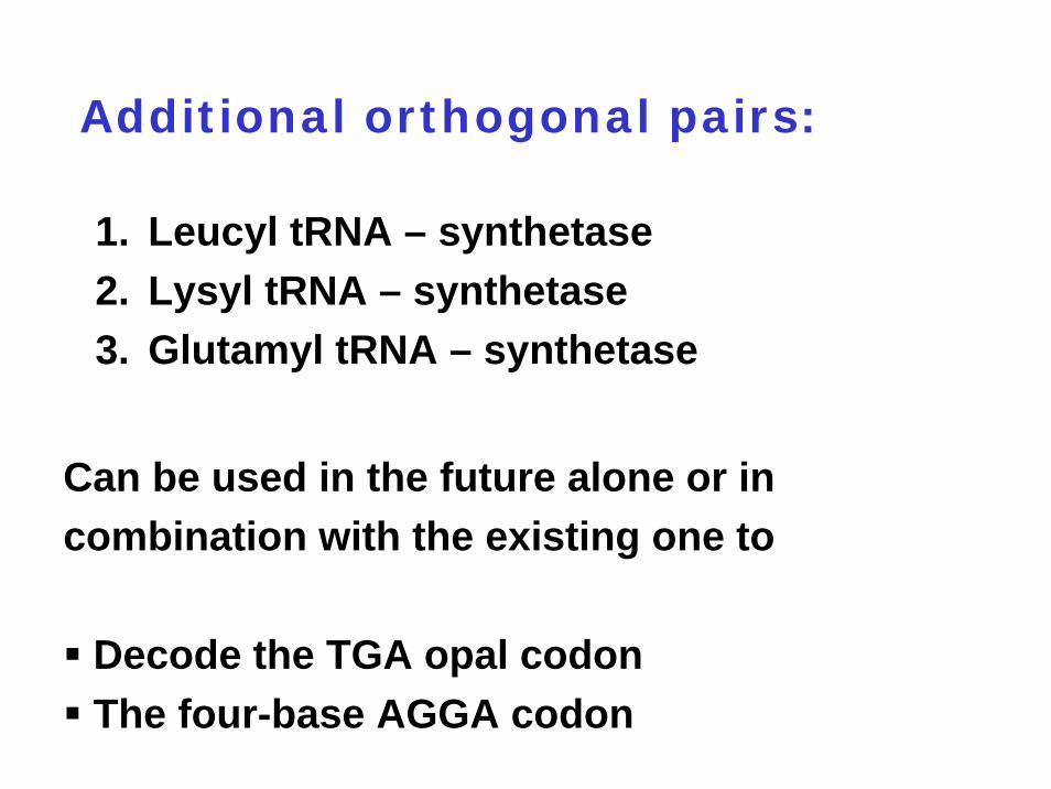
Additional orthogonal pairs:
1. Leucyl tRNA – synthetase 2. Lysyl tRNA – synthetase 3. Glutamyl tRNA – synthetase
Can be used in the future alone or in combination with the existing one to
Decode the TGA opal codonThe four-base AGGA codon
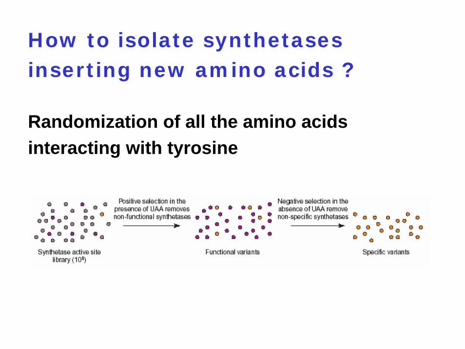
How to isolate synthetases inserting new amino acids ?
Randomization of all the amino acids interacting with tyrosine
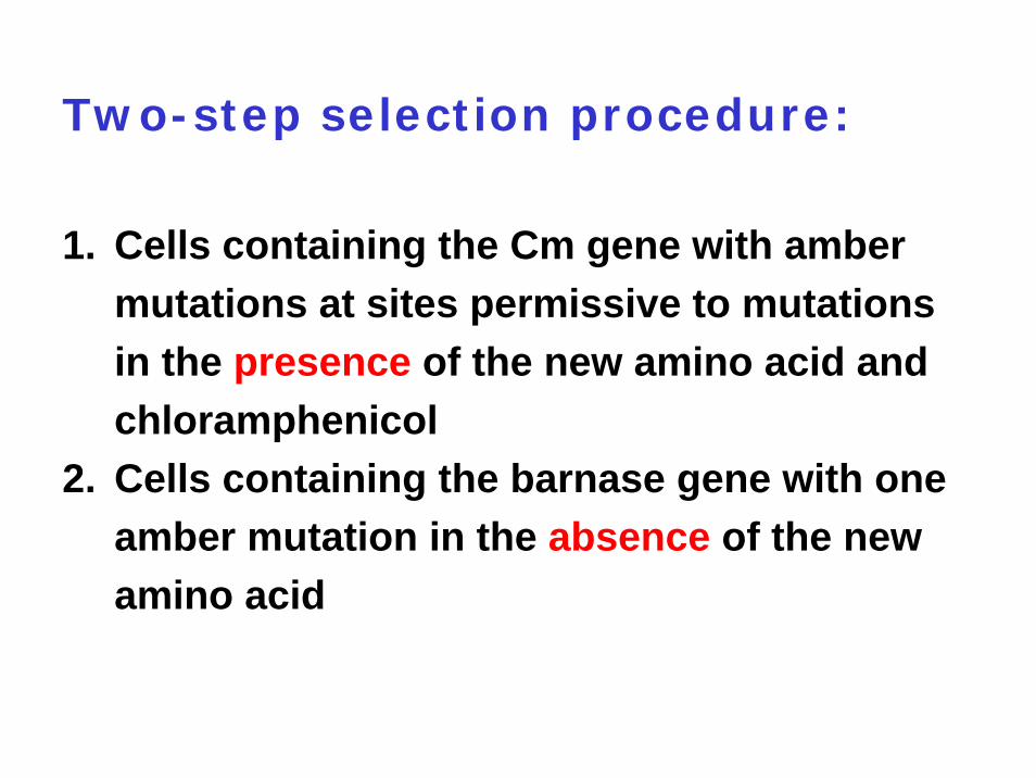
Two-step selection procedure:
1. Cells containing the Cm gene with amber mutations at sites permissive to mutations in the presence of the new amino acid and chloramphenicol
2. Cells containing the barnase gene with one amber mutation in the absence of the new amino acid
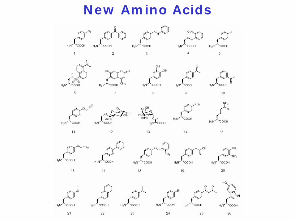
New Amino Acids
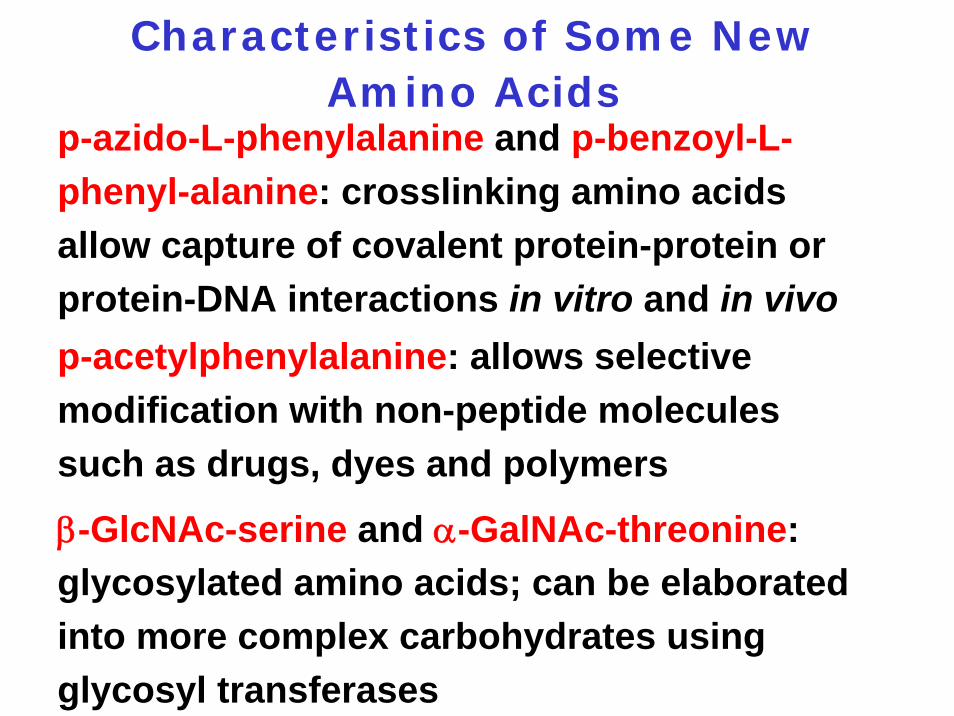
Characteristics of Some New Amino Acids
p-azido-L-phenylalanine and p-benzoyl-L-phenyl-alanine: crosslinking amino acids allow capture of covalent protein-protein or protein-DNA interactions in vitro and in vivop-acetylphenylalanine: allows selective modification with non-peptide molecules such as drugs, dyes and polymers
β-GlcNAc-serine and α-GalNAc-threonine: glycosylated amino acids; can be elaborated into more complex carbohydrates using glycosyl transferases
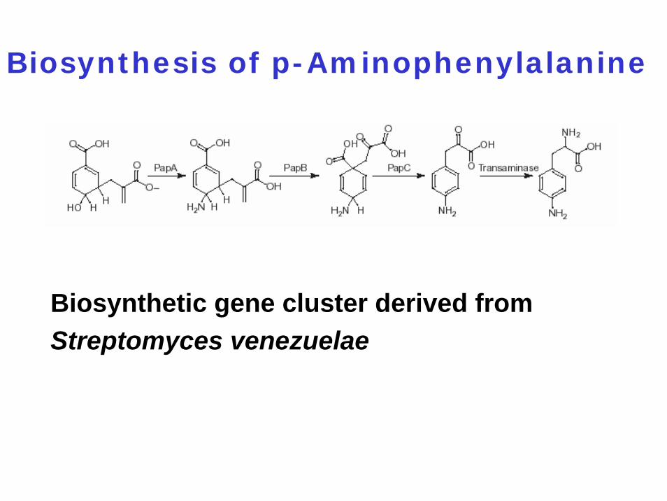
Biosynthesis of p-Aminophenylalanine
Biosynthetic gene cluster derived from Streptomyces venezuelae
Literature:
L Wang et al. (2001) Science 292: 498-500.
TA Cropp and P.G. Schultz (2004) Trends Genetics 20: 625-630.
TL Hendrickson et al. (2004) Annu. Rev. Biochem. 73: 147-176.
CC Liu (2010) Annu. Rev. Biochem. 79: 413