1.1 side a (pbd9: 36; bp9: 4)
TRANSCRIPT

A 70-year-old woman has had a blood pressure of 160/105 mm Hg for many years. Abdominal ultrasound shows the decreased size of one kidney.1. What gross morphologic description applies to the abnormal kidney?2. What is the likely cause of this finding?3. How could this affect the patient’s renal function?4. What cellular organelle plays a major role in this process?5. What cellular protein processing pathway is involved?
1.1 SIDE A (PBD9: 36; BP9: 4)
ANSWERS: SIDE B1. Myocyte cellular atrophy is present as a result of lysosomal autophagy and increased proteasomal degradation.2. Small angulated fibers with occasional central nuclei are grouped together.3. Skeletal muscle fibers in a motor unit are randomly enervated; nerve injury initially leads to scattered myocyte atrophy within any given motor unit. After one nerve is injured, however, an adjacent neuron can branch and reinnervate denervated myocytes. If that neuron is now injured, the result is group atrophy of myocytes.

A 55-year-old man has repeated trauma to his upper arms from operating a jackhammer. He now has hand and forearm weakness. A skeletal muscle biopsy specimen reveals the pattern shown at the right.1. What is the microscopic description of these myocytes?2. What features support the diagnosis?3. Why are smaller angulated fibers grouped together?
1.1 SIDE B (PBD9: 36; BP9: 4)
ANSWERS: SIDE A1. The smaller kidney is atrophic.2. Diminished blood supply (renal artery stenosis as a result of atherosclerosis) with chronic ischemia likely caused this. Although remaining individual cells in the parenchyma are smaller (cellular atrophy), most of the organ shrinkage is attributable to cell dropout because of ischemic injury.3. Diminished blood supply leads to increased renin secretion by the hypoperfused kidney, which causes hypertension.4. Lysosomes play a major role in cellular atrophy through autophagy.5. Cellular atrophy is mediated through increased protein degradation by the ubiquitin-proteasome pathway.
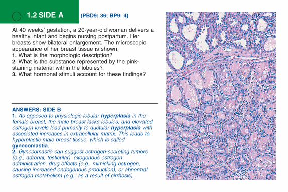
At 40 weeks’ gestation, a 20-year-old woman delivers a healthy infant and begins nursing postpartum. Her breasts show bilateral enlargement. The microscopic appearance of her breast tissue is shown.1. What is the morphologic description?2. What is the substance represented by the pink-staining material within the lobules?3. What hormonal stimuli account for these findings?
1.2 SIDE A (PBD9: 36; BP9: 4)
ANSWERS: SIDE B1. As opposed to physiologic lobular hyperplasia in the female breast, the male breast lacks lobules, and elevated estrogen levels lead primarily to ductular hyperplasia with associated increases in extracellular matrix. This leads to hyperplastic male breast tissue, which is called gynecomastia.2. Gynecomastia can suggest estrogen-secreting tumors (e.g., adrenal, testicular), exogenous estrogen administration, drug effects (e.g., mimicking estrogen, causing increased endogenous production), or abnormal estrogen metabolism (e.g., as a result of cirrhosis).

A 22-year-old man has had bilateral breast enlargement for the past 5 years. The microscopic appearance of his breast tissue is shown.1. This lesion and the tissue shown on Side A represent estrogen effects on breast epithelium. What is the difference between the effects seen in male breasts versus female breasts?2. Why might this man have elevated estrogen levels?
1.2 SIDE B (PBD9: 36; BP9: 4)
ANSWERS: SIDE A1. The female breast lobules have increased cellularity. This is consistent with physiologic hyperplasia from elevated estrogen and progesterone levels during pregnancy.2. The pink secretions within the lobules are milk. The breast is a modified apocrine gland; lipid-rich secretions are formed through the budding of apical portions of epithelial cytoplasm.3. Estrogen and progesterone bind to breast epithelium steroid hormone receptors to cause specific nuclear mRNA transcription. This results in protein translation that promotes cellular proliferation and gain of function.

A 36-year-old man has sudden onset of a high fever. On examination, his temperature is 37.7° C, and a shrill holosystolic murmur is auscultated. A blood culture grows gram-positive cocci in chains that are catalase-negative, that show alpha hemolysis when grown on blood agar, and that are optochin-resistant. Urine microscopic examination reveals RBCs but no WBCs.1. What is suggested by the history along with the blood culture results?2. What complication is shown on this image of the kidney?
1.3 SIDE A (PBD9: 43; BP9: 10)
ANSWERS: SIDE B1. The pale pink region (■) in the adrenal cortex results from a loss of nuclei but preserved cell outlines, which is typical of coagulative necrosis as a consequence of tissue hypoxia and cell death.2. The necrotic cells undergo autolysis and proteolytic degradation by successive waves of neutrophils and then macrophages. The debris is phagocytized by the infiltrating leukocytes, and this is followed by the ingrowth of capillaries with fibroblasts and the eventual replacement of the necrotic zone by a fibrous scar.
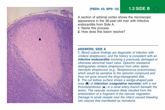
A section of adrenal cortex shows the microscopic appearance in the 36-year-old man with infective endocarditis from Side A.1. Name this process.2. How does this lesion resolve?
1.3 SIDE B (PBD9: 43; BP9: 10)
ANSWERS: SIDE A1. Blood culture findings are diagnostic of infection with viridans streptococci, and the history is consistent with an infective endocarditis involving a previously damaged or otherwise abnormal heart valve. Optochin resistance distinguishes viridans streptococci from other alpha-hemolytic streptococci (e.g., Streptococcus pneumoniae), which would be sensitive to the optochin compound and thus not grow around the drug-impregnated disk.2. The cut kidney surface shows a wedge-shaped pale area (■) of infarction (coagulative necrosis) with a red thromboembolus (▲) in a renal artery branch beneath the lesion. The vascular occlusion likely resulted from the embolization of a fragment of the valvular vegetation. Damage to small vessels near the infarct caused bleeding into calyces that manifested as hematuria.

A 64-year-old man has had increasing dyspnea for the last 12 months. On examination, his blood pressure is 170/105 mm Hg. Heart catheterization reveals elevated right-sided pressures, with a right atrial pressure of 12 mm Hg (nl 3 mm Hg) and a pulmonary capillary wedge pressure of 28 mm Hg (nl 9 mm Hg); the cardiac output is 3.4 L/min (nl 4 to 8 L/min).1. Describe the appearance of this heart.2. What is a likely explanation for the cardiac findings?3. Explain the hemodynamic data.
1.4 SIDE A (PBD9: 34; BP9: 3)
ANSWERS: SIDE B1. Myocyte hypertrophy is reflected by enlarged, boxy, and hyperchromatic nuclei with markedly expanded cytoplasm.2. Congestive heart failure occurs when cardiac compensation fails. Left-sided heart failure leads to pulmonary edema with fluid filling the interstitial and alveolar spaces. Right-sided failure leads to body cavity effusions, visceral organ congestion (hepatomegaly), and peripheral edema (this is most marked in dependent areas, such as the feet when upright).

The ventricular myocardium of the 64-year-old patient from Side A is shown. There are crackles (rales) in both lungs, bilateral pedal edema, and an enlarged liver span.1. Describe the microscopic findings.2. Explain the physical examination findings.
1.4 SIDE B (PBD9: 34; BP9: 3)
ANSWERS: SIDE A1. This heart shows cardiomegaly with severe biventricular hypertrophy and severe biatrial dilation (the latter reflecting stiff, noncompliant ventricles).2. The left ventricular hypertrophy is attributable to chronic pressure overload caused by hypertension. The left ventricle compensates for the increased pressure load through the hypertrophy of myocardial fibers and the thickening of the ventricular wall.3. Left ventricular diastolic dysfunction results in higher left atrial volume and pressure as measured by the pulmonary arterial wedge pressure. The pulmonary arterial and right atrial pressures subsequently increase as well, thereby inducing compensatory right ventricular hypertrophy and subsequent right atrial dilation. The left ventricular cardiac output is reduced because of the poor diastolic filling from decreased compliance with hypertrophy.

The maturation and education of T lymphocytes occurs within the thymus, with more than 97% of proliferating T lymphocytes being eliminated because they either respond to self or express T-cell receptors that interact too weakly or strongly with their relevant antigens. The microscopic appearance of a second-trimester fetal thymus is shown.1. What process is shown?2. What genes play a role in this process?3. How could cell damage by free radicals, toxins, or radiation induce a similar response?
1.5 SIDE A (PBD9: 52; BP9: 18)
ANSWERS: SIDE B1. This is graft-versus-host disease. Cytotoxic donor T lymphocytes kill host cells via an apoptotic pathway that involves the release of granzymes into a cellular target; granzyme proteins directly activate caspases and induce apoptosis (an apoptotic body is seen at the arrow).2. The withdrawal of growth hormones (e.g., estrogen, progesterone) or growth factors may induce the increased synthesis of pro-apoptotic proteins.

After allogeneic bone marrow transplantation with engraftment, a desquamative skin rash occurs. The microscopic findings are shown.1. Name this process, and give the mechanism by which the keratinocytes are eliminated (arrow).2. How might this form of cell death be involved in the cyclic sloughing of the endometrium (menses)?
1.5 SIDE B (PBD9: 53; BP9: 22)
ANSWERS: SIDE A1. This is apoptosis. Thymocytes targeted for deletion undergo tightly regulated cellular fragmentation. Apoptotic bodies are consumed by phagocytes (e.g., macrophages), which gives the appearance of clear spaces filled with granular debris (arrow).2. Regulatory anti-apoptotic proteins such as Bcl-2 are lost or degraded, whereas pro-apoptotic proteins such as Bax are transcribed. Relative amounts of pro-apoptotic and anti-apoptotic proteins regulate mitochondrial cytochrome c release, which controls the activation of intracellular caspases that degrade cellular elements via a proteolytic cascade.
3. DNA damage halts the cell cycle for repair via the accumulation of p53 protein. If DNA is not successfully repaired, p53 triggers apoptosis. Absent or mutated p53 (e.g., as in certain cancers) does not trigger apoptosis, and the defective cell can survive and proliferate.

A 25-year-old man is a cigarette smoker with a chronic cough and frequent severe respiratory infections. The microscopic appearance of his bronchial epithelium is shown.1. What cellular change has occurred?2. Is this change reversible?3. What additional epithelial change can arise?
1.6 SIDE A (PBD9: 37; BP9: 5)
ANSWERS: SIDE B1. Normal squamous epithelium () transitions to dysplasia ( ) with the loss of the normal maturation sequence from basal cells to the surface, hyperchromatic (darker) nuclei, and less cytoplasm (higher N/C ratio).2. Dysplasia is disordered growth with a loss of normal cellular morphology, maturation, and polarity. The appearance reflects increased cell proliferation and decreased differentiation. This can occur with persistent insults in an epithelium, with or without prior metaplastic change.3. Cervical squamous dysplastic lesions are driven by human papillomavirus (HPV) infection. HPV induces cellular proliferation with the concurrent acquisition of increasing numbers of mutations. The greater the number of sexual partners, the greater the risk for acquiring a high-risk strain of HPV.

A 29-year-old woman became sexually active at the age of 14 years, and she has since had 18 male sexual partners. A routine Pap smear showed abnormal cells, which prompted a cervical biopsy. The microscopic appearance of the biopsy specimen is shown.1. Describe the abnormal change in this epithelium.2. Explain the pathophysiology of this disease.3. Why might this patient have developed this disease?
1.6 SIDE B (PBD9: 271; BP9: 165)
ANSWERS: SIDE A1. The pseudostratified ciliated columnar epithelium ( ) is transformed into stratified squamous epithelium (). This is squamous metaplasia.2. Yes. If the injury that produced the change in epithelium is removed, the normal epithelial cell appearance will return.3. If the same injury that caused the metaplasia persists, it can induce malignant transformation. The next step along this pathway is dysplasia, which is also potentially reversible. However, it is further along in the pathogenesis of uncontrolled cell growth (cancer).

A 40-year-old homeless man presents with a 2-month history of fever, purulent productive cough with occasional hemoptysis, soaking night sweats, and weight loss. Physical examination reveals apical rales. His chest radiograph is shown.1. What is present in the right upper lobe?2. What would a sputum sample show?3. Why does this patient have the systemic symptoms of fever, sweats, and weight loss?
1.7 SIDE A (PBD9: 43; BP9: 10)
ANSWERS: SIDE B1. There is central caseation surrounded by enlarged, activated macrophages (so-called “epithelioid” macrophages). A Langhans multinucleated giant cell () is derived from the fusion of multiple activated macrophages.2. This is granulomatous inflammation with central caseous necrosis. Caseation is essentially liquefaction with coagulative necrosis, which in this case is associated with mycobacterial infection.3. The immunosuppression caused by HIV leads to the anergy of cell-mediated immunity and a negative tuberculosis skin test, despite active mycobacterial infection as a result of the reactivation of a previous latent focus or of reinfection caused by a new exposure from the environment.

ANSWERS: SIDE A1. There is upper lobe cavitation ( ) that is typical of secondary tuberculosis.2. His sputum is likely to contain acid-fast bacilli from mycobacterial infection. The cavitation with erosion into the airways of this secondary lesion increases the likelihood of a positive sputum sample and infectivity.3. The systemic symptoms are the consequences of cytokines such as tumor necrosis factor and interleukin-1 being released from activated macrophages.
A 69-year-old man has had increasing dyspnea upon exertion for 3 months. He is known to be HIV-positive. A chest radiograph shows reticulonodular infiltrates in the upper lobes and prominent hilar lymphadenopathy. A skin test with purified protein derivative (PPD) is negative. A lung biopsy is performed.1. What microscopic features are shown?2. What is this pattern of inflammation and tissue necrosis called?3. Why is this patient’s skin test negative to PPD?
1.7 SIDE B (PBD9: 43; BP9: 10)

A 70-year-old woman with a history of rheumatic mitral stenosis has an acute onset of right-sided hemiplegia. Three weeks later, she dies from complications of an aspiration pneumonia.1. What three features are shown here?2. What would have happened during the next 3 months if the patient had lived?3. How could this lesion relate to the patient’s heart disease?
1.8 SIDE A (PBD9: 43; BP9: 10)
ANSWERS: SIDE B1. This cerebral abscess (▲) has highly vascularized granulation tissue around its margin that allows for the bright-appearing concentration of the injected radiocontrast material. With surrounding associated edema (), it is causing a massive ventricular shift to the right ().2. An abscess is a localized collection of degenerating neutrophils; the release of their reactive oxygen species and proteolytic enzymes leads to liquefactive necrosis. Even without abscess formation, central nervous system injury of any form also results in liquefactive necrosis.3. Abscesses are most often caused by bacterial organisms such as staphylococci and streptococci.

A 62-year-old man with a history of rheumatic valvular disease has fever and worsening headaches for 4 days. Physical examination reveals papilledema. A contrast-enhanced MRI of his brain is shown.1. What accounts for the ring enhancement?2. What inflammatory cells are present in the center of the lesion?3. What type of organism is most likely present?
1.8 SIDE B (PBD9: 43; BP9: 10)
ANSWERS: SIDE A1. This is a cerebrovascular accident or “stroke” with cerebral hemispheric softening ( ) caused by liquefactive necrosis secondary to infarction.
2. Microglial cells (central nervous system macrophages) and circulating monocytes would have removed much of the necrotic debris, leaving a cavity (▲) or cortical depression; there also would be peripheral gliosis because of astrocyte enlargement and proliferation.3. Mitral stenosis can lead to left atrial enlargement with subsequent mural thrombosis followed by embolization with infarction. Alternatively, a scarred rheumatic valve is more susceptible to endocarditis; the subsequent embolization of infected vegetation would produce a septic infarct.

A 58-year-old nonsmoker with normal lung function is killed in a hit-and-run accident. The gross appearance of the surface of his lungs is shown.1. What is this pigment?2. How does pigment get to this location?3. What are the risk factors for increased amounts of this pigment?
1.9 SIDE A (PBD9: 64; BP9: 24)
ANSWERS: SIDE B1. This is coal workers’ pneumoconiosis causing restrictive lung disease. The massive amount of carbon pigment leads to the coal macules () shown and their associated fibrosis.2. Restrictive lung diseases lead to cor pulmonale with right heart failure.3. Right heart failure leads to chronic liver passive congestion followed by fibrosis that bridges central veins (this is the opposite of portal cirrhosis). This is not a true cirrhosis, because nodular regeneration is lacking.

A 62-year-old coal miner with a 10-year history of progressive dyspnea develops peripheral edema, pleural effusions, and hepatic “cardiac cirrhosis.” Pulmonary function testing shows decreased FEV1 and decreased FVC. A lung biopsy reveals the findings shown here.1. What is this patient’s diagnosis?2. What cardiac complication has developed?3. Describe the pathogenesis of this patient’s liver disease.
1.9 SIDE B (PBD9: 64; BP9: 24)
ANSWERS: SIDE A1. Anthracotic pigment () within the pleural lymphatics is responsible for the linear (reticular) black appearance of the pleural surface. This amount of pigment has no pathologic consequence.2. Inhaled microscopic carbon particles are ingested by pulmonary macrophages and carried via the lymphatics through interlobular septa to the pleural surfaces and eventually to the hilar lymph nodes.3. Increased amounts of this pigment come from smoking and polluted air.
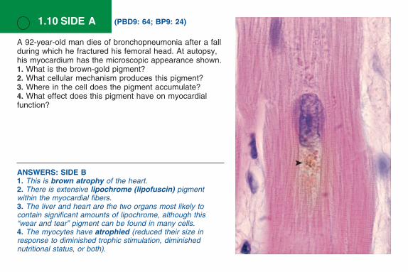
ANSWERS: SIDE B1. This is brown atrophy of the heart.2. There is extensive lipochrome (lipofuscin) pigment within the myocardial fibers.3. The liver and heart are the two organs most likely to contain significant amounts of lipochrome, although this “wear and tear” pigment can be found in many cells.4. The myocytes have atrophied (reduced their size in response to diminished trophic stimulation, diminished nutritional status, or both).
1.10 SIDE A (PBD9: 64; BP9: 24)
A 92-year-old man dies of bronchopneumonia after a fall during which he fractured his femoral head. At autopsy, his myocardium has the microscopic appearance shown.1. What is the brown-gold pigment?2. What cellular mechanism produces this pigment?3. Where in the cell does the pigment accumulate?4. What effect does this pigment have on myocardial function?

ANSWERS: SIDE A1. This is lipochrome (lipofuscin) pigment, which is characteristically found in a perinuclear location ().2. Long-term recurrent free radical damage leads to lipid peroxidation and the cross-linking of polyunsaturated lipids of various cellular membranes, which cannot be catabolized further.3. Lysosomes fuse with autophagic vacuoles to create autophagolysosomes, which are responsible for normal intracellular organellar turnover. Nondigestible debris from abnormally cross-linked cellular molecules persists as residual bodies in lysosomes and manifests as pigmented lipofuscin.4. Some amount of lipofuscin can be found in all individuals with aging. Although it is indicative of past wear and tear, even large amounts have no deleterious effect on cellular function.
A 78-year-old man with progressive dementia has become severely malnourished. At autopsy, his heart is small, with the gross appearance shown.1. What is the name for this appearance?2. What pigment within the myocardial fibers accounts for this appearance?3. In what other organ is this pigment most likely to be found?4. What is the cellular process that accounts for the decreased cardiac size?
1.10 SIDE B (PBD9: 64; BP9: 24)4 cm

A 44-year-old man who has had increasing dyspnea on exertion for 6 years has a loud crescendo-decrescendo systolic ejection murmur and S4. His carotids have pulsus parvus et tardus, and chest radiography shows pulmonary edema. His native aortic valve at replacement surgery is shown.1. What is your diagnosis?2. Why is this valve thickened with nodular densities?3. Although more common during the eighth and ninth decades, this man’s disease became symptomatic when he was 44 years old. Why?4. Where else can this mineral be deposited?
1.11 SIDE A (PBD9: 65; BP9: 25)
ANSWERS: SIDE B1. The dark blue deposits that thicken the interstitium are calcium deposits resulting from metastatic calcification. Elastic tissue, sites of necrosis, and cells with an intracellular alkaline environment (e.g., gastric parietal cells) favor calcium phosphate deposition.2. She has primary hyperparathyroidism; the neck mass is likely a parathyroid adenoma.3. The serum ionized calcium and parathyroid hormone levels would be increased.

A 45-year-old woman had urolithiasis, bone pain, peptic ulcers, constipation, cardiac dysrhythmia, weakness, and depression over the past 4 months. A forearm radiograph showed osteitis fibrosa cystica, and a sestamibi scan showed a solitary neck mass. She died of a sudden cardiac arrhythmia. The microscopic appearance of her lungs is shown.1. What is deposited in her alveolar walls and why?2. What is her underlying disease?3. What other laboratory findings would you expect?
1.11 SIDE B (PBD9: 65; BP9: 26)
ANSWERS: SIDE A1. This patient has calcific degeneration of the aortic valve that leads to aortic stenosis.2. The abnormal valve is subject to more wear and undergoes dystrophic calcification.3. Congenital bicuspid aortic valve affects about 1% of the population and makes degenerative calcification accrue at a much earlier age.4. Crystalline calcium phosphate deposition can occur in damaged or necrotic tissues, as part of atherogenesis, or during the healing of inflammatory processes (e.g., granulomas).