11 into 12 applied science assignment. biology structure
TRANSCRIPT

11 into 12 Applied Science Assignment. Biology Structure and functions of cells and tissues. Name:
Page 1

Specification guidance
B1 Cell structure and function
➔ Know that cell theory is a unifying concept stating that cells are a fundamental unit of structure, function and organisation in all living organisms.
➔ Understand the ultrastructure and function of organelles in the following cells: ➔ prokaryotic cells (bacterial cell) – nucleoid, plasmids, 70S ribosomes, capsule, cell wall
➔ eukaryotic cells (plant and animal cells) – plasma membrane, cytoplasm, nucleus, nucleolus, endoplasmic
reticulum (smooth and rough), Golgi apparatus, vesicles, lysosomes, 80S ribosomes, mitochondria, centriole
➔ eukaryotic cells (plant-cell specific) – cell wall, chloroplasts, vacuole, tonoplast, amyloplasts,
plasmodesmata, pits. Recognise cell organelles from electron micrographs and the use of light microscopes.
➔ Understand the similarities and differences between plant and animal cell structure and function
➔ Understand how to distinguish between gram-positive and gram-negative bacterial cell walls and why each type reacts differently to some antibiotics.
➔ Calculate magnification and size of cells and organelles from drawings or images.
Notes:
Page 2
Page 3
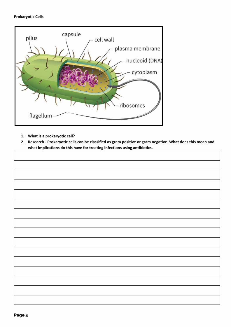
Prokaryotic Cells
1. What is a prokaryotic cell?
2. Research - Prokaryotic cells can be classified as gram positive or gram negative. What does this mean and
what implications do this have for treating infections using antibiotics.
Page 4

Page 5

Eukaryotic cells - Animal
3. Why is an animal cell considered to be eukaryotic?
4. What does it have in common with other types of eukaryotic cells?
Page 6

Eukaryotic Cell - Plant cell
5. Why is this plant cell eukaryotic?
6. How is it different from other types of eukaryotic cells?
Page 7

Endoplasmic reticulum
7. The rough endoplasmic reticulum is a series of membranes that is continuous with the nucleus. Considering
the fact that it is studded with ribosomes, suggest the function of the rough endoplasmic reticulum.
8. Research - Why is it referred to as the rough endoplasmic reticulum? What other reticulums are there and
what do they do?
Page 8

Mitochondria
9. What is the role of the mitochondria
10. Research - What important stage of respiration occurs in the matrix?
11. Research - What happens on the cristae (inner membrane) and what are they so highly folded?
Page 9

Ribosome
12. What is the function of a ribosome?
13. Research - What is the difference between a 70S& 80S ribosome?
Page 10
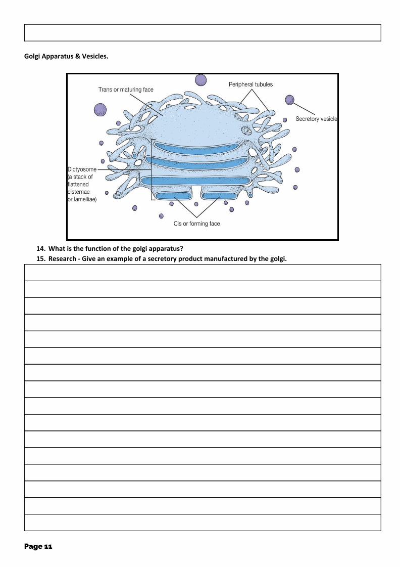
Golgi Apparatus & Vesicles.
14. What is the function of the golgi apparatus?
15. Research - Give an example of a secretory product manufactured by the golgi.
Page 11

Centrioles
16. Research - What role do centrioles play in the cell?
17. Are they found in all cells?
Page 12
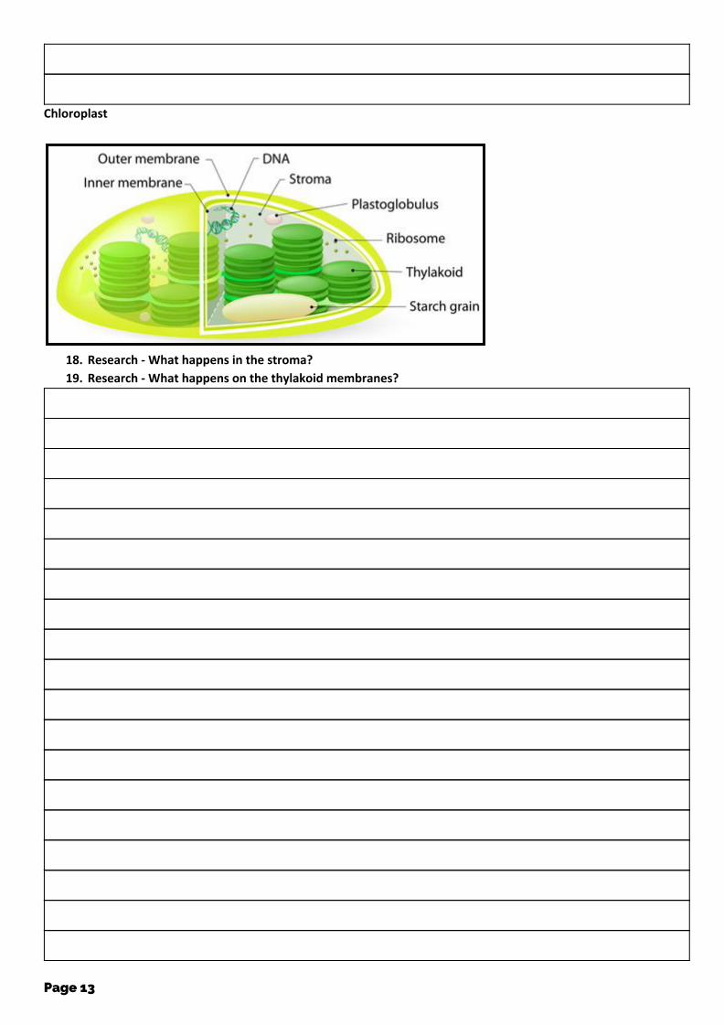
Chloroplast
18. Research - What happens in the stroma?
19. Research - What happens on the thylakoid membranes?
Page 13

Plasmodesmata & Pits
20. Research - What role do plasmodesmata fulfil for plant cells?
Page 14

Amyloplasts
21. Research - Thinking about the name of this organelle (Begins with Amy) what is its function in plant cells?
Page 15

Vacuole & Tonoplast
22. What is the function of the tonoplast and vacuole in plant cells?
Page 16

Magnification
Page 17

Q1. The drawing shows an electron micrograph of parts of epithelial cells from the small intestine.
(a) (i) Name the structures labelled A.
(1) (ii) Explain how these structures help in the absorption of substances from the small intestine.
(1) (b) (i) The scale bar on this drawing represents a length of 0.1μm. Calculate the magnification of the drawing. Show your working.
Magnification .............................................
Page 18

Q2. The drawing shows part of a plant cell as seen with an electron microscope.
(i) Give two features shown in the drawing which are evidence that this cell is eukaryotic.
1.
2.
(2)
(ii) Calculate the actual width of the cell from Y to Z. Give your answer in micrometres (µm) and show your working.
Answer ..................................... µm (2)
(iii) Give one way in which a typical animal cell differs from the cell shown in the drawing.
(1) (Total 5 marks)
Page 19

Q3. The figure shows a section through a palisade cell in a leaf as seen with a light microscope. The palisade has been magnified × 2000.
x 2000
(a) Calculate the actual width of the cell, measured from A to B, in μm. Show your working
Answer ........................................... μm (2)
(b) Palisade cells are the main site of photosynthesis. Explain one way in which a palisade cell is adapted for photosynthesis.
(2) (Total 4 marks)
Page 20

Q4.
(a) Name the process in which cells become adapted for different functions.
(1)
(b) Palisade cells are found in leaves. The diagram shows a palisade cell.
(i) Name structure A.
(1)
(ii) The real length of this cell between X and Y is 20 micrometres (µm). By how many times has it been magnified? Show your working.
Answer ............................................ (2)
(iii) Explain one way in which this cell is adapted for photosynthesis.
(1) (Total 5 marks)
Page 21

Q5. The diagram shows an epithelial cell from the small intestine.
(a) (i) Name organelle Y.
(1)
(ii) There are large numbers of organelle Y in this cell. Explain how these organelles help the cell to absorb the products of digestion.
(2) (b) This diagram shows the cell magnified 1000 times. Calculate the actual length of the cell between points P and Q. Give your answer in µm. Show your working.
Answer ...................................... µm (2)
Page 22

Q6. The diagram shows a cholera bacterium. It has been magnified 50 000 times.
(a) Name A.
(1) (b) Name two structures present in an epithelial cell from the small intestine that are not present in a cholera bacterium.
1.
2.
(2) (d) Calculate the actual width of the cholera bacterium between points B and C. Give your answer in micrometres and show your working.
.................................. µm (2)
Page 23

Q7.The diagram shows a chloroplast as seen with an electron microscope.
(a) Name X and Y.
X
Y
(b) Describe the function of a chloroplast.
(2)
(c) Calculate the maximum length of this chloroplast in micrometres (μm). Show your working.
Answer ............................................... μm (2)
(Total 6 marks)
Page 24

Q8.The diagram shows a eukaryotic cell.
(a) Complete the table by giving the letter labelling the organelle that matches the function. Function of organelle Letter
Protein synthesis
Modifies protein (for example, adds carbohydrate to protein)
Aerobic respiration
(3)
(b) Use the scale bar in the diagram above to calculate the magnification of the drawing. Show your working.
Answer = ................................ (2)
(Total 5 marks)
Page 25