10 t p - aciar.gov.au
TRANSCRIPT

TaroPest – Bacteria 1
109
87
65
43
21
An illustrated guide to pests and diseases of taro in the South Pacific
TaroPest

1
An illustrated guide to pests and diseases of taro in the South Pacific
Amy Carmichael, Rob Harding, Grahame Jackson, Sarlesh Kumar, Sada Lal, Roy Masamdu, Jacqui Wright and Anthony Clarke
Australian Centre for International Agricultural Research
2008
TaroPest

TaroPest: an illustrated guide to pests and diseases of taro in the South Pacific2
The Australian Centre for International Agricultural Research (ACIAR) was established in June 1982 by an Act of the Australian Parliament. Its primary mandate is to help identify agricultural problems in developing countries and to commission collaborative research between Australian and developing-country researchers in fields where Australia has special competence.
Where trade names are used, this constitutes neither endorsement of nor discrimination against any product by the Centre.
ACIAR MONOGRAPH SERIES
This series contains the results of original research supported by ACIAR, or material deemed relevant to ACIAR’s research and development objectives. The series is distributed internationally, with an emphasis on developing countries.
© Commonwealth of Australia 2008
This work is copyright. Apart from any use as permitted under the Copyright Act 1968, no part may be reproduced by any process without prior written permission from the Commonwealth. Requests and inquiries concerning reproduction and rights should be addressed to the Commonwealth Copyright Administration, Attorney-General’s Department, Robert Garran Offices, National Circuit, Barton ACT 2600 or posted at http://www.ag.gov.au/cca.
Published by the Australian Centre for International Agricultural Research (ACIAR) GPO Box 1571, Canberra ACT 2601, Australia Telephone: 61 2 6217 0500 [email protected]
Carmichael A., Harding R., Jackson G., Kumar S., Lal S.N., Masamdu R., Wright J. and Clarke A.R. 2008. TaroPest: an illustrated guide to pests and diseases of taro in the South Pacific. ACIAR Monograph No. 132, 76 pp.
ISBN 978 1 921424 55 6 (print) ISBN 978 1 921434 56 3 (online)
Cover: Giant African snail (Lissachatina fulica) on taro in Samoa. Photo by Amy Carmichael.
Technical editing and production management by Biotext Pty Ltd Design by Design ONE Printing by Finsbury Green

3
ForewordTaro is a major crop of the South Pacific, with a regional production of more than 360,000 tonnes per year, and a wide cultural, economic and food security importance to nearly all Pacific island countries and territories. Given this importance, the Australian Centre for International Agricultural Research (ACIAR) and its partners have invested heavily over the years in enhancing the quality and sustainability of regional taro production. Unfortunately, taro is subject to significant losses from pests and diseases. The most important of these can devastate a previously unchallenged crop. For example, the introduction of taro leaf blight into Samoa in 1993 virtually eliminated that country’s crop, causing economic hardship in rural areas, a destabilisation of internal food security and the loss of lucrative export trade.
To manage pests and diseases effectively, they need to be identified on crops so that biological and control information can be sourced. In the absence of suitable information, unknown exotic pests may establish in a region, or existing pests may be inappropriately treated. During the 2003 ACIAR South Pacific regional consultations, the lack of readily available information on taro pests was identified as a matter of concern. The development of a pest and disease tool kit for taro was subsequently agreed on as a regional priority. This led, in 2004, to the ACIAR project ‘TaroPest: a computer-based information and diagnostics package for taro pests of the South Pacific’ (CP/2004/001). This project combined the expertise of researchers from Fiji, Papua New Guinea and Australia initially, later expanding to include expertise from across the region.
As a summary of knowledge on the regional pests and diseases of taro, this monograph is an outcome of the TaroPest project, which itself built on many earlier taro pest-management projects. ‘TaroPest: an illustrated guide to pests and diseases of taro in the South Pacific’ captures the work and knowledge of many researchers and field officers. We hope it will prove a valuable tool for taro producers, crop advisers and regional quarantine officers.
Peter Core
Chief Executive Officer Australian Centre for International Agricultural Research

TaroPest: an illustrated guide to pests and diseases of taro in the South Pacific4
AcknowledgmentsTaroPest is a collaborative work. It includes contributions from researchers beyond those in the authorship list. Authors of individual fact sheets and suppliers of all photographs are listed in the associated CD. We thank the many regional scientists, extension staff and growers who provided feedback on TaroPest during its development, either through workshop participation, email discussion lists or individual contact. The developers of TaroPest would particularly like to acknowledge the important contributions made by: John Bridge, Fred Brooks, Jeff Daniells, Wolfgang Gerlach, Roger Goebel, Rowland Holmes, Gerald McCormack, Eric McKenzie, Jeri Ooka, George Wall, and Philip Tuivavalagi and other staff of the Samoan Ministry of Agriculture and Fisheries (Nu’u).
The TaroPest project was a joint collaboration between the Papua New Guinea National Agricultural Quarantine and Inspection Agency, the Secretariat of the Pacific Community Plant Protection Service and the Queensland University of Technology in Australia. Core funding was provided by the Australian Centre for International Agricultural Research (ACIAR), under ACIAR project CP/2004/001. Additional information on pests and diseases has been gathered from the following ACIAR projects: CP/1994/043, CP/2000/044, HORT/2007/037 and HORT/2006/053.

5
ContentsForeword . . . . . . . . . . . . . . . . . . . . . . . . . . . . . . . . . . . . . . . . . . . . . . . . . . . . . . . . . . . . . . . . . . . 3
Acknowledgments . . . . . . . . . . . . . . . . . . . . . . . . . . . . . . . . . . . . . . . . . . . . . . . . . . . . . . . . . . . 4
Introduction . . . . . . . . . . . . . . . . . . . . . . . . . . . . . . . . . . . . . . . . . . . . . . . . . . . . . . . . . . . . . . . . 7
Identification key. . . . . . . . . . . . . . . . . . . . . . . . . . . . . . . . . . . . . . . . . . . . . . . . . . . . . . . . . . . . 8
Fact sheets . . . . . . . . . . . . . . . . . . . . . . . . . . . . . . . . . . . . . . . . . . . . . . . . . . . . . . . . . . . . . . . . . 13
4 Bacteria. . . . . . . . . . . . . . . . . . . . . . . . . . . . . . . . . . . . . . . . . . . . . . . . . . . . . . . . . . . . . . . . . 14
Bacterial soft rot Erwinia chrysanthemi. . . . . . . . . . . . . . . . . . . . . . . . . . . . 14
4 Fungi . . . . . . . . . . . . . . . . . . . . . . . . . . . . . . . . . . . . . . . . . . . . . . . . . . . . . . . . . . . . . . . . . . . 16
Corm rot Athelia rolfsii . . . . . . . . . . . . . . . . . . . . . . . . . . . . . . . . . . . 16
Brown leaf spot (or ghost spot) Cladosporium colocasiae . . . . . . . . . . . . . . . . . . . . . . . . 18
Spongy black rot Lasiodiplodia theobromae . . . . . . . . . . . . . . . . . . . . . . . . 20
White spot of taro Leptosphaerulina trifolii . . . . . . . . . . . . . . . . . . . . . . . . . . 22
Corm and leaf spot Marasmiellus stenophyllus . . . . . . . . . . . . . . . . . . . . . . . . 24
Orange leaf spot Neojohnstonia colocasiae . . . . . . . . . . . . . . . . . . . . . . . . 26
Shot hole Phoma spp. . . . . . . . . . . . . . . . . . . . . . . . . . . . . . . . . . . . . 28
Taro leaf blight Phytophthora colocasiae . . . . . . . . . . . . . . . . . . . . . . . . . 30
Leaf blotch Pseudocercospora colocasiae . . . . . . . . . . . . . . . . . . . . . . 32
Corm soft rot Pythium spp. . . . . . . . . . . . . . . . . . . . . . . . . . . . . . . . . . . . 34
4 Insects . . . . . . . . . . . . . . . . . . . . . . . . . . . . . . . . . . . . . . . . . . . . . . . . . . . . . . . . . . . . . . . . . . 36
Spiralling whitefly Aleurodicus dispersus . . . . . . . . . . . . . . . . . . . . . . . . . . . . 36
Aphids Aphis gossypii . . . . . . . . . . . . . . . . . . . . . . . . . . . . . . . . . . 38
Tobacco whitefly Bemesia tabaci . . . . . . . . . . . . . . . . . . . . . . . . . . . . . . . . . 40
Hornworm Hippotion celerio. . . . . . . . . . . . . . . . . . . . . . . . . . . . . . . . 42
Mealybugs Family Pseudococcidae . . . . . . . . . . . . . . . . . . . . . . . . . . 44
Taro beetles Papuana spp. . . . . . . . . . . . . . . . . . . . . . . . . . . . . . . . . . . 46
Taro root aphid Patchiella reaumuri. . . . . . . . . . . . . . . . . . . . . . . . . . . . . . 48
Armyworm Spodoptera litura . . . . . . . . . . . . . . . . . . . . . . . . . . . . . . . 50
Taro planthoppers Tarophagus spp. . . . . . . . . . . . . . . . . . . . . . . . . . . . . . . . . 52
Spider mites Tetranychus spp. . . . . . . . . . . . . . . . . . . . . . . . . . . . . . . . . 54
4 Nematodes. . . . . . . . . . . . . . . . . . . . . . . . . . . . . . . . . . . . . . . . . . . . . . . . . . . . . . . . . . . . . . 56
Miti miti disease Hirschmanniella miticausa . . . . . . . . . . . . . . . . . . . . . . . 56
Root knot nematodes Meloidogyne spp. . . . . . . . . . . . . . . . . . . . . . . . . . . . . . . . 58
Lesion nematode Pratylenchus coffeae. . . . . . . . . . . . . . . . . . . . . . . . . . . . . 60

TaroPest: an illustrated guide to pests and diseases of taro in the South Pacific6
4 Snails . . . . . . . . . . . . . . . . . . . . . . . . . . . . . . . . . . . . . . . . . . . . . . . . . . . . . . . . . . . . . . . . . . . 62
Giant African snail Lissachatina fulica . . . . . . . . . . . . . . . . . . . . . . . . . . . . . . 62
4 Viruses. . . . . . . . . . . . . . . . . . . . . . . . . . . . . . . . . . . . . . . . . . . . . . . . . . . . . . . . . . . . . . . . . . 64
Alomae. . . . . . . . . . . . . . . . . . . . . . . . . . . . . . . . . . . . . . . . . . . . . . . . . . . . . . . . . . . . . . . . . . 64
Colocasia bobone disease virus (CBDV) . . . . . . . . . . . . . . . . . . . . . . . . . . . . . . . . . . . . . . . 66
Dasheen mosaic virus (DsMV) . . . . . . . . . . . . . . . . . . . . . . . . . . . . . . . . . . . . . . . . . . . . . . . 68
Taro badnavirus (TaBV). . . . . . . . . . . . . . . . . . . . . . . . . . . . . . . . . . . . . . . . . . . . . . . . . . . . . 70
Taro vein chlorosis virus (TaVCV) . . . . . . . . . . . . . . . . . . . . . . . . . . . . . . . . . . . . . . . . . . . . . 72
References . . . . . . . . . . . . . . . . . . . . . . . . . . . . . . . . . . . . . . . . . . . . . . . . . . . . . . . . . . . . . . . . . . 74
How to use the TaroPest CD. . . . . . . . . . . . . . . . . . . . . . . . . . . . . . . . . . . . . . . . . . . . . . . . . . 74
Index . . . . . . . . . . . . . . . . . . . . . . . . . . . . . . . . . . . . . . . . . . . . . . . . . . . . . . . . . . . . . . . . . . . . . . . 75

7
IntroductionTaro (Colocasia esculente), which is a major food crop in the South Pacific, is subject to significant losses from pests and diseases. Most South Pacific taro pests have restricted distributions, making effective quarantine critical to their containment and management. Identifying pests already in a country is an ongoing requirement for growers, extension officers and those responsible for trying to gain international market access for the crop. Lack of user-friendly diagnostic tools, however, means that effective quarantine, pest management and pest surveillance are severely hampered. To overcome these problems, TaroPest has been developed as a guide to the pests and diseases of taro in the South Pacific. Its aim is to be a one-stop shop for pests of taro, with keys, fact sheets, photographs and other supporting information. TaroPest consists of a field guide and a self-running CD-ROM, which is interactive and contains information additional to that presented in the manual. The field guide is designed to be a portable printed version to be used in conjunction with the CD-ROM. Users risk missing out on useful information if they only consult the hard-copy part of TaroPest.
The TaroPest project was funded by the Australian Centre for International Agricultural Research (ACIAR). It was initially a collaborative project involving staff of the Secretariat of the Pacific Community Plant Protection Service, the (Papua New Guinea) National Agricultural Quarantine and Inspection Authority and the Queensland University of Technology in Australia. As the project developed, significant and valuable input was provided by private consultants, regionally located research and extension officers, and plant pest and disease specialists from around the world. To the best of our knowledge, TaroPest captures all available information pertinent to the identification and management of taro pests and diseases in the South Pacific region. Although we found that much is known about a small group of the insects and diseases attacking taro, TaroPest also highlights that, for the majority, little or nothing is known about their economic or biological impact, differences in susceptibility between different taro cultivars, and control methods. More research on these topics is needed.
While formal acknowledgments are given elsewhere in TaroPest, the authors wish to recognise here the funding and logistic support of ACIAR and our employer organisations. We would also like to particularly thank the many regional scientists, field officers, extension staff and growers whose photographs, firsthand knowledge and efforts in trialling TaroPest substantially increased its content, usability and accuracy.

TaroPest: an illustrated guide to pests and diseases of taro in the South Pacific8
Identification key1.
a) I can see a pest . . . . . . . . . . . . . . . . . . . . . . . . . . . . . . . . . . . . . . . . . . . . . . . . . . . . . . . . . . . 2
b) The plant is damaged or diseased, but I can’t see a pest. . . . . . . . . . . . . . . . . . . . . . . . . 14
2.
a) I found the pest in or on the corm . . . . . . . . . . . . . . . . . . . . . . . . . . . . . . . . . . . . . . . . . . . 3
b) I found the pest on the leaf blade or petiole. . . . . . . . . . . . . . . . . . . . . . . . . . . . . . . . . . . 5
3.
a) I found the pest inside the corm tissue . . . . . . . . . . . . . . . Taro beetle (Papuana spp.) p. 46
b) I found the pest on the surface of the corm . . . . . . . . . . . . . . . . . . . . . . . . . . . . . . . . . . . 4
4.
a) A very small, soft-bodied insect associated with the plant roots, showing masses of white cottony thread . . . . . . . . . . . . . . . . . . . . Taro root aphid (Patchiella reaumuri) p. 48
b) A soft, segmented insect covered with a white waxy powder, plant roots not covered with white cottony threads . . . . . . . . . . . . . . . . . . Mealybugs (family Pseudococcidae) p. 44
5.
a) It is a caterpillar . . . . . . . . . . . . . . . . . . . . . . . . . . . . . . . . . . . . . . . . . . . . . . . . . . . . . . . . . . 6
b) It is something other than a caterpillar . . . . . . . . . . . . . . . . . . . . . . . . . . . . . . . . . . . . . . . 7
6.
a) A green–brown to red–brown caterpillar with dark markings, without a horn on its tail . . . . . . . . . . . . . . . . . . . . . . . . . . . . . . . . . . . . . . . . . Armyworm (Spodoptera litura) p. 50
b) A bright green (occasionally reddish-brown) caterpillar, with a ‘horn’ on its rear end. It has two spots resembling eyes behind the head . . . . . . . . . . . . . . . . . . . . . . . . . . . . . . . . . . . Hornworm (hawk moth) (Hippotion celerio) p. 42
7.
a) It is a snail . . . . . . . . . . . . . . . . . . . . . . . . . . . . . Giant African snail (Lissachatina fulica) p. 62
b) It is something other than a snail . . . . . . . . . . . . . . . . . . . . . . . . . . . . . . . . . . . . . . . . . . . . 8
8.
a) I cannot see individual organisms without the use of a hand lens or microscope. Organisms smaller than 1 mm . . . . . . . . . . . . . . . . . . . . . . . . . . . . . . . . . . . . . . . . . . . . . . 9
b) I can see individual organisms without the use of a hand lens or microscope . . . . . . . 11
Identification key 9
9.
a) The organisms are green in colour . . . . . . . . . . . . . . . . . . . . . . .Aphids (Aphis gossypii) p. 38
b) The organisms are red in colour . . . . . . . . . . . . . . . . . . Spider mites (Tetranychus spp.) p. 54
c) The organisms are white or whitish-cream in colour. . . . . . . . . . . . . . . . . . . . . . . . . . . . . 10
10.
a) A spiralling pattern is apparent on the leaves or there is an abundance of wax present . . . . . . . . . . . . . . . . . . . . . . . . . . . . . . Spiralling whitefly (Aleurodicus dispersus) p. 36
b) No spiralling pattern is present on the leaves, nor is there lots of wax present . . . . . . . . . . . . . . . . . . . . . . . . . . . . . . . . . . . . . . . . . . Tobacco whitefly (Bemesia tabaci) p. 40
11.
a) The organisms are coloured other than white . . . . . . . . . . . . . . . . . . . . . . . . . . . . . . . . . . 12
b) The organisms are white in colour . . . . . . . . . . . . . . . . . . . . . . . . . . . . . . . . . . . . . . . . . . . 13
12.a) Small and robust insects, adults approximately 4 mm in length, generally black with
broad white patches or markings . . . . . . . . . . . . . Taro planthopper (Tarophagus spp.) p. 52
b) Small, pear-shaped insects with soft, fragile bodies, colour variable from pale green–yellow to dark green, sizes range from 1 mm to 2.5 mm . . . . . . . . . . .Aphids (Aphis gossypii) p. 38
13.a) A soft, highly segmented insect without an outer shell, covered with a white waxy
powder, ovoid in shape, wings absent . . . . . . . . . Mealybugs (family Pseudococcidae) p. 44
b) A spiralling pattern is apparent on the leaves, or there is an abundance of wax present . . . . . . . . . . . . . . . . . . . . . . . . . . . . . . . . . . . . Spiralling whitefly (Aleurodicus dispersus) p. 36
c) Small, distinctly winged insects, wings very white and body pale yellow, no spiralling pattern on the leaves, no free wax . . . . . . . . . . . . . Tobacco whitefly (Bemesia tabaci) p. 40
14.
a) The corm or the roots are affected . . . . . . . . . . . . . . . . . . . . . . . . . . . . . . . . . . . . . . . . . . . 15
b) The leaf blade or petioles are affected. . . . . . . . . . . . . . . . . . . . . . . . . . . . . . . . . . . . . . . . 23
15.a) There are signs of damage and holes in the corms . . . . . . . . .Taro beetles (Papuana spp.) p. 46
b) The roots are distorted with galls or knots. .Root knot nematodes (Meloidogyne spp.) p. 58
c) There are signs of disease and the corm appears rotten . . . . . . . . . . . . . . . . . . . . . . . . . 16
16.
a) The signs of disease were visible in the corms before harvest . . . . . . . . . . . . . . . . . . . . . 17
b) The corms were healthy at harvest, so the rot has occurred since. . . . . . . . . . . . . . . . . . 19
TaroPest: an illustrated guide to pests and diseases of taro in the South Pacific10
17.
a) The plant quickly collapsed and rotten corms were discovered . . . . . . . . . . . . . . . . . . . . . . . . . . . . . . . . . . . . . Bacterial soft rot (Erwinia chrysanthemi) p. 14
b) The plant has shown gradual wilting or other symptoms leading to the discovery of corm rot . . . . . . . . . . . . . . . . . . . . . . . . . . . . . . . . . . . . . . . . . . . . . . . . . . . . . . . . . . . . . . . 18
18.
a) Corms show irregular zones of dry brown rot that originate from the base of the corm, healthy tissue adjacent to the rot is red and corms have the appearance of uncooked fatty meat . . . . . . . . . . . . . . . . . . . . . . . . .Miti miti disease (Hirschmanniella miticausa) p. 56
b) Fans of white mycelia are growing over the infected area and (sometimes) the organic matter surrounding the plant; sclerotia from pale cream to reddish-brown are present at the site of infection . . . . . . . . . . . . . . . . . . . . . . . . . . . . . . . . Corm rot (Athelia rolfsii) p. 16
c) Diseased corms show a rot of varying colour from whitish-yellow, through shades of grey and blue, to dark purple, usually starting at the base of the corm; a sharp line of demarcation can usually be seen between healthy and diseased tissue when the corm is cut open . . . . . . . . . . . . . . . . . . . . . . . . . . . . . . . . . Corm soft rot (Pythium spp.) p. 34
19.
a) The rot is soft and foul smelling . . . . . . . . . . Bacterial soft rot (Erwinia chrysanthemi) p. 14
b) The rot is not soft or foul smelling . . . . . . . . . . . . . . . . . . . . . . . . . . . . . . . . . . . . . . . . . . . 20
20.
a) I am in a country where taro leaf blight (Phytophthora colocasiae) is present (American Samoa, Federated States of Micronesia, Guam, Commonwealth of Northern Mariana Islands, Palau, Papua New Guinea, Samoa, Solomon Islands) . . . . 21
b) I am not in a country where taro leaf blight (Phytophthora colocasiae) is present. . . . 22
21.
a) Light brown, hard rot . . . . . . . . . . . . . . . . . . Taro leaf blight (Phytophthora colocasiae) p. 30
b) White dry rot, often with a dark brown margin and large pink patches ahead of the rot margin . . . . . . . . . . . . . . . . . . . . . . . . . . . . . . . . . . . . . . Corm soft rot (Pythium spp.) p. 34
c) White–cream spongy rot darkening with age and sour smelling . . . . . . . . . . . . . . . . . . . . . . . . . . . . . . . . . Spongy black rot (Lasiodiplodia theobromae) p. 20
d) Pink with white, dense fungal growth after 24 hours at high humidity . . . . . . . . . . . . . . . . . . . . . . . . . . . . . . . . . . . . . . . . . . . . . . . . . . . Corm rot (Athelia rolfsii) p. 16
22.
a) White dry rot, often dark brown at the margin and large pink patches ahead of the rot margin . . . . . . . . . . . . . . . . . . . . . . . . . . . . . . . . . . . . . . Corm soft rot (Pythium spp.) p. 34
b) White–cream spongy rot darkening with age and sour smelling . . . . . . . . . . . . . . . . . . . . . . . . . . . . . . . . . Spongy black rot (Lasiodiplodia theobromae) p. 20

Identification key 11
c) Pink with white, dense fungal growth after 24 hours at high humidity . . . . . . . . . . . . . . . . . . . . . . . . . . . . . . . . . . . . . . . . . . . . . . . . . . . Corm rot (Athelia rolfsii) p. 16
23.
a) The leaf has been chewed . . . . . . . . . . . . . . . . . . . . . . . . . . . . . . . . . . . . . . . . . . . . . . . . . . 24
b) The leaf has not been chewed. . . . . . . . . . . . . . . . . . . . . . . . . . . . . . . . . . . . . . . . . . . . . . . 25
24.
a) The surface of the leaf blade has been chewed . . . . . . Armyworm (Spodoptera litura) p. 50
b) The feeding damage begins from the leaf edge . . . . . . Hornworm (Hippotion celerio) p. 42
25.
a) There are spots on the leaf . . . . . . . . . . . . . . . . . . . . . . . . . . . . . . . . . . . . . . . . . . . . . . . . . 26
b) There are no spots on the leaf; the leaf is damaged some other way. . . . . . . . . . . . . . . 31
26.
a) The spots are minute . . . . . . . . . . . . . . . . . . . . . . . . . . . Spider mites (Tetranychus spp.) p. 54
b) The spots are not minute . . . . . . . . . . . . . . . . . . . . . . . . . . . . . . . . . . . . . . . . . . . . . . . . . . . 27
27.
a) The spots are mostly small (15 mm) on older leaves . . . . . . . . . . . . . . . . . . . . . . . . . . . . 28
b) The spots are not mostly small . . . . . . . . . . . . . . . . . . . . . . . . . . . . . . . . . . . . . . . . . . . . . . 30
28.
a) The spots are white. . . . . . . . . . . . . . . . . . White spot of taro (Leptosphaerulina trifolii) p. 22
b) The spots are yellow, brown or orange (or a combination of these colours) . . . . . . . . . 29
29.
a) There are spots mainly on older leaves; yellowish-brown, circular or irregular blotches on either leaf surface, sometimes surrounded by a yellow halo, or with a brown border; spots are up to 15 mm in diameter, but smaller when there are many spots on one leaf . . . . . . . . . . . . . . . . . . . . . . . . . . . . . . . . . . Orange leaf spot (Neojohnstonia colocasiae) p. 26
b) Brown leaf spot, mainly on older leaves; reddish-brown, circular or irregular, diffuse blotches on either leaf surface, sometimes with a blackish, diffuse centre; sometimes blotches are surrounded by a yellow halo or have a dark brown, diffuse border; spots are up to 15 mm in diameter, but much smaller when there are many spots on one leaf . . . . . . . . . . . . . . . . . . . . . . . . . . . . . . . . . . . . . . . Ghost spot (Cladosporium colocasiae) p. 18
c) There are indistinct, circular spots up to 15 mm in diameter, yellow–reddish discolouration on the upper surface of the leaf, with black mould growth on the corresponding lower surface; on upper leaf surfaces in the early stages, spots appear whitish-green and powdery . . . . . . . . . . . . . . . . . . . . . . . . . . . . . Leaf blotch (Pseudocercospora colocasiae) p. 32
TaroPest: an illustrated guide to pests and diseases of taro in the South Pacific12
30.
a) The first sign of the disease is a small circular speck, brown on the upper leaf surface and water-soaked below; later, larger spots that are circular in shape, dark brown and with yellow margins appear . . . . . . . . . . . . . . . . . Taro leaf blight (Phytophthora colocasiae) p. 30
b) The first symptoms are small, round brown spots on the second or third leaves; as the spots enlarge to 2 cm in diameter, the brown centres fall out; the holes have a narrow, brown margin that is surrounded by an intense yellow ring (halo) . . . . .Shot hole (Phoma spp.) p. 28
31.
a) The taro plant is showing signs of wilting . . . . . . . . . . . . . . . . . . . . . . . . . . . . . . . . . . . . . 32
b) The taro plant is distorted and/or stunted . . . . . . . . . . . . . . . . . . . . . . . . . . . . . . . . . . . . 37
c) The taro leaf has patches of yellow to light green on or between veins. . . . . . . . . . . . . 38
32.
a) There are lots of insects present . . . . . . . . . . . . . . Taro planthopper (Tarophagus spp.) p. 52
b) There are no insects present . . . . . . . . . . . . . . . . . . . . . . . . . . . . . . . . . . . . . . . . . . . . . . . . 33
33.
a) The plant collapsed quickly . . . . . . . . . . . . . . Bacterial soft rot (Erwinia chrysanthemi) p. 14
b) The plant gradually succumbed to a disease. . . . . . . . . . . . . . . . . . . . . . . . . . . . . . . . . . . 34
34.
a) There are no signs of fungal growth at the base of the plant . . . . . . . . . . . . . . . . . . . . 35
b) There are signs of fungal growth at the base of the plant . . . . . . . . . . . . . . . . . . . . . . . . 36
35.
a) Corms show irregular zones of dry brown rot that originate from the base of the corm; healthy tissue adjacent to the rot is red and corms have the appearance of uncooked fatty meat . . . . . . . . . . . . . . . . . . . . . . . . Miti miti disease (Hirschmanniella miticausa) p. 56
b) There are darkened areas of dead tissue on roots and corms . . . . . . . . . . . . . . . . . . . . . . . . . . . . . . . . . . . . . .Lesion nematode (Pratylenchus coffeae) p. 60
36.
a) Leaves collapse due to the development of large brown rots at the base of the plant, associated with white fungal growth; the leaves are often stuck together by fungal threads (mycelia); toadstools form in large numbers on the withered leaves at the soil level . . . . . . . . . . . . . . . . . . . . . . . . Corm and leaf spot (Marasmiellus stenophyllus) p. 24
b) Fans of white mycelia grow over the infected area and (sometimes) the organic matter surrounding the plant; sclerotia from pale cream to reddish-brown in colour are usually present at the site of infection . . . . . . . . . . . . . . . . . . . . . . . . . Corm rot (Athelia rolfsii) p. 16

Identification key 13
37.
a) Severe stunting occurs with distorted, brittle leaves, which sometimes fail to unfurl; in some cases, leaves show dark green wrinkled patches, mostly between the major veins; galls may be present on the petioles and sometimes the larger veins . . . . . . . . . . . . . . . . . . . . . . . . . . . . . . . . . . . . . . Colocasia bobone disease virus (CBDV) p. 66
b) Initial symptoms vary: either plants are similar to those infected with CBDV, showing stunted, thickened, twisted, dark green leaves, or plants are stunted with leaf blades bent under at the tip; in either case, the plants collapse rapidly and the leaves appear splayed (as if they are wilting). . . . . . . . . . . . . . . . . . . . . . . . . . . . . . . . . . . . . . . . Alomae p. 64
c) Plants are small, stunted and have severely distorted leaves; some leaves are reduced to strap-like structures without leaf blades. . . . . . . .Dasheen mosaic virus (severe strain) p. 68
38.
a) Leaves show distinct vein chlorosis; as the leaves age, the chlorosis spreads between the veins, which form a network . . . . . . . . . . . . . . . . . . . . . . . .Taro vein chlorosis virus p. 72
b) Plants show a variety of mosaic patterns: small, irregular, scattered, grey, green or yellow (sometimes white) patches along or between the major veins, or brilliant white or yellow feather-like patterns along the veins . . . . . . . . . . . . . . . . . . . . . . Dasheen mosaic virus p. 68
c) Indistinct areas of vein chlorosis are present, often near the leaf margin; frequently, the leaf blades are bent backwards, and sometimes puckered . . . . . . . . . Taro badnavirus p. 70
Fact sheetsThe text and illustrations in the following sections are abbreviated versions of those available on the CD. For this field guide, we have concentrated on the presentation of field photographs, as an aid to in-field diagnosis. While we hope this will lead to rapid, positive identifications, we encourage users to confirm pest identifications using the CD, which has a more complete set of diagnostic images. The CD also contains significantly more text on a wider range of topics than is presented here (see also ‘How to use the TaroPest CD’ on page 74).

TaroPest: an illustrated guide to pests and diseases of taro in the South Pacific14
BacteriaBacterial soft rotErwinia chrysanthemiWhat is it?
Erwinia chrysanthemi is a bacterium that causes a soft rot of corms in the field and in storage.
Where is it found?
Erwinia chrysanthemi has been recorded on taro in Solomon Islands and on other host plants in Cook Islands and Papua New Guinea.
What does it do?
In the field, infection causes a foul-smelling, creamy-white corm soft rot, and plants wilt suddenly. A similar rot occurs in harvested corms stored at high temperature and humidity. In Solomon Islands, soft rot is associated with plants infected by Pythium myriotylum, sometimes together with P. splendens.
What do I look for?
A sudden collapse of the leaves of mature plants is often indicative of bacterial soft rot of the corm. Leaf collapse occurs in plants that have wilted due to root infection by Pythium spp. At this stage, corms are usually so decayed that plants can topple over in the wind. In storage, in soil-pits or plastic bags, the bacterium can be detected by the presence of soft rot with a strong, unpleasant smell.
How do I control it?
There are no specific measures to prevent field infections of Erwinia chrysanthemi, and the low incidence of the rot in taro planting precludes efforts to find any. However, the ‘tops’—the petiole base with corm piece—from corm-rot affected plants should not be used as propagating material. Rots are more important in corms stored at high humidities, either in soil-pits or in plastic bags. This type of storage would otherwise extend the shelf life by preventing infection from Phytophthora colocasiae, Pythium splendens and Lasiodiplodia theobromae. A reduction in the incidence of these types of rots is possible if corms are pretreated with bleach (1% sodium hypochlorite).

Bacteria 15
98
76
54
32
110
cm
TOP: Taro infected with Erwinia chrysanthemi, before collapse.
BOTTOM: Taro infected with Erwinia chrysanthemi; note collapse of plant.
ABOVE: Erwinia chrysanthemi, a bacterium that causes a soft rot of corms in the field and in storage: left, healthy corm; right, corm infected with bacterial soft rot.
TaroPest: an illustrated guide to pests and diseases of taro in the South Pacific16
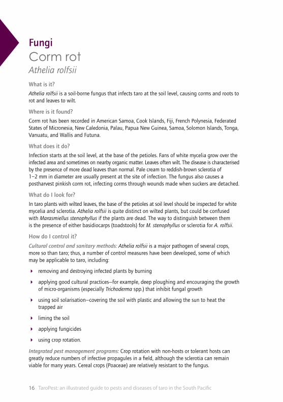
FungiCorm rotAthelia rolfsiiWhat is it?
Athelia rolfsii is a soil-borne fungus that infects taro at the soil level, causing corms and roots to rot and leaves to wilt.
Where is it found?
Corm rot has been recorded in American Samoa, Cook Islands, Fiji, French Polynesia, Federated States of Micronesia, New Caledonia, Palau, Papua New Guinea, Samoa, Solomon Islands, Tonga, Vanuatu, and Wallis and Futuna.
What does it do?
Infection starts at the soil level, at the base of the petioles. Fans of white mycelia grow over the infected area and sometimes on nearby organic matter. Leaves often wilt. The disease is characterised by the presence of more dead leaves than normal. Pale cream to reddish-brown sclerotia of 1–2 mm in diameter are usually present at the site of infection. The fungus also causes a postharvest pinkish corm rot, infecting corms through wounds made when suckers are detached.
What do I look for?
In taro plants with wilted leaves, the base of the petioles at soil level should be inspected for white mycelia and sclerotia. Athelia rolfsii is quite distinct on wilted plants, but could be confused with Marasmiellus stenophyllus if the plants are dead. The way to distinguish between them is the presence of either basidiocarps (toadstools) for M. stenophyllus or sclerotia for A. rolfsii.
How do I control it?
Cultural control and sanitary methods: Athelia rolfsii is a major pathogen of several crops, more so than taro; thus, a number of control measures have been developed, some of which may be applicable to taro, including:
4 removing and destroying infected plants by burning
4 applying good cultural practices—for example, deep ploughing and encouraging the growth of micro-organisms (especially Trichoderma spp.) that inhibit fungal growth
4 using soil solarisation—covering the soil with plastic and allowing the sun to heat the trapped air
4 liming the soil
4 applying fungicides
4 using crop rotation.
Integrated pest management programs: Crop rotation with non-hosts or tolerant hosts can greatly reduce numbers of infective propagules in a field, although the sclerotia can remain viable for many years. Cereal crops (Poaceae) are relatively resistant to the fungus.
Fungi 17
98
76
54
32
110
cm
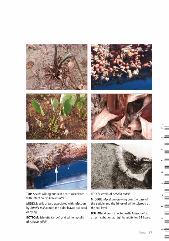
TOP: Sclerotia of Athelia rolfsii.
MIDDLE: Mycelium growing over the base of the petiole and the fringe of white sclerotia at the soil level.
BOTTOM: A corm infected with Athelia rolfsii after incubation at high humidity for 24 hours.
TOP: Severe wilting and leaf death associated with infection by Athelia rolfsii.
MIDDLE: Wilt of taro associated with infection by Athelia rolfsii; note the older leaves are dead or dying.
BOTTOM: Sclerotia (arrow) and white mycelia of Athelia rolfsii.
TaroPest: an illustrated guide to pests and diseases of taro in the South Pacific18
FungiBrown leaf spot (or ghost spot) Cladosporium colocasiaeWhat is it?
Brown leaf spot is a fungal disease of older leaves. It is also called ghost spot because the lesions are often less evident on the opposite surface of the leaf. This leaf spot causes symptoms very similar to those of Neojohnstonia colocasiae (orange leaf spot). In addition, leaf blotch (Pseudocercospora colocasiae) occurs together with Cladosporium colocasiae in Samoa and, on symptoms alone, these are difficult to tell apart.
Where is it found?
Cladosporium colocasiae is widely distributed throughout the Pacific.
What does it do?
Cladosporium colocasiae causes brown leaf spot (or ghost spot) of older leaves—reddish-brown, circular or irregular, diffuse spots or blotches on either leaf surface, sometimes with dark, diffuse centres. The spots are usually less evident on the opposite surface of the leaf. Sometimes the spots are surrounded by a yellow halo or have a dark brown, diffuse border. Spots can be up to 15 mm in diameter, but are usually much smaller when there are many spots on a single leaf.
What do I look for?
Microscopic examination is necessary for identification. Spores can be lifted off the leaf using a scalpel to scrape the surface, or using clear adhesive tape, by pressing a piece gently over the spot and lifting it off the leaf surface. The spores can then be mounted in a drop of water on a microscope slide for identification under a compound microscope. The conidiophores (stalks that bear the spores) are straight or bent, with spores (conidia) that are formed on swellings at the end. The spores are cylindrical to oblong, rounded at the end, often constricted in the middle, with up to three cross walls.
How do I control it?
Phytosanitary measures: Plant quarantine authorities might require certification that consignments of leaves are free from this pathogen when leaves are moved internationally. However, it is not considered to be a pest of ‘potential economic importance’.
Cultural control and sanitary methods: No control is required; however, removal and destruction by burning of infected leaves will reduce inoculum levels.

Fungi 19
98
76
54
32
110
cm
TOP: The spots darken with age and at the margins of the leaf they merge, turn brown and dry out.
MIDDLE: Brown leaf or ghost spots with dark brown, diffuse borders, up to 15 mm in diameter.
BOTTOM: Cladosporium colocasiae conidiophores are straight or bent; spores form on swellings at the ends.
TOP: Brown leaf or ghost spots with dark centres and blackish borders as sporulation occurs and the spots age.
MIDDLE: The pale greenish-yellow spots on the top surface are showing through from spots on the bottom surface of the leaf.
BOTTOM: Yellow–orange ghost spots; spots are variable in colour.

TaroPest: an illustrated guide to pests and diseases of taro in the South Pacific20
FungiSpongy black rotLasiodiplodia theobromaeWhat is it?
Lasiodiplodia theobromae in taro corms causes a postharvest rot that is initially whitish-cream, later becoming blue–black.
Where is it found?
Lasioplodia theobromae has been recorded on taro in Guam, Papua New Guinea, Samoa and Solomon Islands. It has been recorded on other host plants in American Samoa, Australia, Cook Islands, Fiji islands, French Polynesia, Federated States of Micronesia, New Caledonia, New Zealand, Niue, Palau, Papua New Guinea, Tonga, Vanuatu, and Wallis and Futuna.
What does it do?
Lasiodiplodia theobromae is frequently isolated in decayed corm tissues behind advancing rots caused by Phytophthora colocasiae and Pythium splendens. Even in the absence of other fungi, it enters corms through wounds made during harvest and causes complete decay in 10–14 days. Lasioplodia theobromae causes a spongy rot, which occasionally becomes dry and powdery, with an indistinct margin between healthy and diseased tissue.
What do I look for?
Spongy black rot can be detected by cutting the corm to reveal the black interior; it has a strong, sour smell and black spore masses form on the corm surface.
How do I control it?
Chemical control: Dipping corms in bleach (1% sodium hypochlorite) for 2 minutes before storing in polyethylene bags is effective in controlling this fungus.
Traditional practices: The traditional practice of the Sikaiana Island people (of Polynesian descent) is to store taro for up to 4 weeks buried in pits situated in shaded, well-drained soil.

Fungi 21
98
76
54
32
110
cm
TOP: Taro corm rot caused by Lasiodiplodia theobromae; the rot is initially white, later turning black and spongy.
MIDDLE: Lasiodiplodia theobromae in a taro corm.
BOTTOM: Spore-containing structures (pycnidia) form on the corm surface as the rot proceeds.
TOP: Lasiodiplodia theobromae in taro corms, showing advanced decay.
MIDDLE: Lasiodiplodia theobromae in taro corms.
BOTTOM: Initial rot is caused by Pythium sp., a dry and crumbly white rot, which is colonised by Lasiodiplodia theobromae, becoming purple.
TaroPest: an illustrated guide to pests and diseases of taro in the South Pacific22
FungiWhite spot of taroLeptosphaerulina trifoliiWhat is it?
Leptosphaerulina trifolii produces yellow spots on taro leaves. These later turn white. Spots sometimes merge and show ‘shot hole’ symptoms as the centres fall out.
Where is it found?
Leptosphaerulina trifolii has been recorded on taro in American Samoa, Papua New Guinea, Samoa, Solomon Islands and Tuvalu. It is found on other hosts in Fiji, Marshall Islands, Niue, Tonga and Vanuatu.
What does it do?
Infections are initially visible as small, yellow–green spots on the upper leaf. As spots mature, they become edged by a thin (1 mm), reddish-brown border and surrounded by an intense yellow halo, 1–2 mm wide. Mature lesions are 2–5 mm in diameter with paper-white centres. Small, black fruiting bodies can be seen on close observation against the white tissue of mature lesions. Centres often fall out, creating a ‘shot hole’ appearance. In severe infections, spots may coalesce, and the leaves look tattered.
What do I look for?
White spot is visible as small, white spots with yellow haloes on the upper leaf surface. A hand lens will reveal the small, brown-to-black fruiting bodies (pseudothecia). Fruiting bodies of this fungus are easily extracted from lesions. Pseudothecia are relatively small (approximately 125 µm in diameter), asci are usually sac-like (saccate), and most multicelled ascospores have longitudinal and cross septae (dictyospores).
How do I control it?
Control measures are usually not necessary. The impact of this disease is very low. In American Samoa, only a few plants have been seen to be severely infected (25–50% leaf area) and usually the disease is unremarkable.
Fungi 23
98
76
54
32
110
cm
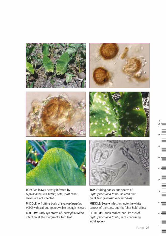
TOP: Fruiting bodies and spores of Leptosphaerulina trifolii isolated from giant taro (Alocasia macrorrhizos).
MIDDLE: Severe infection; note the white centres of the spots and the ‘shot hole’ effect.
BOTTOM: Double-walled, sac-like asci of Leptosphaerulina trifolii, each containing eight spores.
TOP: Two leaves heavily infected by Leptosphaerulina trifolii; note, most other leaves are not infected.
MIDDLE: A fruiting body of Leptosphaerulina trifolii with asci and spores visible through its wall.
BOTTOM: Early symptoms of Leptosphaerulina infection at the margin of a taro leaf.

TaroPest: an illustrated guide to pests and diseases of taro in the South Pacific24
FungiCorm and leaf spot Marasmiellus stenophyllusWhat is it?
Marasmiellus stenophyllus infects taro at the base of the plant, destroying leaves, corms and roots, and commonly producing toadstools on the dying parts.
Where is it found?
Marasmiellus stenophyllus has been recorded on taro in American Samoa, French Polynesia, and Wallis and Futuna, and on other hosts in Fiji.
What does it do?
Corm and leaf spot caused by Marasmiellus stenophyllus leads to leaf collapse due to the development of large brown rots at the base of the plant associated with white fungal growth. The leaves are often stuck together by the fungal threads (mycelia). Toadstools form in large numbers on the withered leaves at soil level. The fungus grows over the roots and kills them, and soil particles become fastened to the roots in the process. Infection with M. stenophyllus can kill the plant, which appears desiccated or mummified. Corms become inedible and, even at an early stage of decay, may be unsightly with mycelium growth causing small ‘pocket’ rots. However, the incidence of infection is low.
What do I look for?
If plants have wilted or are growing slowly compared with others, check for toadstools growing from the dead or dying petioles. The roots will appear dirty with soil, and debris will be adhering to them in clumps that cannot be removed even after gentle washing. Marasmiellus stenophyllus is quite distinct on taro, but could be confused with Athelia rolfsii on completely dead plants. The way to distinguish between the two is to look for the presence of basidiocarps (toadstools) for M. stenophyllus and sclerotia for A. rolfsii.
How do I control it?
Cultural control and sanitary methods: The removal and destruction of infected plants by burning is helpful in controlling the fungus.

Fungi 25
98
76
54
32
110
cm
TOP: Late-stage infection of taro by Marasmiellus stenophyllus, showing matted leaves and mummified corm.
MIDDLE: Toadstool of Marasmiellus stenophyllus.
BOTTOM: ‘Dirty roots’; the mycelia have grown over the roots and corm, trapping soil.
TOP: Toadstools of Marasmiellus stenophyllus growing from decayed leaves at the base of taro.
MIDDLE: Plants with many dead leaves, killed by Marasmiellus stenophyllus.
BOTTOM: Mycelium of Marasmiellus stenophyllus growing over taro roots and corm, trapping soil particles and leading to a ‘dirty’ appearance.

TaroPest: an illustrated guide to pests and diseases of taro in the South Pacific26
FungiOrange leaf spotNeojohnstonia colocasiaeWhat is it?
Orange leaf spot is a fungal disease of older leaves causing symptoms very similar to those of Cladosporium colocasiae (brown leaf spot).
Where is it found?
Neojohnstonia colocasiae has been recorded on taro in American Samoa, Fiji, Federated States of Micronesia, Palau, Papua New Guinea, Samoa, Solomon Islands, Tuvalu, Vanuatu, and Wallis and Futuna.
What does it do?
Neojohnstonia colocasiae causes yellowish-brown, circular or irregular blotches on either leaf surface. These become darker with the onset of sporulation. Spots are sometimes surrounded by a yellow halo or have a brown border. They can be up to 15 mm in diameter, but tend to be smaller when there are many spots on a single leaf.
What do I look for?
On the leaves, the spots can be seen with the naked eye, but microscopic examination is necessary for identification. Spores can be lifted off the leaf using a scalpel to scrape the surface, or using clear adhesive tape by pressing a piece gently over the spot and lifting it off the leaf surface. Spores can then be mounted in a drop of water on a microscope slide under a cover slip. The fungus can also be examined by culturing and inducing sporulation on artificial media. The conidiophores (stalks that bear the spores) are found mainly on the lower leaf surface—they are branched with single round spores (with a cross wall) connected to the conidiophores by a short, thin stalk.
How do I control it?
Phytosanitary measures: Plant quarantine authorities might require certification that consignments of leaves are free from this pathogen when leaves are moved internationally. However, it is not considered to be a pest of ‘potential economic importance’.
Cultural control and sanitary methods: No control measures are recommended; however, removal and destruction by burning of infected leaves will reduce inoculum levels.
Fungi 27
98
76
54
32
110
cm
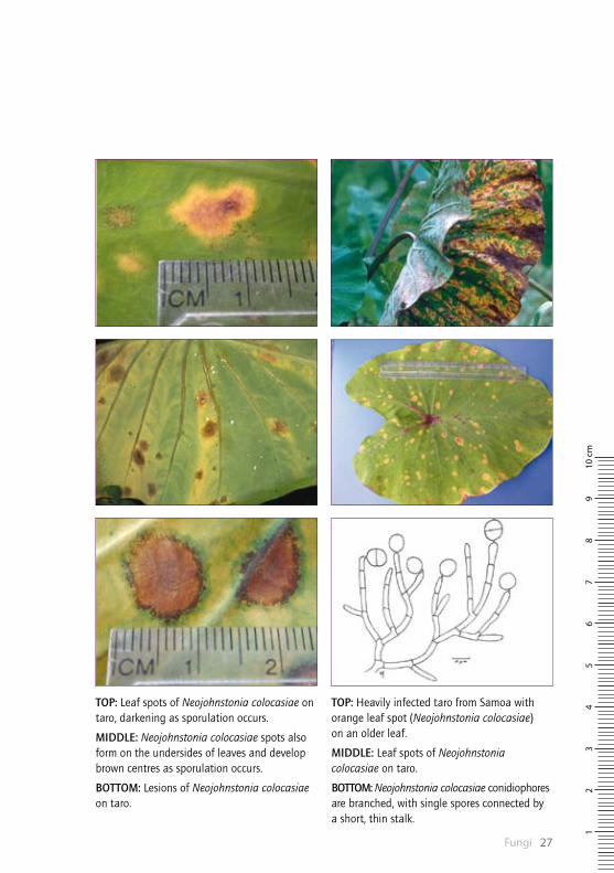
TOP: Heavily infected taro from Samoa with orange leaf spot (Neojohnstonia colocasiae) on an older leaf.
MIDDLE: Leaf spots of Neojohnstonia colocasiae on taro.
BOTTOM: Neojohnstonia colocasiae conidiophores are branched, with single spores connected by a short, thin stalk.
TOP: Leaf spots of Neojohnstonia colocasiae on taro, darkening as sporulation occurs.
MIDDLE: Neojohnstonia colocasiae spots also form on the undersides of leaves and develop brown centres as sporulation occurs.
BOTTOM: Lesions of Neojohnstonia colocasiae on taro.

TaroPest: an illustrated guide to pests and diseases of taro in the South Pacific28
FungiShot holePhoma spp.What is it?
Phoma spp. (Phoma sp. and Phoma colocasiae) produce relatively large lesions on the leaf. As the spots age, their centres fall out, giving the ‘shot hole’ effect.
Where is it found?
Phoma colocasiae has been recorded in Palau and Samoa. The Pacific taro fungus, Phoma sp., has been recorded in American Samoa, Cook Islands, Federated States of Micronesia, Fiji islands, French Polynesia, Marshall Islands, Niue, Palau, Papua New Guinea, Samoa, Solomon Islands, Tokelau, Tonga and Vanuatu.
What does it do?
The first symptoms of a Phoma infestation are small, round, brown spots on the second or third leaves. As the spots enlarge to 2 cm in diameter, the brown centres fall out, resulting in the typical ‘shot hole’ symptom. The holes have a narrow, brown margin, which is surrounded by an intense yellow halo. The holes may merge, so that large areas of the leaf are destroyed. This leads to premature leaf death.
What do I look for?
The leaves are the only part of the plant that show symptoms, so look for the characteristic shot holes. Careful inspection of the leaves and microscopic examination of the pycnidia and spores are necessary. Phoma spp. can be mistaken for taro leaf blight (Phytophthora colocasiae), particularly when infection levels are high. The difference is that Phytophthora colocasiae lesions are often surrounded by a white zone of spores and exude droplets that dry as dark pellets.
How do I control it?
There is no evidence that the disease warrants control. Fungicides may control the disease; however, they cannot be recommended at present, since recent studies have not established that the disease reduces corm yields.

Fungi 29
98
76
54
32
110
cm
TOP: Phoma sp. infection on a taro leaf.
MIDDLE: The centres of Phoma sp. lesions fall out, giving a characteristic ‘shot hole’ effect.
BOTTOM: Phoma sp. conidia are cylindrical to oval in shape.
TOP: Close-up of a ‘shot hole’ lesion caused by Phoma sp.
MIDDLE: Oval spots caused by Phoma sp. are up to 30 mm long, brown with yellow borders, and sometimes merge.
BOTTOM: Some of the lesions caused by Phoma sp. join together at the leaf margins.

TaroPest: an illustrated guide to pests and diseases of taro in the South Pacific30
FungiTaro leaf blight Phytophthora colocasiaeWhat is it?Taro leaf blight is a major disease of taro. In Pacific island countries, taro leaf blight has prevented farmers from growing taro successfully.
Where is it found?Phytophthora colocasiae has been recorded in American Samoa, Federated States of Micronesia, Guam, Northern Mariana Islands, Palau, Papua New Guinea, Samoa and Solomon Islands.
What does it do?A small, circular speck, brown on the upper surface of the leaf and water-soaked below, is the first sign of the disease. Infections often begin on the lobes and sides of the leaf where water collects. The spots enlarge, become irregular in shape, and are dark brown with yellow margins. Initial spots give rise to secondary infections and, soon afterwards, the leaf blade collapses and dies. Spores are produced at night and can be seen around the spots in the morning. Clear, yellow-to-red droplets ooze from the spots and develop into dark brown, hard pellets as they dry. This is a characteristic of the disease. Spores may be trapped inside the pellets.
Usually, petioles are not attacked, but instead collapse as the leaf blade is destroyed. However, in American Samoa and Samoa, petiole infection is common as the taro varieties are very susceptible to the disease. The fungus can also cause a postharvest corm rot that is difficult to detect unless corms are cut open. The rots are light brown and hard.
What do I look for?On the leaves, spots caused by Phytophthora colocasiae can be seen with the naked eye. Microscopic examination of the spore masses is required to identify the spores. Corms can carry spores on the surface (undetectable) and mycelium in postharvest rots. Corms need to be cut open to detect the rots.
How do I control it?Phytosanitary measures: Strict quarantine measures must be observed to prevent the spread of the disease to countries where it does not currently occur. Any movement of planting material between countries should be limited to sterile plantlets growing in a tissue culture medium, and they should be indexed for viruses.
Cultural control and sanitary methods: Selection of sites away from already infected crops, regular removal of diseased leaves and wide spacings between plants are recommended.
Chemical control: Both protectant and systemic fungicides are reported to give control for this fungus. In Samoa, studies done after the outbreak of taro leaf blight recommended phosphoric acid alternated with mancozeb. Corm rots are best controlled by dipping corms in bleach (1% sodium hypochlorite) and storing them in polyethylene bags.
Resistant varieties: Varieties with durable resistance to Phytophthora colocasiae are known from the Philippines and the Federated States of Micronesia and Palau. These have been used successfully in a breeding program in Samoa. Breeding has also been done in Hawaii and Papua New Guinea. Some of the releases from these breeding programs have been pathogen-indexed and are conserved at the Secretariat of the Pacific Community (SPC) Regional Germplasm Centre, Fiji.

Fungi 31
98
76
54
32
110
cm
TOP: Droplets of leaf sap exude from the margin of the lesion caused by Phytophthora colocasiae; this characteristic symptom is seen early in the morning. Later the droplets dry as hard pellets.
MIDDLE & BOTTOM: Initial Phytophthora colocasiae spots have given rise to secondary infections; gradually, the entire leaf blade is succumbing to the disease.
TOP: The lesion caused by Phytophthora colocasiae is beginning to fall out and the black areas are probably signs of secondary fungal infection.
MIDDLE & BOTTOM: Droplets of leaf sap exuded from the margin of the lesion caused by Phytophthora colocasiae.
TaroPest: an illustrated guide to pests and diseases of taro in the South Pacific32
FungiLeaf blotch Pseudocercospora colocasiaeWhat is it?
Leaf blotch is a fungal disease, mostly affecting older leaves. The symptoms are similar to those caused by Neojohnstonia colocasiae (orange leaf spot) and Cladosporium colocasiae (brown leaf spot).
Where is it found?
Pseudocercospora colocasiae has been recorded in American Samoa, Fiji, French Polynesia, New Caledonia, Samoa, Solomon Islands and Vanuatu.
What does it do?
This fungus has little impact of consequence in taro. It causes blotches with indistinct, circular, yellow-reddish to whitish-green discolouration on the upper surface of the leaf, and black mould growth on the corresponding lower surface. The blotches can be up to 1.5 cm in diameter.
What do I look for?
The leaves should be inspected for the presence of leaf blotches as described above. To distinguish between this leaf blotch and leaf spots caused by other fungi (Neojohnstonia colocasiae and Cladosporium colocasiae), the spores should be inspected under a microscope for identification. Spores can be lifted off the leaf using a scalpel to scrape the surface, or using clear adhesive tape, by pressing a piece gently over the spot and lifting it off the leaf surface. The spores can then be mounted in a drop of water on a microscope slide under a cover slip, and viewed under a compound microscope for identification.
The spore-bearing stalks (conidiophores) are dark, unbranched and arise in bundles from the lesions. The spores are pale olive, club-shaped, with a broadly rounded apex and tapering to an inconspicuous scar, almost smooth, mostly four-celled, and solitary.
How do I control it?
This disease is not considered to be of economic importance; therefore, no control measures are necessary. It is a disease of older leaves.
Fungi 33
98
76
54
32
110
cm
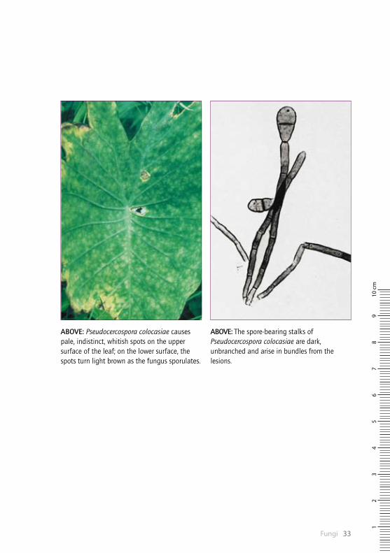
ABOVE: The spore-bearing stalks of Pseudocercospora colocasiae are dark, unbranched and arise in bundles from the lesions.
ABOVE: Pseudocercospora colocasiae causes pale, indistinct, whitish spots on the upper surface of the leaf; on the lower surface, the spots turn light brown as the fungus sporulates.

TaroPest: an illustrated guide to pests and diseases of taro in the South Pacific34
FungiCorm soft rotPythium spp.What is it?
A number of Pythium species have been isolated from the roots and corms of wilted plants in dry and wetland taro.
Where is it found?
Various Pythium species occur throughout the Pacific—see the TaroPest CD for species distribution records.
What does it do?
When infected, the whole plant becomes stunted—the leaf stalks are shortened, the leaf blades become curled or crinkled and, instead of being a deep, healthy green, are yellowish and spotted. The corms show a rot of varying colour from whitish-yellow, through shades of grey and blue, to dark purple. Usually, rot starts at the base of the corm and progresses upward until the whole corm is affected. Occasionally, the disease starts at the side of the corm, 5–7 cm above the base. The skin of a diseased corm becomes softened, usually remaining intact until complete disintegration of the interior of the corm has taken place; then the skin also disintegrates. When the corm is cut open, a sharp line of demarcation can be seen between healthy and diseased tissue.
What do I look for?
Generally, the rot is evident on the corms as it develops from the base. However, if it is an early infection, lesions on the surface of the corm may be observed—if these are found, the corm should be cut open to see what lies beneath. Although few other species of fungi cause rots in the field, there are others that cause postharvest rots. Therefore, it is necessary to isolate the pathogen and identify it by microscopy.
How do I control it?
Cultural control and sanitary methods: Only healthy material that is free from rot should be planted. Removal of diseased plant material from the field at harvest can reduce inoculum levels. Ploughing and drying of wetland taro fields are recommended. Crop rotation with non-host crop plants is also useful. Experiments by taro growers in Halawa (Molokai) gave strong indications that taro rot could be controlled by drying and ploughing the patches, and by applying either lime or coral sand some time before replanting with taro. Calcium has also been implicated in the low incidence of Pythium rot on atolls.
Host plant resistance: In Samoa, the following varieties have shown resistance: Tusi Tusi, Talo Vale, Pute Mu and Pula Sama Sama. Hawaiian taro varieties Pa’lehua, Maui Lehua, Pa’akala and Pauakea are all considered to be resistant to Pythium rot.
Chemical control: Investigations into preventing postharvest rots caused by Pythium splendens (often in a complex with other fungi) in Solomon Islands found bleach (1% sodium hypochlorite) as a corm dip helped to reduce damage. Various fungicides could also reduce rots in early days of storage.
Traditional practices: In Solomon Islands, storage in leaf-lined, shallow soil-pits has been shown to reduce damage.

Fungi 35
98
76
54
32
110
cm
TOP: Root decay and corm soft rot of young taro plants affected by Pythium sp.
MIDDLE: Corm soft rot of young taro plants; note the fringe of healthy roots at the top of the corm.
BOTTOM: Taro plants showing signs of infection with Pythium sp.
TOP: Pythium sp.; note the much reduced number of side or feeder roots and the decay of those present.
MIDDLE: Root decay and corm soft rot of young taro plants affected by Pythium sp.
BOTTOM: Taro corm soft rot caused by Pythium sp.
TaroPest: an illustrated guide to pests and diseases of taro in the South Pacific36
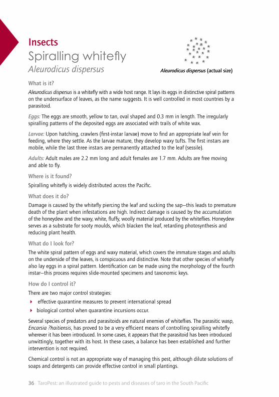
Aleurodicus dispersus (actual size)
InsectsSpiralling whitefly Aleurodicus dispersusWhat is it?
Aleurodicus dispersus is a whitefly with a wide host range. It lays its eggs in distinctive spiral patterns on the undersurface of leaves, as the name suggests. It is well controlled in most countries by a parasitoid.
Eggs: The eggs are smooth, yellow to tan, oval shaped and 0.3 mm in length. The irregularly spiralling patterns of the deposited eggs are associated with trails of white wax.
Larvae: Upon hatching, crawlers (first-instar larvae) move to find an appropriate leaf vein for feeding, where they settle. As the larvae mature, they develop waxy tufts. The first instars are mobile, while the last three instars are permanently attached to the leaf (sessile).
Adults: Adult males are 2.2 mm long and adult females are 1.7 mm. Adults are free moving and able to fly.
Where is it found?
Spiralling whitefly is widely distributed across the Pacific.
What does it do?
Damage is caused by the whitefly piercing the leaf and sucking the sap—this leads to premature death of the plant when infestations are high. Indirect damage is caused by the accumulation of the honeydew and the waxy, white, fluffy, woolly material produced by the whiteflies. Honeydew serves as a substrate for sooty moulds, which blacken the leaf, retarding photosynthesis and reducing plant health.
What do I look for?
The white spiral pattern of eggs and waxy material, which covers the immature stages and adults on the underside of the leaves, is conspicuous and distinctive. Note that other species of whitefly also lay eggs in a spiral pattern. Identification can be made using the morphology of the fourth instar—this process requires slide-mounted specimens and taxonomic keys.
How do I control it?
There are two major control strategies:
4 effective quarantine measures to prevent international spread
4 biological control when quarantine incursions occur.
Several species of predators and parasitoids are natural enemies of whiteflies. The parasitic wasp, Encarsia ?haitiensis, has proved to be a very efficient means of controlling spiralling whitefly wherever it has been introduced. In some cases, it appears that the parasitoid has been introduced unwittingly, together with its host. In these cases, a balance has been established and further intervention is not required.
Chemical control is not an appropriate way of managing this pest, although dilute solutions of soaps and detergents can provide effective control in small plantings.

Insects 37
98
76
54
32
110
cm
TOP & MIDDLE: Spiralling whiteflies on the underside of a taro leaf.
BOTTOM: Spiralling pattern, adult whiteflies and white, waxy filaments.
TOP: Spiral pattern of Aleurodicus dispersus.
BOTTOM: White wax of whiteflies.

TaroPest: an illustrated guide to pests and diseases of taro in the South Pacific38
Aphis gossypii (actual size)
InsectsAphids Aphis gossypiiWhat are they?
Aphids are small, pear-shaped insects with soft, fragile bodies. Often present in large numbers, they pierce leaves to obtain sap.
Eggs: In temperate regions, aphids overwinter as eggs. In tropical regions, reproduction does not involve mating and egg laying.
Nymphs: Immature aphids are called nymphs—they look much like adults, but are smaller and wingless. Females give birth to live female nymphs, which are oval and 0.1 mm in length. There are four nymphal stages, and development is complete in 4–10 days. The fourth-stage nymphs are approximately 1 mm in length.
Adults: Adult females give birth to approximately 50 nymphs. Adults range from 0.9 mm to 2.5 mm in length; they may be winged or wingless. Wingless adult aphids vary in colour from pale green–yellow to dark green. Winged forms are generally darker: the head and thorax are black and the abdomen is yellowish-green, with dark brown veins.
Where are they found?
Aphids have a wide distribution throughout the Pacific.
What do they do?
If aphids are present in high numbers and rainfall is low, leaves senesce faster than normal. In severe cases, the plants wilt and may become stunted. Indirect damage is caused by the accumulation of honeydew produced by the aphids. Honeydew serves as a substrate for sooty moulds, which blacken the leaves, reducing photosynthesis and plant vigour. Aphids are vectors of Dasheen mosaic potyvirus.
What do I look for?
Look for infested leaves, which may appear wilted and curled downwards. The older leaves become distorted and covered in honeydew, which encourages the growth of sooty mould. Ants sometimes tend aphids and feed on the honeydew the aphids excrete. At the same time, they protect the aphids from natural enemies. The presence of ants often indicates that aphids are also present (although ants are also attracted to the honeydew of whiteflies and planthoppers).
How do I control them?
Cultural control: If taro plants are heavily infested, avoid planting new crops downwind—aphids are not strong fliers and are readily blown in the wind so the new planting is likely to become contaminated. It is also good practice to destroy leaves heavily infested with aphids.
Chemical control: It is rarely necessary to use pesticides to control aphids on taro; populations are normally well controlled by predators—ladybird beetles, syrphids and lacewings, in particular. If pesticides are used, these predators will be killed. If it is necessary to use pesticides, a local agriculture extension agent should be asked for advice. Horticultural oils and insecticidal soaps may be considered as alternatives to synthetic pesticides. If ants are present, the best solution may be to destroy the ant colony so that predators and parasites can go about their beneficial acts unhindered.
Insects 39
98
76
54
32
110
cm
Aphis gossypii (actual size)
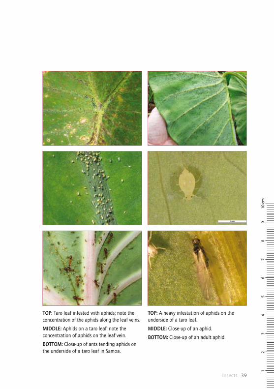
TOP: A heavy infestation of aphids on the underside of a taro leaf.
MIDDLE: Close-up of an aphid.
BOTTOM: Close-up of an adult aphid.
TOP: Taro leaf infested with aphids; note the concentration of the aphids along the leaf veins.
MIDDLE: Aphids on a taro leaf; note the concentration of aphids on the leaf vein.
BOTTOM: Close-up of ants tending aphids on the underside of a taro leaf in Samoa.
TaroPest: an illustrated guide to pests and diseases of taro in the South Pacific40
Bemesia tabaci (actual size)
InsectsTobacco whitefly Bemesia tabaciWhat is it?
The tobacco (or sweet potato) whitefly is commonly seen on taro, but it is not considered a pest. Infestations are rarely large enough to cause feeding damage or sooty mould growth, and this whitefly is not known to transmit viruses of taro.
Eggs: Eggs are laid in groups on the undersurface of the leaf; they are attached to the leaf on a stalk. Eggs are white at first, later becoming brown before hatching. They hatch in 5–7 days.
Nymphs: After hatching, the crawlers (first nymph stage) move a very short distance from the egg. The subsequent three nymphal stages do not move. Nymphs are creamy to light green, flat and oval shaped, and look like small scale insects. This stage lasts 2–4 weeks. The fourth nymph stage (‘pupa’) is used to identify the species.
Adults: Adults are moth-like, small (approximately 0.8 mm), with pale yellow–white bodies and two pairs of wings covered with white, waxy powder. At rest, the wings are held tent-like over the body. Adults usually emerge in the morning and copulate a few hours later. Oviposition (egg-laying) occurs during the first 8 days after mating, and females live for 10–15 days.
Where is it found?
Tobacco whitefly is widely distributed across the Pacific.
What does it do?
The adults and nymphs suck the sap from leaves—this can cause yellowing, wilting and early death. However, symptoms this severe rarely occur on taro as numbers of whitefly are usually insufficient to cause direct damage, although they may increase wilting in times of drought. Indirect damage, that is, the build-up of sooty moulds on honeydew deposits, is not frequently seen.
What do I look for?
Whiteflies are usually detected by examining the undersides of leaves and searching for the tiny yellow–cream, scale-like instars or nymphs. The nymphs also occur occasionally on the upper surfaces of the leaves and can range from being scattered widely to forming dense clusters. Shaking the plant may disturb the small, white adults, which fly out and quickly resettle. Since many whiteflies are similar in appearance, an accurate determination to species usually requires an expert in whitefly taxonomy.
How do I control it?
As Bemesia tabaci is not considered a major pest of taro in Pacific island countries, no control measures specific to that crop are recommended.
Insects 41
98
76
54
32
110
cm
Bemesia tabaci (actual size)
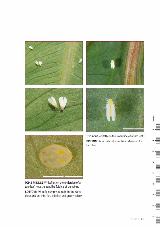
TOP: Adult whitefly on the underside of a taro leaf.
BOTTOM: Adult whitefly on the underside of a taro leaf.
TOP & MIDDLE: Whiteflies on the underside of a taro leaf; note the tent-like folding of the wings.
BOTTOM: Whitefly nymphs remain in the same place and are thin, flat, elliptical and green–yellow.

TaroPest: an illustrated guide to pests and diseases of taro in the South Pacific42
InsectsHornworm Hippotion celerioWhat is it?
Hornworm (hawk moth) caterpillars consume large amounts of leaf, causing conspicuous damage. Hawk moths are the adult stage of the hornworm.
Eggs: Eggs are laid singly on both the upper and lower surface of the leaves as well as on the petioles. Eggs are variable in size and shape, from nearly spherical (1 mm) to oval, and are clear to bluish-green. Before emergence, they become greenish-yellow.
Larvae: The first-instar larvae are approximately 4 mm long with a reddish horn on the posterior end. As they age, they change from pale yellow to glossy green. In the second instar, two spots appear on the first and second abdominal segments, resembling eyes. In the third instar, a yellow, dorso-lateral line appears running from thoracic segment 3 to the base of the horn and the eye spots assume their final colouration. Finally, the larvae grow to 80–90 mm, becoming mid to dark brown, although a few remain green, before pupation.
Adults: Hawk moths have a wing span of 40–90 mm. They are streamlined and robust in flight, with a conspicuous head and large eyes.
Where is it found?
Hippotion celerio is widespread throughout the Pacific region.
What does it do?
Symptoms are very noticeable—infested plants have large areas of leaf missing and the leaf appears ragged. The caterpillars are voracious feeders. Hornworms can defoliate taro when numbers are high.
What do I look for?
The larvae can be found on the leaves during the day, often on the underside. The leaves should be inspected for damage and larval stages, and any specimens compared to photographs. If there is any doubt, the caterpillars can be raised to maturity and the adult stage identified. It is advisable to raise several caterpillars, as some may be parasitised. .
How do I control it?
Physical control: The larvae are large and relatively easily seen; they can be picked off the plants by hand. In small taro plantings, this is the best means of control.
Chemical control: Applications of chemical sprays may help control populations of hornworm. Present recommendations in Pacific island countries include: indoxacarb (e.g. Steward), spinosad (e.g. Success), Bt (e.g. Delfin, Thuricide, Dipel) and imidacloprid (e.g. Confidor, Mustang). However, it is best to consult a local agricultural extension agent for up-to-date information on pesticides and methods of application.
Hippotion celerio, late- instar larvae (actual size)
Hippotion celerio , adult (actual size)

Insects 43
98
76
54
32
110
cm
TOP: Feeding by hornworm larvae, causing severe defoliation to a taro plant.
MIDDLE: Hornworm larva; note the variation in colour of the caterpillars.
BOTTOM: Close-up of an adult hornworm (hawk moth).
TOP: Young hornworm larvae on a taro leaf.
MIDDLE: Hornworm larva feeding on a taro leaf; note extensive damage to the leaf tissue.
BOTTOM: Hornworm larva feeding on a taro leaf; note the ragged margin on the leaf.

TaroPest: an illustrated guide to pests and diseases of taro in the South Pacific44
Mealybug adult (actual size)
InsectsMealybugs Family PseudococcidaeWhat are they?
Mealybugs belong to the insect group that is commonly known as scale insects (family Pseudococcidae). They have soft, segmented, oval bodies, but without an outer shell. They are covered with a white, waxy powder and may have long or short filaments projecting from the margin; some have none. Although they have legs, most mealybugs remain in the same place throughout their life.
Eggs: Some mealybugs lay eggs (often many hundred) within an egg sac made from waxy secretions that encloses most of the female; others produce hundreds of living young.
Larvae: The young are referred to as crawlers, nymphs or larvae. As crawlers, males and females look alike. After 10–14 days, and after several stages, the crawlers pupate for 7 days.
Adults: Males emerge as tiny (1 mm) winged insects; they are seldom seen, short-lived, and do not feed. Females live for approximately 30 days. Most mealybugs can reproduce sexually, but reproduction without mating is common. There are a number of generations per year, depending on temperature, which often overlap.
Where are they found?
Many species of mealybugs have been recorded from the Pacific islands.
What do they do?
Mealybugs have a long feeding tube that is used to pierce plant parts and suck the sap—in doing so, they cause a variety of symptoms. Direct feeding results in distorted foliage, yellowing, stunting and wilting; indirectly, mealybugs cause a build-up of sooty mould fungi, which grows on the honeydew excreted as they feed. They also transmit viruses. In these ways, mealybugs are similar to aphids. On taro, mealybugs rarely harm the plants or promote sooty mould growth. However, some transmit taro badnavirus (TaBV), also known as taro bacilliform badnavirus.
What do I look for?
On taro, mealybugs occur on the undersurface of the leaves, on and between the petioles, and on the roots and corms. On the roots, they occur as cotton-like masses, containing males and females, which are sometimes difficult to see clearly with the naked eye. Mealybugs can be detected using a physical examination of plant parts for the insects, the white fluffy wax and the presence of sooty mould growing on excreted honeydew.
How do I control them?
Mealybugs rarely cause direct damage to taro from their feeding or indirect damage by promoting the growth of sooty moulds; numbers are rarely sufficient. If control is required, the best course of action is to check if ants are present and take action against them, so that the activities of natural enemies are not curtailed. If this is not sufficient, then a spray of horticultural oil or soap should be considered.
Insects 45
98
76
54
32
110
cm
Mealybug adult (actual size)
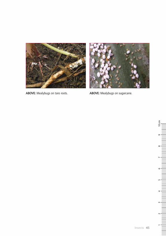
ABOVE: Mealybugs on sugarcane.ABOVE: Mealybugs on taro roots.
TaroPest: an illustrated guide to pests and diseases of taro in the South Pacific46
Papuana spp. (actual size)
InsectsTaro beetles Papuana spp.What are they?Taro beetles are shiny black beetles, measuring approximately 25 mm long and 12 mm wide, which feed on taro corms. The males have a horn on their head and a tubercle just behind the head. The females sometimes have a small horn and a short tubercle. Newly emerged beetles are brown in colour, but turn black as they get older.
Where are they found?Taro beetles are native and widespread throughout Papua New Guinea, and some species are also present in Vanuatu, Solomon Islands, Kiribati and Fiji.
What do they do?
The adult beetle feeds on underground taro corms, creating tunnels while feeding. The impact of feeding is considerable as export markets do not tolerate any damage and more than 15% damage makes the crop unacceptable for local markets. Damage may be such that the corms cannot be used for home consumption or livestock feed. Above ground, symptoms vary with the age of the plants: young plants may be killed as the beetle invades the shoot, while older plants grow more slowly and a few or all of the leaves will wilt.
What do I look for?There are three methods of early detection in an area or incursion:
4 dig up taro plants that are wilting or looking weak and examine them for taro beetle damage symptoms
4 use a light trap at night, particularly on moonless and rainy nights, to catch the beetle
4 sample other plant species and materials for the beetle: banana, sugarcane, rotting logs, compost heaps, sawdust, grassland where Paspalum spp. and Brachiaria mutica (para grass) are dominant, especially along river banks.
How do I control them?
Strict quarantine measures must be observed to prevent the spread of taro beetles into new areas within countries where they already occur and into countries where they do not occur. Planting material, soil, taro and alternative hosts of the beetle must not be moved from infested areas to uninfested areas. A combination of measures should be used to manage populations of taro beetles:
4 cultural control—crop rotation, clean planting material (i.e. free from soil) and destruction of breeding sites near farms
4 chemical control—imidacloprid, bifenthrin
4 biological control—Metarhizium anisopliae
4 phytosanitary procedures.
New chemicals and the efficacy of Metarhizium anisopliae are being researched in Fiji and Papua New Guinea. No single method is effective; therefore, an integrated crop management approach is recommended, except in large commercial taro production systems where corms are produced for the urban or export market, and chemical control can be effective and economic.

Insects 47
98
76
54
32
110
cm
Papuana spp. (actual size)
TOP: Damage to a young taro plant caused by taro beetles.
MIDDLE: Male Papuana woodlarkiana beetle.
BOTTOM: Taro beetle larvae, commonly found in the soil around damaged plants.
TOP: Damage to a taro corm by Papuana uninodis.
MIDDLE: Damage to a taro corm by taro beetles; note the tunnelling in the corm.
BOTTOM: Damage to a taro corm by Papuana uninodis.

TaroPest: an illustrated guide to pests and diseases of taro in the South Pacific48
Patchiella reaumuri (actual size)
InsectsTaro root aphid Patchiella reaumuriWhat is it?
The taro root aphid infests the roots and, when populations are high, the lower parts of the petioles of taro leaves.
Where is it found?
Within the Pacific region, the aphid is known only from the islands of Hawaii and O’ahu in the State of Hawaii, in the United States. It is also recorded in Europe on Arum spp., where it occurs on the roots, and on Tilia spp. (lime or linden trees), where it causes a gall of the shoot tip.
What does it do?
The taro root aphid feeds on the roots of taro, causing them to rot. Damage to the roots produces stunted plants with yellowish, small leaves. The effect of the aphid is greater on young plants than on mature plants, and worse on all stages during periods of drought.
What do I look for?
Infestations appear as a white mould on the fibrous taro roots. Because the aphid is on the roots, it may not be detected until above-ground symptoms occur. The base of the petioles, corm and roots should be inspected to see if masses of white, cottony, waxy threads are present (these cover the insects). The presence of ants on the soil and lower parts of taro (and sometimes on the leaves when aphid numbers are high) can be an indication that plants are infested with Patchiella reaumuri.
How do I control it?
There is quarantine on transport of planting materials from the island of Hawaii to other Hawaiian islands. It is recommended that where heavy infestations occur, the crop should be harvested and destroyed, and the land ploughed and left fallow for a year or planted with a different crop. Insecticidal soaps (1% active ingredient) have been recommended for the control of infestations of Patchiella reaumuri in the field and as a treatment for planting material before planting. However, because the aphid is in the soil and has a waxy covering, soaps may not be effective for control in the field, and are probably better used to rid planting material of aphids. Tests using hot water at 49°C for 6 minutes, followed by immersion in cold water, have been effective in disinfesting planting material without damage. It is important that planting material is free from the aphid, otherwise yields will be unsatisfactory.

Insects 49
98
76
54
32
110
cm
ABOVE: When taro root aphid populations are high, colonies can be found both on roots and on the petioles, just above the corm.
ABOVE: Signs of taro root aphid infestation appear as white mould on the fibrous taro roots.

TaroPest: an illustrated guide to pests and diseases of taro in the South Pacific50
Spodoptera litura (actual size)
InsectsArmyworm Spodoptera lituraWhat is it?
Spodoptera litura is a pest of Asia and the Pacific. It is usually well controlled biologically, although outbreaks occur occasionally, particularly in the aftermath of cyclones or in newly cleared isolated areas. Spodoptera litura has a wide host range.
Eggs: Eggs are laid in masses and are covered with hair scales from the tip of the abdomen of the female moth. Egg masses usually measure 4–7 mm in diameter and are a cream to golden-brown colour.
Larvae: Larvae are variable in colour: young larvae are pale green, whereas later instars are dark green to brown. Although overall colour is variable, bright yellow stripes along the dorsal surface are characteristic of Spodoptera litura larvae.
Adult: The adult moth has a grey–brown body, which is 15–20 mm long, and a wingspan of 30–38 mm. The forewings are grey to reddish-brown, with a strongly variegated pattern and paler lines along the veins. The hindwings are greyish-white with grey margins.
Where is it found?
Spodoptera litura is widespread throughout the Pacific region.
What does it do?
The early larval stages remain together at first, later radiating out from the egg mass, stripping the interveinal leaf surface and skeletonising the leaves as they advance. Later (solitary) stages eat all parts of the leaf, including the petioles.
What do I look for?
The egg masses are relatively easily seen against the dark green of the leaves. The presence of newly hatched larvae can be detected by the ‘scratch’ marks they make on the leaf surface. Older larvae are night feeders. Armyworms chew large areas of the leaf and, when numerous, can defoliate a crop. In such cases, the larvae migrate in large groups from one field to another in search of food.
How do I control it?
Biological control: The use of natural enemies and applications of Bt pesticide (biologically derived from the bacterium Bacillus thuringiensis) can help reduce Spodoptera litura populations.
Chemical control: Chemical sprays may be needed when biological control of Spodoptera litura is insufficient, but should be used with caution so as not to disrupt the balance established between natural enemies and this pest—otherwise, they may do more harm than good. In support of this caution, in Samoa, more damage has been noted in insecticide-sprayed fields than in unsprayed fields. Only products that are nontoxic to beneficial insects should be considered, such as biologically derived pesticides: spinosad (derived from the soil actinomycete Saccharopolyspora spinosa) and Bt. Neem extracts have been used in India.
Physical control: Removal and destruction of leaves infested with egg masses or young larvae will help reduce populations of Spodoptera litura.

Insects 51
98
76
54
32
110
cm
TOP & MIDDLE: Feeding damage caused by the armyworm, Spodoptera litura.
BOTTOM: The adult moth of the armyworm, Spodoptera litura.
TOP: A recently hatched egg mass of the armyworm, Spodoptera litura.
MIDDLE & BOTTOM: Fourth-instar larvae of the armyworm, Spodoptera litura, showing the variation in colour.

TaroPest: an illustrated guide to pests and diseases of taro in the South Pacific52
Taro planthopper (actual size)
InsectsTaro planthoppers Tarophagus spp.What are they?There are three named species of taro planthoppers (Tarophagus colocasiae, T. persephone and T. prosperpina) and one unnamed species in Pacific island countries. They are important pests, directly damaging plants through their feeding and indirectly through spread of viruses.
Eggs: Eggs are deposited in holes made by female taro planthoppers in the midrib on the underside of the leaf, and in the base of the leaf stalks or petioles.
Nymphs: Young nymphs are creamy white, and the third to fifth nymphal stages are predominantly black with white markings.
Adults: Adult taro planthoppers are 4 mm in length, and black with broad white patches on the back of the thorax and abdomen. Adults are of two types; they have either short wings for most of the year or long wings for the cooler periods as the plant matures and starts to die. Taro planthoppers normally move sideways, but both nymphs and adults hop readily if disturbed.
Where are they found?
Tarophagus spp. have a wide distribution throughout the Pacific.
What do they do?Taro planthoppers feed mostly on taro, although they have been observed on Alocasia spp. and Cyrtosperma spp. Heavy infestations of taro planthopper cause plants to wilt and, in exceptional cases, to die. Sap sucking and the laying of eggs cause sap exudation, which forms small red encrustations on the petioles, particularly at the base. Older leaves are affected by severe infestations during dry weather: the petioles bend down giving the plants a splayed appearance, and the leaves die prematurely. Taro planthoppers transmit Colocasia bobone disease ?rhabdovirus, and possibly a related virus, taro vein chlorosis rhabdovirus. These viruses are associated with the alomae and bobone virus diseases reported from Solomon Islands and Papua New Guinea.
What do I look for?The leaves should be inspected, particularly underneath. Early nymphal stages are minute, almost clear and difficult to see. Later instars are white, whitish brown, brownish and predominantly black with white markings (they get progressively darker as they age). Adults are blackish brown. Taro planthoppers are readily found on the undersurface of the leaves, within leaves that are starting to unfurl and at the base of the younger petioles. Older petioles appear dirty because sap oozes from the punctured cells and dries to form a brown crust.
How do I control them?Biological control: The egg predator Cyrtorhinus fulvus has successfully controlled Tarophagus spp. in many parts of the Pacific, but C. fulvus is unlikely to reduce populations sufficiently to prevent the spread of alomae and bobone virus diseases.
Chemical control: Pesticides such as imidacloprid (Confidor or Mustang) should only be used when there are no natural enemies present in the field. Other recommended pesticides are malathion, carbaryl and acephate. If pesticides are being considered, a local agriculture extension agent should be asked for advice.

Insects 53
98
76
54
32
110
cm
TOP & MIDDLE: Nymphal stages of the taro planthopper.
BOTTOM: Adult taro planthopper.
TOP: Nymphal stage of the taro planthopper.
MIDDLE: Large cluster of taro planthoppers on the underside of a taro leaf.
BOTTOM: Cyrtorhinus fulvus (arrowed) eating Tarophagus spp. eggs on the underside of a taro leaf.

TaroPest: an illustrated guide to pests and diseases of taro in the South Pacific54
Tetranychus spp. (actual size)
InsectsSpider mites Tetranychus spp.What are they?
Spider mites are difficult to see with the unaided eye—a hand lens at least is needed. Tiny spots on the upper leaf surface and the occurrence of webs are signs of their presence. They are reddish-brown to yellowish-green, depending on the species.
Where are they found?
Tetranychus spp. probably occur in all Pacific island countries, but there are few official records from taro or any other host.
What do they do?
Spider mites are common plant pests—sucking of leaf tissue results in characteristic white to pale yellow speckling, usually between the main veins. When infestations are severe, the speckling is seen all over the leaf. Webs occur on the undersurface. As the infestation advances, the leaves turn yellow and die prematurely. Many crops are host to spider mites in Pacific island countries, among them okra, papaya, sweet potato, cassava, tomato, beans, and cucumber and other cucurbits.
What do I look for?
The underside of leaves, particularly near the veins, should be examined with a hand lens or a microscope for the presence of mites. When found, mites should be identified using photographs and description keys. Often, spider mite infestations can be detected by the presence of webbing, but this can only be seen with the naked eye when mites are present in large numbers.
How do I control them?
Managing spider mites requires the preservation of natural enemies; in most cases, this means doing nothing to harm them; for instance, using pesticides. If pesticides are used, they should be applied with care. It is important to rotate between different chemical groups, to prevent resistance developing to any one of them. It should be noted that not all insecticides kill mites, and even those products that do may not kill all stages. Eggs are particularly resistant to pesticides, as are larvae and nymphs, especially during moulting when they do not feed. More than one application is needed at 5–10 days apart. Note that horticultural oil and soaps may be effective —if they are used, they should be tested on a few taro first, to check phytotoxicity. Spider mite outbreaks commonly occur on water-stressed plants, so direct hosing of plants can suppress mite populations. However, this is not a practical solution for a large planting of taro.

Insects 55
98
76
54
32
110
cm
TOP: Heavy infestation of spider mites on taro.
MIDDLE: Close-up of a spider mite; note the length of the mite is less than 0.5 mm.
BOTTOM: Heavy infestation of spider mites; note the frass and dust collected in the webbing.
TOP: Spider mites and webbing are often found near the veins on the undersides of leaves.
MIDDLE: Close-up of the speckling of the leaf surface, typical of spider mite infestation.
BOTTOM: Speckling of the leaf surface, typical of spider mite infestation.
TaroPest: an illustrated guide to pests and diseases of taro in the South Pacific56
NematodesMiti miti disease Hirschmanniella miticausaWhat is it?
Miti miti disease is caused by a nematode, Hirschmanniella miticausa. It infects the roots and corms, sometimes causing the plants to wilt.
Where is it found?
Hirschmanniella miticausa has been recorded on taro in Papua New Guinea (southern highlands) and Solomon Islands (recorded from only some islands).
What does it do?
The nematode causes a corm rot. Corms show irregular zones, 1–10 mm wide, of dry brown rot, originating from the base of the corm, initially confined to the vascular tissue. Healthy tissue alongside the rot is red, giving the corms the appearance of uncooked fatty meat (hence the name in Solomon Islands pidgin of ‘miti miti’). Often, the basal parts of the corms are completely decayed by secondary brown soft rots. Rots are often not apparent until the taro is harvested, although some chlorosis and wilting of leaves may occur. This is more pronounced in wetland taro where infected plants become stunted and die.
What do I look for?
The nematodes need to be extracted from corms, roots and soil in order to be identified. Most nematodes will be found in the corms. Symptoms are usually only apparent when corms are harvested and cut open to observe the unique characteristics (the dry rot and miti miti symptoms).
How do I control it?
The restricted occurrence of this disease—it is confined to Papua New Guinea and Solomon Islands—means that strict quarantine measures should be followed when germplasm is moved internationally. Guidelines suggest that transfers should only be with sterile, pathogen-indexed plantlets growing in a tissue culture medium. At a local level, the main source of infection in new land is planting material infested with Hirschmanniella miticausa. Therefore, the most effective and practical control measure is to ‘clean’ the planting material. The nematodes will be removed if the outer leaves and corm are trimmed back to white, healthy tissue, leaving a few centimetres without roots. It is best to avoid planting corms or cormels as this increases the risk of spreading nematodes. Where taro is grown intensively with continuous production, it is not always possible or practical to clean up the planting material or use other hygiene measures. In this case, the use of resistant varieties and crop rotation with non-host plants are the better options. However, little is known about host resistance or the length of survival of H. miticausa in the soil between consecutive crops of taro.
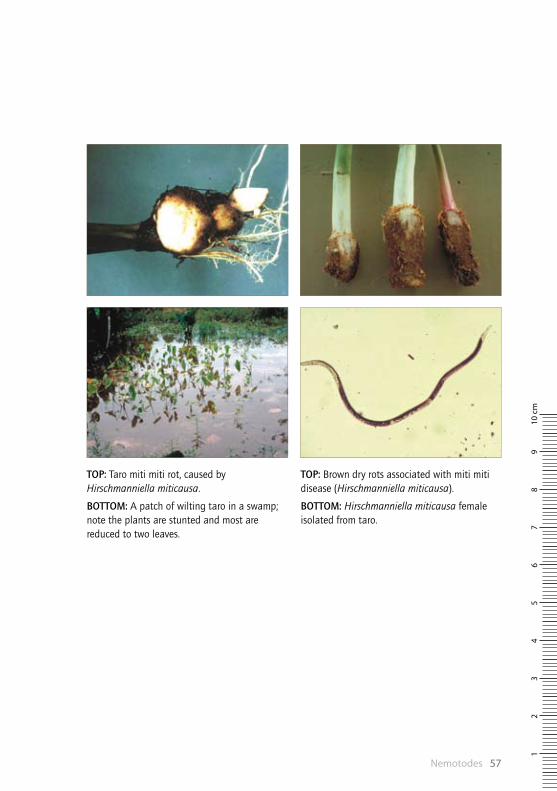
Nemotodes 57
98
76
54
32
110
cm
TOP: Brown dry rots associated with miti miti disease (Hirschmanniella miticausa).
BOTTOM: Hirschmanniella miticausa female isolated from taro.
TOP: Taro miti miti rot, caused by Hirschmanniella miticausa.
BOTTOM: A patch of wilting taro in a swamp; note the plants are stunted and most are reduced to two leaves.

TaroPest: an illustrated guide to pests and diseases of taro in the South Pacific58
NematodesRoot knot nematodes Meloidogyne spp.What are they?
Root knot nematodes are commonly found associated with the roots of taro. However, surveys in the Pacific have found little evidence of damage.
Where are they found?
Meloidogyne nematodes have a wide distribution throughout the Pacific.
What do they do?
Surveys of nematodes in Pacific island countries suggest that damage to taro caused by Meloidogyne spp. is relatively mild compared with damage caused on other crops. It is suggested that Colocasia and Xanthosoma are more tolerant of Meloidogyne incognita than other crops, and high populations of the nematode have to be present in the field soil before planting for damage to occur. Typically, roots are slightly swollen without knots—only very rarely have swollen, deformed roots with galls been reported.
What do I look for?
The presence of Meloidogyne spp. in infested soils can be determined by extraction of the second-stage juveniles using a standard nematode extraction procedure for free-living nematodes. External symptoms on corms are obvious only in cases of heavy infestations. Where the nematode is suspected, but numbers are low or infection is in its early stages, clearing, staining and microscopic examination will be required.
How do I control them?
Cultural control: The use of cultural control methods to manage root knot nematodes is the most environmentally sustainable and potentially most successful method for limiting damage. Planting material should be free from soil and roots. It is best if the ‘tops’ are selected from fields without any history of infection. Cover crops such as marigolds produce chemicals that are toxic to nematodes. Cover crops have the added benefits of stabilising topsoil and improving soil quality.
Chemical control: Nematocides (e.g. dazomet, oxamyl and fenamiphos) are commonly applied as preplanting treatments to reduce nematode numbers. They should be used with extreme caution and only by licensed operators. Their use on taro in Pacific island countries is thought to be limited.
Biological control: The bacteria Pasteuria penetrans and Paecilomyces lilacinus have been used as biological control agents. The most significant problem in developing effective biocontrol agents is an inability to generate the amount of biological material necessary for application over large areas. However, P. penetrans can be cultured easily by planting root-knot infected tomatoes where the bacterium is found. The roots can then be harvested, chopped and dried, and incorporated into the soil where there is a nematode problem.
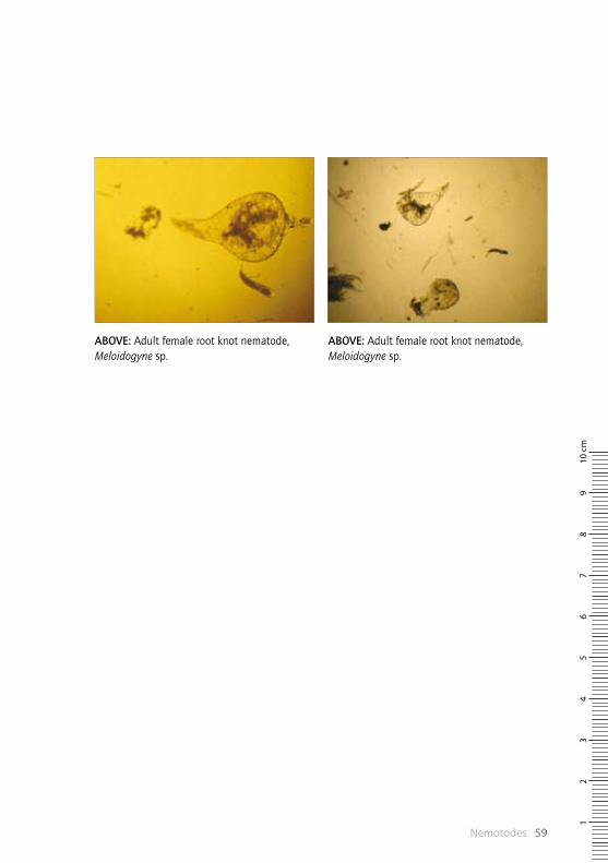
Nemotodes 59
98
76
54
32
110
cm
ABOVE: Adult female root knot nematode, Meloidogyne sp.
ABOVE: Adult female root knot nematode, Meloidogyne sp.
TaroPest: an illustrated guide to pests and diseases of taro in the South Pacific60
NematodesLesion nematode Pratylenchus coffeaeWhat is it?
The root lesion nematode Pratylenchus coffeae is commonly found associated with the roots of taro. However, only in Japan has this nematode been found to be the cause of a disease.
Where is it found?
Pratylenchus coffeae has been recorded associated with taro in American Samoa, Fiji and Papua New Guinea, and associated with other host plants in Cook Islands, Federated States of Micronesia, Kiribati, Niue, Samoa, Tonga and Vanuatu.
What does it do?
In Japan, Pratylenchus coffeae has consistently been found associated with root decay, reduced number of cormels, stunting and death in taro. The problem appears to be exacerbated by continuous cropping. It is not clear whether this nematode damages taro in Pacific island countries sufficiently to cause above-ground symptoms. Symptoms are reported only from Papua New Guinea, where P. coffeae is reported to cause localised necrosis (darkened areas of dead tissue) on roots and corms. In American Samoa, high numbers of P. coffeae have been found on taro, with the plants appearing unhealthy.
What do I look for?
The lesion nematode can be detected by extraction from root and corm tissues, using standard methods, and examination at high magnification. It is unlikely that symptoms will be seen on the plant, except for localised necrosis of feeder roots.
How do I control it?
The four most important control measures are:
Quarantine: Strict quarantine measures should be followed when germplasm is moved internationally, in order to prevent further spread of Pratylenchus coffeae.
Treatment of planting material: Necrotic tissues should be removed from the tops (approximately 40 cm of petiole and 1–2 cm of corm) and suckers, which should then be washed with running water and allowed to dry before planting. Care should be taken to ensure that no infected materials or trimmings are taken into the field.
Reduction of populations in the field: Crop rotations will reduce populations but, because of its wide host range, damage from Pratylenchus coffeae may not always be avoided.
Chemical control: Nematocides (e.g. dazomet, oxamyl and fenamiphos) are commonly applied as preplanting treatments to reduce nematode numbers. They should be used with extreme caution and only by licensed operators. Their use on taro in Pacific island countries is thought to be limited. Even in Japan, nematocides are considered to be uneconomic and crop rotation is regarded as a better strategy for control of Pratylenchus coffeae.

Nemotodes 61
98
76
54
32
110
cm
ABOVE: Root symptoms of Pratylenchus coffeae.
TOP: Microscopic, thread-like nematodes, Pratylenchus coffeae, found in and around soil, roots and corms.
BOTTOM: Field symptoms of Pratylenchus coffeae.
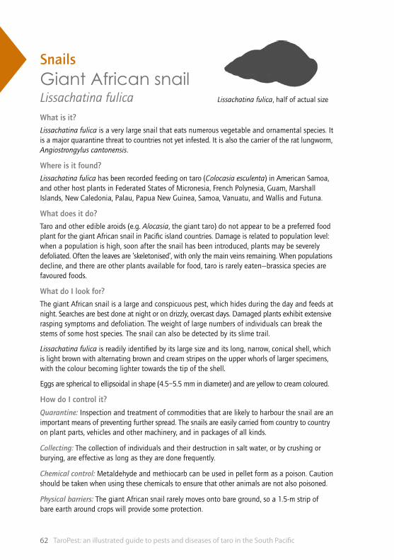
TaroPest: an illustrated guide to pests and diseases of taro in the South Pacific62
Lissachatina fulica, half of actual size
SnailsGiant African snail Lissachatina fulicaWhat is it?
Lissachatina fulica is a very large snail that eats numerous vegetable and ornamental species. It is a major quarantine threat to countries not yet infested. It is also the carrier of the rat lungworm, Angiostrongylus cantonensis.
Where is it found?
Lissachatina fulica has been recorded feeding on taro (Colocasia esculenta) in American Samoa, and other host plants in Federated States of Micronesia, French Polynesia, Guam, Marshall Islands, New Caledonia, Palau, Papua New Guinea, Samoa, Vanuatu, and Wallis and Futuna.
What does it do?
Taro and other edible aroids (e.g. Alocasia, the giant taro) do not appear to be a preferred food plant for the giant African snail in Pacific island countries. Damage is related to population level: when a population is high, soon after the snail has been introduced, plants may be severely defoliated. Often the leaves are ‘skeletonised’, with only the main veins remaining. When populations decline, and there are other plants available for food, taro is rarely eaten—brassica species are favoured foods.
What do I look for?
The giant African snail is a large and conspicuous pest, which hides during the day and feeds at night. Searches are best done at night or on drizzly, overcast days. Damaged plants exhibit extensive rasping symptoms and defoliation. The weight of large numbers of individuals can break the stems of some host species. The snail can also be detected by its slime trail.
Lissachatina fulica is readily identified by its large size and its long, narrow, conical shell, which is light brown with alternating brown and cream stripes on the upper whorls of larger specimens, with the colour becoming lighter towards the tip of the shell.
Eggs are spherical to ellipsoidal in shape (4.5–5.5 mm in diameter) and are yellow to cream coloured.
How do I control it?
Quarantine: Inspection and treatment of commodities that are likely to harbour the snail are an important means of preventing further spread. The snails are easily carried from country to country on plant parts, vehicles and other machinery, and in packages of all kinds.
Collecting: The collection of individuals and their destruction in salt water, or by crushing or burying, are effective as long as they are done frequently.
Chemical control: Metaldehyde and methiocarb can be used in pellet form as a poison. Caution should be taken when using these chemicals to ensure that other animals are not also poisoned.
Physical barriers: The giant African snail rarely moves onto bare ground, so a 1.5-m strip of bare earth around crops will provide some protection.

Snails 63
98
76
54
32
110
cm
TOP: Giant African snails are often seen on taro petioles.
MIDDLE: The giant African snail is a large and conspicuous pest.
BOTTOM: Colour and size variations of the giant African snail.
TOP & MIDDLE: Giant African snails are often seen around the base of taro plants.
BOTTOM: Giant African snails damage plants by rasping the leaf tissue.
TaroPest: an illustrated guide to pests and diseases of taro in the South Pacific64
VirusesAlomaeWhat is it?
Three viruses are associated with alomae: Colocasia bobone disease ?rhabdovirus, taro vein chlorosis rhabdovirus and taro badnavirus. They are not necessarily the cause of the disease.
Where is it found?
Alomae virus has been recorded in Solomon Islands and Papua New Guinea.
What does it do?
Initial symptoms of alomae vary. Symptoms are similar to those in plants infected with Colocasia bobone disease ?rhabdovirus (stunted, thickened, twisted, dark green leaves) or the plants are stunted with leaf blades bent under at the tip. Symptoms differ depending on whether the taro is ‘male’ or ‘female’—a distinction made by farmers in Solomon Islands based on the size of the plants and their reaction to alomae. In either case, the plants collapse rapidly and the leaves appear splayed (as if they are wilting). After this stage of the disease, growth ceases and plants rot from a systemic necrosis.
What do I look for?
Preliminary identification is based on the examination of leaves for typical symptoms. However, since the symptoms vary depending on variety, accurate diagnosis requires further testing. It is very difficult to differentiate between early symptoms of alomae and bobone. If plants die, then they had alomae and they were male. If plants recover, there are two possibilities:
(i) infection from Colocasia bobone disease ?rhabdovirus (CBDV) alone (as alomae is thought to be caused by a virus complex) and plants are male
(ii) infection from CBDV, causing bobone in taro resistant to alomae, and plants are female.
How do I control it?
Traditionally, taro growers in Solomon Islands and Papua New Guinea controlled alomae by growing taro in small, secluded patches within mature forests, and by roguing diseased plants as they appeared. Today, as a great deal of forest cover has been removed, the disease is more readily spread between taro gardens. Therefore, it is even more crucial for growers to remove and burn or bury infected plants that may otherwise serve as sources of infection. Present recommendations are that growers in a community synchronise roguing to minimise virus spread between gardens.

Viruses 65
10 c
m9
87
65
43
21
TOP: Taro in final stages of alomae; only the youngest leaves remain and the leaf blades are chlorotic.
MIDDLE: Alomae causing the youngest leaves to roll, and other leaves to be short with tips that curl under.
BOTTOM: Taro plant with dark green, stunted and twisted leaves, similar to those with bobone.
TOP: Early symptoms of alomae, showing a stunted plant with a backward curled young leaf.
MIDDLE: A later stage of alomae; young leaves are rolled, others have collapsed and are rotting.
BOTTOM: Alomae infection causes chlorosis of the veins; this appears as a feather-like pattern.
TaroPest: an illustrated guide to pests and diseases of taro in the South Pacific66
VirusesColocasia bobone disease virus (CBDV)What is it?
Colocasia bobone disease ?rhabdovirus causes leaf distortions that are sometimes severe. It is spread by a planthopper, Tarophagus proserpina.
Where is it found?
Colocasia bobone disease ?rhabdovirus is widespread throughout Papua New Guinea and Solomon Islands.
What does it do?
Symptoms differ between taro varieties, whether they are ‘male’ or ‘female’—a distinction made by farmers in Solomon Islands based on the size of the plants and their reaction to alomae. In female varieties, Colocasia bobone disease ?rhabdovirus (CBDV) causes severe stunting with distorted, thickened and brittle leaves, which sometimes fail to unfurl. Galls may be present on the petioles and sometimes on the larger veins. Plants usually recover after three to five leaves have been affected. Sometimes symptoms recur after recovery. In Solomon Islands, plants with these symptoms are said to have bobone. Translated, this means ‘the plant grows small’.
Male taro contains many varieties on which CBDV causes only mild symptoms: leaves show localised dark green wrinkled patches and petioles are normal in appearance. This is a mild form of bobone from which the plants recover. There is no local name for this phenomenon.
What do I look for?
Preliminary identification is based on examination of leaves for typical symptoms. However, as symptoms can vary depending on taro variety and environmental conditions, accurate diagnosis requires further testing. Diagnostic tests have been developed at Queensland University of Technology that use molecular (polymerase chain reaction) methods. These have proven to be both sensitive and robust.
How do I control it?
Growers do not normally apply control measures against this disease as the plants recover and produce symptomless leaves. However, the removal and subsequent burning or burial of infected plants is recommended, as these plants may serve as sources of infection for alomae. It is not yet clear which viruses interact to cause alomae, but Colocasia bobone disease ?rhabdovirus is suspected to be one of them.
Care must be taken to ensure that all insects on the infected plants are killed before removal of the plants for destruction, or that the plants are pulled up carefully so the insects are not spread. The virus is reportedly eliminated from taro plants if meristem tip culture is used. The restricted occurrence of this disease—confined to Papua New Guinea and Solomon Islands—means that strict quarantine measures should be followed when germplasm is moved internationally. Guidelines suggest that all transfers should be as sterile, pathogen-indexed plantlets growing in a tissue culture medium.

Viruses 67
10 c
m9
87
65
43
21
TOP & MIDDLE: Galls on the petioles and stems of CBDV-infected taro plants.
BOTTOM: A taro plant infected with CBDV.
TOP & MIDDLE: A CBDV-infected plant showing stunted, thickened leaves, which sometimes fail to unfurl.
BOTTOM: Leaf from a CBDV-infected plant showing thickened veins and dark green, wrinkled patches.

TaroPest: an illustrated guide to pests and diseases of taro in the South Pacific68
VirusesDasheen mosaic virus (DsMV)What is it?
Dasheen mosaic virus causes patterns of various colours, shapes and sizes to appear on the leaf. The disease may reduce corm yield.
Where is it found?
Dasheen mosaic virus is widespread throughout the Pacific; however, only in French Polynesia have severe distortions been reported.
What does it do?
Plants show a variety of mosaic patterns: small, irregular and scattered grey, green, yellow and sometimes white patches along or between the major veins, or brilliant white or yellow feather-like patterns along the veins and sometimes throughout the leaf blade. Invariably, plants recover from the symptoms, producing leaves that are healthy in appearance. Plants in French Polynesia have been reported with small, stunted and severely distorted leaves. Some leaves are reduced to strap-like structures without leaf blades. These plants often fail to recover.
What do I look for?
Preliminary identification is based on examination of leaves for typical symptoms. However, as the symptoms can vary depending on variety and environmental conditions, accurate diagnosis requires further testing. Diagnostic tests have been developed at Queensland University of Technology that use molecular (polymerase chain reaction) methods. These have proven to be both sensitive and robust.
How do I control it?
There are no effective control measures for dasheen mosaic virus (DsMV). Removal and subsequent burning or burial of infected plants has been recommended, as these plants may serve as sources of infection. However, farmers do not do this as it is not clear that yields are reduced by DsMV infection. In any case, it is likely that most and perhaps all taro in Pacific island countries carries DsMV as latent infections—the virus is transmitted easily by aphids and is then retained in the plants due to vegetative propagation techniques. Therefore, roguing plants displaying symptoms is not a useful control technique. The virus is reportedly eliminated from taro plants using meristem tip culture, but reinfection is rapid. Although DsMV is widespread, caution is still required when moving taro (or other aroids) internationally, especially as there appear to be severe forms of the disease. Strict quarantine measures should be followed. Guidelines suggest that all transfers should be as sterile, pathogen-indexed plantlets growing in a tissue culture medium.
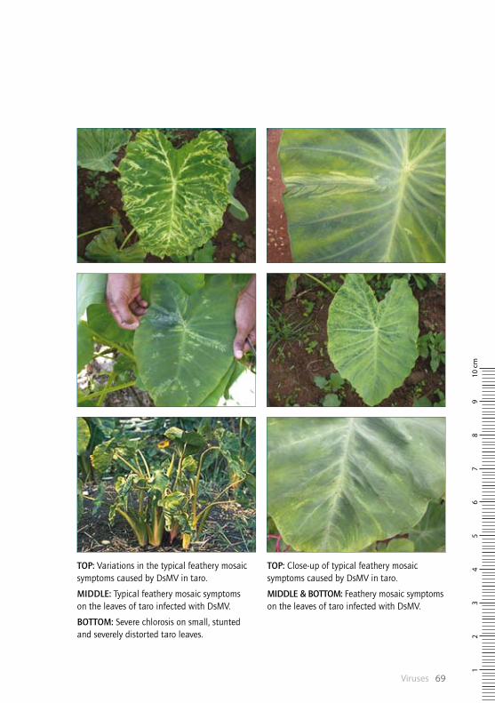
Viruses 69
10 c
m9
87
65
43
21
TOP: Close-up of typical feathery mosaic symptoms caused by DsMV in taro.
MIDDLE & BOTTOM: Feathery mosaic symptoms on the leaves of taro infected with DsMV.
TOP: Variations in the typical feathery mosaic symptoms caused by DsMV in taro.
MIDDLE: Typical feathery mosaic symptoms on the leaves of taro infected with DsMV.
BOTTOM: Severe chlorosis on small, stunted and severely distorted taro leaves.

TaroPest: an illustrated guide to pests and diseases of taro in the South Pacific70
VirusesTaro badnavirus (TaBV)What is it?
There is no widely accepted common name for taro badnavirus disease, although ‘vein clearing’ has been suggested.
Where is it found?
Taro badnavirus is widespread in American Samoa, Australia, Cook Islands, Fiji, Papua New Guinea, Samoa, Solomon Islands, Tonga and Vanuatu, and is present in Federated States of Micronesia, Marshall Islands and New Caledonia.
What does it do?
Taro badnavirus (TaBV) is often latent. Where symptoms do occur, they appear as intermittent, indistinct areas of vein chlorosis, often near the leaf margin. Frequently, the leaf blades are bent backwards, and sometimes the leaves are puckered. Rarely (but particularly on the Philippine variety PSB-G2 in Samoa), distinct vein chlorosis occurs throughout the leaf. Plants may be stunted, but eventually recover, producing apparently healthy leaves. Alone, TaBV appears to have a minimal effect on taro plant growth. However, in association with another virus, Colocasia bobone disease ?rhabdovirus (and possibly taro vein chlorosis virus), in Solomon Islands and Papua New Guinea, TaBV is thought to be responsible for causing alomae.
What do I look for?
Preliminary identification is based on examination of leaves for typical symptoms. However, as most infections are latent and virus symptoms can vary depending on cultivar and environmental conditions, accurate diagnosis requires further testing. Interestingly, a virus sequence similar to taro badnavirus has been found in all taro tested. It is unknown whether this sequence is part of the taro genome or is another badnavirus.
How do I control it?
No control measures are known to prevent the spread of taro badnavirus in the field. The virus is latent. Although the virus is widespread, caution is necessary when moving taro (and other aroids) internationally, as sequence variability has been observed. Strict quarantine measures should be followed. Guidelines suggest that all transfers should be as sterile, pathogen-indexed plantlets growing in a tissue culture medium.

Viruses 71
10 c
m9
87
65
43
21
ABOVE: A TaBV-infected taro leaf showing the backward bending of the leaf blade.
ABOVE: Distinct vein chlorosis caused by TaBV on the taro variety PSB-G2 in Samoa.

TaroPest: an illustrated guide to pests and diseases of taro in the South Pacific72
VirusesTaro vein chlorosis virus (TaVCV)What is it?
Taro vein chlorosis virus causes chlorosis (yellowing) of the veins, often near the leaf margin. The effect of the virus on corm yield is unknown, but there is a possibility that it causes alomae when in complexes with other viruses.
Where is it found?
Taro vein chlorosis virus has been recorded in Federated States of Micronesia, Fiji, New Caledonia, Palau, Papua New Guinea, Solomon Islands, Tuvalu and Vanuatu.
What does it do?
Unlike other taro virus diseases, symptoms of taro vein chlorosis virus (TaVCV) are commonly seen on plants when they are at maximum growth rather than after planting or at maturity. Leaves show distinct vein chlorosis, which is more pronounced than vein chlorosis sometimes associated with taro badnavirus. As the leaves age, the chlorosis spreads between the veins, which then form a network. In some cases, the veins later become necrotic and the leaf margins have a tattered appearance. The number of leaves showing symptoms varies, but in most cases it is three or four. Leaves formed subsequently appear healthy. In contrast to Colocasia bobone disease virus, which is also a rhabdovirus, infection with TaVCV does not lead to galls on the leaf blades and petioles, and plants are generally not stunted.
What do I look for?
Preliminary identification is based on examination of leaves for typical symptoms. However, as the symptoms can vary depending on taro variety and environmental conditions, accurate diagnosis requires further testing. Diagnostic tests have been developed at Queensland University of Technology that use molecular (polymerase chain reaction) methods. These have proven to be both sensitive and robust.
How do I control it?
Growers do not normally apply control measures for this disease as the taro plants recover and produce symptomless leaves. There is no indication that infection affects corm yield. However, removal and subsequent burning or burial of infected plants are recommended in Papua New Guinea and Solomon Islands, as these plants may serve as sources of infection for alomae. It is not yet clear which viruses interact to cause alomae, but taro vein chlorosis virus (TaVCV) is a candidate.
Care must also be taken to ensure that either all insects are killed before removal of the infected plants for destruction, or the plants are pulled up carefully so that the insects are not spread. The virus is reportedly eliminated from taro by using meristem tip culture. The restricted occurrence of this virus, to only nine countries, means that strict quarantine measures should be followed when germplasm is moved internationally. It is possible that mixed infection of taro by TaVCV, Colocasia bobone disease ?rhabdovirus and taro badnavirus causes the lethal alomae disease present in Papua New Guinea and Solomon Islands.

Viruses 73
10 c
m9
87
65
43
21
TOP & MIDDLE: Older leaves from a TaVCV-infected taro plant, showing the veins becoming necrotic.
BOTTOM: Severe TaVCV on taro, showing symptoms of yellowing throughout the leaf.
TOP & MIDDLE: Variations of the distinct vein chlorosis on the leaves of taro infected with TaVCV.
BOTTOM: TaVCV causes feathering symptoms as the veins of the taro leaf appear yellow.
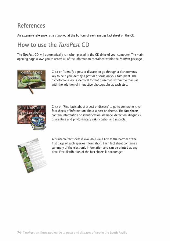
TaroPest: an illustrated guide to pests and diseases of taro in the South Pacific74
ReferencesAn extensive reference list is supplied at the bottom of each species fact sheet on the CD.
How to use the TaroPest CDThe TaroPest CD will automatically run when placed in the CD drive of your computer. The main opening page allows you to access all of the information contained within the TaroPest package.
Click on ‘Identify a pest or disease’ to go through a dichotomous key to help you identify a pest or disease on your taro plant. The dichotomous key is identical to that presented within the manual, with the addition of interactive photographs at each step.
Click on ‘Find facts about a pest or disease’ to go to comprehensive fact sheets of information about a pest or disease. The fact sheets contain information on identification, damage, detection, diagnosis, quarantine and phytosanitary risks, control and impacts.
A printable fact sheet is available via a link at the bottom of the first page of each species information. Each fact sheet contains a summary of the electronic information and can be printed at any time. Free distribution of the fact sheets is encouraged.
75
IndexAleurodicus dispersus . . . . . . . . . . . . . . . . . . . . . . . . . . . . . . . . . . . . . . . . . . . . . . . . . . . . . . . 36–37
alomae . . . . . . . . . . . . . . . . . . . . . . . . . . . . . . . . . . . . . . . . . . . . . . . . . . . . . . . . . . . . . . . . . . . 64–65
aphids . . . . . . . . . . . . . . . . . . . . . . . . . . . . . . . . . . . . . . . . . . . . . . . . . . . . . . . . . . . . . . . . . . . . 38–39
Aphis gossypii . . . . . . . . . . . . . . . . . . . . . . . . . . . . . . . . . . . . . . . . . . . . . . . . . . . . . . . . . . . . . 38–39
armyworm . . . . . . . . . . . . . . . . . . . . . . . . . . . . . . . . . . . . . . . . . . . . . . . . . . . . . . . . . . . . . . . . . 50–51
Athelia rolfsii . . . . . . . . . . . . . . . . . . . . . . . . . . . . . . . . . . . . . . . . . . . . . . . . . . . . . . . . . . . . . . 16–17
bacteria . . . . . . . . . . . . . . . . . . . . . . . . . . . . . . . . . . . . . . . . . . . . . . . . . . . . . . . . . . . . . . . . . . . 14–15
bacterial soft rot . . . . . . . . . . . . . . . . . . . . . . . . . . . . . . . . . . . . . . . . . . . . . . . . . . . . . . . . . . . 14-15
Bemesia tabaci . . . . . . . . . . . . . . . . . . . . . . . . . . . . . . . . . . . . . . . . . . . . . . . . . . . . . . . . . . . . 40–41
brown leaf spot (or ghost spot) . . . . . . . . . . . . . . . . . . . . . . . . . . . . . . . . . . . . . . . . . . . . . . . 18–19
Cladosporium colocasiae . . . . . . . . . . . . . . . . . . . . . . . . . . . . . . . . . . . . . . . . . . . . . . . . . . . . 18–19
Colocasia bobone disease virus (CBDV) . . . . . . . . . . . . . . . . . . . . . . . . . . . . . . . . . . . . . . . . 66–67
corm and leaf spot . . . . . . . . . . . . . . . . . . . . . . . . . . . . . . . . . . . . . . . . . . . . . . . . . . . . . . . . . 24–25
corm rot . . . . . . . . . . . . . . . . . . . . . . . . . . . . . . . . . . . . . . . . . . . . . . . . . . . . . . . . . . . . . . . . . . 16–17
corm soft rot . . . . . . . . . . . . . . . . . . . . . . . . . . . . . . . . . . . . . . . . . . . . . . . . . . . . . . . . . . . . . . . 34–35
Dasheen mosaic virus (DsMV) . . . . . . . . . . . . . . . . . . . . . . . . . . . . . . . . . . . . . . . . . . . . . . . . 68–69
Erwinia chrysanthemi . . . . . . . . . . . . . . . . . . . . . . . . . . . . . . . . . . . . . . . . . . . . . . . . . . . . . . . 14–15
fungi . . . . . . . . . . . . . . . . . . . . . . . . . . . . . . . . . . . . . . . . . . . . . . . . . . . . . . . . . . . . . . . . . . . . . 16–35
ghost spot . . . . . . . . . . . . . . . . . . . . . . . . . . . . . . . . . . . . . . . . . . . . . . . . . . . . . . . . . . . . . . . . . 18–19
giant African snail . . . . . . . . . . . . . . . . . . . . . . . . . . . . . . . . . . . . . . . . . . . . . . . . . . . . . . . . . . 62–62
hawk moth . . . . . . . . . . . . . . . . . . . . . . . . . . . . . . . . . . . . . . . . . . . . . . . . . . . . . . . . . . . . . . . . 42–43
Hippotion celerio . . . . . . . . . . . . . . . . . . . . . . . . . . . . . . . . . . . . . . . . . . . . . . . . . . . . . . . . . . . 42–43
Hirschmanniella miticausa . . . . . . . . . . . . . . . . . . . . . . . . . . . . . . . . . . . . . . . . . . . . . . . . . . . 56–57
hornworm (hawk moth) . . . . . . . . . . . . . . . . . . . . . . . . . . . . . . . . . . . . . . . . . . . . . . . . . . . . . 42–43
identification key . . . . . . . . . . . . . . . . . . . . . . . . . . . . . . . . . . . . . . . . . . . . . . . . . . . . . . . . . . . 8–13
insects . . . . . . . . . . . . . . . . . . . . . . . . . . . . . . . . . . . . . . . . . . . . . . . . . . . . . . . . . . . . . . . . . . . . 36–55
Lasiodiplodia theobromae . . . . . . . . . . . . . . . . . . . . . . . . . . . . . . . . . . . . . . . . . . . . . . . . . . . 20–21
leaf blotch . . . . . . . . . . . . . . . . . . . . . . . . . . . . . . . . . . . . . . . . . . . . . . . . . . . . . . . . . . . . . . . . 32–33
Leptosphaerulina trifolii . . . . . . . . . . . . . . . . . . . . . . . . . . . . . . . . . . . . . . . . . . . . . . . . . . . . . 22–23
lesion nematode . . . . . . . . . . . . . . . . . . . . . . . . . . . . . . . . . . . . . . . . . . . . . . . . . . . . . . . . . . . 60–61
Lissachatina fulica . . . . . . . . . . . . . . . . . . . . . . . . . . . . . . . . . . . . . . . . . . . . . . . . . . . . . . . . . . 62–62
Marasmiellus stenophyllus . . . . . . . . . . . . . . . . . . . . . . . . . . . . . . . . . . . . . . . . . . . . . . . . . . . 24–25
mealybugs . . . . . . . . . . . . . . . . . . . . . . . . . . . . . . . . . . . . . . . . . . . . . . . . . . . . . . . . . . . . . . . . 44–45
Meloidogyne spp. . . . . . . . . . . . . . . . . . . . . . . . . . . . . . . . . . . . . . . . . . . . . . . . . . . . . . . . . . . . 58–59
miti miti disease . . . . . . . . . . . . . . . . . . . . . . . . . . . . . . . . . . . . . . . . . . . . . . . . . . . . . . . . . . . 56–57

TaroPest: an illustrated guide to pests and diseases of taro in the South Pacific76
nematodes . . . . . . . . . . . . . . . . . . . . . . . . . . . . . . . . . . . . . . . . . . . . . . . . . . . . . . . . . . . . . . . . 56–61
Neojohnstonia colocasiae . . . . . . . . . . . . . . . . . . . . . . . . . . . . . . . . . . . . . . . . . . . . . . . . . . . . 26–27
orange leaf spot . . . . . . . . . . . . . . . . . . . . . . . . . . . . . . . . . . . . . . . . . . . . . . . . . . . . . . . . . . . . 26–27
Papuana spp. . . . . . . . . . . . . . . . . . . . . . . . . . . . . . . . . . . . . . . . . . . . . . . . . . . . . . . . . . . . . . . 46–47
Patchiella reaumuri . . . . . . . . . . . . . . . . . . . . . . . . . . . . . . . . . . . . . . . . . . . . . . . . . . . . . . . . . 48–49
Phoma spp. . . . . . . . . . . . . . . . . . . . . . . . . . . . . . . . . . . . . . . . . . . . . . . . . . . . . . . . . . . . . . . . . 28–29
Phytophthora colocasiae . . . . . . . . . . . . . . . . . . . . . . . . . . . . . . . . . . . . . . . . . . . . . . . . . . . . . 30–31
Pratylenchus coffeae . . . . . . . . . . . . . . . . . . . . . . . . . . . . . . . . . . . . . . . . . . . . . . . . . . . . . . . . 60–61
Pseudocercospora colocasiae . . . . . . . . . . . . . . . . . . . . . . . . . . . . . . . . . . . . . . . . . . . . . . . . . 32–33
Pseudococcidae . . . . . . . . . . . . . . . . . . . . . . . . . . . . . . . . . . . . . . . . . . . . . . . . . . . . . . . . . . . . 44–45
Pythium spp. . . . . . . . . . . . . . . . . . . . . . . . . . . . . . . . . . . . . . . . . . . . . . . . . . . . . . . . . . . . . . . 34–35
root knot nematodes . . . . . . . . . . . . . . . . . . . . . . . . . . . . . . . . . . . . . . . . . . . . . . . . . . . . . . . . 58–59
shot hole . . . . . . . . . . . . . . . . . . . . . . . . . . . . . . . . . . . . . . . . . . . . . . . . . . . . . . . . . . . . . . . . . . 28–29
snails . . . . . . . . . . . . . . . . . . . . . . . . . . . . . . . . . . . . . . . . . . . . . . . . . . . . . . . . . . . . . . . . . . . . . 62–62
spider mites . . . . . . . . . . . . . . . . . . . . . . . . . . . . . . . . . . . . . . . . . . . . . . . . . . . . . . . . . . . . . . . 54–55
spiralling whitefly . . . . . . . . . . . . . . . . . . . . . . . . . . . . . . . . . . . . . . . . . . . . . . . . . . . . . . . . . . 36–37
Spodoptera litura . . . . . . . . . . . . . . . . . . . . . . . . . . . . . . . . . . . . . . . . . . . . . . . . . . . . . . . . . . . 50–51
spongy black rot . . . . . . . . . . . . . . . . . . . . . . . . . . . . . . . . . . . . . . . . . . . . . . . . . . . . . . . . . . . 20–21
Taro badnavirus (TaBV) . . . . . . . . . . . . . . . . . . . . . . . . . . . . . . . . . . . . . . . . . . . . . . . . . . . . . . 70–71
taro beetles . . . . . . . . . . . . . . . . . . . . . . . . . . . . . . . . . . . . . . . . . . . . . . . . . . . . . . . . . . . . . . . 46–47
taro leaf blight . . . . . . . . . . . . . . . . . . . . . . . . . . . . . . . . . . . . . . . . . . . . . . . . . . . . . . . . . . . . . 30–31
taro planthoppers . . . . . . . . . . . . . . . . . . . . . . . . . . . . . . . . . . . . . . . . . . . . . . . . . . . . . . . . . . 52–53
taro root aphid . . . . . . . . . . . . . . . . . . . . . . . . . . . . . . . . . . . . . . . . . . . . . . . . . . . . . . . . . . . . . 48–49
Taro vein chlorosis virus (TaVCV) . . . . . . . . . . . . . . . . . . . . . . . . . . . . . . . . . . . . . . . . . . . . . . 72–73
Tarophagus spp.. . . . . . . . . . . . . . . . . . . . . . . . . . . . . . . . . . . . . . . . . . . . . . . . . . . . . . . . . . . . 52–53
Tetranychus spp. . . . . . . . . . . . . . . . . . . . . . . . . . . . . . . . . . . . . . . . . . . . . . . . . . . . . . . . . . . . 54–55
tobacco whitefly . . . . . . . . . . . . . . . . . . . . . . . . . . . . . . . . . . . . . . . . . . . . . . . . . . . . . . . . . . . 40–41
viruses . . . . . . . . . . . . . . . . . . . . . . . . . . . . . . . . . . . . . . . . . . . . . . . . . . . . . . . . . . . . . . . . . . . . 64–73
white spot of taro . . . . . . . . . . . . . . . . . . . . . . . . . . . . . . . . . . . . . . . . . . . . . . . . . . . . . . . . . . 22–23

www.aciar.gov.au