10. formulation of ointment 10.1. introduction · during the preparation, the oil is heated to...
TRANSCRIPT
179
10. FORMULATION OF OINTMENT
10.1. Introduction
Herbal drugs are formulated in the form of ointment and are
used topically for several purposes, e.g. as protectants, antiseptics,
emollients, antiprurit ic, keratolytics and astringents. Ointment bases
are always anhydrous and generally contain one or more
medicaments in suspension or solution or dispersion. Ointment
bases may be hydrocarbon (oleaginous), absorption, water
removable and water soluble type. On the basis of their level of
action, they are classified as: epidermatic , endodermatic and
diadermatic (Carter, 1987). A wound healing ointment is aimed to
heal the incised and excised wounds. In an earlier study, medicinal
plants have been reported to be very beneficial in wound care,
promoting the rate of wound healing with minimal pain, discomfort ,
and scarring to the patient (Odimegwu et al . , 2008)
The objective of the study was to formulate and evaluate the
original herbal extract ointment and Nanotised herbal extract from
the local medicinal plant for its wound healing property.
Ointments are semisolid preparations that are thicker than
creams but thinner than pastes. The ointment formulation consists of
active and inactive ingredients added to a base. For herbal
formulations, an ointment is made by emulsifying oil pha se in water
while the mixture is warm, then letting the formulation congeal at
room temperature. During the preparation, the oil is heated to
approximately 70 C and the water to 75 C. Usually the water phase
180
contains an extract of the herb to be incorporated into the mixture.
When added together and mixed in the presence of an emulsifying
agent, the two phases quickly form a stable emulsion. The mixing
continues at room temperature until the formulation begins to
congeal. Upon mixing the two phases together, the mixture initially
looks milky in appearance, and unless colouring agents are added to
the formulation, the final ointment is normally white to off -white in
colour. Since the ointment is kept over a period of up to several
weeks, preservatives such as benzoic acid or its salt, sodium
benzoate, must be used in ointment preparations. Dispensation may
be in a plastic, but preferably, glass jar. Shelf life can be improved
by storing the ointment in a refrigerator or in a cool place. If heat is
deleterious to the herbal components, then the ointment can be
prepared without heat by incorporation method. A possible method
for incorporating an aqueous herbal extrac t into an ointment base is
to mix the extract with a small quantity of lanolin (wool fat) using a
porcelain mortar and then incorporating the result ing mixt ure into
white petrolatum (petroleum jelly) using the same mortar for
mixing.
The herbal extract and nano herbal extract were incorporated
to the base separately and named as extract ointment (EO) and Nano
ointment (NO).
The ointment of the Urena lobata was prepared by using the simple
ointment base I.P.
181
Table: 10.1 Composition for the formulation of Urena lobata
Extract Ointment
(EO)
Quantity
(g)
Nano extract Ointment
(NO)
Quantity
(g)
White bees wax 2 White bees wax 2
Hard paraffin 3 Hard paraffin 3
Cetosteryl alcohol 5 Cetosteryl alcohol 5
White soft
paraffin 90 White soft paraffin 90
Urena lobata
alcoholic extract 5
Urena lobata Nanotised
alcoholic extract 5
Procedure
The extract and the nano extract separately were incorporated
into the molten simple ointment base and allowed to congeal by
stirring. After the ointment was formulated, they we re packed in
collapsible tube separately.
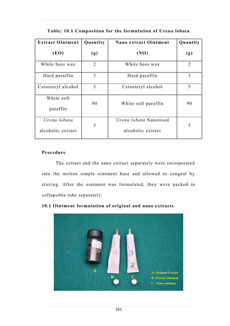
10.1 Ointment formulation of original and nano extracts
A B C
A - Original Extract
B - Extract ointment
C - Nano ointment
182
10.2. Quality control of the formulated Ointment
Physical evaluations: Preliminary evaluation of formulations at
different concentrations was carried ou t as follows:
Organoleptic parameters : Organoleptic parameters like colour,
odour of the formulations were carried out by visual examination.
Loss on drying
This is employed in IP and USP. Although the loss in weight,
in the sample so tested, principally is due to water and small amount
of other volatile material will be contribute the weight loss 1gm of
ointment is placed digital moisture balance instrument set the
temperature 105oC and run the instrument up to constant weight.
Finally read out the percen tage loss on drying automatically.
pH: The pH of various formulations was determined by using
Digital pH meter (Digital pH meter 335, Systronics, Noroda,
Ahmedabad). The 0.5 g of the weighed formulation was dispersed in
50 mL of distilled water and the pH was (Panigrahi et al . , 1997)
noted.
Homogeneity: All the developed ointments were tested for
homogeneity by visual inspection. They were tested for their
appearance with no lumps (Panigrahi et al . , 1997).
Viscosity: The measurement of viscosity of prepared ointments was
carried out with Brookfield Viscometer (model LV -DV-II, Helipath
spindle type S-96). The values of each formulation were done in
triplicate and average values were depicted in Table 10.2. The

183
viscosity values are expressed as Mean ± Stan dard deviation
(Kim et al . , 2003).
Spreadability: Spreadabili ty of the formulation was determined by
an apparatus suggested by Mutimer et al . (1956) which was suitably
modified in the laboratory and used for the study. The experiment
was performed as described by Wood et al . (1963).
Spreadability was determined by using the formula
S = MxL/T.
Where S = spreadability, M = Weight tied to upper slide, L =
Length of glass slides and T = Time taken to separate the slides
completely from each other. In this present experiment, M = 80 g,
L = 10 cm and T was recorded (Ehrlich and Hunt, 1968).
Acute skin irritation study: This test was performed on albino rats
weighing between 150-200g. The animals were given standard
animal feed and had free access to water ad libitum . The total mass
was separated into four groups, each batch containing five animals.
Dorsal hair at the back of the rats were removed one day prior to the
commencement of the study and kept individually in cages to avoid
contact with the other rats. Two groups of each were used for
control and standard irritant. Other two groups were used as test.
The 50mg of the each formulation were applied over one s quare
centimetre area of whole and abraded skin to different animals.
Aqueous solution of 0.8 % formalin was used as standard irri tant.
The animals were observed for seven days for any signs of oedema
and erythema (Marzulli and Maibach, 1997).

184
Stability studies: The stability studies were carried out in all
formulations at different temperature conditions (4°, 25° and 37°C)
for 3 months. All the evaluation parameters i.e. , pH, viscosity,
spreadabili ty, consistency and phase separation were studied at
different t ime intervals i .e., 15, 30, 60 and 90th days ( Shinde et al . ,
2005; Mohanta et al . , 2007).
Table: 10.2 Stability studies of the ointment of the Urena lobata extracts
Physicochemical parameters Formulation
Colour Light green
Odour Characteristic
Loss on Drying 9.5 % w/w
pH 6.5 (5.95 - 6.56)
Spreadability(Seconds) 15 (between 18 sec)
Diffusion study 0.9 cm (between 1 cm)
Skin irritation study No skin irritation was observed
Storage( 4 °,24°,37° C) Stable
In-vitro drug release study:
In In-vitro diffusion study of the ointment was carried out on
Franz diffusion cell having 57 ml capacity. Whatman filter paper
no.41 was used as diffusion membrane. Pieces of Whatman filter
paper no.41 were soaked in phosphate buffer (PB) pH 6.0 for 24
hours, prior to experiment. Diffusion cell was filled with phosphate
buffer pH 6.0, Whatman filter paper no.41 was mounted on cell. The

185
temperature was maintained at 37 ± 0.5°C. The formulation was
spread on the filter paper as a thin layer. The time point fo r
ointment was different. A sample of 1ml was withdrawn at
predetermined time intervals, the solution was filter ed with 0.45
micron filter paper and make up the volume with 5 ml of PB pH 6.0
and equivalent amount of fresh dissolution fluid equil ibrated at
same temperature was replaced. The sample was diluted to 5 ml with
PB at pH 6.0. The standard also was prepared as the same
concentration of that of sample. The amount of drug permeated was
determined using a UV- spectrophotometer at 340 nm. (Linearity
range = 10-20µg/ml, R2 = 0.9988)
Weight of the ointment-10 mg
Volume of solution (7.4 pH): 100 ml
Amount of sample withdrawn: 5 ml
Absorbance: 304 nm
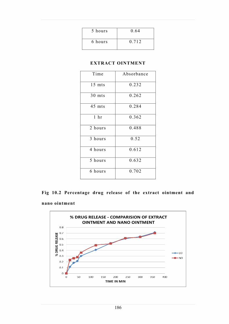
Table: 10.3 In-vitro drug release study
NANO OINTMENT
Time Absorbance
15 mts 0.114
30 mts 0.183
45 mts 0.214
1 hour 0.302
2 hours 0.41
3 hours 0.522
4 hours 0.602
186
5 hours 0.64
6 hours 0.712
EXTRACT OINTMENT
Time Absorbance
15 mts 0.232
30 mts 0.262
45 mts 0.284
1 hr 0.362
2 hours 0.488
3 hours 0.52
4 hours 0.612
5 hours 0.632
6 hours 0.702
Fig 10.2 Percentage drug release of the extract ointment and
nano ointment

187
10.3. RESULTS AND DISCUSSION
The various physicochemical parameters utilized to evaluate
the prepared ointment formulations are shown in Table 10.2.
The pH of the formulations lies in the normal pH range of the
human skin (6.5 ± 1).
All the formulations did not produce any skin irritation, i .e. ,
erythema and edema for about a week when applied over the
skin.
The rheological behaviours of the different formulations of
ointments in Rotational Brookfield Viscometer indicated that
the speed of spindle increases viscosity decre ases. A
comparative study of viscosity and spreadabili ty showed that
the viscosity of the formulations increases, spreadability
decreases and vice versa.
These formulations did not produce any skin irritat ion for
about a week when applied over the skin.
From the stabili ty studies, Ointments showed no changes in
pH, viscosity, spreadabili ty, consistency and phase separation
after keeping at different temperatures for 90 days.
Discussion
The mechanical evaluation parameters like pH, viscosity,
spreadabili ty, homogeneity are important tests to evaluate
pharmaceutical ointment formulations. The result of all the
formulations near to pH 6.5 ± 1 indicates better chemical
compatibility of ointments with skin. The results of viscosity gives
188
an idea about measurement of strength and the result of
spreadabili ty denote the extent of area to which the prepared
formulations readily spreads on application to skin or affected part
and homogeneity confirms no lumps. The results of stability study
indicate that there was no change in results of evaluation parameters
of prepared ointments during the treatment period. The absence of
erythema and edema for about a week when the ointments are
applied over the skin for skin irritat ion test indicates patient
compliance and fewer s ide effects. The results of the physical
evaluation of ointment preparation with ethanolic extract and the
nano extract of Urena lobata indicated the suitabili ty of method for
the production of ointments (Ansel et al . , 2005).
189
References:
Pharmaceutical Students: Ointments, Pastes and Jellies. 12 th
Edition, CBS Publishers and Distributors, India,
Chakole CM, Shende MA, Khadatkar SN. Formulation and
evaluation of novel combined halobetasol propionate and
fusidic acid ointment . International Journal of Chem-Tech
Research . 2009 Jan-March ;1(1):103-106
Ehrlich, H.P. and T.K. Hunt, 1968. Effect of cortisone and
Vitamin A on wound healing . Ann. Surg . , 167: 324-328.
Fites et. al . J. Pharm. Sci . 1970; 59(2): 610-11.
Gupta GD and Gaud RS. Release rate of Nimesulide from
different gellants . Ind. J. Pharm. Sci . 1999; 61(4): 227-30.
Indian pharmacopoeia, microbiological test and assay, 1996,
1, Appendix 4: A. 88-A.11.
Kim, J.Y., J .Y. Song, E.J. Lee and S.K. Park, 2003.
Rheological properties and microstructure of carbopol gel
network system . Colloid Polymer Sci . , 281: 614-623.
Marzulli, F.N. and H.I. Maibach, 1997. Advance in Modern
Toxicology . Hemisphere Publishing Corporation, London,
pp: 193-210.
Mohanta, G.P., M. Jamal and S. Umadevi, 2007. Formulation
and evaluation of a poly herbal wound healing cream . Indian
Drugs, 44: 281-281.

190
Mutimer, M.N., C. Riffikin, J .A. Hill , E. Marry and C.N.G.
Glickman, 1956. Synthesis of methylsilyl derivat ives of
procaine and their diffusion . J. Am. Pharm. Assoc. Sci . , 45:
212-212.
Odimegwu, D.C., Ibezim, E.C., Esimone, C.O ., Nworu, C.S.,
Okoye, F.B.C., Wound Healing and Antibacterial Activit ies
of the Extract of Dissotis theifolia (Melastomataceae) Stem
Formulated in a Simple Ointment Base . J Medicinal Plant
Res 2008,2(1): 011-016.)
Panigrahi, L., T. Jhon, A. Shariff and R.S. Shobanirani,
Formulation and evaluat ion of lincomycin HCL gels . Ind. J.
Pharm. Sci . , 1997, 59: 330-332.
Peruru D. et. al. Isolation of eumelanin from Sepia
officinalis and investigation of its antimicrobial activity by
ointment formulation . Int .J.Pharm .2012;2(2):67-72.192-210.
Purushothamrao K, Khaliq K, Sagare P, Patil SK, Kharat SS,
Alpana.K. Formulation and evaluation of vanishing cream
for scalp psoriasis . International Journal of Pharma Sci
Tech . 2010; 4(1):33-41.
Shinde, A.J., S.B. Bhise, R .J. Jarag and N.R. Jadhav, 2005.
Preparation of cream containing Tridax procumbens,
Curcuma longa and Azadirachta indica and its evaluation for
wound healing property . Indian Pharm . , 4:107-110.
191
United States Pharmacopoeial Convention. Pharmacopoeial
Forum, Rockville (MD). Topical and transdermal drug
products-product quality tests 3, 2009; 35(3): 1-26.
United States Pharmacopoeial Convention. Pharmacopoeial
Forum, Rockville (MD). Topical and transdermal drug
products-product performance tests 725, 2009; 35(3): 1-28.
Wei Liu, Meiling Hu,Wenshuang Liu, Chengbin Xue, Huibi
Xu, XiangLiang Yang , International Journal of
Pharmaceutics . , 2008, 364, 135 141.
Wood, J.H., G. Catacalos and S.V. Liberman, 1963.
Adaptation of commercial viscometers for special
applications in pharmaceutical rheology II. Severs extrusion
rheometer . J. Pharm. Sci . , 52: 375-378

192
11. WOUND HEALING STUDY OF THE FORMULATIONS OF
Urena lobata
11.1. Introduction
Wound healing processes are well organized biochemical and
cellular events leading to the growth and regeneration of wounded
tissue in a special manner. Healing of wounds is an important
biological process involving tissue repair and regeneration. It
involves the activity of an intricate network of blood cells,
cytokines, and growth factors which ultimately leads to the
restoration to normal condition of the injured skin or tissue (Clark,
1991). The aim of wound care is to promote wound healing in the
shortest time possible, with minimal pain, discomfort, and scarring
to the patient and must occur in a physiologic environment
conducive to tissue repair and regeneration (Bowler et al . , 2001).
Wound healing are conveniently classified into any of three
types, healing by first intention, healing by second intention and
healing by third intention, depending on the nature of the edges of
the healed wounds. Whereas the edges of wounds healed by first
intention are smoothly closed that no scar is left, wounds healed by
second intention involve formation of granulation tissues which fill
up the gaps between the wound edges and are associated with
significant loss of tissue, leaving little scars. Wounds healed by
third intention are usually those wounds left for three to five days
until granulation bed falls before they are sutured re sulting in
extensive scars formation (Thomas, 1997). Four distinct stages of

193
wound healing have also been identified -inflammatory,
debridement, proliferation, and remodelling maturation stages.
Wound healing processes are known to be influenced by
among other factors by infections, nutritional status, drugs and
hormones, type and sites of wound, and wasting diseases like
diabetes (Karl et al , 1995). In folklore medicine, medicinal plants
have been used widely in facil itating wound healing with high
degree of successes. This has inspired many researches which are
aimed at validating the claims and discovering mechanisms which
possibly explains the potentials of these herbs on wound repair
processes.
In our investigation the two different formulations in the form
of Ointments (original extract ointment (EO) and nanotised extract
ointment (NO) from the plant Urena lobata were evaluated using the
incision and excision models with Swiss albino rats.
Chemicals
All chemicals and reagents used were of analytical grade.
Experimental animals
Swiss Albino rats of 200 to 250 g body weight were used in
the study. Animals were procured from BiogenLaboratory Animal
Facility (CPCSEA -Reg no. 971 /bc/ 06), Bangalore.
All animals were kept in polyacrylic cages and main tained
under standard housing conditions (room temperature 24 27°C and
humidity 60 65% with 12:12 light: dark cycles. Food was provided
in the form of dry pellets and water ad libitum.
194
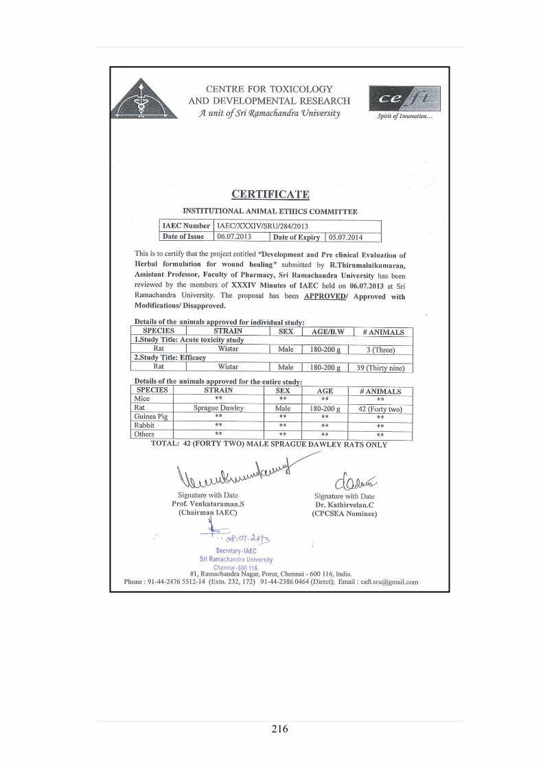
All experiments involving animals complies with the ethical
standards of animal handling and approved by Institutional Animal
Ethics Committee (IAEC/XXXIV/SRU/284/2013).
Model design
The design of wound healing activity was performed by two models
1) Excision wound model
2) Incision wound model
11.2. Materials and Methods
Male Wister rats were used for excision and incision wound
models and the ointments were applied topically and animal were
divided into the following groups.
Group I : No wound was created and served as control .(6 animals)
Group II: wound was created and served as posit ive control.
(6 animals)
Group III: 5%w/w Extract ointment (EO) was applied once daily.
i) Excision-4 animals
ii ) Incision-4 animals
Group IV: 5%w/w Nanotised extract ointment (NO) was
applied once daily.
i) Excision- 4 animals
ii ) Incision- 4 animals
Group V: Betadine ointment (5 gm) was applied once daily
i) Excision-4animals
ii ) Incision-4 animals

195
1) Excision wound model
The back of each rat was shaven under ether anaesthesia
and prepared for operation. Thereafter open circular wound of
500 mm2 area was produced in each rat by excising the skin.
For this purpose a marker was used to mark the area to be
excised.
The wounded animals were kept separately.
Rats wound were left undressed to the open environment; this
model was used to monitor wound contraction and epithelisation
time. The standard drug (Betadine ointment (0.5 g m), simple
ointment; ethanolic extract ointment (EO) 5 %w/w and 3 %w/w
Nanotised extract ointment (NO) were applied everyday t ill the
wound was completely healed.
Incision Wound Model
Animals were grouped which was divided into four groups
same as followed in excision wound model. The incision wound
model was studied under light ether anesthesia the anim al was
secured to operation table in its natural position. A longitudinal
paravertebral incision of 6 cm long was made through the skin and
cutaneous tissue on the back. After complete haemostasis, the
wound was closed by means of interrupted sutures place d at
equidistance points about 1 cm apart . Animals were treated daily
with drugs, as mentioned above under excision wound model from
0th day to 9th post wounding day. Wounds were cleaned with 70 %
alcohol soaked with cotton swabs. They were kept in separat e cages.
196
All the sutures were removed on the 9 th post wounding day. On 10 th
day the tensile strength was measured by continuous constant water
supply technique.
All the above mentioned treatments were started from the day
of operation and continued ti ll 20th day of healing. On 2n d, 4th , 8th ,
10th , 12th , 14th , 16t h , 18th and 20th days the wound area of each
rat was traced on a graph paper. Animals were sacrificed by
cervical dislocation on day 30. Liver, spleen, stomach and skin were
collected and processed for histopathological studies.
The following physical and biochemical parameters were studied
11.2.1. Physical Evaluation
Body weight
Wound size and area
Tensile strength
11.2.2 Antioxidant Studies
Superoxide Dismutase (SOD) activity (Mark lund and
Marklund, 1974)
Glutathione Peroxidase activity (GPX) (Moren et al . , 1973)
Estimation of Reduced glutathione (GSH) (Moren et al . ,
1979)
Activity of Catalase (Asru K Sinha, 1987)
11.2.3 Biochemical studies
Myeloperoxidase activity (Bradley et al . , 1982)
Total protein by Biuret method (Biuret et al . , 1948)
Hydroxy proline content (R.E.Newman et al , 1950
197
11.2.1 Physical evaluation
1. Measurement of wound area
The progressive changes in wound area were measured planim
etrically by tracing the wound margin on a graph paper every altern
ate dayThe changes in healing of wound i .e the measurement of wou
nd on graph paper was expressed as unit (mm2).
Wound contraction was expressed as percentage reduction of
original wound size.
Wound area - Unhealed Area
% Wound Contraction = ----------- X 100
Wound area
2. Measurement of Wound Breaking Strength of Incised Wounds
Measurement of wound breaking strength was pe rformed by
using the following method with certain modifications. A board was
placed on the table, on which the anaesthetized animal was made to
lie on its abdomen. Two clamps were clamped on either sides of
healed wound at a distance 0.5 cm. the left clam p was fastened
tightly to stand by means of thread. The right clamp was connected
to a leak proof polythene container through a pulley, by means of a
thread. A reservoir containing water was placed at a suitable height
and connected to a polythene bag by m eans of a rubber tube. The
position of the board was adjusted so that , the polythene bag was
hanging freely. Water was added to polythene bag rapidly at
constant rate from the reservoir until the wound opened. Amount of
water in polythene bag was measured (in ml) and was considered as
tensile strength of the wound.

198
11.2.2 Antioxidant Studies
1. Superoxide Dismutase (SOD)
0.05 ml of sample is added to 0.3 ml of sodium pyrophosphate
buffer (0.025 M, pH 8.3), 0.025 ml of phenazonium methosulphate
(186 µM) and 0.075 ml of Nitroblue tetrazolium chloride (300 µM
in buffer of pH 8.3).The reaction was started by addition of 0.075
ml of reduced nicotinamide adenine dinucleotide (780 µM in buffer
of pH 8.3).After incubation at 30º C for 90 seconds, the reaction
mixture was stirred vigorously and shaken with 2.0 ml of n -butanol.
The mixture was allowed to stand for 10 minutes and centrifuged.
1.5 ml of n-butanol alone was served as blank. The colour intensity
of the chromogen was read at 560 nm using thermo scientific
multiscan spectrometer, USA (Kakkar et al . , 1984).
Enzyme activity (1 unit) =50% inhibit ion/minute
2. Glutathione Peroxidase (GPX)
Glutathione peroxidise (GPX) was assayed by taking 200 µl of
tris HCl buffer (0.4 M), 200µl K.EDTA (0.4 mM) along with 100 µl
of sodium azide and 200 µl of sample and mixed well . Thereafter,
200µl of reduced glutathione solution (2 mM) followed by 0.1ml of
H2O2 were added and allowed to incubate for 10 min at 37º C. The
overall reaction was then arrested by adding 0.5 ml of 10 %
trichloroacetic acid (TCA).The precipitate was removed by
centrifugation at 1500 rpm for 10 minutes. To 0.2 ml of the
supernatant, 0.5 ml of saline and 1.0 ml of di -thio nitro benzoic acid
(DTNB) were added and the colour intensity formed was absorb ed at
199
412 nm using Thermo Scientific Multiskan Spectrophotometer,
USA, (Rotruck,et al . ,1973)
3. Reduced Glutathione (GSH)
Reduced glutathione was assayed by taking 0.25 ml of sample
to equal volume of ice cold 5 % TCA. The precipitate was removed
by centrifugation at 3500 rpm for 10 minutes.1 ml of the
supernatant was mixed with 0.25 ml of 0.2 M phosphate buffer, pH
8.0 and 0.5 ml of DTNB (0.6 mM in 0.2 M phosphate buffer, pH
8.0) were added and mixed well.The absorbance was read at 412 nm
using Thermo Scientific Multiskan Spectrophotometer,USA (Moren
et al . ,1979).
4. Activity of Catalase :
The assay mixture contained 100 µl of sample, 0.4 ml of H2O2
(2 mM) and 0.5 ml of phosphate buffer (10mM, pH 7.4).The above
mixture was stirred well and incubated at 37° C for 5 min and then
dichromate acetic acid reagent (5 % potassium dichromate in water,
glacial acetic acid mixed in 1:3 ratio) was added and absorbance
was taken at 570 nm using Thermo Scientific Multiskan
spectrophotometer, USA. 2 ml of dichromate acetic acid reagent
acts as blank (Asru K Sinha , 1987).
5. Myeloperoxidase activity
1ml of sample was mixed with equal volume of hexadecyl
trimethyl ammonium bromide (HTAB) buffer and centrifuge at 4ºC
for 15 minutes at 3000 rpm. Then 0.5 ml of supernatant was taken
and added to 3.5 ml of phosphate buffer (pH 6.0) followed by 1.5 ml

200
of dinizidine. The absorbance was read at 450nm at 0 sec and 60 sec
against blank using Thermo Scientific Multiskan spectrophotometer,
USA (Bradley et al . , 1982).The results were expressed in terms of
units (Nanomoles H2O2 lib/min/mg protein).
6. Total Protein by Biuret method
0.6 ml of saline was mixed with 50 µl of sample followed by
addition of 1.25 ml of working biuret reagent. The tubes were
incubated at room temperature for 15 min.The color
intensity was read at 540nm using Thermo Scientific
Multiskanspectrophotometer, USA, (Lubran et al . , 1948).
7. Hydroxyproline (HPR) (R.E.Newman et.al ., 1950).
To each tube, 0.3 mL of hydrolysate, 2.5 N NaOH, 0.01 M
CuSO4, and 6 % H2O2 were added. Tubes were shaken vigorously
and placed immediately in water bath at 80 ° C. After 15 minutes,
tubes were removed and cooled for 5 minutes in cold water. 0.6 mL
of freshly prepared 5 % solution of paradimethyl amino -
benzaldehyde in n-propanol and 1.2 ml of 3 N H2SO4 was added.
The test tubes were once again placed in a hot water bath at 75 ° C
for 15 minutes and then cooled for 5 minutes under running stream
of water. Color intensity was measured at 540 nm against the blank.
Hydroxyproline content in the tissue was estimated as per standard
curve prepared with standard 4 -Hydroxy-L-proline (HiMedia
Laboratories Pvt. Ltd., Mumbai, India), from 75 to 900 g/0.3 mL
using 3 mg/mL working solution

201
Results
Table: 11.1 Tensile Strength
NO= Nano extract ointment
EO= original extract ointment
All values are mean SEM ± n =6, **P < 0.001 indicates
extremely significant compared to the control

202
Fig: 11.1 Tensile strength of the tissue of wound treated rats
0
100
200
300
400
500
600
Control STD NO EO
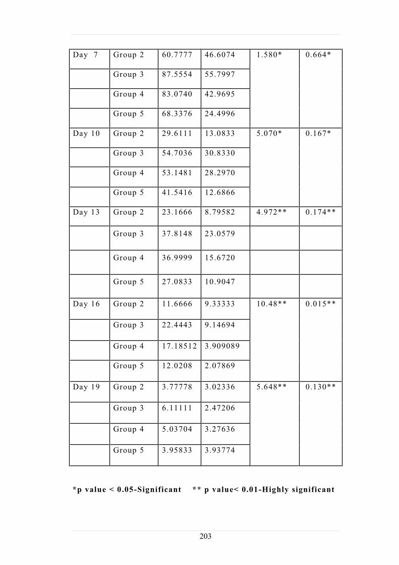
Table: 11.2 Comparison of wound area Using
-
Days Groups Mean SD Chi
Square
P value
Day 1 Group 2 136.166 95.1155 5.261*
0.154*
Group 3 173.148 55.4694
Group 4 252.703 112.347
Group 5 188.750 63.6295
Day 4 Group 2 104.777 64.0765 2.842*
0.417*
Group 3 143.036 63.6552
Group 4 161.333 77.7134
Group 5 113.041 37.1494
203
Day 7 Group 2 60.7777 46.6074 1.580* 0.664*
Group 3 87.5554 55.7997
Group 4 83.0740 42.9695
Group 5 68.3376 24.4996
Day 10 Group 2 29.6111 13.0833 5.070* 0.167*
Group 3 54.7036 30.8330
Group 4 53.1481 28.2970
Group 5 41.5416 12.6866
Day 13 Group 2 23.1666 8.79582 4.972** 0.174**
Group 3 37.8148 23.0579
Group 4 36.9999 15.6720
Group 5 27.0833 10.9047
Day 16 Group 2 11.6666 9.33333 10.48**
0.015**
Group 3 22.4443 9.14694
Group 4 17.18512 3.909089
Group 5 12.0208 2.07869
Day 19 Group 2 3.77778 3.02336 5.648**
0.130**
Group 3 6.11111 2.47206
Group 4 5.03704 3.27636
Group 5 3.95833 3.93774
*p value < 0.05-Significant ** p value< 0.01-Highly significant

204
1. Effect of Extract ointment and Nano ointment on Protein and
Hydroxy Proline content on Dry Connective Tissue
Table: 11.3 Estimation of Protein & Hydroxy proline
Groups Protein
mg / g dry tissue
Hydroxyproline
g / mg protein
Control 198.1 ± 12.4 132.2 ± 10.6
Std 272.3 ± 11.3* 197.2 ± 9.02**
NO 297.5 ± 15.0* 193.1 ± 10.2**
EO 305.6±11.3* 203.6±9.9**
Values are mean ± SEM of 6 rats in each group. * <0.05, **<0.01
compared to respective control group (statistical analysis was done
by using Mann Whitney test).
1. Effect of Extract ointment and Nano ointment on Protein and
Myeloperoxidase content on Wet granulation Tissue
Table: 11.4 Estimation of Protein & Myeloperoxidase
Groups Protein mg / g wet
tissue
Myeloperoxidase
mU/ mg protein
Control 38.6 ± 2.25 22.1 ± 0.57
Std 53.7 ± 2.12* 17.9 ± 0.29**
NO 62.1 ± 2.98* 13.8 ± 0.37**
EO 66.20±2.66* 12.9±0.29**
Values are mean ± SEM of 6 rats in each group. *<0.05, and
** <0.001 compared to respective control group (statistical analysis
was done by using Mann Whitney test).
205
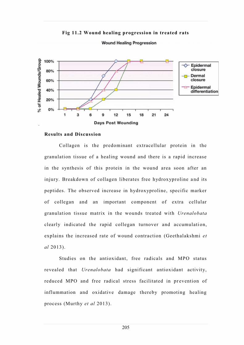
Fig 11.2 Wound healing progression in treated rats
Results and Discuss ion
Collagen is the predominant extracellular protein in the
granulation tissue of a healing wound and there is a rapid increase
in the synthesis of this protein in the wound area soon after an
injury. Breakdown of collagen liberates free hydroxyproline a nd its
peptides. The observed increase in hydroxyproline, specific marker
of collegan and an important component of extra cellular
granulation tissue matrix in the wounds treated with Urenalobata
clearly indicated the rapid collegan turnover and accumulati on,
explains the increased rate of wound contraction (Geethalakshmi et
al 2013).
Studies on the antioxidant, free radicals and MPO status
revealed that Urenalobata had significant antioxidant activity,
reduced MPO and free radical stress facilitated in pr evention of
inflummation and oxidative damage thereby promoting healing
process (Murthy et al 2013).
206
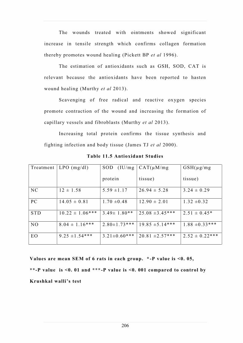
The wounds treated with ointments showed significant
increase in tensile strength which confirms collagen formation
thereby promotes wound healing (Pickett BP et al 1996).
The estimation of antioxidants such as GSH, SOD, CAT is
relevant because the antioxidants have been reported to hasten
wound healing (Murthy et al 2013).
Scavenging of free radical and reactive oxygen species
promote contraction of the wound and increasing the formation of
capillary vessels and fibroblasts (Murthy et al 2013).
Increasing total protein confirms the t issue synthesis and
fighting infection and body t issue (James TJ et al 2000).
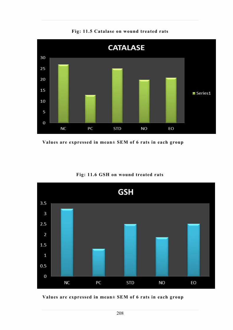
Table 11.5 Antioxidant Studies
Treatment LPO (mg/dl) SOD (IU/mg
protein
CAT(µM/mg
tissue)
GSH(µg/mg
tissue)
NC 12 ± 1.58 5.59 ±1.17 26.94 ± 5.28 3.24 ± 0.29
PC 14.05 ± 0.81 1.70 ±0.48 12.90 ± 2.01 1.32 ±0.32
STD 10.22 ± 1.06*** 3.49± 1.80** 25.08 ±3.45*** 2.51 ± 0.45*
NO 8.04 ± 1.16*** 2.80±1.73*** 19.85 ±5.14*** 1.88 ±0.33***
EO 9.25 ±1.54*** 3.21±0.60*** 20.81 ±2.57*** 2.52 ± 0.22***
Values are mean SEM of 6 rats in each group. * -P value is <0. 05,
**-P value is <0. 01 and ***-P value is <0. 001 compared to control by
207
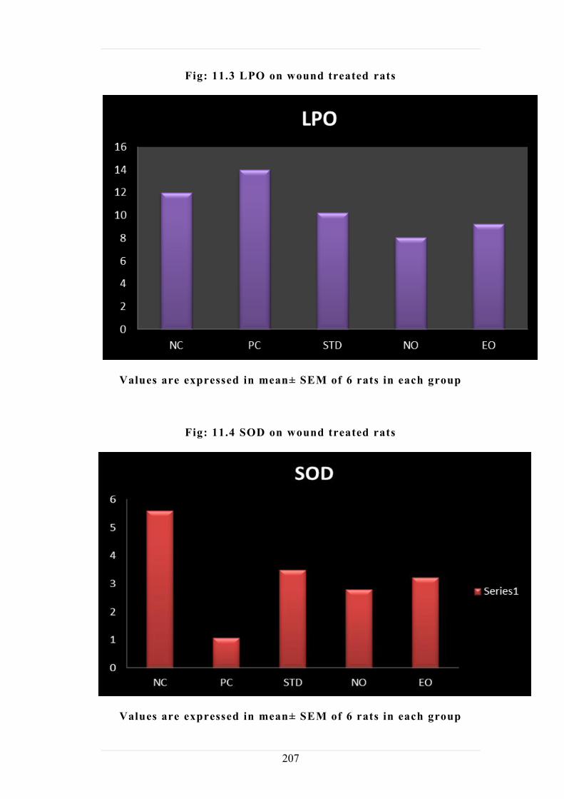
Fig: 11.3 LPO on wound treated rats
Values are expressed in mean± SEM of 6 rats in each group
Fig: 11.4 SOD on wound treated rats
Values are expressed in mean± SEM of 6 rats in each group
208
Fig: 11.5 Catalase on wound treated rats
Values are expressed in mean± SEM of 6 rats in each group
Fig: 11.6 GSH on wound treated rats
Values are expressed in mean± SEM of 6 rats in each group

209
11.3. Histopathology
Procedure:
Wound was created in the rat skin by incision and excision
method. On day 30, all rats from each group were euthanized using
anesthetic ether. The skin from the incised and excised region were
collected and fixed in 10% neutral buffered formalin for 48 hours.
The skin tissues were subjected to dehydration in series of graded
alcohol and embedded in paraffin wax. Tissue sections of 4 to 5
micron thickness were obtained and stained with Hematoxylin and
Eosin (Bancroft and Gamble, 2008 ) for l ight microscopic
exa
staining for specific evaluation of fibrous tissue proliferation.
Histopathology studies of treated rats for both extract and nano
ointment
Fig: 11.7
Control group MST 4x Control group Skin H & E 10x
Dermis
Hair follicles Muscle
Hypodermis
210
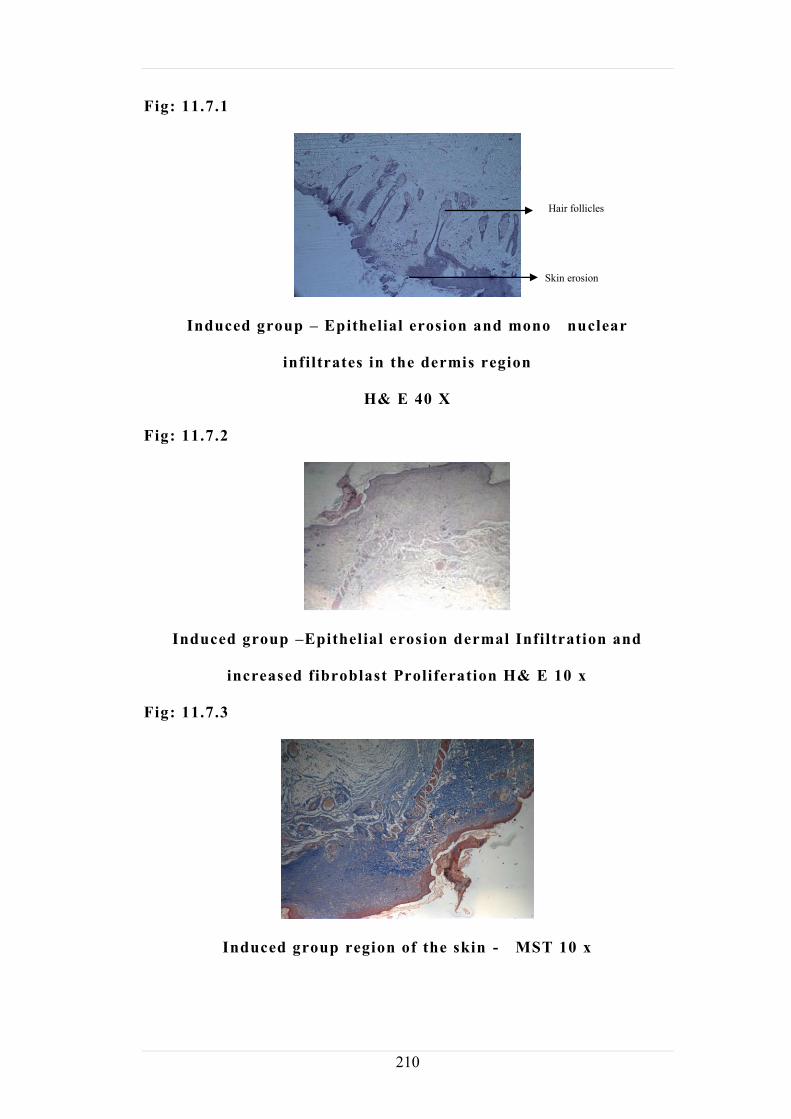
Fig: 11.7.1
Induced group Epithelial erosion and mono nuclear
infiltrates in the dermis region
H& E 40 X
Fig: 11.7.2
Induced group Epithelial erosion dermal Infiltration and
increased fibroblast Proliferation H& E 10 x
Fig: 11.7.3
Induced group region of the skin - MST 10 x
Hair follicles
Skin erosion
211
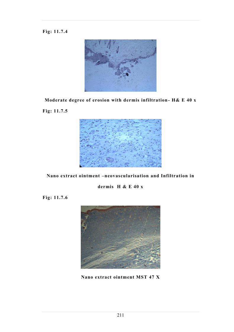
Fig: 11.7.4
Moderate degree of erosion with dermis infiltration - H& E 40 x
Fig: 11.7.5
Nano extract ointment neovascularisation and Infiltration in
dermis H & E 40 x
Fig: 11.7.6
Nano extract ointment MST 47 X
212
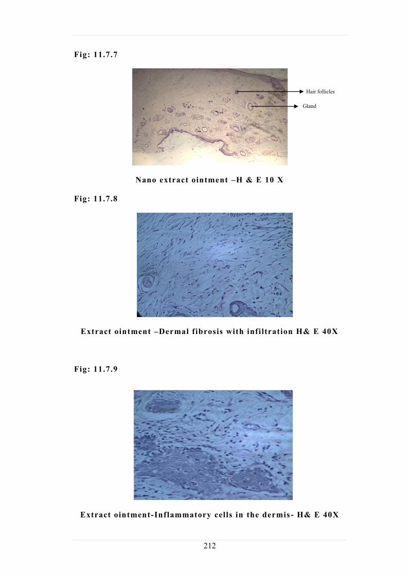
Fig: 11.7.7
Nano extract ointment H & E 10 X
Fig: 11.7.8
Extract ointment Dermal fibrosis with infiltration H& E 40X
Fig: 11.7.9
Extract ointment-Inflammatory cells in the dermis - H& E 40X
Hair follicles
Gland

213
Fig: 11.7.10
Extract ointment MST 4x
Fig: 11.7.11
Extract ointment Fibrosis in healed region-H&E 10x
Histopathology report on the evaluation of wound healing
activityof herbal ointment in rats
Wound was created in the rat skin by incision and excision
method. On day 30, all rats from each group were euthanized using
anesthetic ether. The skin from the incised and excised region were
collected and fixed in 10 % neutral buffered formalin for 48 hours.
The skin tissues were subjected to dehydration in series of graded
alcohol and embedded in paraffin wax. Tissue sections of 4 to 5
micron thickness were obtained and stained with Hematoxylin and
Eosin (Bancroft and Gamble, 2008 ) for l ight microscopic
Epidermis
Dermis
Secretory glands
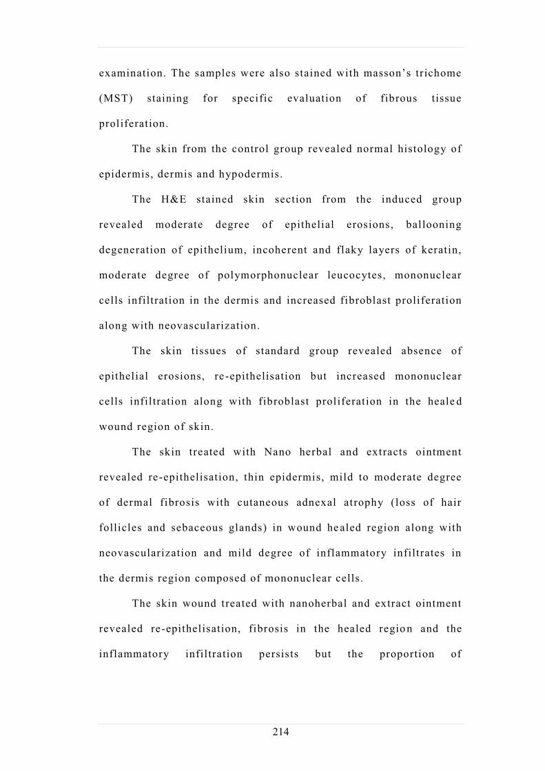
214
(MST) staining for specific evaluation of fibrous tissue
proliferation.
The skin from the control group revealed normal histology of
epidermis, dermis and hypodermis.
The H&E stained skin section from the induced group
revealed moderate degree of epithelial erosions, ballooning
degeneration of epithelium, incoherent and flaky layers of keratin,
moderate degree of polymorphonuclear leucocytes, mononuclear
cells infiltration in the dermis and increased fibroblast proliferation
along with neovascularization.
The skin tissues of standard group revealed absence of
epithelial erosions, re -epithelisation but increased mononuclear
cells infil tration along with fibroblast proliferation in the heale d
wound region of skin.
The skin treated with Nano herbal and extracts ointment
revealed re-epithelisation, thin epidermis, mild to moderate degree
of dermal fibrosis with cutaneous adnexal atrophy (loss of hair
follicles and sebaceous glands) in wound he aled region along with
neovascularization and mild degree of inflammatory infiltrates in
the dermis region composed of mononuclear cells.
The skin wound treated with nanoherbal and extract ointment
revealed re-epithelisation, fibrosis in the healed regio n and the
inflammatory infil tration persists but the proportion of
215
inflammatory cells was reduced than those observed in the standard
and induced group.
These histopathology findings suggest that nanoherbal and
extract ointment treated group revealed bet ter wound healing
property as evident by fibrosis and the reduced inflammatory
infiltrates when compared to that of the standard. The fibrosis was
induced, standard, nanoherbal and extract ointment treated groups.
216

217
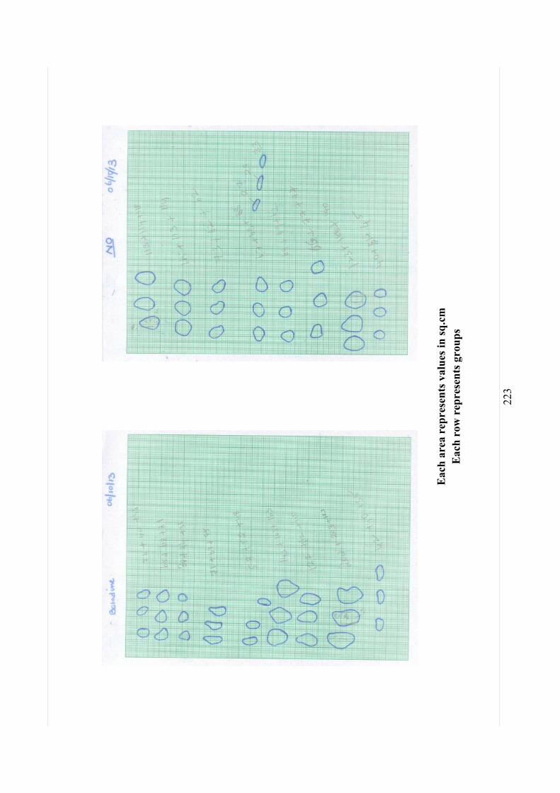
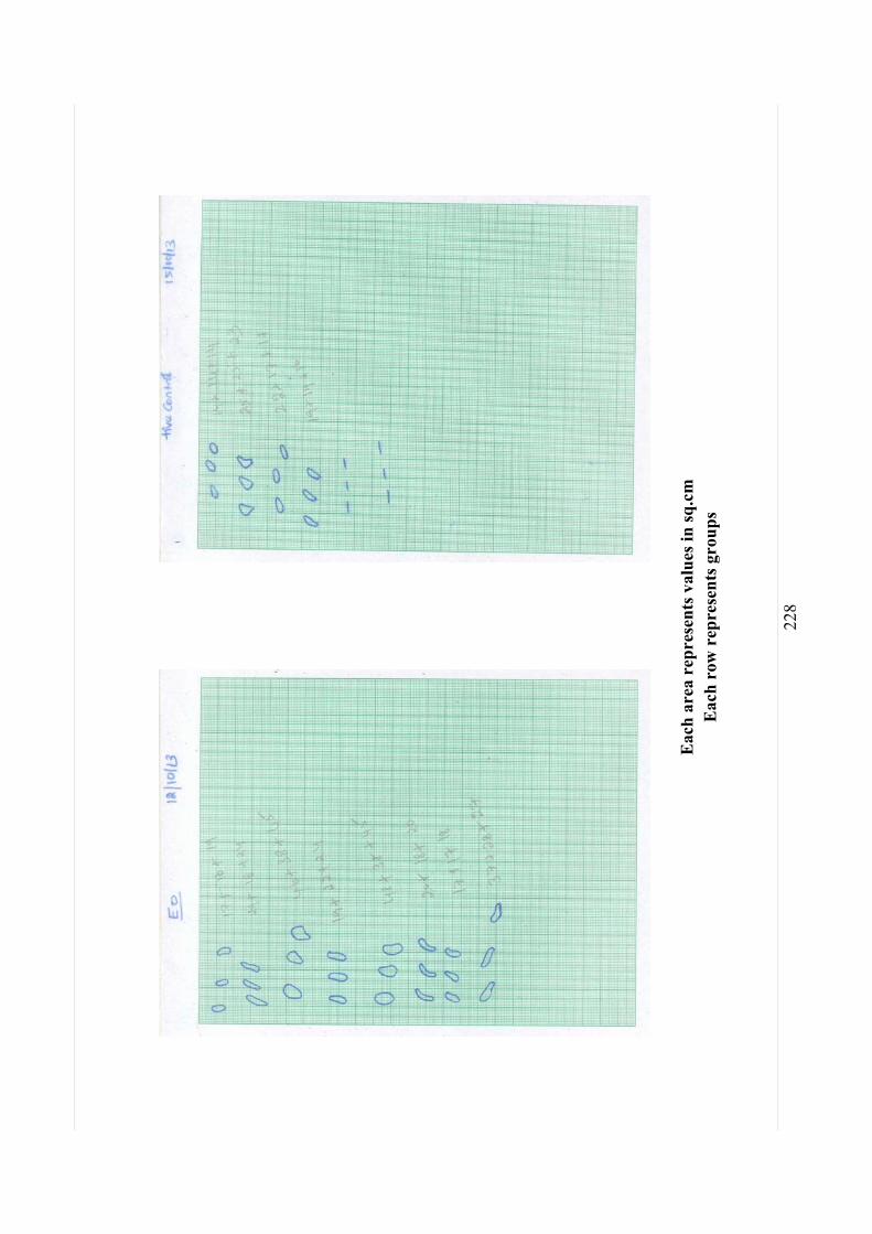
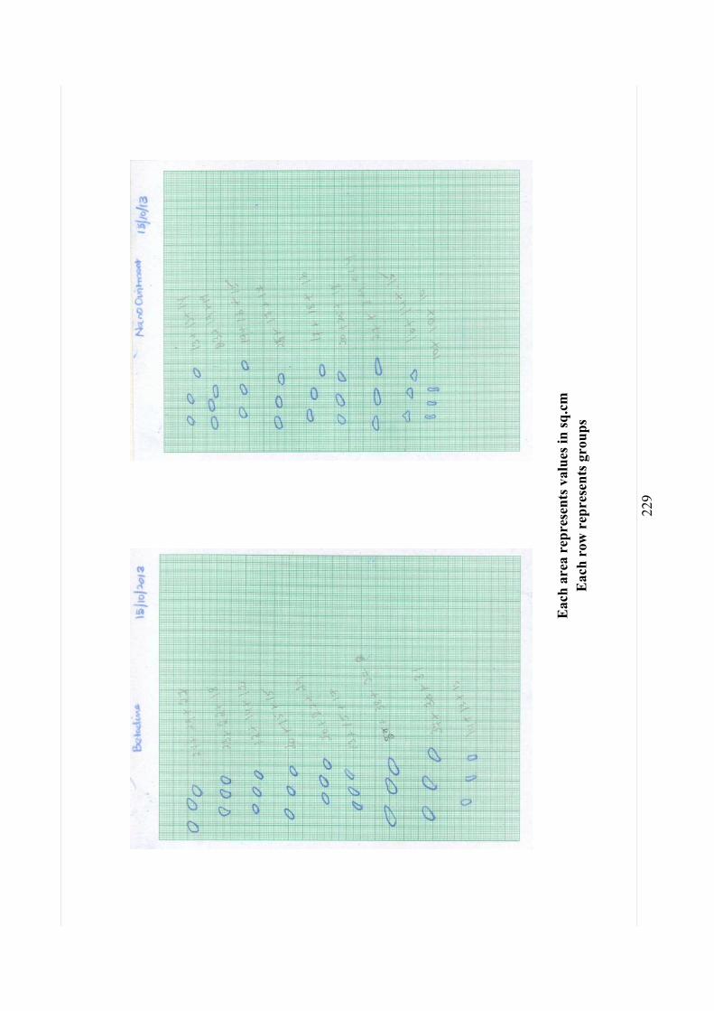
Eac
h ar
ea r
epre
sent
s val
ues i
n sq
.cm
E
ach
row
rep
rese
nts g
roup
s
SCO
RIN
G O
F W
OU
ND
FR
OM
0T
H D
AY
TO
21ST
DA
Y

218
Eac
h ar
ea r
epre
sent
s val
ues i
n sq
.cm
E
ach
row
rep
rese
nts g
roup
s

219
Eac
h ar
ea r
epre
sent
s val
ues i
n sq
.cm
E
ach
row
rep
rese
nts g
roup
s

220
Eac
h ar
ea r
epre
sent
s val
ues i
n sq
.cm
E
ach
row
rep
rese
nts g
roup
s

221
Eac
h ar
ea r
epre
sent
s val
ues i
n sq
.cm
E
ach
row
rep
rese
nts g
roup
s
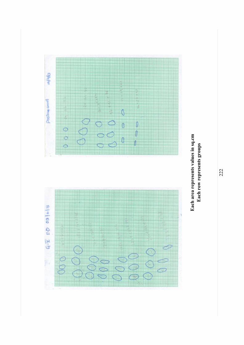
222
Eac
h ar
ea r
epre
sent
s val
ues i
n sq
.cm
E
ach
row
rep
rese
nts g
roup
s
223
Eac
h ar
ea r
epre
sent
s val
ues i
n sq
.cm
E
ach
row
rep
rese
nts g
roup
s
224
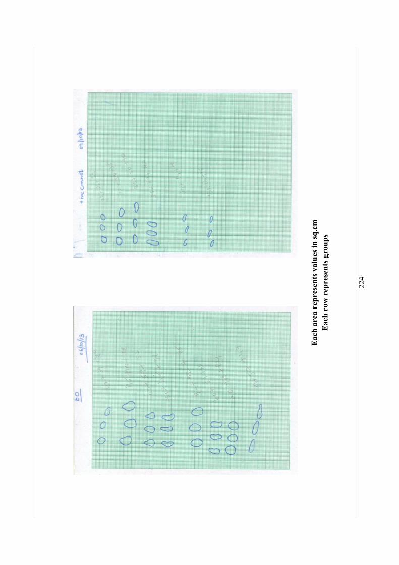
Eac
h ar
ea r
epre
sent
s val
ues i
n sq
.cm
E
ach
row
rep
rese
nts g
roup
s
225
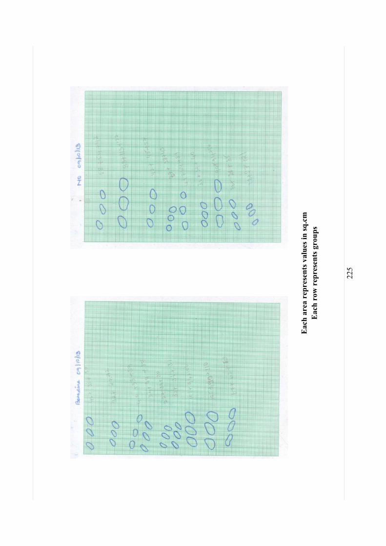
Eac
h ar
ea r
epre
sent
s val
ues i
n sq
.cm
E
ach
row
rep
rese
nts g
roup
s
226
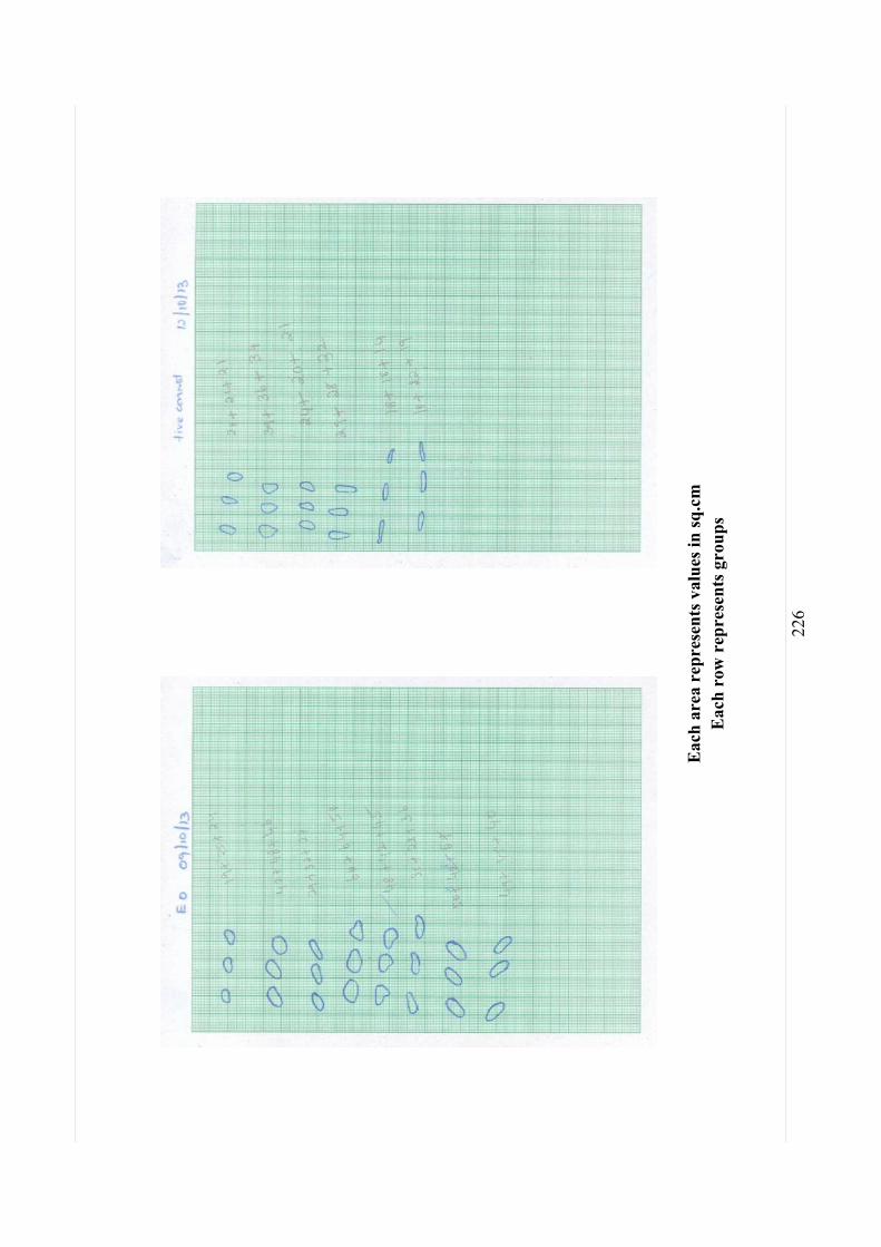
Eac
h ar
ea r
epre
sent
s val
ues i
n sq
.cm
E
ach
row
rep
rese
nts g
roup
s

227
Eac
h ar
ea r
epre
sent
s val
ues i
n sq
.cm
E
ach
row
rep
rese
nts g
roup
s
228
Eac
h ar
ea r
epre
sent
s val
ues i
n sq
.cm
E
ach
row
rep
rese
nts g
roup
s
229
Eac
h ar
ea r
epre
sent
s val
ues i
n sq
.cm
E
ach
row
rep
rese
nts g
roup
s

230
Eac
h ar
ea r
epre
sent
s val
ues i
n sq
.cm
E
ach
row
rep
rese
nts g
roup
s

231
Eac
h ar
ea r
epre
sent
s val
ues i
n sq
.cm
E
ach
row
rep
rese
nts g
roup
s
232
Eac
h ar
ea r
epre
sent
s val
ues i
n sq
.cm
E
ach
row
rep
rese
nts g
roup
s

233
References:
Ansel, H.C., L.V. Allen and G.N. Popovich, 2005. Ansel
Pharmaceuticals Dosage form and Drug Delivery System. 8th
Edn., Lippincott Williams and Wilkins, London, UK., pp:
279-282.
Asru,K. Sinha,1972. Calorimetric assay of catalase ,
Analytical biochemistry ,47,389-394.
Baie S, Hj, Sheikh KA: The wound healing properties of
Channa striatuscetrimide cream -tensile strength
measurement . J Ethnopharmacol 2000, 71:93 10.
Bancroft JD, Gamble M. 2008. Theory and practice of
histological techniques. 6th ed. London: Churchill
Livingstone
Bonte F, Dumas M, Chaudagne C, Meybeck A Influence of
Asiatic acid, madecassic acid, and asiaticoside on human
collagen I synthesis . Planta Med 1993, 60:133 135.
Bowler PG, Duerden BI, Armstrong DG. 2001. Wound
microbiology and associated approaches to wound
management . Clin Microbiol Rev .14:244 269. doi:
10.1128/CMR.14.2.244-269.2001
Bowler PG, Duerden BI, Armstrong DG. 2001. Wound
microbiology and associated approaches to wound
management . Clin Microbiol Rev . 2001, 14:244 269.
Bradley PP, Priebat DA, Christensen RD and Rothstein G, ,
Measurement of cutaneous inflammation: Estimation of

234
neutrophil content with an enzyme marker. J. Invest.
Dermatol. 1982,78(3): 206-209
Charde MS. Fulzele SV. Satturwar PM. Joshi SB.
Kasture AV. Dorle AK. Study on wound healing activity of
Mulathiaadi Ghrita Journal of Pharmaceutical Research
2005, 4(1): 08 12.
Clark RAF .1991. Cutaneous wound repairs , In Goldsmith
LA (ed.) Physiology, Biochemistry and Molecular Biology of
Skin. New York: Oxford University Press, p.576.
Dunphy, J. E. The Fibroblast-A Ubiquitous Ally for the
Surgeon. Shattuck Lecture . New Engl. J. Med.,
1963,268:1367,
Esimone CO, Nworu CS, Jackson CL, Cutaneous wound
healing activity of a herbal ointment containing the leaf
extract of Jatropha curcas L. (Euphorbiaceae) , International
Journal of Applied Research in Natural Products 1963.1(4),
1-4.
Fiore-Donati, L. & Moltke, E. (1960). Effect of reserpine, 5-
hydroxytryptamine and polymyxin B on mast cells and tensile
strengths of healing wounds . Acta endocr. (Kbh. ), 34, 430-
436.
Gross, J ., Highberger, J . H. & Schmrrr, F. O. (1952). Some
factors involved in the fibrogenesis of collagen in vitro
Proc. Soc. exp. Biol . (N.Y.), 80, 462-465.

235
Harkness, R. D, Biological Functions of Collagen . Biol.
Rev . , 36:399, 1961.
Howes, E. L., W. J. Sooy and S. C. Harvey The Healing of
Wounds as Determined by their Tensile Strength J. A. M. A . ,
1929, 92:42.
Karl M, Lacrix JV, Peterson HH, Canine Sugery, 4 th edn,
American Veterinary Publications, California ,1995, 42-45.
Lu L, Ying K, Wei S, Fang Y, Liu Y, Lin H, Ma L, Mao Y:
Asiaticoside induction for cell-cycle progression,
proliferation and collagen synthesis in human dermal
fibroblasts Int . J Dermatol 2004, 43:801 807.
Lubran,M.,and Moss, D. W., The determination of small
albumin concentrations using 311 -labeled albumin .
Clin.C/tim. Acta 2,246-251(1957)
Majumdar MR, Kamath JV. Herbal concept on wound
healing Journal of Pharmaceutical Research : 2005,4(1):
01 07.
Moren, M.S., Desplerra, J .W. and Mannervik, B. 1979,
Biochem Biophysic, Acta, 585, 67 .
Mortan Evaluation of Vulnerary by an open
procedure in rats . Arch. Int. Pharmacodyn : 1972. 196 117.
Mukherjee PK, Verpoorte R, Suresh B. Evaluation of
in vivo wound healing activity of Hypericum patulum
(Family:Hypericaceae) leaf extract on different wound model
in rats Journal of Ethnopharmacology , 2000, 70: 315 321.
236
Mustafa MR, Mahmood AA, Sidik K, Noor SM. Evaluation o
f wound healing potentia l of Ageratum conyzoides leaf
extract in combination with honey in rats as animal model .
Interational Journal of Molecular Medicine and Advance
Science : 2005. 1(4): 406 410.
Neuman, R.E., Logan, M.A., 1950. The determination of
hydroxyproline . Journal of Biological Chemistry 184, 299
306.
Odimegwu DC, Ibezim EC, Esimone CO, Nworu CS, Okoye
FBC, Wound healing and antibacterial activit ies of the
extract of Dissotis theifolia (Melastomataceae) stem
formulated in a simple Ointment base J Medicinal Plant Res,
2008 , 2(1): 011-016.
hydrox- The Journal of Biological Chemistry , 184,
1, 299 306, 1950.)
Ritu Sanwal, Amrendra Kumar Chaudhary, Wound healing
and antimicrobial potential of Carissa spinarum Linn.in
albino mice , Journal of Ethnopharmacology 2011, 135,792
796
Rotruck JTAL, Pope HE and Ganther AB Swanson
Biochemical role as acomponent of glutathione peroxidase
J science , 1973; 179: 588-590.
Rozaini, M.Z., Zuki, A.B.Z., Noordin, M., N orimah, Y.,
Hakim, A.N., The effects of different types of honey on

237
tensile strength evaluation of burn wound t issue healing .
International Journal of Applied Research in Veterinary
Medicine 2004,2, 290 296.
S. Murthy, M. K. Gautam, Shalini Goel, V. Puro hit , H.
Sharma, and R. K. Goel, Evaluation of In Vivo Wound
Healing Activity of Bacopa monniera on Different Wound
Model in Rats , BioMed Research International Volume 2013,
Article ID 972028, 9 pages
Stefan Marklund and Gudrun Marklund, Involvement of the
Superoxide Anion Radical in the Autoxidation of Pyrogallol
and a Convenient Assay for Superoxide Dismutase ,
Eur.J.biochem , 1974, 47,469-474
Sunita Shailajan, Sasikumar Menon, Suhas Pednekar, Ashish
Singh, Wound healing efficacy of Jatyadi Taila: In v ivo
evaluation in rat using excision wound model , Journal of
Ethnopharmacology , 2011, 138, 99 104
and Wilkin, Maryland, USA, p 150 -156
World Health Organization: National policy on traditional
medicine and regulation of herbal medicine -report of a WHO
global survey. http://apps.who.int/medicinedocs/en/d/Js7916e/
(accessed 10 March 2012).