1 title: influence of insertion site on central venous catheter (cvc
TRANSCRIPT
Influence of insertion site on central venous cathetercolonization and bloodstream infection rates
Author
Gowardman, John, K. Robertson, Iain, Parkes, Scott, Rickard, Claire
Published
2008
Journal Title
Intensive Care Medicine
DOI
https://doi.org/10.1007/s00134-008-1046-3
Copyright Statement
© 2008 Springer-Verlag. This is the author-manuscript version of this paper. Reproduced inaccordance with the copyright policy of the publisher. The original publication is available atwww.springerlink.com
Downloaded from
http://hdl.handle.net/10072/21668
Link to published version
http://www.springer.com/medicine/journal/134
Griffith Research Online
https://research-repository.griffith.edu.au

1
Title: Influence of insertion site on central venous catheter (CVC) colonization and
bloodstream infection rates.
Authors:
1, 2 John R Gowardman, FRACP, FJFICM
3 Iain K Robertson, M.Med.Sci
4 Scott Parkes, FRACP, FJFICM
5 Claire M Rickard, PhD
1Senior Specialist, Intensive Care Medicine, Intensive Care Unit, Launceston General
Hospital, Tasmania, 7250
Senior Lecturer, School of Medicine, University of Tasmania, AUSTRALIA, 7000.
2 Currently , Senior Specialist, Royal Brisbane and Woman’s Hospital, Herston,
Brisbane, 4029, AUSTRALIA, Senior Lecturer, University Of Queensland, QLD,
AUSTRALIA.
3Senior Research Fellow, School of Human Life Sciences, University of Tasmania,
Launceston, and Biostatistician, Clifford Craig Medical Research Trust, Launceston
General Hospital. Tasmania, 7250, AUSTRALIA
4 Senior Specialist, Intensive Care and Respiratory Medicine, Launceston General
Hospital, Tasmania, 7250, AUSTRALIA.
Associate Professor, School of Medicine, University of Tasmania, Tasmania,
AUSTRALIA 7000.
5 Professor of Nursing, Griffith University Research Centre for Clinical Practice
Innovation, School of Nursing and Midwifery, Brisbane, QLD, AUSTRALIA

2
Address for correspondence:
Dr John Gowardman, Senior Specialist, Department of Intensive Care Medicine
Level 3, Ned Hanlon Building Royal Brisbane and Woman’s Hospital, Herston,
Brisbane, QLD 4029 E mail: [email protected]
Word count, Abstract 255, Body Text 3379
3
ABSTRACT
Objective: To compare colonization and catheter related bloodstream infection (CR-
BSI) rates amongst three insertion sites (SC, IJ, FEM) used for central venous catheter
(CVC) placement.
Design: 24 month prospective study, with relative effects analyzed by Cox proportional hazards regression.
Setting: 8-bed ICU/HDU.
Patients: 410 critically ill patients requiring CVC placement.
Measurements and results: All short term multi-lumen CVCs, including
antimicrobial coated (AM) were studied. CVC management was standardized. Six
hundred and five CVCs (4,040 catheter days) were analyzed. Colonization and CR-BSI
incidence was 15.1 (95% CI 13.5; 21.0) and 1.8 (95% CI 1.2; 4.2) per 1,000 catheter-
days. Colonization was higher at the IJ (HR 3.64; 95% CI 1.32; 10.00; p= 0.01) and
FEM (HR 5.15; 95% CI 1.82; 14.51, p=0.004) sites compared with the SC. IJ v FEM
sites were not different, p= 0.34.The FEM site carried a greater risk of being colonized
by non S.epidermidis species compared with the SC and IJ sites combined (HR 4.15;
95% CI 1.79; 9.61, p=0.001). CVCs inserted in Department of Emergency Medicine
(DEM) were more colonized than those inserted in the ICU or operating theatre (OT)
(HR 2.66; 95% CI 1.27; 5.56; p= 0.01) and CVCs were less colonized in females
4
compared to males (HR 0.49; 95% CI 0.26; 0.89; p=0.02). No difference in CR-BSI
rates was noted between the three sites.
Conclusions:
Colonization was lowest at the SC site. Regional differences exist with respect to type
of pathogen isolated. Colonization was influenced by insertion location and gender. The
incidence of CR -BSI was not different.
Descriptor: 45
Key Words:
Catheterization, CVC, Central Venous Catheter, Intensive Care, Sepsis,
Colonization.

5
Introduction
Complications of intravascular access devices (IAD) in particular central venous
catheters (CVCs) can be classified as mechanical and infective [1, 2]. Increasing
awareness of factors that influence CVC related infection has resulted in evidence based
practice guidelines which have been shown to be effective in reducing rates of CVC
related sepsis [3]. Despite this, CVCs continue to remain one of the leading causes of
nosocomial sepsis in the critically ill.
The anatomical insertion sites commonly used for CVC placement are the Internal
Jugular (IJ), Subclavian (SC) and Femoral (FEM) veins. In terms of complications,
several studies have compared one site with another [2, 4, 5] and others the three sites
concurrently [6, 7, 8, 9, 10]. The Hospital Infection Control Advisory Committee
(HICPAC) [11] has consistently given selection of the SC site an IA recommendation
for preventing infection. A number of publications however have suggested the FEM
site is on par with upper body sites in terms of both sepsis and mechanical complication
rate [12, 13, 14, 15]. Some of these studies however were in particular subgroups of
patients such as children and burns injury [15, 16] and in others the conclusions
controversial [14, 17].
In our ICU all 3 vascular access sites were routinely used additionally accurate data on
IAD infection rates was prospectively collected. Due to the conflicting reports in the
literature we sought to investigate the use of CVCs in relation to placement site and to
compare infective outcomes and risk factors between CVCs placed at all three sites.

6
Materials & Methods
This prospective observational study was carried out over 24 months in an 8-bed
combined general intensive care unit (ICU) and co-located high dependency unit (HDU)
of a 350-bed regional Australian teaching hospital. The ICU treated all forms of acute
illnesses with the exception of post cardiothoracic surgical and acute neurosurgical
cases. Admission and treatment rights in the ICU were limited to attending intensivists
and the unit staffed by critical care registered nurses.
All short term non-tunneled CVCs (including peripherally inserted central lines), both
regular and antimicrobial coated (AM) that presented to, or were inserted in, the ICU
were included in the study. Neither pulmonary artery catheters, their introducer sheaths
nor long term access devices (e.g. Hickman’s catheters) were studied. The study was
conducted without clinical interference amongst the physicians inserting CVCs and was
intended to be a true reflection of clinical practice at that time. Institutional ethics
committee approval was obtained for using the non-identified data.
7
Data collection
For study entry the CVC must have been inserted within the departments of emergency
medicine (DEM), operating theatre (OT) or the ICU. CVCs inserted in other hospitals
were not included. During the study, CVCs were excluded if their removal, and
microbiological sampling, was not according to the study protocol. On admission to the
ICU, CVCs were identified with a unique identifier label which was attached to an
external lumen. Data collected and entered included: CVC insertion details (time, place,
and operator level of experience), CVC type (regular or AM, lumen number),
anatomical insertion site (FEM, SC, IJ, cubital) and CVC removal details (date, time,
reason and location).These data were completed for each CVC inserted. The clinical
nurse followed up the patient and completed the data entry in cases where discharge
from the ICU occurred prior to CVC removal. Other data collected included 24 hour
APACHE and SAPS II scores, APACHE II diagnostic codes, age, and sex. Data on
patient co morbidity or thrombotic events was not recorded. Microbiological details
including all catheter tip culture, blood culture and microorganisms isolated were
collected concurrently.
CVC management
Insertion of CVCs was performed by ICU personnel (intensivist, registrar, senior
resident). CVCs inserted in the OT or DEM were likewise inserted by trained operators
ideally under the same conditions. All regular ( non AM) CVCs used were multi-lumen
20cm polyurethane (Arrow® International, Reading, PA,USA) inserted using a standard
8
Seldinger approach under maximum sterile barrier precautions (sterile gloves, gown,
large drapes, mask and cap). Chlorhexidine 0.5% in ethanol was used as skin antisepsis.
AM catheters were ARROWg+ard Blue®, (Arrow® International, Reading, PA, USA).
These were also 20cm multi-lumen devices inserted under identical conditions. No
antibiotic coated CVCs were used. AM CVCs were used at the discretion of the medical
team. In general these CVCs were placed if the clinician expected the CVC in- situ
duration to exceed 7days or if the patient was clinically judged at high risk for
developing CR-BSI however insertion was not subject to protocol. All patients had
optimal CVC tip placement confirmed by plain CXR. For both types of CVC (regular
and AM) no specific anatomical insertion site was mandated by policy rather, insertion
into the IJ, SC, or FEM veins was based on patient variables such as risk of
pneumothorax and level of operator experience.
The CVC insertion site was inspected daily as part of the multidisciplinary ICU ward
round however superficial skin cultures were not taken. All line manipulations
including pressure transducers, giving sets, and site dressings were performed by ICU
nursing staff. All site dressings and giving sets were changed according to current
guidelines [11]. CVCs were not used routinely for blood sampling and guide-wire
exchange was not performed. The CVC was not changed on a scheduled basis but
removed for clinical suspicion of sepsis (with culture of the catheter tip and peripheral
blood), mechanical failure, or when no longer required. All patients if possible had their
CVC removed prior to discharge to the general wards and peripheral IV access inserted
if intravenous therapy was still deemed necessary [18].
9
Microbiological Sampling
CVCs were removed by the bedside ICU nurse.. The distal 3 to 5cm end of the CVC tip
was removed using a sterile dressing pack which included sterile forceps and scissors,
taking care not to contaminate the tip on removal. The tip was then immediately
transferred to a sterile container and transported to the microbiology department for
analysis [19].
Microbiological Definitions
The following definitions of CVC infection were applied [11]; Catheter Colonization:
tip culture > 15 CFU in the absence of BSI and CR-BSI: Catheter tip culture >15 CFU
plus a positive blood culture taken before or within 48 hours of CVC removal with the
same micro-organism and antibiogram with no other obvious source of infection
apparent.
Statistical analysis
The reported rates per 1,000 catheter days of colonization and CR-BSI were calculated
using Poisson regression. These were reported after adjustment for age, gender,
APACHE and SAPSII scores, insertion location (ICU, OT, DEM) and CVC type
(regular or AM) in order to remove these sources of confounding when assessing these
rates within the study. Poisson regression and simpler comparable methods for
calculation of incidence rates assume that these events were occurring at random
throughout the period each CVC is in-situ. However, colonization and CR-BSI are
10
terminating events, either because they are recorded only at the time of CVC removal or
because the CR-BSI provokes the removal of the catheter. The relative rates were
therefore compared using Cox proportional hazards regression, which is based on the
variable time of occurrence of each terminating event. A multivariate Cox proportional
hazards regression model was constructed by stepwise removal of insignificant (p>0.2)
variables (anatomical site of insertion, regular and or AM CVC, ICU/OT/DEM location
of insertion, specialist/registrar/RMO operator, diagnostic category, age, gender,
APACHEII and SAPSII scores). The same variables were then analyzed separately in
multivariate Cox proportional hazards regression models for the three sites of insertion.
All analyses were adjusted where required for multiple comparisons by the Holm
method. Time-to-event graphs were drawn to illustrate the occurrence of these events
over time. Statistical analyses were performed using STATA™ Statistics/Data Analysis
Version 9.2 (Stata Corp, College Station, Texas USA).
Results
General:
During the entire study period 618 CVCs were sited in 410 patients (226 (55.1%)
males). These patients had a mean age of 61.5 ± (SD) 16.5 years and APACHE II score
21.4 ± 17.9. Mean duration of catheterization was 6.5 ± 5.5 days. Ninety-five (23.2%)
patients died in hospital. Primary admission diagnoses were: 69 (15.3%) sepsis or other
major infection; 121 (26.9%) Post GI surgery; 55 (12.2%) multi-organ failure and
multiple trauma; 47 (10.4%) non-septic respiratory failure; 11 (2.4%) non-surgical GI

11
disease; 37 (8.2%) neurological disease; 31 (6.9%) cardiac failure or instability; 43
(9.6%) non-surgical malignancy; 36 (8.0%) other major surgery. Forty patients were
readmitted at later dates with alternate diagnoses.
In 13 CVC records the site was not recorded, leaving 605 CVCs (413 regular, 176 AM
and 16 cubital) in 410 patients for further analysis. No CVCs were excluded for missing
microbiological data. In total, these 605 CVCs were observed for 4,040 CVC days.
Detail of these 605 CVCs with rates of colonization and CR-BSI and total CVC in situ
time at each site is seen in table1. The overall incidence of microbial colonization and
CR-BSI was 15.1 (95% CI 13.5-21.0) and 1.8 (95% CI 1.2-4.2) per 1,000 catheter-days.
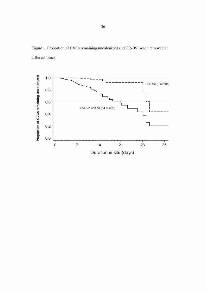
Inspection of fig 1 demonstrates the cumulative incidence of CVC colonization at the
time of catheter removal increased over time.
CR-BSI and Colonization at each anatomical insertion site:
Further analysis of colonization and CR-BSI incidence was performed only on the 589
CVCs that were sited at the IJ (regular 204, AM 75), SC (regular 59, AM 43) and FEM
(regular 150, AM 58) sites. Mean duration of catheterization at IJ, SC and FEM sites for
regular and AM catheters respectively was 5.5, 7.4 and 5.4 days and 8.5, 9.2, and 5.7
days with a significant difference noted in dwell time between IJ sites only ( p=0.001).
There were 9 episodes of CR-BSI. Number of CR-BSI events by CVC type can be seen
in table1. When using the SC as a reference CR-BSI rate between the three sites was
not significantly different (table2). Of all CVCs studied, IJ AM CVCs were associated
with the greatest CR-BSI rate (HR 7.02; 95% CI 0.35; 1.43, p=0.205).
12
Table 3a reports colonization rates and multivariate estimates of the simultaneous
effects of differing risk factors for CVC colonization at all sites combined and table 3b
at individual sites, after stepwise removal of insignificant (p>0.2) variables. The final
model found the only factors significantly associated with colonization were non SC
insertion site, DEM insertion location, male gender and use of regular CVCs.
Colonization at the IJ (HR 3.64; 95% CI 1.32; 10.00, p=0.01) and FEM (HR 5.15; 95%
CI 1.82; 14.51, p=0.004) sites was significantly greater than that at the SC (fig 2).
Colonization at the FEM site was not different from that at the IJ site (HR 1.31; 95% CI
0.54-3.21; p=0.34). CVCs inserted in the DEM were significantly more colonized than
those inserted in the ICU or OT (p= 0.01). CVCs were significantly less colonized in
females compared to males (p=0.02) an effect was most marked at the IJ site (p= 0.003).
There was a significant reduction in colonization when AB CVCs were used compared
with regular CVCs (p= 0.02). At individual sites this effect was greatest at the IJ site
(p=0.03) but not significant at FEM site (p=0.50). Colonization of CVCs was not
significantly different if inserted by registrars and residents (HR 1.22, 95% CI 0.31;
3.59 p=0.723) compared to specialists. Table 4 displays colonization and admitting
diagnosis. Colonization was lower in those patients with a primary diagnosis of sepsis
or other major infection (p=0.05).
A total of 81 microorganisms were responsible for the 68 CVC colonization’s (table5),
of which 62 (76.5%) were Gram-positive bacteria, 14 (17.3%) were Gram-negative
bacteria and 5 (6.2%) were yeasts. Isolated from the 81 microorganisms were: 50

13
(61.7%) Staphylococcus epidermidis; 12 (14.8%) Enterococcus faecalis; 9 (11.1%) S.
aureus; 5 (6.2%) Candida albicans; 3 (3.7%) Corynebacterium sp; and 2 (2.5%)
Klebsiella sp. The SC site was associated with the lowest level of isolates (3.7%).
At the IJ site 83% of isolates were S. epidermidis compared with 34% at the FEM site.
The likelihood of heavy colonization with non S. epidermidis organisms was
significantly greater at the FEM site v the SC and IJ sites combined ( HR 4.15;95% CI
1.79;9.61,p=0.001) whereas the SC and IJ sites were similar ( HR 2.01;95% CI
0.23;17.6, p=0.52).
Discussion
We have shown that in an environment of consistent CVC care after adjusting for the
effect of AM CVCs and CVC in situ time, that catheter tip colonization ( CTC) was
significantly different between the 3 commonly used CVC insertion sites in favor of the
SC. IJ and FEM sites were not different. Differences in colonization were also
observed with respect to insertion location, gender and type of pathogen isolated at
individual and all sites combined. For all CVCs no significant difference was detected
in CR-BSI rate between sites.
The HICPAC guidelines [11] for the prevention of intravascular catheter related
infections recommend that the SC site be used preferentially for CVC catheterization to
reduce the incidence of catheter related infection. This is based on 4 studies including
Merrer’s randomized controlled trial comparing FEM and SC access sites [2] and
Goetz’s prospective observational study [7]. The former study found that catheterization

14
at the FEM site was associated with a 5 fold increased incidence density in catheter
related infection over the SC site. In particular when the endpoints of colonization plus
CR-BSI were combined this difference was highly significant. Goetz [7] also found
catheter contamination to be associated with FEM location (HR 4.2; p=0.0001) and a
trend towards greater clinical infection at the same site (HR 4.7; p=0.08). Colonization
rates in this study were comparable to our own at 28.8/1,000 and 5.8/1,000 CVC days
for IJ and SC sites respectively but appeared lower than ours at the FEM site
(12.6/1,000 CVC days). Other studies have produced conflicting results. Although
Collignon [6] found a significantly higher colonization rate with catheters inserted at the
FEM site compared with the SC site, Richet [8] found the IJ and not the FEM site to be
independently associated with positive CVC tip culture. Despande [14] found that there
was no significant difference in the rate of infection including BSI or colonization at
three concurrently studied sites. These data led the authors to conclude all three sites
were safe as regards risk of infection providing site selection was chosen carefully,
trained personnel inserted the CVC and appropriate infection control measures were in
place. One of the only other studies to examine infection rates at all 3 sites concurrently
[10] found catheter related local infection (signs of local infection plus CTC) incidence
density was statistically higher for FEM than for IJ (15.83 versus 7.65, p < 0.001) and
SC (15.83 versus 1.57, p < 0.001) accesses, and higher for IJ than for SC access (7.65
versus 1.57, p < 0.001). CR-BSI incidence density was also statistically higher for FEM
than for IJ (8.34 versus 2.99, p = 0.002) and SC (8.34 versus 0.97, p < 0.001) accesses.

15
A common theme thru all of these studies is that the SC site is remains the lowest risk in
terms of both CTC and BSI rates. Our results support this assertion in that the SC site
was significantly less colonized that either the IJ or FEM sites which appeared
equivalent. CTC would appear to be a valid surrogate end point for BSI correlating
powerfully with the subsequent development of CR-BSI [20]. The difference we
observed at the IJ and FEM sites would support these two positions as second choice to
the SC site for routine CVC insertion. The salient issues of patient and operator
variables need consideration in this. In those at risk of complication with SC or IJ
access the FEM approach may be safest. The perception that SC access is more prone to
complication than the IJ may not be warranted with one study suggesting no difference
in the incidence of haemopnuemothorax between the two. These results must be
interpreted with caution however because they represent a meta analysis of non
randomized studies and exclude certain high risk sub groups such as patients with
COPD or ARDS [21]. Although our results suggest the SC is preferable in terms of
colonization the clinical end point of CR- BSI was not different at each three sites. As
suggested previously [14, 17] with optimal insertion and aftercare technique clinically
meaningful outcomes such as BSI may be equivalent between the three sites, insertion
site in this context being influenced by operator experience and risk of complication.
Varying factors influenced the colonization rates at differing sites. Differences in
colonization patterns of anatomical areas have been described previously [22]. We
observed a clear colonization benefit in favor of females at the IJ site but not SC or
FEM sites. The higher rate in males may in part be explained by the presence of facial

16
hair and beards which extends down to around the usual insertion sites of the IJ CVCs
increasing the risk of contamination. Whilst the IJ site cannot be recommended above
the SC for routine catheterization, our data suggests in females this site is likely to
remain significantly less colonized and may pose less of an infective risk. Although the
overall numbers of CVCs studied was small, devices inserted in the DEM, in particular
FEM CVCs were significantly more colonized than those inserted in either the ICU or
OT environments. This can be explained by the often emergent insertion in this
environment where sterility may be suboptimal. CVCs inserted in these conditions
should be replaced as soon as is practicable.
It has been suggested that anatomical insertion site may influence the type of bacteria
isolated from catheter tip culture and as a cause of CR-BSI [22, 23] few studies
however have compared three sites concurrently. Lorente [23] recently demonstrated
that the FEM site is an independent risk factor for BSI due to yeasts and gram negative
organisms. Our results also suggest that the FEM site carries a significantly greater risk
of infection than either the IJ or SC sites for non S. epidermis organisms. This may have
implications for treatment of suspected CR-BSI arising from the FEM site where
organisms of significantly greater virulence may be responsible.
Our study has a number of limitations which need to be considered when interpreting
the results. Despite the fact that clinical practice was uniform as regards insertion, use
and maintenance of the CVCs studied this was not a randomized comparison, therefore

17
FEM site selection may be biased toward more junior operators and emergent insertions
both of which will lead to higher colonization. Additionally despite the fact we
controlled for severity of illness in our analysis it is possible that bias may have also
occurred in patient selection with certain subgroups of patients more prone to FEM
insertion (and avoidance of SC) e.g. severe respiratory failure .
Although CTC which is a valid surrogate of BSI [21] remains unequivocally different
between the 3 sites, due to the very low rate of CR-BSI, our study was under powered
to detect differences in this outcome measure. With quality improvement initiatives the
use of CR-BSI may become problematic as the overall incidence of bloodstream
infection continues to reduce. Unless the background incidence of BSI is high, CTC
may therefore become a more valid and practically achievable end point in future
studies
Overall, although AM CVCs have been associated with up to 50% reductions in both
colonization rates and incidence of CR-BSI in multiple studies [24] controversy exists
with regard to their role [25,26 ]. Our results are consistent with those previously
reported with significant reductions in colonization compared with regular CVCS. Our
study was however not designed to demonstrate an outcome benefit for the use of AB
CVCs and analysis was limited by small numbers and confounded by microbiological
technique used to analyze CVC tips in particular the impact of external antiseptic
coating on colonization using the roll plate technique. We support the concept that AM

18
CVCs should be used selectively where the rates of CR-BSI remain unacceptably high
despite adherence to standard infection control practices [27].
In conclusion when CTC is used as an end point our study suggests that the SC site
remains the lowest risk of the three commonly used anatomical insertion sites for
routine CVC catheterization with no difference being found between the IJ and FEM
sites. Our results also suggest regional differences may exist with respect to insertion
location, gender and type of pathogen isolated.
19
Acknowledgments:
The authors thank the nursing and medical staff of the Launceston General Hospital,
ICU for their cooperation in the execution of the study. We would like to also thank
Mr Andy Brown ( Nurse Educator ,ICU) for database development, data acquisition
and assistance with study implementation. Statistical analysis was funded by the
Clifford Craig Medical Research Trust, Launceston, Tasmania, Australia. The authors
have no conflict of interest to declare.

20
References:
1. Polderman KH, Girbes AJ (2002) Central venous catheter use. Part 1: mechanical
complications. Intensive Care Med 28:1-17
2. Merrer J, De Jonghe B, Golliot F, Lefrant JY, Raffy B, Barre E, Rigaud JP, Casciani
D, Misset B, Bosquet C, Outin H, Brun-Buisson C, Nitenberg G (2001) French
Catheter Study Group in Intensive Care. Complications of femoral and subclavian
venous catheterization in critically ill patients: a randomized controlled trial. JAMA
286:700-7
3. Eggimann P, Harbarth S, Constantin MN, Touveneau S, Chevrolet JC, Pittet D (2000)
Impact of a prevention strategy targeted at vascular-access care on incidence of
infections acquired in intensive care. Lancet 355:1864-8
4. Sadoyama G, Gontijo Filho PP (2003) Comparison between the jugular and
subclavian vein as insertion site for central venous catheters: microbiological aspects
and risk factors for colonization and infection. Braz J Infect Dis 7:142-8.
5. Ruesch S, Walder B, Tramer MR (2002) Complications of central venous catheters:
internal jugular versus subclavian access-a systematic review.Crit Care Med 30:454-60.

21
6. Collignon P, Soni N, Pearson I, Sorrell T, Woods P (1988) Sepsis associated with
central vein catheters in critically ill patients. Intensive Care Med 14: 227-31
7. Goetz AM, Wagener MM, Miller JM, Muder RR (1998) Risk of infection due to
central venous catheters: effect of site of placement and catheter type. Infect Control
Hosp Epidemiol 19: 842-5
8. Richet H, Hubert B, Nitemberg G, Andremont A, Buu-Hoi A, Ourbak P, Galicier C,
Veron M, Boisivon A, Bouvier AM ( 1990) Prospective multicenter study of vascular-
catheter-related complications and risk factors for positive central-catheter cultures in
intensive care unit patients. J Clin Microbiol 11: 2520-5
9. Nagashima G, Kikuchi T, Tsuyuzaki H, Kawano R, Tanaka H, Nemoto H, Taguchi K,
Ugajin K (2006) To reduce catheter-related bloodstream infections: is the subclavian
route better than the jugular route for central venous catheterization? J Infect Chemother
12:363-5
10. Lorente L, Henry C, Martin MM, Jimenez A, Mora ML ( 2005) Central venous
catheter-related infection in a prospective and observational study of 2,595 catheters.
Crit Care 9: 631-5

22
11.O'Grady NP, Alexander M, Dellinger EP, Gerberding JL, Heard SO, Maki DG,
Masur H, McCormick RD, Mermel LA, Pearson ML, Raad II, Randolph A, Weinstein
RA (2002) Guidelines for the prevention of intravascular catheter-related infections.
Centers for Disease Control and Prevention. MMWR Recomm Rep 51:1-29
12. Durbec O, Viviand X, Potie F, Vialet R, Albanese J, Martin C (1997) A prospective
evaluation of the use of femoral venous catheters in critically ill adults. Crit Care Med
25: 1986-9
13.Williams JF, Seneff MG, Friedman BC, McGrath BJ, Gregg R, Sunner J,
Zimmerman JE (1991) Use of femoral venous catheters in critically ill adults:
prospective study. Crit Care Med. 19:550-3
14. Deshpande KS, Hatem C, Ulrich HL, Currie BP, Aldrich TK, Bryan-Brown CW,
Kvetan V (2005) The incidence of infectious complications of central venous catheters
at the subclavian, internal jugular, and femoral sites in an intensive care unit population.
Crit Care Med 33:13-20
15 Venkataraman ST, Thompson AE, Orr RA (1997) Femoral vascular catheterization
in critically ill infants and children. Clin Pediatr (Phila) 36:311-9
23
16. Murr MM, Rosenquist MD, Lewis RW 2nd, Heinle JA, Kealey GP ( 1991) A
prospective safety study of femoral vein versus non femoral vein catheterization in
patients with burns. J Burn Care Rehabil 12:576-8
17. O'Grady NP, Dezfulian C (2005) The femoral site as first choice for central venous
access? Not so fast. Crit Care Med 33:234-5
18. Gowardman JR, Kelaher C, Whiting J, Collignon PJ (2005) Impact of a formal
removal policy for central venous catheters on duration of catheterisation. Med J Aust
182:249-50
19.Maki DG, Weise CE, Sarafin HW (1977) A semi quantitative culture method for
identifying intravenous-catheter-related infection. N Engl J Med 296: 1305-9
20. Ruesch S, Walder B, Tramer MR ( 2002) Complications of central venous catheters:
Internal jugular versus subclavian access-A systematic review. Crit Care Med 30:454-
460
21 Rijnders BJ, Van Wijngaerden E, Peetermans WE (2002) Catheter-tip colonization
as a surrogate end point in clinical studies on catheter-related bloodstream infection:
how strong is the evidence? Clin Infect Dis 35:1053-8

24
22. Lorente L, Jimenez A, Iribarren JL, Jimenez JJ, Martin MM, Mora ML (2006) The
micro-organism responsible for central venous catheter related bloodstream infection
depends on catheter site. Intensive Care Med 32:1449-50
23. Lorente L, Jimenez A, Santana M, Iribarren JL, Jimenez JJ, Martin MM, Mora ML
(2007) Microorganisms responsible for intravascular catheter-related bloodstream
infection according to the catheter site. Crit Care Med; 21 [E pub ahead of print]
24. Crnich CJ, Maki DG (2004) Are antimicrobial-impregnated catheters effective?
Don't throw out the baby with the bathwater. Clin Infect Dis 38:1287-92
25. McConnell SA, Gubbins PO, Anaissie EJ (2003) Do antimicrobial-impregnated
central venous catheters prevent catheter-related bloodstream infection? Clin Infect Dis
37: 65-72
26. Maki DG, Kluger DM, Crnich CJ (2006) The risk of bloodstream infection in adults
with different intravascular devices: a systematic review of 200 published prospective
studies. Mayo Clin Proc 81: 1159-71
27.Crnich CJ, Maki DG (2005) Are antimicrobial-impregnated catheters effective?
When does repetition reach the point of exhaustion? Clin Infect Dis 41: 681-5

25
Table1. Total numbers of CVCs at different insertion sites; with associated rates of colonization and CR-BSI
CVC (n)* CVC Time
(CVC days)
Colonization‡ CR-BSI
Site n % Rate n % Rate
IJ 204 1,118 25 12.3% 22.0 1 0.49% 0.7
SC 59 439 4 6.8% 8.7 0 0.00% 0.0
FEM 150 802 17 11.3% 20.1 1 0.67% 1.3
Cubital 16 203 1 6.3% 4.4 0 0.00% 0.0
IJ AM§ 75 635 11 14.7% 18.2 5 6.67% 6.9
SC AM§ 43 395 3 7.0% 7.5 2 4.65% 4.5
FEM AM§ 58 330 7 12.1% 19.1 0 0.00% 0.0
All sites 605 4,040 68 11.0% 15.1 9 1.46% 1.8

26
Legend table 1
* Of the 618 CVCs, the site was not recorded in 13 catheter records
‡Colonization (> 15 CFU): n= number observed and as % of total CVCs colonized at time of CVC removal with associated rate,
calculated by Poisson regression adjusted for age, gender, APACHE and SAPSII scores.
CR-BSI: n=number observed and as % of total CVCs inserted at each site
§ Anti-microbial CVC

27
Table 2: Adjusted rates of CR-BSI for each site for both regular and AM CVCs (n =589§) combined: the risks at the FEM and IJ sites
are indexed to the risk at the SC site.
Site Rate* HR † 95% CI PΩ
SC 1.26 1.00
IJ 2.19 2.82 (0.50; 15.8) 0.47
FEM 0.75 1.39 (0.13; 15.2) 0.79
28
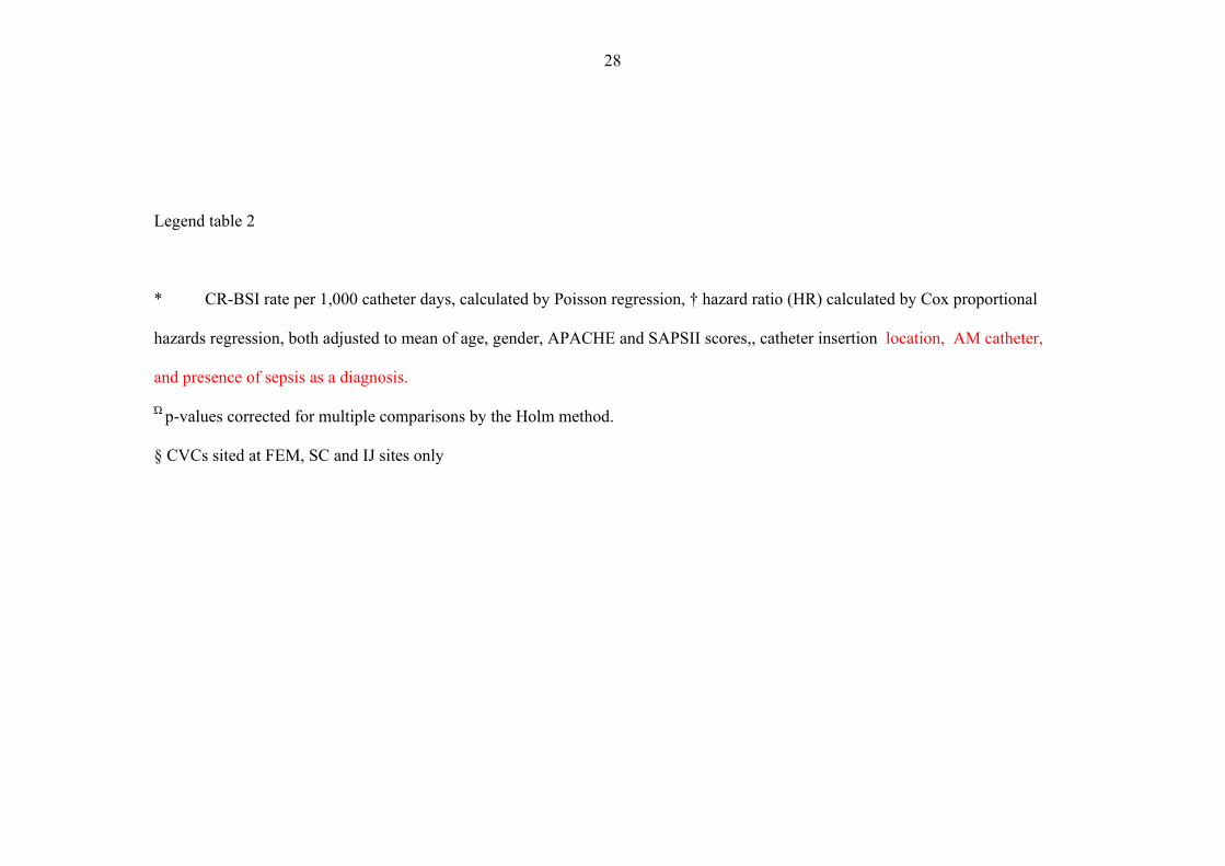
Legend table 2
* CR-BSI rate per 1,000 catheter days, calculated by Poisson regression, † hazard ratio (HR) calculated by Cox proportional
hazards regression, both adjusted to mean of age, gender, APACHE and SAPSII scores,, catheter insertion location, AM catheter,
and presence of sepsis as a diagnosis.
Ώ p-values corrected for multiple comparisons by the Holm method.
§ CVCs sited at FEM, SC and IJ sites only

29
Table 3a. Comparison of colonization between IJ, FEM and SC sites N (colonized) Rate* HR† 95% CI p‡
Site§ SC 102 (6) 8.1 1.00
IJ 279 (33) 19.7 3.64 (1.32 ; 10.03) 0.01
FEM 208 (24) 26.4 5.15 (1.82 ; 14.51) 0.004
CVC type Regular 413 (45) 18.3 1.00
AM 176 (18) 13.8 0.47 (0.25 ; 0.89) 0.02
Where
inserted
ICU 443 (45) 16.0 1.00
OR 117 (13) 16.8 1.17 (0.60 ; 2.30) 0.63
DEM 29 (5) 32.8 2.66 (1.27 ; 5.56) 0.01
Gender Male 349 (41) 20.1 1.00
Female 240 (22) 12.9 0.49 (0.26 ; 0.89) 0.02
Sepsis Absent 415 (51) 21.6 1.00
Present 120 (8) 13.4 0.60 (0.31 ; 1.19) 0.14
30
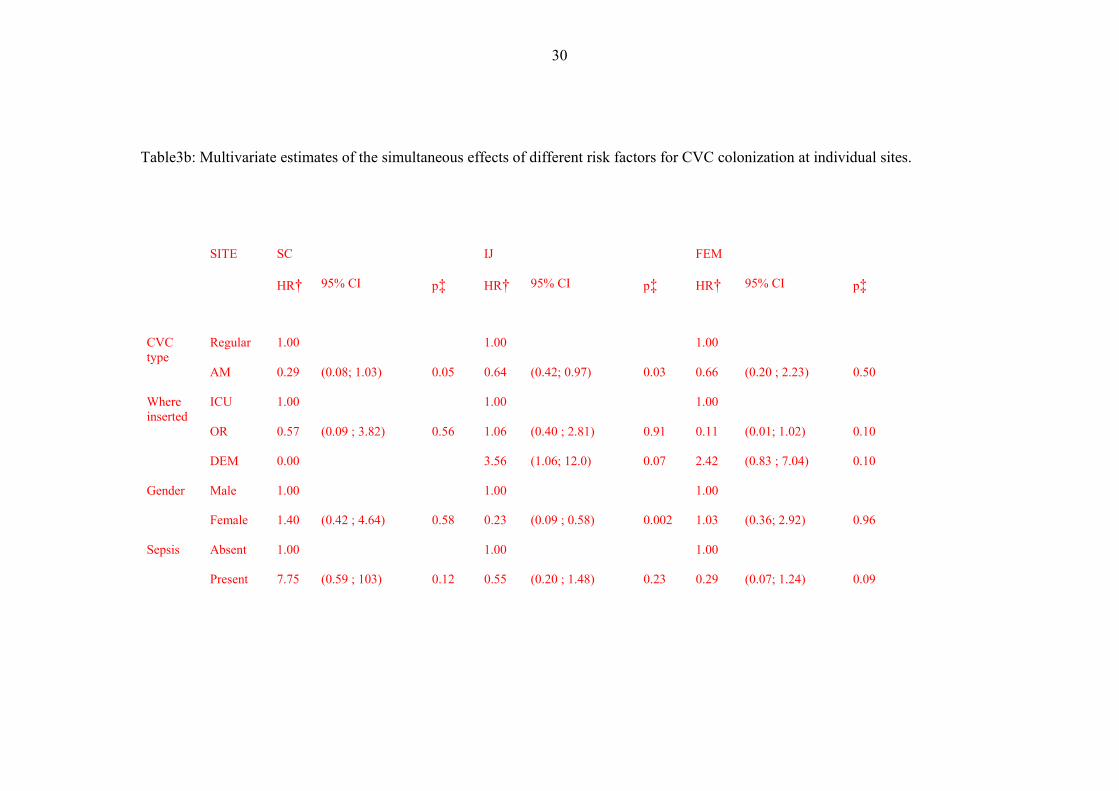
Table3b: Multivariate estimates of the simultaneous effects of different risk factors for CVC colonization at individual sites.
SITE SC IJ FEM
HR† 95% CI p‡ HR† 95% CI p‡ HR† 95% CI p‡
CVC type
Regular 1.00 1.00 1.00
AM 0.29 (0.08; 1.03) 0.05 0.64 (0.42; 0.97) 0.03 0.66 (0.20 ; 2.23) 0.50
Where inserted
ICU 1.00 1.00 1.00
OR 0.57 (0.09 ; 3.82) 0.56 1.06 (0.40 ; 2.81) 0.91 0.11 (0.01; 1.02) 0.10
DEM 0.00 3.56 (1.06; 12.0) 0.07 2.42 (0.83 ; 7.04) 0.10
Gender Male 1.00 1.00 1.00
Female 1.40 (0.42 ; 4.64) 0.58 0.23 (0.09 ; 0.58) 0.002 1.03 (0.36; 2.92) 0.96
Sepsis Absent 1.00 1.00 1.00
Present 7.75 (0.59 ; 103) 0.12 0.55 (0.20 ; 1.48) 0.23 0.29 (0.07; 1.24) 0.09

31
Legend table 3a and b § Colonization at the IJ and FEM sites was compared to colonization at the SC site * Colonization per 1,000 CVC days calculated by multivariate Poisson regression, † Hazard ratio and 95% confidence intervals
estimated by multivariate Cox proportional hazards regression, The model included variables in the table adjusted to mean of age, and severity of illness.
‡ P-values corrected for multiple comparisons by the Holm method

32
Table 4: Absolute and relative rates of CVC colonization in patients with different reasons for admission: each disease is compared
with the rates in all the remaining patients.
n* N* Rate
95% CI HR† 95% CI p
Organ failure 7 45 16.1 (7.31; 35.4) 2.84 (0.94 ; 8.58) 0.06
GI disease 2 10 31.2 (11.1; 88.0) 2.46 (0.13 ; 46.02) 0.54
Respiratory disease 6 44 17.6 (8.83 ; 35.2) 1.58 (0.45; 5.61) 0.47
Neurological disease 5 31 25.1 (10.3; 61.1) 1.54 (0.21 ; 11.35) 0.67
Cancer 8 38 17.7 (8.82 ; 35.3) 1.16 (0.25; 5.43) 0.85
GI surgery 16 102 21.7 (13.1;35.8) 0.88 (0.33; 2.36) 0.80
Sepsis 7 63 9.27 (4.57 ;18.8) 0.32 (0.10; 1.04) 0.05
Cardiovascular shock 3 24 16.8 (5.69; 49.7) 0.23 (0.01; 4.20) 0.32
Major surgery 0 18 8.7 (1.33; 57.7) 0.20 (0.03; 1.50) 0.11

33
Legend table 4
* Number of colonization’s (n) in patients (N) with the condition
† Hazard Ratio (HR) or relative rate calculated by Cox proportional hazards regression, adjusted for age, gender, APACHE and
SAPSII scores.
34
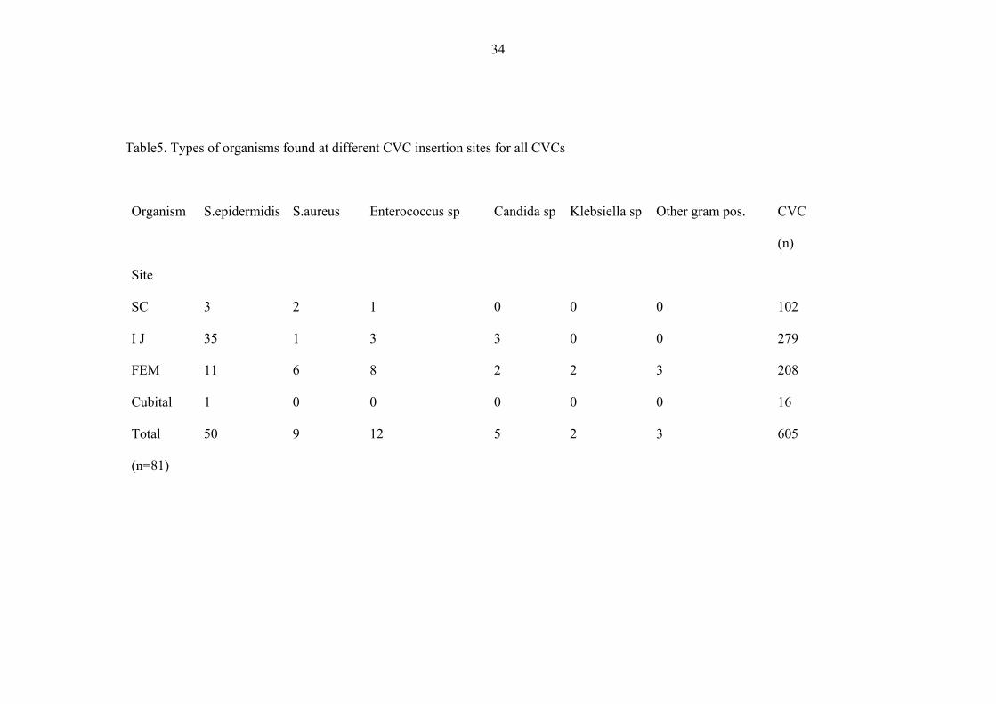
Table5. Types of organisms found at different CVC insertion sites for all CVCs
Organism
Site
S.epidermidis S.aureus Enterococcus sp Candida sp Klebsiella sp Other gram pos. CVC
(n)
SC 3 2 1 0 0 0 102
I J 35 1 3 3 0 0 279
FEM 11 6 8 2 2 3 208
Cubital 1 0 0 0 0 0 16
Total
(n=81)
50 9 12 5 2 3 605

35
Legend table 5
The likelihood of heavy colonization with non S. epidermidis organisms was greater at the FEM site v the SC and IJ sites (HR 4.15;
95% CI 1.79; 9.61, p=0.001).
36
Figure1. Proportion of CVCs remaining uncolonized and CR-BSI when removed at
different times

37
Figure 2. Proportion of CVCs remaining uncolonized versus duration in situ at three
sites.

38
Legend fig 2
Both plain and AM CVCs are represented. Colonization at the IJ (HR 3.64; 95% CI
1.32; 10.00, p=0.01) and FEM (HR 5.15; 95% CI 1.82; 14.51, p=0.004) sites was
significantly greater than that at the SC. Colonization at the FEM site was not different
from that at the IJ site (HR 1.31; 95% CI 0.54-3.21; p=0.34).