1 © the author 2012. published by oxford university press. all
TRANSCRIPT

Nxnl2 splicing results in dual functions in neuronalcell survival and maintenance of cell integrity
Celine Jaillard1,2,3, Aurelie Mouret4,5, Marie-Laure Niepon1,2,3, Emmanuelle Clerin1,2,3,
Ying Yang1,2,3, Irene Lee-Rivera1,2,3, Najate Aıt-Ali1,2,3, Geraldine Millet-Puel1,2,3,
Therese Cronin6, Tina Sedmak7, Wolfgang Raffelsberger8, Bernd Kinzel9, Alain Trembleau10,
Olivier Poch8, Jean Bennett6, Uwe Wolfrum7, Pierre-Marie Lledo4,5, Jose-Alain Sahel1,2,3,∗
and Thierry Leveillard1,2,3,∗
1Institut de la vision, INSERM, U968, Paris F-75012, France, 2UPMC Universite Paris 06, UMR-S 968, Institut de la
Vision, Paris F-75012, France, 3CNRS, UMR_7210, Paris F-75012, France, 4Institut Pasteur, Paris F-75015, France,5CNRS URA2182, Paris F-75015, France, 6Scheie Eye Institute, University of Pennsylvania, Philadelphia, PA, USA,7Johannes Gutenberg University of Mainz, Institute of Zoology, Cell and Matrix Biology, Muellerweg 6, D-55099 Mainz,
Germany, 8Laboratoire de Bioinformatique et Genomique Integratives, IGBMC, Illkirch, France, 9Novartis Pharma,
Basel, Switzerland and 10Universite Pierre et Marie Curie, UMR 7102, CNRS, Paris F-75005, France
Received December 7, 2011; Revised and Accepted February 10, 2012
The rod-derived cone viability factors, RdCVF and RdCVF2, have potential therapeutical interests for thetreatment of inherited photoreceptor degenerations. In the mouse lacking Nxnl2, the gene encodingRdCVF2, the progressive decline of the visual performance of the cones in parallel with their degeneration,arises due to the loss of trophic support from RdCVF2. In contrary, the progressive loss of rod visual functionof the Nxnl22/2 mouse results from a decrease in outer segment length, mediated by a cell autonomousmechanism involving the putative thioredoxin protein RdCVF2L, the second spliced product of the Nxnl2gene. This novel signaling mechanism extends to olfaction as shown by the progressive impairment of olfac-tion in aged Nxnl22/2 mice and the protection of olfactory neurons by RdCVF2. This study shows that Nxnl2is a bi-functional gene involved in the maintenance of both the function and the viability of sensory neurons.
INTRODUCTION
Inherited retinal degenerations (IRDs) constitute a group ofgenetically heterogeneous diseases that are generally untreat-able and commonly lead to blindness. The most commonform of IRD, retinitis pigmentosa (RP), is characterized clin-ically by an initial loss of night vision resulting from the dys-function and death of rod photoreceptors, followed by aprogressive non-cell autonomous loss of cones. Through theinvestigation of medical approaches to prevent secondarycone death in RP patients, we have demonstrated that rodssecrete trophic factors essential for cone viability, and setabout identifying such factors by high content screening fortrophic support of cone-enriched primary cultures (1–3).From this screen rod-derived cone viability factor (RdCVF),
one of the products of the Nucleoredoxin-like 1 (Nxnl1)gene was isolated. Injection of the RdCVF protein protectsthe cones of two rodent models of RP, the rd1 mouse andthe Pro23His rat (4).
In silico, we have identified a paralogue gene Nxnl2 thatshares most of the characteristics of the gene encodingRdCVF. Nxnl2, such as Nxnl1, encodes for a short isoform(respectively, RdCVF2 and RdCVF) and shows similartrophic effects on cone photoreceptors (5). These trophicmolecules are produced by the absence of splicing of theintron in between the two coding exons and of a stop codonin frame of the first exon. The splicing of this intron leadsto the production of long isoforms (RdCVF2L andRdCVFL) that possess an entire thioredoxin fold (6), unlikethe short isoforms. This novel signaling pathway, involving
∗To whom correspondence should be addressed at: Institut de la vision, INSERM, U968, UPMC Universite Paris 06, UMR-S 968, CNRS, UMR 7210,Paris F-75012, France. Tel: +33 153462548; Fax: +33 153462502; Email: [email protected] (T.L.); [email protected] (J.A.S.)
# The Author 2012. Published by Oxford University Press. All rights reserved.For Permissions, please email: [email protected]
Human Molecular Genetics, 2012, Vol. 21, No. 10 2298–2311doi:10.1093/hmg/dds050Advance Access published on February 15, 2012
Downloaded from https://academic.oup.com/hmg/article-abstract/21/10/2298/2900729by gueston 21 February 2018
bi-functional thioredoxin-like genes, is suggested by thefinding that Nxnl2 expression is not restricted to the retina(5), to be further implicated in neurodegenerative diseasesoutside the eye. We confirm the importance of Nxnl2 in main-taining cone photoreceptors throughout the life of the animalby showing a gradual loss of cones preceded by the loss oftheir function in the Nxnl22/2 mice. Delivering the trophicfactor RdCVF2 by an adeno associated viral (AAV) vectorto these mice prevented the loss of cone function. The rodfunction was also affected in the Nxnl22/2 retina as charac-terized by shortening of rod outer segments. In this case, treat-ing these mice with a vector expressing the alternative Nxnl2splice form, AAV-RdCVF2L, prevented this shortening. Inter-estingly, the messengers for RdCVF2 and RdCVF2L werealso detected in olfactory neurons, and olfactory function wasimpaired in Nxnl22/2 aged mice. Taken together, these obser-vations demonstrate that this novel signaling mechanism in-volving the non-cell autonomous effects of RdCVF2 and thecell autonomous effects of the second Nxnl2 isoformRdCVF2L supports two essential sensory systems: the retinalphotoreceptor function and the non-retinal olfactory function.
RESULTS
Construction of Nxnl22/2 mouse strain
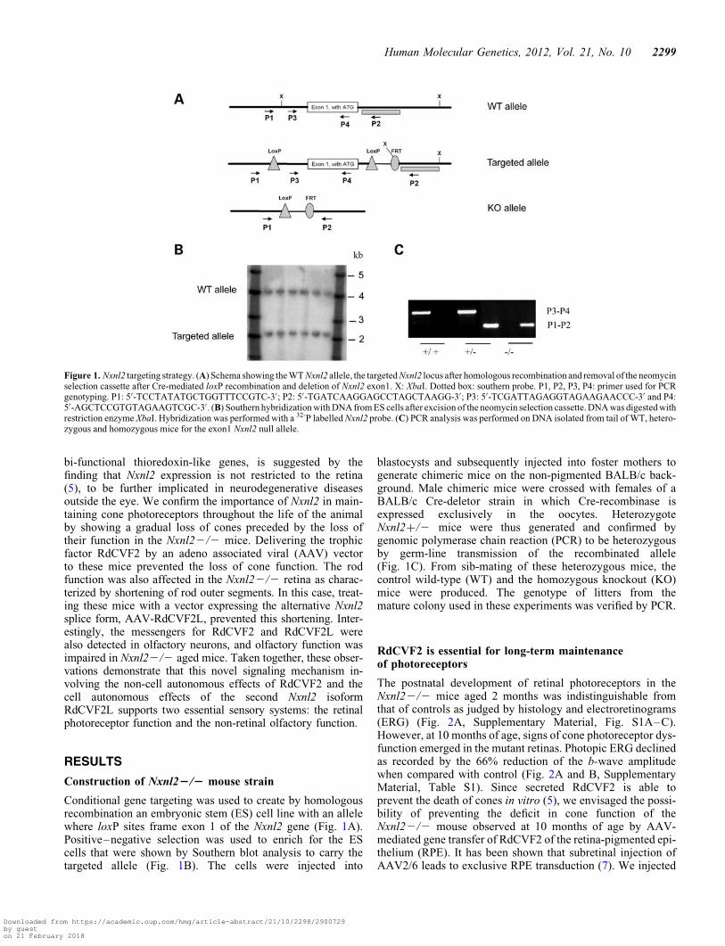
Conditional gene targeting was used to create by homologousrecombination an embryonic stem (ES) cell line with an allelewhere loxP sites frame exon 1 of the Nxnl2 gene (Fig. 1A).Positive–negative selection was used to enrich for the EScells that were shown by Southern blot analysis to carry thetargeted allele (Fig. 1B). The cells were injected into
blastocysts and subsequently injected into foster mothers togenerate chimeric mice on the non-pigmented BALB/c back-ground. Male chimeric mice were crossed with females of aBALB/c Cre-deletor strain in which Cre-recombinase isexpressed exclusively in the oocytes. HeterozygoteNxnl2+/2 mice were thus generated and confirmed bygenomic polymerase chain reaction (PCR) to be heterozygousby germ-line transmission of the recombinated allele(Fig. 1C). From sib-mating of these heterozygous mice, thecontrol wild-type (WT) and the homozygous knockout (KO)mice were produced. The genotype of litters from themature colony used in these experiments was verified by PCR.
RdCVF2 is essential for long-term maintenanceof photoreceptors
The postnatal development of retinal photoreceptors in theNxnl22/2 mice aged 2 months was indistinguishable fromthat of controls as judged by histology and electroretinograms(ERG) (Fig. 2A, Supplementary Material, Fig. S1A–C).However, at 10 months of age, signs of cone photoreceptor dys-function emerged in the mutant retinas. Photopic ERG declinedas recorded by the 66% reduction of the b-wave amplitudewhen compared with control (Fig. 2A and B, SupplementaryMaterial, Table S1). Since secreted RdCVF2 is able toprevent the death of cones in vitro (5), we envisaged the possi-bility of preventing the deficit in cone function of theNxnl22/2 mouse observed at 10 months of age by AAV-mediated gene transfer of RdCVF2 of the retina-pigmented epi-thelium (RPE). It has been shown that subretinal injection ofAAV2/6 leads to exclusive RPE transduction (7). We injected
Figure 1. Nxnl2 targeting strategy. (A) Schema showing the WT Nxnl2 allele, the targeted Nxnl2 locus after homologous recombination and removal of the neomycinselection cassette after Cre-mediated loxP recombination and deletion of Nxnl2 exon1. X: XbaI. Dotted box: southern probe. P1, P2, P3, P4: primer used for PCRgenotyping. P1: 5′-TCCTATATGCTGGTTTCCGTC-3′; P2: 5′-TGATCAAGGAGCCTAGCTAAGG-3′; P3: 5′-TCGATTAGAGGTAGAAGAACCC-3′ and P4:5′-AGCTCCGTGTAGAAGTCGC-3′. (B) Southern hybridization with DNA from ES cells after excision of the neomycin selection cassette. DNA was digested withrestriction enzyme XbaI. Hybridization was performed with a 32-P labelled Nxnl2 probe. (C) PCR analysis was performed on DNA isolated from tail of WT, hetero-zygous and homozygous mice for the exon1 Nxnl2 null allele.
Human Molecular Genetics, 2012, Vol. 21, No. 10 2299
Downloaded from https://academic.oup.com/hmg/article-abstract/21/10/2298/2900729by gueston 21 February 2018

subretinally a group of 6-month-old mice with AAV2/6-RdCVF2 in one eye and another group with AAV2/6-EGFP. A third group of mice was not injected. The functionalrescue was evaluated 4 months later (i.e. at 10 months of age)using ERG recording. At 10 months of age, photopic ERGshows dramatic decrease b-wave in untreated (53+ 13 mV)or treated mice with AAV-GFP (59+ 13 mV), in agreementwith Figure 1A (Fig. 2C). In contrast, Nxnl22/2 micetreated with AAV-RdCVF2 have higher ERG amplitude(135+ 32 mV) at 10 months. These results show that the
short trophic isoform encoded by the Nxnl2 gene is sufficientto prevent the loss of function brought into play by cone photo-receptors.
In order to investigate possible deficit in cone survival, westained cones using the lectin Peanut Agglutinin (PNA) (8). At2 months of age, no deficit in cone density was observed forthe Nxnl22/2 mouse retina, in agreement with the absenceof cone dysfunction. However, by 10 months of age, thecone density was reduced by 23% in the Nxnl22/2 retina(Fig. 2D). Furthermore, in accordance to previous results
Figure 2. Cone degeneration and dysfunction at 10 months in Nxnl22/2 mice. (A) Photopic ERG tracing from WT and Nxnl22/2 at 2 and 10 months of age.(B) Summarized photopic ERG data from 10 months of age mice (n ¼ 10). (C) Photopic ERG from Nxnl22/2 mice retina treated at 6 months of age withAAV-GFP, AAV-RdCVF2 or untreated. AAV-RdCVF2 delivery to the retina prevents the loss of cone function observed at 10 months of age in the animaltreated with AAV-GFP. (D) Number of cones was evaluated by counting PNA-positive cells at 10 months of age. (E) Immunohistochemistry using PNA labelingand S-Opsin antibody on Nxnl2+/+ and Nxnl22/2 flat-mounted retina. In the Nxnl22/2 retina, the presence of cones labeled with PNA without expression ofS-opsin is indicated with an arrow (scale bar: 5 mm). (F) Immunohistochemistry using PNA labeling and M-Opsin antibody on Nxnl2+/+ and Nxnl22/2 cross-sections in ventral part of retina. Scale bar: 20 mm.
2300 Human Molecular Genetics, 2012, Vol. 21, No. 10
Downloaded from https://academic.oup.com/hmg/article-abstract/21/10/2298/2900729by gueston 21 February 2018

(9), we observed a decrease in S-cones density in Nxnl22/2flat-mounted retina in the ventral part of the retina. We alsonotice specifically in the Nxnl22/2 retina the presence ofcones labeled with PNA, a marker of the cone extracellularmatrix sheet, without expression of S-opsin in the ventralretina (Fig. 2E). This observation indicates that the loss ofcone outer segments may precede their degeneration. Analysisof the ventral region showed that M-cones are affected to thesame extent in the Nxnl22/2 retina (Fig. 2F). This result sug-gests that cone photoreceptors were undergoing progressivedegeneration preceded by functional loss, supporting the factthat Nxnl2 encodes for a cone viability factor, RdCVF2,involved in the maintenance of cones in the adult animal. Itshould be noted that the degeneration of cones was observedin the presence of the potentially compensating gene Nxnl1.
Absence of RdCVF2L induces shorteningof the outer segment length
Since both RdCVF2 and RdCVF2L are expressed by rods (5),we also evaluated rod function of the Nxnl22/2 mouse. Adecrease in the Nxnl22/2 a-wave amplitudes in mice aged10 months was shown to increase with light intensity (Fig. 3Aand B, Supplementary Material, Table S1). However, the reduc-tion in rod function does not result from a reduced rod viabilitysince the outer nuclear layer (ONL) compose of 97% rods is not
decreased in aged Nxnl22/2 mice compared with controls(Fig. 3C and D). We decided to explore cone morphology inNxnl2+/+ and Nxnl22/2 retina mice. Whereas a disruptionof outer segment of Nxnl12/2 mice was shown using tomog-raphy electron microscopy, no extracellular space between seg-mented stacks was observed in rod outer segment of Nxnl22/2mice at 12 months of age (Fig. 4A–C). However, scanning elec-tron microscopy revealed a decrease in length of Nnll22/2outer segment compared with control (8.7+ 1.2 versus 12+1.3 mm) (Fig. 4D and E). Then, we envisaged an alternativecause for dysfunction of rods and considered a possible reduc-tion in the outer segments. To address this directly, we isolatedthe photoreceptor sensory cilium (PSC) complexes from WTand Nxnl22/2 mouse retinas (10). The isolated PSC complexesstained with anti-rhodopsin and anti-RPGRIP antibodies, bothexpressed by rods, consist essentially of rod-photoreceptorouter segments (Fig. 4F). At 3 months of age, there is no differ-ence in length of outer segment between Nxnl2+/+ andNxnl22/2 retina mice (Fig. 4H). However, we observed adecrease in outer segment length in Nxnl22/2 comparedwith Nxnl2+/+ at 10 months (8.36+ 0.35 versus 10.57+0.24 mm) (Fig. 4G and H). The rod outer segments were purifiedusing the laser capture microdissection by cutting them as indi-cated (Fig. 4I). Western blotting analysis of these preparationsshowed that opsin content of the rod outer segment is reducedin the absence of the Nxnl2 gene (Fig. 4I, left panel), whereas
Figure 3. Impaired rod function at 10 months in Nxnl22/2 mice. (A) Scotopic ERG tracing from WT and Nxnl22/2 at 10 months of age. (B) Summarizedscotopic ERG data from 10 months of age (n ¼ 10). (C) Spidergram showing ONL thickness in Nxnl22/2 and control mice at 8 months of age (n ¼ 6).(D) Thinning of Nxnl22/2 and control ONL mice at 3, 8 and 18 months (n ¼ 6).
Human Molecular Genetics, 2012, Vol. 21, No. 10 2301
Downloaded from https://academic.oup.com/hmg/article-abstract/21/10/2298/2900729by gueston 21 February 2018

rhodopsin content is not affected in the whole retina (Fig. 4I,right panel).
In order to check whether RdCVF2L is implicated in themaintenance of the outer segment, we injected subretinallya group of 7-month-old Nxnl22/2 mice with AAV2/8-RdCVF2L or AAV2/8-EGFP. The AAV2/8 serotype was
chosen as it efficiently achieves expression within photorecep-tor cells. Mice were sacrificed 3 months later in the sameperiod of the light cycle and the presence of GFP fluorescencein the photoreceptor layer in the AAV-GFP-injected animalswas confirmed (Fig. 4J). The length of the outer segment ofNxnl22/2 injected with AAV-GFP was not significantly
Figure 4. RdCVF2 is essential to the maintenance of outer segment length. (A–C) Ultrastructure of the outer retina in WT, Nxnl12/2 and Nxnl22/2 mice at12 months of age. The transmission electron microscopy images show the stacked outer segments of the ONL. Scale bar: 2 mm. (D and E) Scanning electronmicroscopy on Nxnl2+/+ and Nxnl22/2 retina at 12 months of age showed outer segment morphology. (F) The PSC complex was purified and stained withRho4d2 and RPGRIP antibodies. (G) Outer segments from Nxnl22/2 and control mice at 10 months of age were stained with Rho4D2 antibody. (H) Length ofouter segments was evaluated from Nxnl22/2 and control mice at 3 and 10 months of age (n ¼ 3). Scale bars represent 5 mm. (I) Outer segments from controland Nxnl22/2 mouse retina were dissected using laser capture microdissection. Dissected outer segments and whole retina were immunoblotted with rhodopsinand actin antibodies. Quantification was evaluated using Image J software. (J) Representative fluorescence patterns in cryosections 3 months after subretinalinjection with AAV2/8-GFP. ONL, outer nuclear layer. Scale bar represents 20 mm. (K) Length of outer segments of Nxnl22/2 mice treated with AAV2/8RdCVF2L.
2302 Human Molecular Genetics, 2012, Vol. 21, No. 10
Downloaded from https://academic.oup.com/hmg/article-abstract/21/10/2298/2900729by gueston 21 February 2018

different from that of non-injected Nxnl22/2 mice (7.88+0.40 versus 8.36+ 0.35 mm) (Fig. 4K). However, retinainjected with AAV-RdCVF2L exhibit longer outer segment(12.58+ 0.81 versus 7.88+ 0.40 mm). These results demon-strate that one of the products of the Nxnl2 gene, the thiore-doxin protein RdCVF2L, is involved in maintenance ofouter segment.
The inactivation of Nxnl2-induced stress, TAUhyperphosphorylation and down-regulationof the Wnt pathway
Microarray profiling of retinal RNA from WT and Nxnl22/2mice at post-natal day 40 (PN40) was performed to identifymolecular events implicated in the Nxnl2 signaling pathway(11). The largest significant fold change apart from Nxnl2itself was observed for Endothelin 2 (Edn2) which is increased43-fold in the Nxnl22/2 retinas when compared with con-trols (Supplementary Material, Table S2, http://lbgi.igbmc.fr/Nxnl2/). We have previously reported Edn2 induction (sameprobe-set 1449161_at) in the Nxnl12/2 retinas at PN40, amarker of stress induced in most models of photoreceptordisease or injury (12). Cumulative stress in the Nxnl22/2retina is also evidenced by the increased expression of glial fi-brillary acidic protein (GFAP) from 7 to 18 months (Fig. 5Aand Supplementary Material, Fig. S1A–C). We also observedthat the probe-sets for the transcription factor Sox30(1440509_at) and Transgelin 2 (Tagln2, 1439407_x_at) thatencodes for a protein involved in the organization and dynam-ics of the actin cytoskeleton are upregulated in bothNxnl22/2 and Nxnl12/2 retinas (Supplementary Material,Table S2). They may represent markers of the stress generatedby a deficit in RdCVF signaling that may cause the activationof microglial cells in the retina (11). Interestingly, transgelinwas shown to be overexpressed in the brains of patientsaffected by Alzheimer’s disease (13), a fact that may berelated to the observation that the microtubule-associatedprotein t is hyperphosphorylated and aggregated in theretina of the Nxnl12/2 mouse, a model with progressiverod loss (14). We therefore analyzed the status of TAU phos-phorylation using AT8 antibody in 10-month-old Nxnl22/2mice. Whereas no difference was observed in the overallexpression of TAU using the tau5 antibody, the Nxnl22/2retina exhibits a hyperphosphorylation of TAU comparedwith control throughout the three different layers of theretina (Fig. 5B). Overall, the comparison of markers asso-ciated with the mouse carrying an inactivation of eitherNxnl1 or Nxnl2 does not account for the lack of rod celldeath in the Nxnl2 retina when compared with the Nxnl1retina. However, the genes whose expression is shown bythe microarray data to be reduced in the Nxnl22/2 retinamay offer mechanistic insights into RdCVF2’s role in theretina. Among these are genes involved in the Wnt signalingpathway: secreted frizzled-related protein 1 (Sfrp1,1428136_at) and the homologue of beta-catenin, armadillorepeat containing 9 (Armc9, 1454213_at, Fig. 5D) which aredown-regulated by 2.3- and 4-fold, respectively. The analysisof Nxnl22/2 retinal lysates by western blotting confirms thatbeta-catenin is downregulated (Fig. 5C), indicating a possibleresemblance with the nucleoredoxin pathway (15). We also
noticed that the expression of Nxnl2 is reduced in theNxnl12/2 retina (Fig. 5D). The expression of Nxnl1 isslightly increased in the retina of the Nxnl22/2 mouse(data not shown).
Nxnl2 is expressed by olfactory sensory neurons
We have previously reported that in addition to beingexpressed by the photoreceptors of the retina, Nxnl2 is alsoexpressed in the brain (5). The transcriptomic data availablein the SymAtlas database (http://biogps.gnf.org) reveal Nxnl2(gnf1m10508_a_at) to be expressed at several orders of mag-nitude higher in the retina and the olfactory epithelium overthe 78 other mouse tissues examined. To localize the expres-sion of Nxnl2 in the olfactory epithelium, we performedin situ hybridization with riboprobes specific for the two
Figure 5. Signaling pathways in the Nxnl22/2 retina. (A) Immunolabeling ofcryosections from control and Nxnl22/2 mice at 18 months of age with GFAP(green) antibody. (B) Distribution of phosphorylated TAU (AT8) and TAU(Tau5) in 10 months mouse retina. Immunostaining of control and Nxnl22/2was carried out using AT8 and Tau5 antibodies. GC, ganglion cells; INL,inner nuclear layer; ONL, outer nuclear layer. (C) Western blotting on retinallysates from control and Nxnl22/2 mice using beta-catenin antibody. (D) Rela-tive expression based on the microarray data of a selection of the target genesidentified by the false discovery rate method in the Nxnl1+/+, Nxnl12/2Nxnl2+/+ and Nxnl22/2 retinas. Scale bar represents 50 mm.
Human Molecular Genetics, 2012, Vol. 21, No. 10 2303
Downloaded from https://academic.oup.com/hmg/article-abstract/21/10/2298/2900729by gueston 21 February 2018

alternative mRNAs encoding RdCVF2 and RdCVF2L. In situhybridization revealed that RdCVF2 and RdCVF2L mRNAsare expressed throughout the olfactory sensory neuron(OSN) layer of the nasal epithelium (Fig. 6A–D). Afterbulbectomy treatment (OBX) (16,17), expressions of bothRdCVF2 and RdCVF2L mRNAs were found to be sharplydecreased establishing that both isoforms are expressed byolfactory neurons (Fig. 6E and F).
Impaired olfactory discrimination of the Nxnl22/2mice with age
The expression of Nxnl2 by olfactory neurons indicates a pos-sible implication of this gene in the maintenance of the olfac-tory function in the adult mouse, paralleling its functionalimpact on vision. To explore olfactory function of theNxnl22/2 mouse, we performed olfactory discriminationlearning tests. We trained Nxnl22/2 mice and controls with
an odor pair (two enantiomers) using a go/no-go olfactory con-ditioning task (Fig. 7A). In this paradigm, water-deprived miceare trained to discriminate between a water-rewarded odorantstimulus [odor S+, (+)-Carvone] and a non-rewardedodorant stimulus [odor S2, (2)-Carvone]. Mice are rewardedwith water for licking in response to odor S+, while correctwithholding of licking to odor S2 is not rewarded.Nxnl22/2 and control mice reached a learning criterion (setto 85% of correct responses), on average within 400 trials(Fig. 7B). This procedure showed that both young (2 months)and old (12 months) Nxnl22/2 mice were able to detect theodors present in both solutions. This eliminated any problemin gross olfactory sensitivity. We noticed that the training of12-month-old Nxnl22/2 mice was more tedious, althoughthe test was not sensitive enough to identify any difference inolfactory discrimination ability. To increase the sensitivity ofthe task, we then trained mice to discriminate between twobinary mixtures of carvone enantiomers. Over training sessions,
Figure 6. Expression of both Nxnl2 mRNAs by olfactory neurons. (A and B) In situ hybridization on olfactory epithelium sections with a digoxigenin-labeledRdCVF2 and RdCVF2L antisense riboprobes. The specificity of staining was shown with the sense probes (C and D). (E) Quantification of RdCVF2 andRdCVF2L mRNA 6 days post-bulbectomy (OBX).
2304 Human Molecular Genetics, 2012, Vol. 21, No. 10
Downloaded from https://academic.oup.com/hmg/article-abstract/21/10/2298/2900729by gueston 21 February 2018

the two mixtures became progressively more similar, whichincreased the task difficulty. For 2-month-old mice, correctresponses were similar between Nxnl22/2 and control micefor each mixture of (+)-Carvone and (2)-Carvone used(from easy to difficult tasks) (Fig. 7C). This result is alsoshown in the mean percentage of correct responses with eachmixture (Fig. 7D). In sharp contrast with these observations,at 12 months of age, we observed a decline in the ability ofmice to perform fine odor discrimination, independently ofthe genotype. These results are in agreement with anotherstudy, showing a progressive reduction in mice fine olfactorydiscrimination performance with aging (18). When exposedto a 99/1 mixture, old WT mice failed to perform the task cor-rectly, whereas young animals were still able to discriminateboth solutions (Fig. 7C). Interestingly, the performance ofNxnl22/2 mice was worse, since they already failed toreach the performance criterion with the 98/2 mixture(Fig. 7C). The difference in discrimination responses betweenNxnl22/2 and control mice became significant for the 80/20
mixture (Fig. 7D). Taken together, these results show that theNxnl22/2 mice present a stronger age-dependent impairmentof fine odor discrimination.
RdCVF2 promotes survival of OSNs in vitro
The reported trophic activity of RdCVF2 on cultured cone photo-receptors (5) suggested that the age-dependant impairment of odordiscrimination of the Nxnl22/2 mouse may result, at least in part,from a dysfunction resulting from the absence of trophic support toOSNs. However, it should be noted that in contrast to photorecep-tors, OSNs regenerate throughout the life of the animal thus com-pensating for any gradual loss of neurons (19). We neverthelessevaluated the trophic activity of the two products of the Nxnl2gene, RdCVF2 and RdCVF2L, on adult cultures of b-tubulin III-positive OSNs. Primary cultures of purified adult OSN wereperformed according to a previous report (20). These authorshave shown that after 1 day of culture, two main cell populationsare found in cultures: OSNs and epithelioid cells, including
Figure 7. Olfactory discrimination performances of Nxnl22/2 mice decrease with age. (A) Go/no-go procedure in the olfactometer. (B) Mean percentage ofcorrect responses in each block of 2 days (D1, D2) of training (n ¼ 7–10 mice/group). S+ was (+)-Carvone and S2 was (2)-Carvone. Fifty percent representschance level (dashed line) and 85% represents performance criterion (dotted line). (C) Mean percentage of correct responses for each training block of the 7 days(D1 to D7) of training. Mice were trained to discriminate between (+)-Carvone (S+) and a mixture of (+)-Carvone and (2)-Carvone (S2). The concentrationof (2)-Carvone in the S2 mixture was reduced each day from 2 × 1024 to 5 × 1025, 2 × 1025, 1.5 × 1025, 1025, 1026 and 1028
M. Thus, (2)-Carvone suc-cessively represented 20, 5, 2, 1.5, 1, 0.1 and 0.001% of the mixture. (D) Mean percentage of correct responses for all training blocks for each day of the 7 days(D1 to D7). ∗P , 0.005, ∗∗P ¼ 0.01, ∗∗∗P , 0.001 (n ¼ 7–10). Error bars indicate the SEM. 2 M-WT: wild-type 2-month-old mice (n ¼ 9), 2 M-KO:Nxnl22/2 2-month-old mice (n ¼ 9), 12 M-WT: wild-type 12-month-old mice (n ¼ 10), 12 M-KO: Nxnl22/2 12-month-old mice (n ¼ 7).
Human Molecular Genetics, 2012, Vol. 21, No. 10 2305
Downloaded from https://academic.oup.com/hmg/article-abstract/21/10/2298/2900729by gueston 21 February 2018

supporting and basal cells. After 5 days in vitro, neurons died andonly supporting and basal cells survived. We prepared OSN cul-tures from a WT mouse (BALB/c) and incubated them for 5days in the presence of conditioned media from COS-1 cells trans-fected with the empty vector pcDNA3, or alternatively withpcDNA-RdCVF2 or pcDNA-RdCVF2L. The number of surviv-ing b-tubulin III-positive cells is higher with cells transfectedwith RdCVF2 or RdCVF2-L than in controls (Fig. 8A–C).We also tested the survival activity toward OSNs ofpurified RdCVF2 and RdCVF2L as a fusion protein withgluthatione-S-transferase (GST). The addition of GST-RdCVF2and GST-RdCVF2L resulted in a significant increase in thenumber of OSNs when compared with GST (Fig. 8D). Sincethis effect may reflect an enhanced differentiation of epithelioidcells to olfactory neurons in the presence of RdCVF2 proteins, cul-tures were established in serum-free medium without any growthfactor for 5 days in vitro (at this time, no more OSNs survived, onlybasal cells survived) and RdCVF2 was added for 3 days. Differen-tiation was no significant increase in b-tubulin III-positive cells incultures treated with GST-RdCVF2 or GST-RdCVF2-L com-pared with GST showing no differentiation of basal cells intoneurons. These results demonstrate the existence of a trophiceffect directed specifically to OSNs. Notably, this trophic effectwas more pronounced for the short trophic isoform RdCVF2,the truncated thioredoxin-like protein.
DISCUSSION
The Nxnl2 gene was originally identified through its homologywith Nxnl1, the gene encoding the trophic factor RdCVF. The
therapeutic potential of this latter gene is being investigatedfor patients suffering from RP, an inherited retinal diseasecharacterized by the sequential loss of rod and cone photore-ceptors (2). The short isoforms of the nucleoredoxin-likegenes, the RdCVF and RdCVF2 proteins are bona fidetrophic factors whose action is relayed by the activation ofan as yet unidentified cell surface receptor that mediates acascade of events leading to the survival of the target cells.Within the thioredoxin family, the short RdCVF proteins arecomparable to TRX80, the truncated product of TRX1,which acts as a cytokine of the immune system and does notrequire the cysteines of the thioredoxin catalytic site (21). Itis possible that RdCVF and RdCVF2 prevent the death ofcones by maintaining their functionality, and indirectly acti-vating a survival pathway. We observed here that the decreasein function of the cones of the Nxnl22/2 mouse is of higheramplitude than the actual cone cell loss and consequently theimpairment in function precedes the cell death (Fig. 1A–C).We also showed that the short RdCVF2 protein whendelivered into the retina by an AAV vector achieves almostcomplete rescue of cone photoreceptor function of theNxnl22/2 mouse. This demonstrates that the cone dysfunc-tion arises in this model due to the lack of trophic supportfrom the short protein RdCVF2.
We consider the possibility that the production of theRdCVF trophic factors is a result of fortuitous inhibition ofsplicing of an ancestral thioredoxin gene. The resultinggenes would be bi-functional with one secreted productaimed at protecting the cones and another product, an activethioredoxin involved in an unrelated process. In this regard,it is worth remembering that retinal diseases are part of the
Figure 8. RdCVF2 promotes survival of adult OSNs in vitro. Survival of OSNs in culture by adding, respectively, media from COS-1 cells transfected withempty vector pcDNA3, pcDNA-RdCVF2, pcDNA-RdCVF2L (A–C) and GST, GST-RdCVF2, GST-RdCVF2L (D).
2306 Human Molecular Genetics, 2012, Vol. 21, No. 10
Downloaded from https://academic.oup.com/hmg/article-abstract/21/10/2298/2900729by gueston 21 February 2018

group commonly termed neurodegenerations, for which awealth of studies have highlighted the role of oxidativestress as a causative or accelerating factor. Oxidative stressmay trigger an RdCVF-based redox signaling detected bythe long isoforms produced by splicing of the two exons ofthe nucleoredoxin-like genes: RdCVFL and RdCVF2L. Wehave formulated the hypothesis that both isoforms of thesegenes participate in the same signaling pathway, in whichthe long isoform, an enzyme, would be sensor of the oxidativestress coordinating an adaptive trophic response from the shortisoform (2). However, thiol-oxydoreductase activity has notbeen directly demonstrated for these proteins. Instead, the re-cently identified protein–protein interaction between RdCVFLand TAU may serve as an indirect measure of oxidative stressand hence as the environmental cue for a trophic response.This interaction has led us to demonstrate hyperphosphoryla-tion of TAU in the Nxnl12/2 (14), and now in theNxnl22/2 retinas. Furthermore, RdCVFL was shown toinhibit TAU phosphorylation in vitro and to prevent its deg-radation by oxidation. Our interpretation of these results isthat the RdCVFL protein exerts a cell autonomous functionwithin the rod photoreceptors, which in its absence causes arod degeneration accompanied by TAU aggregation (11).This long isoform encoded by the Nxnl1 gene may thus beinvolved in the defense of rod photoreceptors against photo-oxidative stress. Interestingly, the main difference in thevisual phenotype of the Nxnl22/2 mouse when comparedwith the Nxnl12/2 mouse is the absence of thinning of theONL in the Nxnl22/2 retina, a cellular layer composed of97% rods (Fig. 2C and D). Both mouse models exhibit a dys-function of rod photoreceptors, but only the Nxnl12/2 showsa progressive loss of rod cells, whereas Nxnl22/2 exhibit adefect in rhodopsin transport. We have examined the tran-scriptome of the retina of the Nxnl22/2 at PN40 before theloss of function starts and compared it with that of theNxnl12/2 under the same conditions. We could not findany striking differences that could explain the lack of deathof the rods in the absence of Nxnl2. The transcriptomes ofboth mouse models display sign of injury response and micro-glial activation.
When the RdCVF2L and RdCVFL protein sequences arecompared, the most striking difference is the absence of con-servation of the most C-terminal cysteine of the catalytic siteof thioredoxin in RdCVF2L. This cysteine has been replacedby a serine residue in the mammalian Nxnl2 genes (5).Without this critical cysteine residue, it is unlikely thatRdCVF2L would have a thiol-oxydoreductase activitytoward protein substrates, and consequently would not partici-pate directly in the direct defense mechanisms against oxida-tive stress (22). Given these considerations, we looked atalternatives that may explain the dysfunction of rod photore-ceptors in the Nxnl22/2 mouse. We detected a reduction inthe length of rod outer segments. We demonstrated that thedeficit can be reverted by reintroducing the RdCVF2Lprotein using an AAV vector. The mislocalization of rhodop-sin in the cell bodies of the ONL and microtubule disorganiza-tion has been described in other forms of murine retinaldegeneration such as in mice lacking Rp1, Bbs2 and Bbs4 ormyosin 7A genes (23–26). In these models, protein transportis impaired. In addition, Bbs12/2 and Bbs42/2 mice have
deficits in smell which resemble the olfactory deficit reportedhere in the Nxnl22/2 mouse (27). Therefore, the Nxnl2 geneencoding for a short RdCVF2 protein is necessary for neuronalsurvival, while the long isoform RdCVF2L may act in thetransport of rhodopsin protein to the outer segments of rodcells. Understanding the mechanism by which RdCVF2L isparticipating in the maintenance of outer segment length willrequire further investigation, and may involve its interactionwith the Wnt/beta-catenin pathway (15). It is also knownthat thiorexdoxin proteins have chaperone activity that doesnot rely on the catalytic site (28).
We have further demonstrated that RdCVF signaling extendsto other sensory organs in addition to the eye. Expression of theNxnl2 gene was observed in the olfactory epithelium (Fig. 6) andmore specifically by OSNs. It is presently unknown whether dis-tinct sub-types of OSNs exist corresponding to the two classes ofphotoreceptors, the rods and the cones. However, some differ-ences clearly exist within these cells as different OSNsexpress distinct classes of G-protein-coupled receptors, thewell-characterized protein superfamily that includes opsins(29). Some of these sensory neurons can be protected in vitroby RdCVF2, and to a lesser extent by RdCVF2L (Fig. 8). Arole of the Nxnl2 gene in maintaining the function of the OSNneurons throughout the life is supported by the deficit in olfac-tory discrimination in the aged Nxnl22/2 mice (Fig. 7). Hereagain, as for the cone function, the phenotype is linked to theage of the animal. It is possible that the Nxnl22/2 mouse repre-sents a model of accelerated aging of sensory systems. SinceOSNs regenerate throughout the life, their increased death inthe absence of protection by the trophic factor RdCVF2 wouldfinally, at a late age, saturate the regenerative process leadingto the observed dysfunction. Alternatively, and based on apossible deficit in rhodopsin transport to the outer segmentsand on the fact that TAU is found to be hyperphosphorylatedin olfactory epithelium (Supplementary Material, Fig. S2), thedysfunction in olfactory discrimination may result from a pro-gressive defect in the transport of odorant receptor moleculesthroughout the cilium of these neurons. It remains to character-ize what molecular mechanisms in aging create such conditions.
We have described here the phenotype of the mouse lackingthe Nxnl2 gene, the paralogue of the Nxnl1 gene that encodesthe therapeutic RdCVF protein. Our results show that Nxnl1and Nxnl2 belong to two distinct signaling pathways, althoughthey may possibly interact as shown by the reduction in theexpression of Nxnl2 in the Nxnl12/2 retina (Fig. 5D). Thebi-functional nature of these genes encoding two protein pro-ducts participating in a coordinated action is demonstratedhere by the deficit in the maintenance of outer segments attrib-uted to RdCVF2L. The extension of this novel signaling to theolfactory system opens the possibility that it extends morebroadly in the nervous system and may be involved in arange of neurodegenerative diseases that include but are notrestricted to IRD.
MATERIALS AND METHODS
Animals
Experiments were conducted in accordance with the ARVOStatement for the Use of Animals in Ophthalmic and Vision
Human Molecular Genetics, 2012, Vol. 21, No. 10 2307
Downloaded from https://academic.oup.com/hmg/article-abstract/21/10/2298/2900729by gueston 21 February 2018

Research and with protocols approved by the National Eye In-stitute Animal Care and Use Committee. Animals were housedunder a 12 h light/12 h dark cycle and given ad-libitum accessto food and water.
Generation of Nxnl2 KO mice
Nxnl2 genomic sequences corresponding to Nxnl2 5′UTR,exon 1 and intron 1 were amplified from BALB/c mousegenomic DNA and subcloned into a modified targetingvector containing a loxP element and an flip-recombinasetarget-flanked neomycin cassette. Subcloned sequenceswere compared with sequences available from the MouseEnsembl database (gene ID: ENSMUSG00000021396).Finally, a loxP element was inserted into the 5′UTR up-stream of exon 1, resulting in the plasmid pNxnl2 target.BALB/c mouse ES cell culture was performed withprimary X-ray-inactivated embryonic fibroblasts derivedfrom DR4 mice. ES cells were transfected by electroporationusing 12 mg of linearized pNxnl2 target. Transfected ES cellswere selected for neomycin resistance using 0.2 mg/mlgeneticin (Invitrogen). Ten days after transfection,G418-resistant ES cell clones were isolated and analyzedby PCR for homologous recombination as well as for thepresence of the loxP element integrated into the Nxnl25′UTR. To remove the neomycin selection cassette, targetedES cells were transfected with an Flpe expression plasmid.Individual ES cell clones were subsequently screened forneomycin sensitivity. DNA was prepared from selectedneomycin-sensitive ES cell clones and analyzed by PCRfor the loss of the selection cassette. Southern blotting wasperformed on 12 mg of genomic DNA, and digested with30 units of the XbaI as above. Southern blotting was per-formed on 12 mg of genomic DNA, digested with 30 unitsof the HindIII or MunI/Asp718 restriction enzymes and sepa-rated on a 1% agarose gel. After denaturation, the DNA wasblotted on a Hybond N+ membrane (GE Healthcare) fol-lowed by UV crosslinking. Hybridization with the32P-labeled DNA probe (Rediprime II Random prime label-ing kit, GE Healthcare) was performed in Perfect Plus Hy-bridization buffer (Sigma, St Louis, MO, USA) at 658Covernight. After washing of the hybridized membrane,image analysis was performed using a phosphoimager. Tar-geted BALB/c ES cells were injected into C57Bl/6 host blas-tocysts, which were then transferred into pseudopregnantCB6F1 foster mothers. Chimeric offspring were identified bycoat pigmentation (white BALB/c on a black C57Bl/6 back-ground). White offspring indicated the germline transmissionof the targeted ES cells and were further analyzed for theircorrect genotype. In order to generate Nxnl2 knock-out mice,targeted mice were mated with BALB/c Cre deleter females[C-TgN(CMV-Cre)#Cgn] (30), resulting in Cre-mediatedloxP recombination and the excision of the floxed exon 1. -Offspring were analyzed for their genotype by PCR, and per-formed on genomic DNA prepared from tail biopsiesusing following primers: P1: 5′-TCCTATATGCTGGTTTCCGTC-3′; P2: 5′-TGATCAAGGAGCCTAGCTAAGG-3′;P3: 5′-TCGATTAGAGGTAGAAGAACCC-3′ and P4: 5′-AGCTCCGTGTAGAAGTCGC-3′.
Cone counting
Cone counting in whole retina was performed on mice at 10months of age according to the protocol described previously.Briefly, retinas were dissected, fixed and labeled with thelectin PNA (1/40) and Opsin (1/250) (8,9). Counting wasperformed on automatic platform (9).
Immunohistochemistry
Mouse eyes were fixed by immersion in 4% paraformaldehydein phospahte buffer saline (PBS) for 4 h at 48C, cryoprotectedin 30% sucrose and embedded in OCT. Antibodies werediluted in blocking buffer (5% BSA in PBS-Tween 0.05%),at dilutions of 1/250 for the rhodopsin antibody (Rho-4D2,gift from David Hicks, Strasbourg, France), 1/1000 for reco-verin (Millipore, MA, USA), Q2, RPE65 (Abcam, Cambridge,UK), GFAP (Dako, Glostrup, Denmark) and glutamine synthe-tase (Chemicon, Millipore, MA, USA). A concentration of 1/100 for RPGRIP (generous gift from Aziz El Amraoui, Paris,France). Primary antibodies were detected with Alexa 488- orAlexa 594-conjugated goat anti-mouse or goat anti-rabbit anti-bodies.
In situ hybridization
The expression of RdCVF2 and RdCVF2L mRNA in theolfactory epithelium was analyzed by in situ hybridizationwith a digoxigenin-labeled murine antisense riboprobe. Afterdefrosting and drying at room temperature, sections were post-fixed on ice for 10 min in 4% paraformaldehyde and washed inPBS at room temperature for 10 min. Sections were hybridizedwith sense and antisense RdCVF2 and RdCVF2L riboprobesgenerated from SP6 or T7 promoters and labeled withdigoxigenin-UTP (Boehringer, Mannheim, Germany) asdescribed previously (5).
Isolation of mouse PSC and dissection of outer segment
Retinas were dissected and transferred to 1 ml of PBS withcalcium. They were vortexed for 30 s. Using a wide-openpipette, PSC solution was transferred on a superfrost slideand fixed with 1:1 methanol and acetone for 10 min. Toisolate outer segment, slides were mounted on a Leica microdissection laser system DM 6000 (Leica, Germany) with thesection facing downwards. Using a ×63 objective, cutting in-tensity, aperture and velocity were adjusted as follows: aper-ture 20, intensity 45, speed 1 and offset 45. Then, the pulsedUV laser beam was carefully directed along the borders ofouter segment. Outer segments were then transferred bygravity into a microcentrifuge tube cap placed directly under-neath the section.
Western blot analysis
Cell lysate was homogenized by sonication in a lysis buffercontaining 50 mM Tris–HCl, pH 7.5, 1 mM phenyl methanesulfonyl fluoride, 1 mM ethylene diaminete traacetic acid,1 mM dithiothreitol, 1% Triton X-100, protease inhibitors,50 mg/ml TLCK, 1 mM sodium fluoride and 1 mM sodium
2308 Human Molecular Genetics, 2012, Vol. 21, No. 10
Downloaded from https://academic.oup.com/hmg/article-abstract/21/10/2298/2900729by gueston 21 February 2018

orthovanadate. Ten micrograms of proteins were resolvedby 12% sodium duodecyl sulfate–polyacrylamide gel electro-phoresis and transferred onto nitrocellulose. The membranewas saturated with PBS, 0.05% Tween-20, 5% non-fat drymilk for 1 h at room temperature and then incubated overnightat 48C with antibodies. The membrane was then washed andincubated with the peroxidase-conjugated goat anti-rabbit oranti-mouse secondary antibody (1/15 000; Jackson ImmunoR-esearch Laboratories, Hamburg, Germany) for 1 h. Antibodybinding was detected by Enhanced Chemiluminescencesystem and hyperfilm-ECL X-ray film (ECL+, AmershamPharmacia Biotech) as recommended by the manufacturer.
Microarray analysis
Using purified retinal RNA from PN40 mice, cDNA probeswere subsequently generated and hybridized to Affymetrixgene chips (mouse genome 430 2.0 array). Three replicateswere performed for each experiment. Quality control was per-formed using RReportGenerator (31) and Affymetrix raw datawere summarized and normalized using genehip robust multi-array analysis (gcrma) using R/Bioconductor, and filtered toremove genes with very low signal intensities in all samplesas described previously. Testing for differential geneexpression was performed (i) using an empirical Bayes shrink-age (package ‘limma’) and (ii) using the fdr2d procedure(package ‘OCplus’) (32). The final selection of probe-setscharacterizing the Nxnl2+/+ and Nxnl22/2 transcriptomeswas performed based on local false discovery rate values(GEO Series GSE21863).
Scanning electron microscopy
The mouse was fixed using paraformaldehyde 4% and glutar-aldehyde 2% in cacodylate sodium buffer 0.2 M, pH 7.4. Eyeswere enucleated and cut in several pieces. They were incu-bated overnight in the perfusion solution. Then they wererinsed in cacodylate sodium buffer. Eyes were post-fixed inosmium tetroxide 2% in cacodylate buffer 1 h and rinsedin ultra pure water. Samples were dehydrated in a grade ofalcohol. They were dried by the critical point dryingmethod. Samples were dried by evaporation of carbon mon-oxide. Then they were fixed on an aluminum support andplaced in Scancoat six Edwards and metal coated with goldby spray. Observations were made using a scanning electronmicroscope Cambridge S260 at 10 kV under a pressureof 1027 torr.
Transmission electron microscopy
The eye cups were fixed in 2.5% glutharaldehyde at room tem-perature 2 h, extensively washed overnight and post-fixed inosmium tetraoxide 1% for 1 h at room temperature. Sampleswere washed in Ringer–Krebs buffer (140 mM NaCl; 4.5 mM
KCl, 2.2 mM CaCl2,12 mM MgSO4, 12 mM NaHCO3,0.44 mM KH2PO4, 5.55 mM glucose, pH 7.4) followed bydehydration in graded ethanol and acetone. They were embed-ded in epoxy resin, and ultrathin sections (400 to 600 nm)were cut and stained with uranyl acetate and lead citrate,
and observed under an electron microscope (Met Zeiss 912,at 80 kV).
Semithin sectioning and ONL measurement
Mice (n ¼ 6) were anesthetized by a mixture of ketamine(160 mg/kg)/xylazine (32 mg/kg) followed immediately byvascular perfusion of glutharaldehyde 2.5% and formaldehyde2% in PBS. The eyes were embedded in epoxy resin and histo-logical sections of 1 mm thick were made along the sagitalaxis at the optic nerve level as previously described (37).Briefly, in each of the superior (dorsal) and inferior (ventral)hemispheres, ONL thickness was measured in nine sets ofthree measurements each (total of 27 measurements in eachhemisphere). Each set was centered on adjacent 250 mmlengths of the retina, with the first set centered 250 mm fromthe optic nerve head and subsequent sets located more periph-erally. Within each 250 mm length, the three measurementswere made at defined points separated from one anotherby 50 mm. The 54 measurements in the two hemispheressampled are representative of the entire retina.
Generation of recombinant AAV vectors
The AAV2/6.1 vector was created by mutating a particularAAV2 capsid residue (the lysine residue at position 531 wasmutated to glutamic acid), thereby ablating the heparinbinding motif of the AAV2 capsid (33,34). The modifiedcapsid sequence was cloned into an AAV packaging construct.Both for AAV2/6.1 and AAV2/8, the AAV cis-plasmid con-tains AAV2 inverted terminal repeats bordering the transgenecassette. The cassette consists of the RdCVF2, RdCVF2L orGFP cDNA driven by the CMV promoter and carrying anSV40 poly(A). Recombinant AAV was generated by tripletransfection of 293 cells with the cis-plasmid, adenovirushelper plasmid and the packaging construct as described pre-viously (35). Recombinant AAV was purified by CsCl2 sedi-mentation, and genome copy (GC) titers of the vectors weredetermined by TaqMan (Applied Biosystems) analysis byusing probes and primers targeting the SV40 poly(A) region.
Delivery of the AAV constructs to the Nxnl22/2mouse retina
Animals were anesthetized, and intraocular injections wereperformed with a technique similar to that described earlierby inserting a needle into the eye posterior to the limbus (4).Nxnl22/2 mice were injected with 1 ml of AAVRdCVF2,AAVRdCVF2L or AAVGFP at 3.1012 genome copies(gc/ml) into the right eye.
ERG recording
Following overnight dark adaptation, animals were preparedfor recording. Under intramuscular anesthesia with a mixtureof ketamine (100 mg/kg) and xylazine (10 mg/kg), pupilswere dilated with 0.5% tropicamide or 1% atropine and thecornea was locally anesthetized with oxbuprocaine applica-tion. Body temperature was maintained near 378C with aheating pad. An electrode was placed on the corneal surface.
Human Molecular Genetics, 2012, Vol. 21, No. 10 2309
Downloaded from https://academic.oup.com/hmg/article-abstract/21/10/2298/2900729by gueston 21 February 2018

A reference electrode was inserted subcutaneously on the headof the mice and a needle electrode inserted subcutaneously inthe back served as ground. The light stimulus was provided bya 150 watt xenon lamp in a Ganzfeld stimulator (MultilinerVision, Jaeger Toennies, Germany). Following overnight,dark-adaptation rod responses were determined to flash inten-sities between 100 and 10 000 mcds/m2. Each scotopic ERGrepresents the average of five responses from a set of fiveflashes of stimulation. To isolate cone responses, a 10 minlight saturation at 25 cds/m2 was used to desensitize therods. The cone photopic ERGs shown represents the averageof 10 responses from 10 consecutive flashes at 10 cds/m2 in-tensity.
Production of GST-RdCVF2 and GST-RdCVF2L
RdCVF2 and RdCVF2L were cloned into the pGEX-2TKplasmid (GH Healthcare), expressed and purified as describedpreviously (1).
Bulbectomy and real-time RT–PCR
Two-month-old mice were given a survival dose of anesthetic(ketamine 60 mg/kg, xylazine 20 mg/kg) by intraperitonealinjection. A rostral to caudal incision was made above thenose to behind the ears. With the skin held open, a smallhole was made in the frontal bone over the right olfactorybulb using a drill and the bulb was removed by aspiration.The skin was sutured and animals were housed singly duringrecovery. After 6 days, animals were killed by cervical dis-location and olfactory epithelium was dissected, and theright and left sides were separated and placed in guanidineHCl buffer (Promega). RNA was purified from a cesium chlor-ide gradient (36). Real-time RT–PCR (Light Cycler, Roche)was performed as described previously (37). RdCVF2 andRdCVF2L were amplified using, respectively, the followingprimers: 5′-CCGTGCTATTGTTTCAGAGCCCTTAACTTTCTATC-3′, 5′-CTGACACTCCAATCGTAA-AAGGCAGAAAACGC-3′, 5′-CATCACCAAC AAAGGGCG GAAG-3′ and5′-CATTCCTCAGCAGAGAAG GGAA C-3′.
Odor discrimination task
Mice were trained using an operant conditioning go/no-go pro-cedure in home-made computer-controlled olfactometers (38).In this paradigm, the presence of the positive odor stimulus(S+) was associated with a water reward obtained by lickinga water delivery tube. For the negative odor stimulus (S2),mice received no water reward and had to refrain fromlicking. In each trial, a single stimulus (S+ or S2) was pre-sented, and the percentage of correct responses was determinedfor each block of 20 trials. All mice underwent a session of 10blocks per day. Olfactory discrimination performances weredetermined with monomolecular odor compounds and binaryodor mixtures (see Supplementary Methods).
Olfactory neuron cell culture
After removal from the nasal septum, the mouse olfactory epi-thelium was separated from the underlying lamina propria and
cultured (39). Briefly, the olfactory mucosa was incubated for45 min at 378C in 5% CO2 in a 2.4 U/ml dispase II solution(Roche) until the olfactory epithelium could be carefully dis-sected away from the lamina propria. The olfactory epitheliumwas then gently triturated to separate cells. Cells were thenplated at a density of 2000 cells per well in a plastic 24-welltissue culture plate (Nunc, Naperville, IL, USA), which hadbeen coated at 5 mg/cm2 with collagen type IV (Sigma).Cells were cultured in serum-free Dulbecco’s modified Eaglemedium-ITS in the presence of conditioned media fromCOS-1 cells transfected with empty vector pcDNA3,pcDNA-RdCVF2, pcDNA-RdCVF2L or GST-RdCVF2,GST-RdCVF2L or GST. The medium was changed every48 h. After 5 days, cells were counted after labeling withanti b-tubulin III antibody.
SUPPLEMENTARY MATERIAL
Supplementary Material is available at HMG online.
ACKNOWLEDGEMENTS
We thank Aziz El Amraoui for providing us generously theRPGRIP antibody, Luk Vandenberghe and Jim Wilsonfor providing the AAV2/6.1-RdCVF2, AAV2/8-RdCVF2LAAV2/6.1-GFP and AAV2/8-GFP vectors. Isabelle Renault,Paul Bureau, Simon Bourgery and Quenol Cesar for technicalassistance in the animal facility. We thank Francois Feron foradvice in adult olfactory neuron cell culture. Philippe Kastnerfor hybridization of cDNA probes to Affymetrix gene chips.Thanks to Stephane Fouquet for technical assistance inconfocal microscopy.
Conflict of Interest statement. C.J., J.-A.S. and T.L. have apatent on Nxnl2 for neurological diseases.
FUNDING
This work was supported by Inserm, ANR-Neuro 2005 andANR-MNP 2008 grants.
REFERENCES
1. Leveillard, T., Mohand-Said, S., Lorentz, O., Hicks, D., Fintz, A.C.,Clerin, E., Simonutti, M., Forster, V., Cavusoglu, N., Chalmel, F. et al.(2004) Identification and characterization of rod-derived cone viabilityfactor. Nat. Genet., 36, 755–759.
2. Leveillard, T. and Sahel, J.A. (2010) Rod-derived cone viability factor fortreating blinding diseases: from clinic to redox signaling. Sci. Transl.Med., 2, 26ps16.
3. Mohand-Said, S., Deudon-Combe, A., Hicks, D., Simonutti, M., Forster,V., Fintz, A.C., Leveillard, T., Dreyfus, H. and Sahel, J.A. (1998) Normalretina releases a diffusible factor stimulating cone survival in the retinaldegeneration mouse. Proc. Natl Acad. Sci. USA, 95, 8357–8362.
4. Yang, Y., Mohand-Said, S., Danan, A., Simonutti, M., Fontaine, V.,Clerin, E., Picaud, S., Leveillard, T. and Sahel, J.A. (2009) Functionalcone rescue by RdCVF protein in a dominant model of retinitispigmentosa. Mol. Ther., 17, 787–795.
5. Chalmel, F., Leveillard, T., Jaillard, C., Lardenois, A., Berdugo, N.,Morel, E., Koehl, P., Lambrou, G., Holmgren, A., Sahel, J.A. et al. (2007)Rod-derived Cone Viability Factor-2 is a novel
2310 Human Molecular Genetics, 2012, Vol. 21, No. 10
Downloaded from https://academic.oup.com/hmg/article-abstract/21/10/2298/2900729by gueston 21 February 2018

bifunctional-thioredoxin-like protein with therapeutic potential. BMCMol. Biol., 8, 74.
6. Lillig, C.H. and Holmgren, A. (2007) Thioredoxin and relatedmolecules—from biology to health and disease. Antioxid. Redox Signal.,9, 25–47.
7. Yang, G.S., Schmidt, M., Yan, Z., Lindbloom, J.D., Harding, T.C.,Donahue, B.A., Engelhardt, J.F., Kotin, R. and Davidson, B.L. (2002)Virus-mediated transduction of murine retina with adeno-associated virus:effects of viral capsid and genome size. J. Virol., 76, 7651–7660.
8. Blanks, J.C. and Johnson, L.V. (1984) Specific binding of peanut lectin toa class of retinal photoreceptor cells. A species comparison. Invest.Ophthalmol. Vis. Sci., 25, 546–557.
9. Clerin, E., Wicker, N., Mohand-Said, S., Poch, O., Sahel, J.A. andLeveillard, T. (2012) e-conome: an automated tissue counting platform ofcone photoreceptors for rodent models of retinitis pigmentosa. BMCOphthalmol., 11, 38.
10. Liu, Q., Tan, G., Levenkova, N., Li, T., Pugh, E.N. Jr, Rux, J.J., Speicher,D.W. and Pierce, E.A. (2007) The proteome of the mouse photoreceptorsensory cilium complex. Mol Cell Proteomics, 6, 1299–1317.
11. Cronin, T., Raffelsberger, W., Lee-Rivera, I., Jaillard, C., Niepon, M.L.,Kinzel, B., Clerin, E., Petrosian, A., Picaud, S., Poch, O. et al. (2010). Thedisruption of the rod-derived cone viability gene leads to photoreceptordysfunction and susceptibility to oxidative stress. Cell Death Differ., 17,1199–1210.
12. Rattner, A. and Nathans, J. (2005) The genomic response to retinal diseaseand injury: evidence for endothelin signaling from photoreceptors to glia.J. Neurosci., 25, 4540–4549.
13. Muller, T., Concannon, C.G., Ward, M.W., Walsh, C.M., Tirniceriu, A.L.,Tribl, F., Kogel, D., Prehn, J.H. and Egensperger, R. (2007) Modulation ofgene expression and cytoskeletal dynamics by the amyloid precursorprotein intracellular domain (AICD). Mol. Biol. Cell, 18, 201–210.
14. Fridlich, R., Delalande, F., Jaillard, C., Lu, J., Poidevin, L., Cronin, T.,Perrocheau, L., Millet-Puel, G., Niepon, M.L., Poch, O. et al. (2009) Thethioredoxin-like protein rod-derived cone viability factor (RdCVFL)interacts with TAU and inhibits its phosphorylation in the retina. Mol. CellProteomics, 8, 1206–1218.
15. Funato, Y. and Miki, H. (2010) Redox regulation of Wnt signalling vianucleoredoxin. Free Radic. Res., 44, 379–388.
16. Schwartz Levey, M., Chikaraishi, D.M. and Kauer, J.S. (1991)Characterization of potential precursor populations in the mouse olfactoryepithelium using immunocytochemistry and autoradiography.J. Neurosci., 11, 3556–3564.
17. Matulionis, D.H. (1975) Ultrastructural study of mouse olfactoryepithelium following destruction by ZnSO4 and its subsequentregeneration. Am. J. Anat., 142, 67–89.
18. Enwere, E., Shingo, T., Gregg, C., Fujikawa, H., Ohta, S. and Weiss, S.(2004) Aging results in reduced epidermal growth factor receptorsignaling, diminished olfactory neurogenesis, and deficits in fine olfactorydiscrimination. J. Neurosci., 24, 8354–8365.
19. Mackay-Sim, A. and Beard, M.D. (1987) Hypothyroidism disrupts neuraldevelopment in the olfactory epithelium of adult mice. Brain Res., 433,190–198.
20. Newman, M.P., Feron, F. and Mackay-Sim, A. (2000) Growth factorregulation of neurogenesis in adult olfactory epithelium. Neuroscience,99, 343–350.
21. Pekkari, K., Avila-Carino, J., Gurunath, R., Bengtsson, A., Scheynius, A.and Holmgren, A. (2003) Truncated thioredoxin (Trx80) exerts uniquemitogenic cytokine effects via a mechanism independent of thioloxido-reductase activity. FEBS Lett., 539, 143–148.
22. Arner, E.S. and Holmgren, A. (2000) Physiological functions ofthioredoxin and thioredoxin reductase. Eur. J. Biochem., 267, 6102–6109.
23. Gao, J., Cheon, K., Nusinowitz, S., Liu, Q., Bei, D., Atkins, K., Azimi, A.,Daiger, S.P., Farber, D.B., Heckenlively, J.R. et al. (2002) Progressivephotoreceptor degeneration, outer segment dysplasia, and rhodopsinmislocalization in mice with targeted disruption of the retinitispigmentosa-1 (Rp1) gene. Proc. Natl Acad. Sci. USA, 99, 5698–5703.
24. Nishimura, D.Y., Fath, M., Mullins, R.F., Searby, C., Andrews, M., Davis,R., Andorf, J.L., Mykytyn, K., Swiderski, R.E., Yang, B. et al. (2004)Bbs2-null mice have neurosensory deficits, a defect in social dominance,and retinopathy associated with mislocalization of rhodopsin. Proc. NatlAcad. Sci. USA, 101, 16588–16593.
25. Kim, J.C., Badano, J.L., Sibold, S., Esmail, M.A., Hill, J., Hoskins, B.E.,Leitch, C.C., Venner, K., Ansley, S.J., Ross, A.J. et al. (2004) TheBardet-Biedl protein BBS4 targets cargo to the pericentriolar region and isrequired for microtubule anchoring and cell cycle progression. Nat.Genet., 36, 462–470.
26. Liu, X., Udovichenko, I.P., Brown, S.D., Steel, K.P. and Williams, D.S.(1999) Myosin VIIa participates in opsin transport through thephotoreceptor cilium. J. Neurosci., 19, 6267–6274.
27. Kulaga, H.M., Leitch, C.C., Eichers, E.R., Badano, J.L., Lesemann, A.,Hoskins, B.E., Lupski, J.R., Beales, P.L., Reed, R.R. and Katsanis, N.(2004) Loss of BBS proteins causes anosmia in humans and defects inolfactory cilia structure and function in the mouse. Nat. Genet., 36,994–998.
28. Berndt, C., Lillig, C.H. and Holmgren, A. (2008) Thioredoxins andglutaredoxins as facilitators of protein folding. Biochim. Biophys. Acta,1783, 641–650.
29. Dulac, C. and Axel, R. (1995) A novel family of genes encoding putativepheromone receptors in mammals. Cell, 83, 195–206.
30. Schwenk, F., Baron, U. and Rajewsky, K. (1995) A cre-transgenic mousestrain for the ubiquitous deletion of loxP-flanked gene segments includingdeletion in germ cells. Nucleic Acids Res., 23, 5080–5081.
31. Raffelsberger, W., Krause, Y., Moulinier, L., Kieffer, D., Morand, A.L.,Brino, L. and Poch, O. (2008) RReportGenerator: automatic reports fromroutine statistical analysis using R. Bioinformatics, 24, 276–278.
32. Ploner, A., Calza, S., Gusnanto, A. and Pawitan, Y. (2006)Multidimensional local false discovery rate for microarray studies.Bioinformatics, 22, 556–565.
33. Limberis, M.P., Vandenberghe, L.H., Zhang, L., Pickles, R.J. and Wilson,J.M. (2009) Transduction efficiencies of novel AAV vectors in mouseairway epithelium in vivo and human ciliated airway epithelium in vitro.Mol. Ther., 17, 294–301.
34. Vandenberghe, L.H., Breous, E., Nam, H.J., Gao, G., Xiao, R., Sandhu,A., Johnston, J., Debyser, Z., Agbandje-McKenna, M. and Wilson, J.M.(2009) Naturally occurring singleton residues in AAV capsid impactvector performance and illustrate structural constraints. Gene Ther., 16,1416–1428.
35. Gao, G.P., Alvira, M.R., Wang, L., Calcedo, R., Johnston, J. and Wilson,J.M. (2002) Novel adeno-associated viruses from rhesus monkeys asvectors for human gene therapy. Proc. Natl Acad. Sci. USA, 99,11854–11859.
36. Glisin, V., Crkvenjakov, R. and Byus, C. (1974) Ribonucleic acid isolatedby cesium chloride centrifugation. Biochemistry, 13, 2633–2637.
37. Reichman, S., Kalathur, R.K., Lambard, S., Ait-Ali, N., Yang, Y.,Lardenois, A., Ripp, R., Poch, O., Zack, D.J., Sahel, J.A. et al. (2009) Thehomeobox gene CHX10/VSX2 regulates RdCVF promoter activity in theinner retina. Hum. Mol. Genet., 19, 250–261.
38. Slotnick, B. and Restrepo, D. (2005) Olfactometry with mice. Curr.Protoc. Neurosci., Chapter 8, Unit 8 20.
39. Feron, F., Mackay-Sim, A., Andrieu, J.L., Matthaei, K.I., Holley, A. andSicard, G. (1999) Stress induces neurogenesis in non-neuronal cellcultures of adult olfactory epithelium. Neuroscience, 88, 571–583.
Human Molecular Genetics, 2012, Vol. 21, No. 10 2311
Downloaded from https://academic.oup.com/hmg/article-abstract/21/10/2298/2900729by gueston 21 February 2018