1 skeleton development patricia ducy hhsc1616 x5-9299 [email protected]
TRANSCRIPT


2
• ≥ 200 elements
• Two tissues: cartilage, bone
• Three cell types:chondrocytes, osteoblasts, osteoclasts
• Three “environments”: marrow, blood, SNS
Skeleton
Growth Formation Resorption

3
Embryonic origin of the skeleton
Chondrocytes & Osteoblasts Osteoclasts
Cranial neuralcrest cells
Somiticmesoderm
Lateral plate mesoderm
Craniofacialskeleton
Axialskeleton
AppendicularSkeleton
Monocytelineage
4
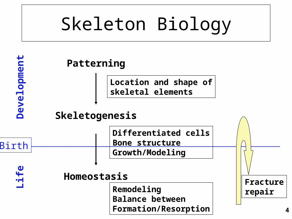
Skeleton Biology
Patterning
Skeletogenesis
Homeostasis
Dev
elop
men
tL
ife
Location and shape ofskeletal elements
Differentiated cellsBone structureGrowth/Modeling
RemodelingBalance betweenFormation/Resorption
Fracturerepair
Birth

5
Skeleton Pathologies
Patterning
Skeletogenesis
Homeostasis
Dev
elop
men
tL
ife
Dysostoses
Dysplasia
Mineralization defectsDegenerative diseases

6
Genetic defects associated with skeleton development
(RUNX2)

7
Skeleton patterning
• Condensation of mesenchymal cells to form the scaffold of each future skeletal element– Migration– Adhesion– Proliferation
• Early steps use signaling molecules and pathways generally involved in patterning other tissues (FGFs, Wnts, BMPs)
• Orchestrated by specific set of genes acting as territories organizers
• When not embryonic lethal disorders often localized

8
Hox transcription factors
• First described in Drosophila where they control body plan organization
• Arranged in 4 genomic clusters in mammals
• Expression patterns follow the cluster arrangement

9
Homeotic transformations in absence of Hox transcription factors
Wellik, Dev. Dynamics 236 (2007)
10
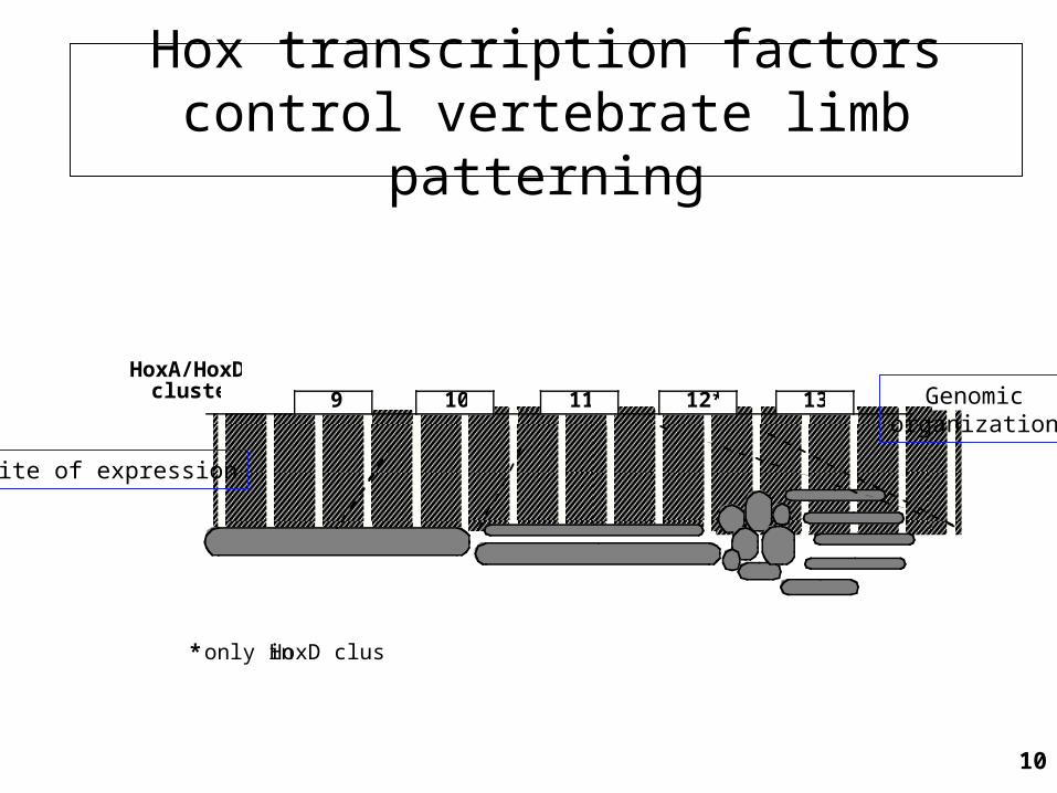
Hox transcription factors control vertebrate limb patterning
HoxA/HoxDclusters
* only in HoxD cluster
9 10 11 12* 13 Genomicorganization
Site of expression

11
Mutations in HOXD13 cause synpolydactyly*in humans
Muragaki et al., Science 272 (1996)
*OMIM 18600, 186300
Patient with increased number of Ala repeat in
HOXD13

12
Skeletogenesis
• Cell differentiation– Chondrocytes, osteoblasts, osteoclasts
• Bone morphogenesis– Formation of growth plate cartilage, bone shaft and
marrow cavity– Vascular invasion and innervation
• Defects generalized

13
Two skeletogenetic mechanisms
• Endochondral ossification– Differentiation of a cartilaginous scaffold (chondrocytes)
later replaced by bone (osteoblasts)– Most of the skeletal elements
• Intramembranous ossification– Direct differentiation of the condensed mesenchymal cells
into osteoblasts– Many bones of the skull, clavicles

14
Patterning ProliferationChondrocytedifferentiation
Chondrocytematuration
Vascular invasionOsteoblast differentiationOsteoclast differentiation
Endochondral ossification
Hypertrophy

15
Sox9
• Transcription factor of the HMG family
• Regulates the expression of chondrocyte-specific genes
• Sox9 haploinsufficiency causes Campomelic dysplasia (OMIM 114290)
• Earliest known regulator of chondrocyte differentiation

16
Sox9-deficient cells cannot differentiate into chondrocytes
Bi et al., Nat. Genet 22 (1999)

17
Patterning ProliferationChondrocytedifferentiation
Chondrocytematuration
Vascular invasionOsteoblast differentiationOsteoclast differentiation
Endochondral ossification
Hypertrophy
Sox9Sox5, 6

18
Accelerated chondrocyte hypertrophy in PTHrP-deficient mice
Karp et al., Development 127 (2000)
+/+ PTHrP -/-

19
PTHrP
• Ubiquitously expressed growth factor
• Shares the same receptor with PTH
• Mice “knockout” only phenotype is a generalized growth plate cartilage defect
• PTHrP protein signals to its receptor in the prehypertrophic chondrocytes and blocks their hypertrophic differentiation

20
Dwarfism in Ihh-deficient mice
Ihh -/-
St-Jacques et al. , Genes Dev. 13 (1999)
+/+

21
Indian hedgehog (Ihh)
• One of 3 members of the Hedgehog family of growth factors
• Widely expressed during development
• Expression positively regulated by the transcription factor Runx2

22
Reduced chondrocyte proliferation and delayed chondrocyte hypertrophy in Ihh-deficient mice
Ihh -/-
St-Jacques et al. , Genes Dev. 13 (1999)
+/+ Ihh -/-+/+
23
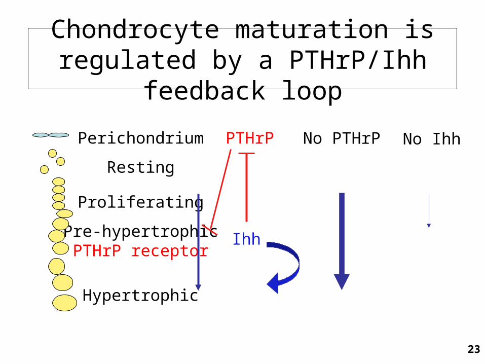
Chondrocyte maturation is regulated by a PTHrP/Ihh feedback loop
Perichondrium
Resting
Proliferating
Pre-hypertrophicPTHrP receptor
Hypertrophic
PTHrP
Ihh
No PTHrP No Ihh

24
Mutations in the PTH/PTHrP receptor cause Jansen and Bloomstrand chondrodysplasia
Jansen metaphysealchondrodysplasia
OMIM 156400
Blomstrand's lethalchondrodysplasia
OMIM 215045
Activatingmutations
Loss-of-functionmutations
Schipani & Provost. Brith Defects Res. 69 (2003)

25
Patterning ProliferationChondrocytedifferentiation
Chondrocytematuration
Vascular invasionOsteoblast differentiationOsteoclast differentiation
Endochondral ossification
Hypertrophy
Sox9Sox5, 6
Ihh/PTHrPRunx2
VEGF

26
MesenchymeBone collarCartilageBoneHypertrophicChondrocyte
OsteoblastProgenitorEndothelial
CellOsteoclastCalcified CartilageOsteoblastMIGRATION CartilageCartilage
resorption resorption
VascularizationVascularization
OsteogenesisOsteogenesis
Endochondral ossification
27
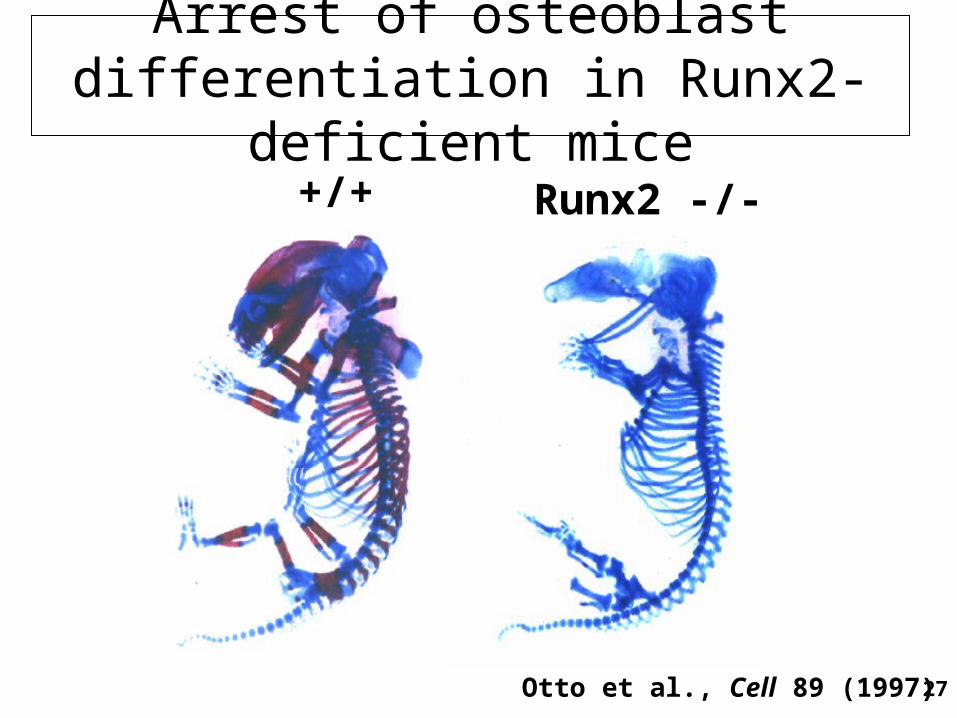
Arrest of osteoblast differentiation in Runx2-deficient mice
+/+ Runx2 -/-
Otto et al., Cell 89 (1997)
28
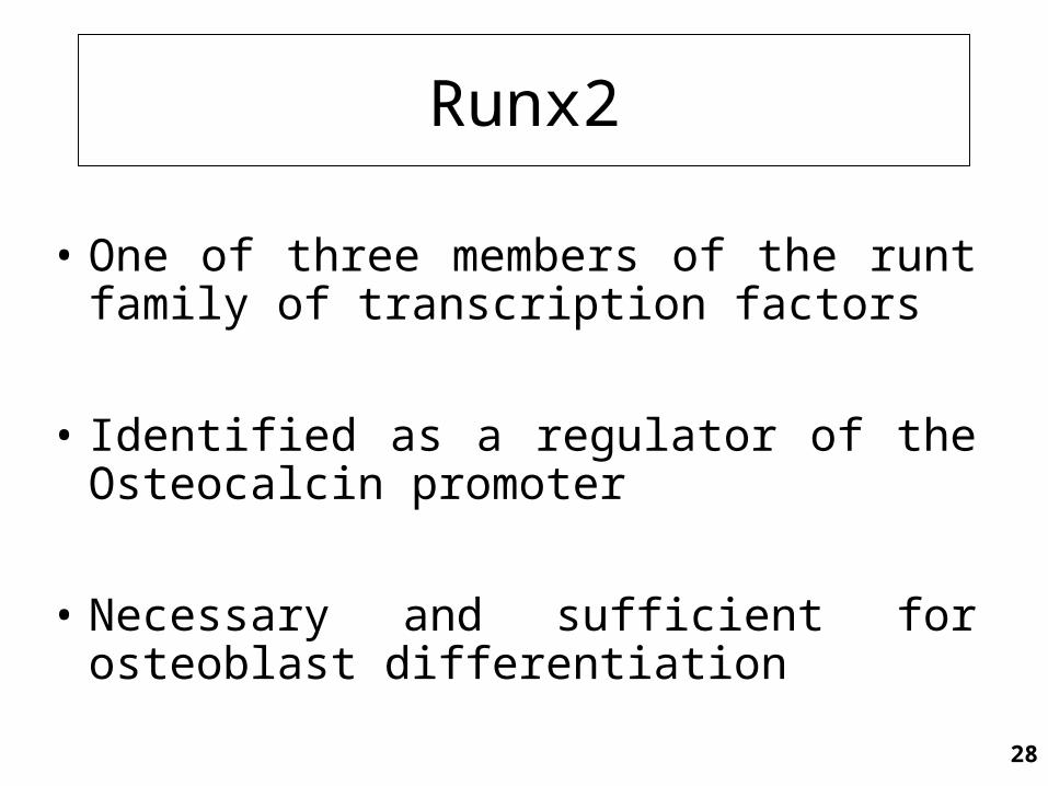
Runx2
• One of three members of the runt family of transcription factors
• Identified as a regulator of the Osteocalcin promoter
• Necessary and sufficient for osteoblast differentiation

29
Cleidocranial dysplasia (CCD, OMIM 119600)is caused by Runx2 haploinsufficiency
+/+
+/-
Mundlos et al., Cell 89 (1997)Lee et al. , Nat Genetics 16 (1997)

30
Intramembranous ossification
Growth
Closure
Sutureformation

31
Disorders of suture fusion
Delay
Acceleration = craniosynostosis
Msx2, Runx2 haploinsufficiency
FGFR1, 2, 3 activating mutationsMsx2 activating mutationsTwist haploinsufficiency

32
Osteoblast differentiation
Osteoprogenitor Osteoblast
Osx, ATF4
Pre-osteoblast
Runx2
Twist (Saethre-ChotzenSyndrome OMIM 101400)

33
Atf4-/-WT
E14
Delayed osteogenesisin absence of Atf4
Yang et al., Cell 117 (2004)

34
Delayed osteogenesis inAtf4-deficient mice
WT
Atf4 -/-
P0E16E15
Yang et al., Cell 117 (2004)
35
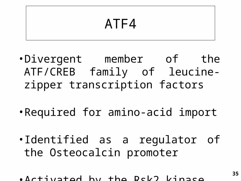
ATF4
• Divergent member of the ATF/CREB family of leucine-zipper transcription factors
• Required for amino-acid import
• Identified as a regulator of the Osteocalcin promoter
• Activated by the Rsk2 kinase

36
ATF4
• Lack of ATF4 phosphorylation by inactivating mutations in Rsk2 causes the skeletal defects associated with Coffin-Lowry syndrome (OMIM 303600)
• Increased ATF4 phosphorylation by Rsk2 causes the skeletal defects associated with Neurofibromatosis Type I (OMIM 162200)

37
ATF4
• Divergent member of the ATF/CREB family of leucine-zipper transcription factors
• Required for amino-acid import
• Identified as a regulator of the Osteocalcin promoter
• Activated by the Rsk2 kinase

38
A high protein diet normalizes bone formation in Atf4-/- and Rsk2-/- mice
BV/TV
BFR
Ob.S/BS
High protein diet
Elefteriou et al., Cell Metab. 4 (2006)

39
BV/TV
BFR
ObS/BS
Nf1ob-/-wt
15.3±1
153.3±11
19.6±0.4*
313.9±7.0*
Nf1ob-/-wt
14.8±0.7
157.0±11
15.3±0.6
186.2±22
Normal diet
19.1±0.8 31.8±1.6* 19.0±0.5 19.7±1.0
A low protein diet normalizes bone formation in a mouse model of Neurofibromatosis type I
Low protein diet
Elefteriou et al., Cell Metab. 4 (2006)

articular cartilage (chondrocytes)
secondary ossification centre(osteoblasts/osteoclasts)
reserve cartilage
proliferating cells
hypertrophic cells
trabecular bone (osteoblasts/osteoclasts)
cortical bone (osteoblasts)
calcified cartilage
Structure of a growing long bone
Growth plateCartilage
(chondrocytes)

41
PU.1Myeloid precursor cellc-fmsOsteoclastprogenitorMonocyte/MacrophageFunctional osteoclast
OPG/OCIFc-fosc-srcNFκB -Cathepsin K-M CSFmi ?SurvivalRANKL /ODF/RANKL ODFRANK-6Traf-c cbl2Pyk
Control of osteoclast differentiation and function

42
Osteopenia in OPG-deficient mice
Bucay et al., Genes Dev. 12 (1998)

43
Osteopetrosis in RANK-L deficient mice
Lacey et al., Cell 93 (1998)
44
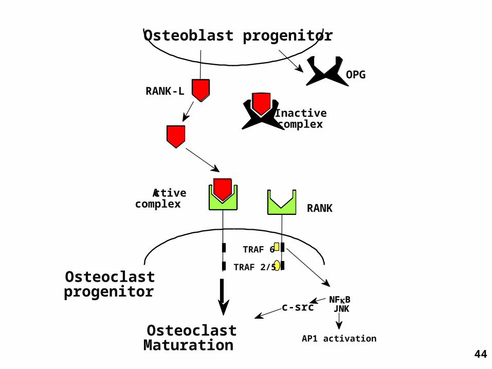
Osteoblast progenitor
Osteoclastprogenitor
RANK-L
OPG
RANK
Inactivecomplex
Activecomplex
TRAF 6
TRAF 2/5
NFκB JNK
OsteoclastMaturation
AP1 activation
NFκB JNKc-src
45
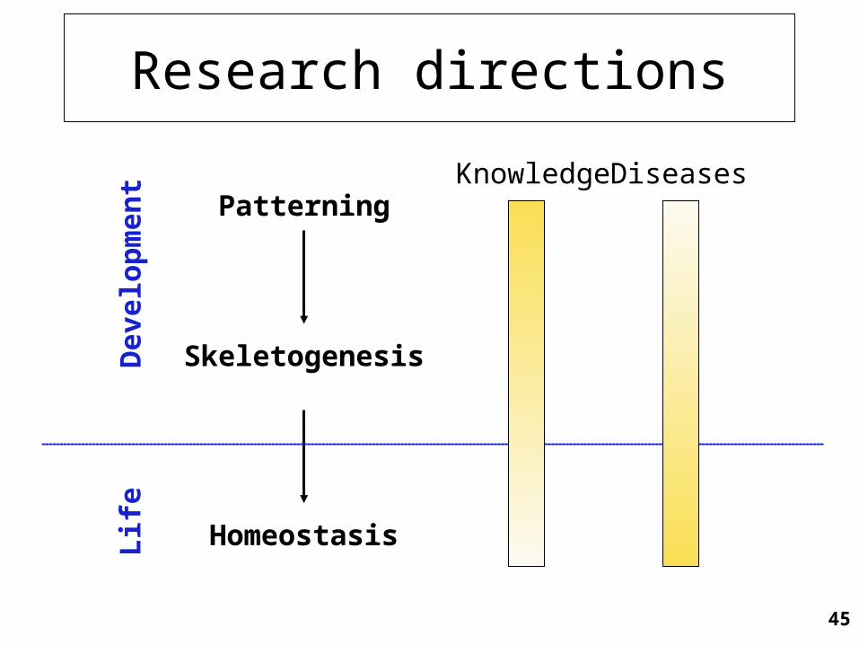
Research directions
Patterning
Skeletogenesis
Homeostasis
Dev
elop
men
tL
ife
DiseasesKnowledge