1-s2.0-s0955067414001410-main
TRANSCRIPT
7/26/2019 1-s2.0-S0955067414001410-main
http://slidepdf.com/reader/full/1-s20-s0955067414001410-main 1/6
PPARs and ERRs: molecular mediators of
mitochondrialmetabolismWeiwei
Fan1 and
Ronald
Evans1,2
Since
the
revitalization
of
‘the
Warburg
effect’,
there
has
been
great interest in mitochondrial oxidative metabolism, not only
from
the
cancer
perspective
but
also
from
the
general
biomedical science field. As the center of oxidative
metabolism,
mitochondria
and
their
metabolic
activity
are
tightly controlled to meet cellular energy requirements under
different physiological conditions. One such mechanism is
through
the
inducible
transcriptional
co-regulators
PGC1a
and
NCOR1, which respond to various internal or external stimuli to
modulate mitochondrial function. However, the activity of such
co-regulators
depends
on
their
interaction
with
transcriptional
factors that directly bind to and control downstream target
genes.
The
nuclear
receptors
PPARs
and
ERRs
have
beenshown
to
be
key
transcriptional
factors
in
regulating
mitochondrial
oxidative
metabolism
and
executing
the
inducible
effects
of
PGC1a
and
NCOR1.
In
this
review,
we
summarize recent gain-of-function and loss-of-function studies
of
PPARs
and
ERRs
in
metabolic
tissues
and
discuss
their
unique roles in regulating different aspects of mitochondrial
oxidative metabolism.
Addresses1Gene Expression Laboratory, The Salk Institute for Biological Studies,
La Jolla, CA 92037, USA 2Howard Hughes Medical Institute, The Salk Institute for Biological
Studies, La Jolla, CA 92037, USA
Corresponding author: Evans, Ronald ( [email protected] )
Current Opinion in Cell Biology 2015, 33:49–54
This review comes from a themed issue on Cell regulation
Edited by Jodi Nunnari and Johan Auwerx
For a complete overview see the Issue and the Editorial
Available online 6th December 2014
http://dx.doi.org/10.1016/j.ceb.2014.11.002
0955-0674/ # 2014 The Authors. Published by Elsevier Ltd. This is an
open access article under the CC BY-NC-ND license ( http://creative-
commons.org/licenses/by-nc-nd/3.0/ ).
IntroductionEnergy
is
vital
to
all
living
organisms.
In
humans
and
other mammals,
the
vast
majority
of
energy
is
produced
by oxidative
metabolism
in
mitochondria
[1].As
a
cellular
organelle, mitochondria
are
under
tight
control
of
the
nucleus.
Although
the
majority
of
mitochondrial
proteins
are encoded
by
nuclear
DNA
(nDNA)
and
their
expres-
sion regulated
by
the
nucleus,
mitochondria
retain
their
own genome, mitochondrial DNA (mtDNA), encoding13 polypeptides
of
the
electron
transport
chain
(ETC)
in
mammals.
However,
all
proteins
required
for
mtDNA
replication, transcription, and translation, as well as factors
regulating such
activities, are
encoded
by
the
nucleus
[2].
The cellular
demand
for
energy
varies
in
different
cells
underdifferent
physiological
conditions.
Accordingly,
the
quantity
and
activity
of
mitochondria
are
differentially
controlled by
a transcriptional
regulatory
network
in
both
the basal
and
induced
states.
A
number
of
components
of
this network
have
been
identified,
including
members
of
the nuclear
receptor
superfamily,
the
peroxisome
prolif-
erator-activated
receptors
(PPARs)
and
the
estrogen-
related receptors
(ERRs)
[3–5].
The Yin-Yang co-regulatorsA
well-known
inducer
of
mitochondrial
oxidative
metab-
olism is
the
peroxisome
proliferator-activated
receptor g
coactivator
1a (PGC1a) [6],
a
nuclear
cofactor
which
is
abundantly
expressed
in
high
energy
demand
tissues
such
as heart, skeletal muscle, and brown adipose tissue (BAT)
[7]. Induction
by
cold-exposure,
fasting,
and
exercise
allows PGC1a to
regulate
mitochondrial
oxidative
metab-
olism by
activating
genes
involved
in
the
tricarboxylic
acid cycle
(TCA
cycle),
beta-oxidation,
oxidative
phos-
phorylation (OXPHOS),
as
well
as
mitochondrial
bio-
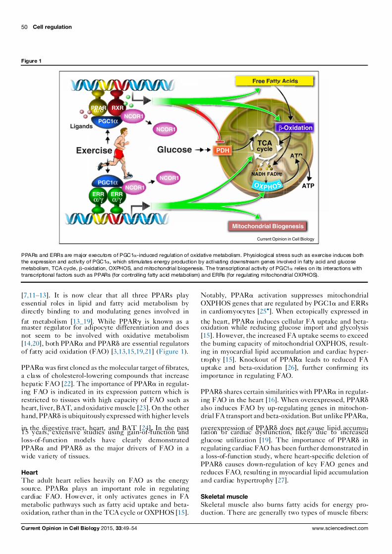
genesis [6,8] (Figure 1).
The effect
of
PGC1a on
mitochondrial
regulation
is
antagonized
by
transcriptional
corepressors
such
as
the
nuclear
receptor
corepressor
1
(NCOR1)
[9,10]. In
con-
trast to
PGC1a, the
expression
of
NCOR1
is
suppressed
in conditions
where
PGC1a
is
induced
such
as
during
fasting,
high-fat-diet
challenge,
and
exercise
[9,11].
Moreover, the knockout of NCOR1 phenotypicallymimics PGC1a
overexpression
in
regulating
mitochon-
drial oxidative metabolism [9]. Therefore, coactivators
and corepressors
collectively
regulate
mitochondrial
metabolism in a Yin-Yang fashion.
However,
both PGC1a
and
NCOR1
lack
DNA
binding
activityand
rather
act via
their
interaction
with
transcription
factors that direct the
regulatory program. Therefore the
transcriptional
factors
that
partnerwith
PGC1a
and
NCOR1
mediate the
molecular
signaling cascades
and
execute their
inducible
effects
on
mitochondrial
regulation.
PPARs: master executors controlling fatty acid oxidationBoth PGC1a and NCOR1 are co-factors for the peroxi-some proliferator-activated
receptors
(PPARa, g, and d)
Available online at www.sciencedirect.com
ScienceDirect
www.sciencedirect.com Current Opinion in Cell Biology 2015, 33:49–54
7/26/2019 1-s2.0-S0955067414001410-main
http://slidepdf.com/reader/full/1-s20-s0955067414001410-main 2/6
[7,11–13].
It
is
now
clear
that
all
three
PPARs
play
essential roles
in
lipid
and
fatty
acid
metabolism
by
directly binding
to
and
modulating
genes
involved
in
fat metabolism [13–19]. While PPARg is known as a
master regulator
for
adipocyte
differentiation
and
does
not seem
to
be
involved
with
oxidative
metabolism
[14,20], both
PPARa and
PPARd are
essential
regulators
of fatty
acid
oxidation
(FAO)
[3,13,15,19,21]
(Figure
1).
PPARawas
first
cloned
as
the
molecular
target
of
fibrates,
a class
of
cholesterol-lowering
compounds
that
increase
hepatic FAO [22]. The importance of PPARa in regulat-ing FAO
is
indicated
in
its
expression
pattern
which
is
restricted to tissues with high capacity of FAO such as
heart, liver,BAT, and
oxidative
muscle
[23].
On
the
otherhand, PPARd
is
ubiquitously
expressed
with
higher
levels
in the
digestive
tract,
heart,
and
BAT
[24].
In
the
past15 years,
extensive
studies
using
gain-of-function
and
loss-of-function models have clearly demonstrated
PPARa and
PPARd
as
the
major
drivers
of
FAO
in
a
wide variety
of
tissues.
Heart
The
adult
heart
relies
heavily
on
FAO
as
the
energy
source. PPARa
plays
an
important
role
in
regulating
cardiac FAO. However,
it
only
activates
genes
in
FA
metabolic pathways such as fatty acid uptake and beta-oxidation, rather
than
in
the
TCA
cycle
or
OXPHOS
[15].
Notably,
PPARa
activation
suppresses
mitochondrial
OXPHOS
genes
that
are
regulated
by
PGC1a and
ERRs
in cardiomyocytes
[25].
When
ectopically
expressed
in
the heart, PPARa induces cellular FA uptake and beta-oxidation
while
reducing
glucose
import
and
glycolysis
[15]. However,
the
increased
FA
uptake
seems
to
exceed
the burning
capacity
of
mitochondrial
OXPHOS,
result-
ing in
myocardial
lipid
accumulation
and
cardiac
hyper-
trophy [15].
Knockout
of
PPARa leads
to
reduced
FA
uptake and
beta-oxidation
[26],
further
confirming
its
importance in
regulating
FAO.
PPARd shares
certain
similarities
with
PPARa in
regulat-
ing FAO in the heart [16]. When overexpressed, PPARd
also induces
FAO
by
up-regulating
genes
in
mitochon-drial FA
transport
and
beta-oxidation.
But
unlike
PPARa,
overexpression
of
PPARd
does
not
cause
lipid
accumu-lation or
cardiac
dysfunction,
likely
due
to
increased
glucose utilization [19]. The importance of PPARd in
regulating
cardiac
FAO
has
been
further
demonstrated
in
a loss-of-function
study,
where
heart-specific
deletion
of
PPARd causes
down-regulation
of
key
FAO
genes
and
reduces FAO, resulting
in
myocardial
lipid
accumulation
and cardiac
hypertrophy
[27].
Skeletal muscle
Skeletal muscle also burns fatty acids for energy pro-duction. There
are
generally
two
types
of
muscle
fibers:
50
Cell
regulation
Figure 1
PPARα/δ RXR
ERRα/γ
ERRα/γ
PGC1α
PGC1α
Exercise
Ligands
Glucose
Mitochondrial Biogenesis
Free Fatty Acids
TCAcycle
NADH FADH2
ATP
β-Oxidation
PDH
ATP
NCOR1
NCOR1
NCOR1
NCOR1
Current Opinion in Cell Biology
O X P H O S
PPARs and ERRs are major executors of PGC1a-induced regulation of oxidative metabolism. Physiological stress such as exercise induces both
the expression and activity of PGC1a,
which stimulates energy production by activating downstream genes involved in fatty acid and glucose
metabolism, TCA cycle, b-oxidation, OXPHOS, and mitochondrial biogenesis. The transcriptional activity of PGC1a
relies on its interactions with
transcriptional factors such as PPARs (for controlling fatty acid metabolism) and ERRs (for regulating mitochondrial OXPHOS).
Current Opinion in Cell Biology 2015, 33:49–54 www.sciencedirect.com

7/26/2019 1-s2.0-S0955067414001410-main
http://slidepdf.com/reader/full/1-s20-s0955067414001410-main 3/6
type
I
oxidative
fibers
that
are
rich
in
mitochondria
andpredominantly powered
by
oxidation
of
glucose
and
FA;
and type II glycolytic fibers that contain less mitochondria
and heavily
rely
on glycolysis
for
energy
[28].
Both PPARa and
PPARd play
important
roles
in
regulat-ing muscle
FAO.
Overexpression
of
either
in
muscle
induces FAO
by
up-regulating
genes
involved
in
fatty
acid utilization
and
beta-oxidation
[17,18,29,30].
How-
ever, PPARa overexpression
also
promotes
a
fiber-type
transition
toward
more
glycolytic
fibers
which
have
lower
mitochondrial OXPHOS
activity.
This
induces
lipid
accumulation
and
drives
the
development
of glucose
intolerance
and
insulin
resistance
[18,30].
PPARadeletion, on
the
other
hand,
increases
the
number
of
oxidative fibers
and
improves
glucose
homeostasis,
despite
the
reduction
in
muscle
FAO
[18,30].
In contrast to PPARa, overexpression of PPARd does not
cause lipid accumulation or glucose intolerance [17,29].
In addition to induced FAO, PPARd also increases the
proportion of
oxidative
fibers
that
are
rich
in
mitochon-dria, thus
dramatically
boosting
mitochondrial
oxidative
metabolism. This
is
associated
with
90%
increase
in
endurance capacity
and
resistance
against
diet-induced
diabetes [17,30]. Conversely, muscle-specific PPARddepletion leads
to
an
oxidative-to-glycolytic
fiber-type
switch with
reduced
FAO
and
OXPHOS.
The
mutant
mice gain
more
weight
on
high
fat
diet
and
are
more
susceptible
to
insulin
resistance
[31].
The ability
of
PPARa
and
PPARd to
transform fiber
types in opposite directions seem to be mediated bytwo microRNAs, miR-208b
and miR-499 [30],
which
directly
activate the
oxidative and
repress the
glycolytic
myofibril gene program [32].
Liver
In
the
liver
PPARa is
the
predominant
PPAR
isoform
[24]. It is essential in regulating hepatic FA uptake, beta-oxidation, and
ketogenesis,
especially
during
fasting.
PPARa knockout suppresses the expression of genes
involved in
FA
uptake
and
FAO,
resulting
in
decreased
basal-state hepatic FA uptake and beta-oxidation [33]. In
addition, fasting-induced
hepatic
responses,
including
elevatedFA
oxidation,
gluconeogenesis,
and
ketogenesis,are all
impaired
in
PPARa-null mice.
As
a
result, the
fasted mutant
mice
develop
hypoketogenesis,
hypogly-
cemia, and
liver
steatosis
[26,34].
PPARd seems
to
play
a
different
role
in
regulating
hepatic
energy metabolism.
Unlike
PPARa, PPARd
deletion
reduces the
expression
of
genes
involved
in
lipogenesis
and glucose
utilization
instead
of
FA
metabolism.
More-
over, fasted
PPARd-null mice
have
a
normal
ketogenic
response, increased serum glucose levels and no sign of liver steatosis
[35].
When
overexpressed
in
the
liver,
PPARd increases
hepatic
glycogen
and
lipid
storage
asa result
of
up-regulation
of
genes
in
glucose
utilization
and lipogenesis [36]. In addition, hepatic PPARd acti-
vates muscle
FA
oxidation
through
a
lipid
molecule
PC
(18:0/18:1), which is produced by a PPARd-dependent
lipogenic pathway
[37
].
Fat
PPARa is
highly
expressed
in
BAT
but
not
in
white
adipose tissue
(WAT)
[24]. Its
primary
function
in
BAT
seems to
be
the
regulation
of
PGC1a and
UCP1
(a
mitochondrial
uncoupling
protein)
expression.
PPARadeletion
reduces
expression
of
PGC1a
and
UCP1
under
both basal
and
cold-exposure
conditions
[21,38].
How-
ever,unlike
in
other
tissues,
FA
metabolism
in
BAT
is
not
affected
by
PPARa knockout, suggesting
the
contri-
bution of
other
PPARs
[39].
When
activated
in
human
and mouse
adipocytes,
PPARa induces
the
expression
of
FAO genes and increases energy expenditure [40,41].
PPARd is expressed in both BAT and WAT. While its
function in
WAT
is
unknown,
it
is
clear
that
PPARd
playsan essential
role
in
regulating
FA
oxidation
and
thermo-
genesis in
BAT
[13,42,43].
When
ectopically
expressed
in
adipose
tissue,
PPARd dramatically
induces
the
expres-
sion of genes in FAO, OXPHOS, and thermogenesis,
which results
in
increased
FAO
in
BAT,
reduced
systemic
adiposity
and
improved
serum
lipid
profiles
[13,44].
Con-
versely,
deletion
of
PPARd
in
BAT
reduces
the
expres-
sionof
FAO
and
thermogenic
genes,
which
impairs in vivo
thermogenesis [13,43].
ERRS: master executors controllingmitochondrial OXPHOSERRs
are essential regulators of
mitochondrial energy
metabolism
[4]. ERRa
is ubiquitously
expressed
but
particularly
abundant
in tissues with
high
energy demands
such as brain, heart, muscle, and
BAT.
ERRb
and
ERRghave similar expression
patterns,
both
are selectively
expressed in highly oxidative tissues including brain, heart,and oxidative muscle
[45]. Instead
of
endogenous
ligands,
the transcriptional activity of ERRs is primarily regulated by
co-factors
such
as
PGC1a and
NCOR1
[4,46]
(Figure
1).
Ofthe
three
ERRs,
ERRb
is
the
least
studied
and
its
role in
regulating mitochondrial
function
is
unclear
[4,47].
Incontrast,
when
PGC1a
is
induced,
ERRa
is
the
master
regulator of
the
mitochondrial
biogenic
gene
network.
As
ERRa binds
to
its
own
promoter,
PGC1a
can
also
induce
an autoregulatory
loop
to
enhance
overall
ERRa
activity
[48]. Without
ERRa,
the
ability
of
PGC1a
to
induce
the
expression of
mitochondrial
genes
is
severely
impaired.
However,
the
basal-state
levels
of
mitochondrial
target
genes are
not
affected
by
ERRa
deletion,
suggesting
induced mitochondrial
biogenesis
is
a
transient
process
andthat other transcriptional factors such as ERRg may beimportant
maintaining
baseline
mitochondrial
OXPHOS
PPARs
and
ERRs:
molecular
mediators
mitochond
metabolism Fan and Evans 51
www.sciencedirect.com Current Opinion in Cell Biology 2015, 33:49–54

7/26/2019 1-s2.0-S0955067414001410-main
http://slidepdf.com/reader/full/1-s20-s0955067414001410-main 4/6
[41,42,43].
Consistent
with
this
idea,
ERRg
(which isactive even
when PGC1a
is
not
induced)
shares
many
target genes with ERRa [49,50].
Genome-wide analysis of ERRa and ERRg has con-
firmed their
direct
and
overlapping
binding
in
promoterregions of
a
large
number
of
mitochondrial
genes,
many
of
which are
PGC1a
targets
[8,49].
These
genes
cover
many
aspects of
mitochondrial
oxidative
metabolism,
ranging
from glucose
utilization,
FA
oxidation,
the
TCA
cycle,
and OXPHOS.
About
a quarter
of
the
binding
sites
are
shared by
both
ERRs,
indicating
their
cooperative
regu-
lation of
those
genes.
Recently
several
gain-of-function
and loss-of-function
studies
have
revealed
a better
un-
derstanding of
their in vivo
roles
[44–50].
Heart
Both
ERRa
and
ERRg
are
highly
expressed
in
the
heart
[45]. Knockout of ERRa causes down-regulation of a
number of genes in mitochondrial oxidative metabolism,
some of which are direct ERRa targets. Interestingly,
another set
of
genes
in
mitochondrial
oxidative
metab-olism, including
PGC1a and
ERRg,
are
up-regulated
by
ERRa deletion,
indicating
a
compensatory
effect
of
ERRg [49].
The
ERRa-null
heart
only
shows
minimal
defects with normal mitochondrial function in the basal
state. However,
when
challenged
with
pressure
overload
(a common
method
to
induce
cardiac
hypertrophy),
the
knockout
mice
exhibit
more
severe
phenotypes
with
dilated hypertrophy
and
early
heart
failure,
possibly
due to
a
defect
in
energy
reserve,
indicating
the
require-
ment of
ERRa
during
stressed
conditions
[51].
Similar
to
ERRa,
ERRg
deletion
also
causes
both
down-
regulation and
up-regulation
of
genes
in
mitochondrial
oxidativemetabolism,
as
well as
up-regulated
PGC1a and
ERRa. The
altered
expression
of
OXPHOS
genes
causes
severe mitochondrial
defects:
increased
mtDNA
copy
number,
reduced
ETC
complex
I
activity,
and
increased
complex IV activity. As a result, most ERRg-null mice diewithin the
first
week
of
their
life
due
to
heart
failure
[50].
Skeletal muscle
ERRa is uniformly expressed in both oxidative and
glycolytic muscles
[52].
The
knockout
of
ERRa
in
skeletal muscle
causes
no
phenotypic
change
in
the
basalstate [53,54].
However,
in
a
cardiotoxin
induced
injury
model, its
deficiency
reduces
mitochondrial
activity
and
impairs muscle
regeneration
[54].
Unlike
ERRa,
ERRg
is
selectively
expressed
in
oxi-
dative muscle
[52].
When
ectopically
expressed
in
glycolytic muscle,
ERRg
drives
a
fiber-type
switch
from
glycolytic to
oxidative
fibers,
with
dramatically
induced
mitochondrial
biogenesis
and
vascularization.
This
is
accompanied by the induction of genes in pathways suchas FA
oxidation,
TCA
cycle,
and
OXPHOS,
many
of
which
are
direct
ERR
targets
[49,52,55].
However,whether ERRg
is
required
for
basal-sate
mitochondrial
function in oxidative muscle remains unclear.
Fat
Both
ERRa
and
ERRg
are
expressed
higher
in
BAT
thaninWAT
[45].
While
the
role
of
ERRg
in
fat
is
still
under
investigation,
ERRa
seems
to
be
required
in
regulating
stress-induced metabolism
in
both
WAT
and
BAT.
Whole-body
knockout
of
ERRa
causes
reduced
fat
mass
which becomes
more
striking
when
challenged
with
a
high-fat diet.
The
ERRa-null
WAT
has
altered
expres-
sion of
metabolic
genes
including
direct
ERR
targets
[56].
When
challenged
with
cold-exposure,
the
mutant
mice
display impaired
thermogenesis,
accompanied
by
decreased mitochondrial
biogenesis
and
increased
lipid
accumulation in
BAT. Gene
expression
analysis
reveals
repression
of
mitochondrial
oxidative
metabolism
genes,
many of which are direct ERR targets [57].
Therefore, ERRa and ERRg cooperatively regulate oxi-
dative metabolism
by
directly
controlling
the
expressionof genes
in
this
pathway.
Despite
their
shared
target
genes, each
appears
to
have
its
own
unique
function,
with ERRa
more
involved
in
stress-induced
responses
and ERRg required for maintaining the baseline mito-
chondrial
integrity.
Conclusion and perspectivesTaken
together,
recent studies
have clearly demonstrated
the
essential roles
of
PPARs
and ERRs
in regulating
mitochondrial
oxidative metabolism
and executing the
inducible effects of PGC1a (Figure 1). Both PPARa andPPARd are
key
regulators
for
FA
oxidation.
While
the
function of
PPARa
seems
more
restricted
in FA
uptake,
beta-oxidation,
and
ketogenesis, PPARd
plays a
broader
role in
controlling oxidative
metabolism
and fuel
prefer-
ence, with
its
target genes involved in FA
oxidation,
mito-
chondrialOXPHOS,
andglucose
utilization.
However, it is
stillnot clear how much redundancy exists between PPARaand
PPARd,
a
question
which
may
require the
generationof
adouble knockoutmodel. In addition, moreeffort is needed
to fully understand
how
PPARa
and PPARd
control
their
target genes in response to environmental changes.
Likewise, ERRa
and
ERRg
have
been
shown
to
be
keyregulators of
mitochondrial
OXPHOS.
Knockout
studies
ofERRa
suggest
it
to
be
the
principal
executor
of
PGC1ainduced
up-regulation
of
mitochondrial
genes,
though
its
role in
exercise-dependent
changes
in
skeletal
muscle
needs further
investigation.
Transgenic
models
have
demonstrated
ERRg’s
powerful
induction
of
mitochon-
drial biogenesis
and
its
ability
to
act
in
a
PGC1a-inde-
pendent manner.
However,
it
remains
to
be
elucidated
whether
ERRg
is
sufficient
for
basal-state
mitochondrial
function in general, and whether ERRa can compensatefor its
function.
52
Cell
regulation
Current Opinion in Cell Biology 2015, 33:49–54 www.sciencedirect.com

7/26/2019 1-s2.0-S0955067414001410-main
http://slidepdf.com/reader/full/1-s20-s0955067414001410-main 5/6
Acknowledgements
We thank C. Brondos for administrative support, R. Yu and M. Downes fordiscussion and comments. R.M.E. is
an HHMI Investigator at the SalkInstitute forBiological Studies andMarch of Dimes Chair.W.F. andR.M.E.are supported by NIH grants (DK057978, DK090962, HL088093,HL105278 and ES010337), the Glenn Foundation for Medical Research,
the Leona M.
and Harry B. Helmsley Charitable Trust, Ipsen/Biomeasure,CIRM, and The Ellison Medical Foundation.
References and recommended readingPapers of particular interest, published within the period of review,have been highlighted as:
of special interest of outstanding interest
1. Wallace DC, Fan W, Procaccio V:Mitochondrial energetics andtherapeutics. Annu Rev Pathol 2010, 5:297-348.
2. Scarpulla RC, Vega RB, Kelly DP: Transcriptional integration ofmitochondrial biogenesis. Trends Endocrinol Metab 2012,23:459-466.
3. Poulsen L, Siersbaek M, Mandrup S: PPARs: fatty acid sensorscontrolling metabolism.
Semin Cell Dev Biol 2012, 23:631-639.
4. Eichner LJ,GiguereV: Estrogen relatedreceptors (ERRs): a newdawn in transcriptional control of mitochondrial genenetworks. Mitochondrion 2011, 11:544-552.
5. Fan W,Downes M,Atkins A,Yu R,EvansRM:Nuclear receptorsandAMPK:resettingmetabolism.
Cold SpringHarbSympQuant Biol 2011, 76:17-22.
6. Handschin C, Spiegelman BM: Peroxisome proliferator-activated receptor gamma coactivator 1 coactivators, energy homeostasis, and metabolism.
Endocr Rev 2006, 27:728-735.
7. Puigserver
P,
Wu Z, Park CW, Graves
R,
Wright
M,Spiegelman BM: A cold-inducible
coactivator
of nuclearreceptors
linked to adaptive thermogenesis.
Cell
1998,92:829-839.
8. Wu Z, Puigserver P, Andersson U, Zhang C, Adelmant G,Mootha V, Troy A, Cinti S, Lowell B, Scarpulla RC et al.:Mechanisms controlling mitochondrial biogenesis andrespiration through the thermogenic coactivator PGC-1. Cell 1999, 98:115-124.
9. Yamamoto H, Williams EG, Mouchiroud L, Canto C, Fan WW,Downes M, Heligon C, Barish GD, Desvergne B, Evans RM et al.:NCoR1 is a conserved physiological modulator of musclemass and oxidative function. Cell 2011, 147:827-839.
10. Li P, Fan W, Xu J, Lu M, Yamamoto H, Auwerx J, Sears DD,Talukdar S, Oh D, Chen A et al.:
Adipocyte NCoR knockoutdecreases PPARgamma phosphorylation and enhancesPPARgamma activity and insulin sensitivity .
Cell 2011,147:815-826.
11. Perez-Schindler J, Summermatter S, Salatino S, Zorzato F,Beer M, Balwierz PJ, van Nimwegen E, Feige JN, Auwerx J,Handschin C: The corepressor NCoR1 antagonizes PGC-
1alpha and estrogen-related receptor alpha in the regulationof skeletalmuscle function andoxidativemetabolism.
MolCell Biol 2012, 32:4913-4924.
12. VegaRB,HussJM, Kelly DP:Thecoactivator PGC-1cooperateswith peroxisome proliferator-activated receptor alpha intranscriptional control of nuclear genes encodingmitochondrial fatty acidoxidation enzymes.
Mol Cell Biol 2000,20:1868-1876.
13. Wang YX, Lee CH, Tiep S, Yu RT, Ham J, Kang H, Evans RM:Peroxisome-proliferator-activated receptordeltaactivates fatmetabolism to prevent obesity . Cell 2003, 113:159-170.
14. Rosen ED, Sarraf P, Troy AE, Bradwin G, Moore K, Milstone DS,Spiegelman BM, Mortensen RM: PPAR gamma is required forthedifferentiation ofadiposetissuein vivo andin vitro.MolCell 1999, 4:611-617.
15.
Finck BN, Lehman JJ, Leone TC, Welch MJ, Bennett MJ,Kovacs A, Han X, Gross RW, Kozak R, Lopaschuk GD et al.:
Thecardiac phenotype induced by PPARalpha overexpressionmimics that caused by diabetes mellitus.
J Clin Invest 2002,109:121-130.
16. Gilde AJ, van der Lee KA, Willemsen PH, Chinetti G, van derLeij FR, van der Vusse GJ, Staels B, van Bilsen M: Peroxisome
proliferator-activated receptor (PPAR) alpha and PPARbeta/ delta, butnotPPARgamma, modulate theexpression ofgenesinvolved in cardiac lipidmetabolism.
Circ Res2003, 92:518-524.
17. Wang YX, Zhang CL, Yu RT, Cho HK, Nelson MC, Bayuga-Ocampo CR, Ham J, Kang H, Evans RM: Regulation of musclefiber type and running endurance by PPARdelta.
PLoS Biol 2004, 2:e294.
18. Finck BN,Bernal-MizrachiC, HanDH, Coleman T, SambandamN,LaRiviereLL,Holloszy JO,SemenkovichCF,KellyDP: A potentiallink between muscle peroxisome proliferator-activatedreceptor-alpha signaling and obesity-related diabetes.
Cell Metab 2005, 1:133-144.
19. Burkart EM, Sambandam N, Han X, Gross RW, Courtois M,Gierasch CM, Shoghi K, Welch MJ, Kelly DP: Nuclear receptorsPPARbeta/delta and PPARalpha direct distinct metabolicregulatory programs in the mouse heart.
J Clin Invest 2007,117:3930-3939.
20. Wang F, Mullican SE, DiSpirito JR, Peed LC, Lazar MA:Lipoatrophy and severe metabolic disturbance in mice withfat-specific deletion of PPARgamma.
ProcNatlAcadSci U S A2013, 110:18656-18661.
21. Barbera MJ, Schluter A, Pedraza N, Iglesias R, Villarroya F,Giralt M: Peroxisome proliferator-activated receptor alphaactivates transcription of the brown fat uncoupling protein-1gene. A link between regulation of the thermogenic and lipidoxidation pathways in the brown fat cell. J Biol Chem
2001,276:1486-1493.
22. Issemann I, Green S: Activation of a member of the steroidhormone receptor superfamily by peroxisome proliferators.Nature 1990, 347:645-650.
23. Braissant O, Foufelle F, Scotto C, Dauca M,Wahli W:Differentialexpression of peroxisome proliferator-activated receptors(PPARs): tissue distribution of PPAR-alpha, -beta, and -gamma in the adult rat.
Endocrinology 1996, 137:354-366.
24. Escher P, Braissant O, Basu-Modak S, Michalik L, Wahli W,Desvergne B: Rat PPARs: quantitative analysis in adult rattissues and regulation in fasting and refeeding. Endocrinology 2001, 142:4195-4202.
25.
Oka S, Alcendor R,Zhai P,Park JY, ShaoD, Cho J, Yamamoto T,Tian B, Sadoshima J: PPARalpha-Sirt1 complex mediatescardiac hypertrophy and failure through suppression of theERR transcriptional pathway . Cell Metab 2011, 14:598-611.
Demonstrates the negative impact of Ppara
on cardiac mitochondrialfunction by directly recruiting Sirt1 to ERR-response elements in mito-chondrial genes.
26. Leone TC, Weinheimer CJ, Kelly DP: A critical role for theperoxisome proliferator-activated receptor alpha (PPARalpha) in the
cellular fasting response: the PPAR alpha-nullmouse as a model of fatty acid oxidation disorders. Proc Natl Acad Sci U S A 1999, 96:7473-7478.
27. Cheng L, Ding G, Qin Q, Huang Y, Lewis W, He N, Evans RM,SchneiderMD, Brako FA,XiaoY etal.:
Cardiomyocyte-restrictedperoxisome proliferator-activated receptor-delta deletionperturbs myocardial fatty acid oxidation and leads tocardiomyopathy . Nat Med 2004, 10:1245-1250.
28. FanW, AtkinsAR, YuRT,DownesM,EvansRM:Roadto exercisemimetics: targeting nuclearreceptors in skeletalmuscle.
J Mol Endocrinol 2013, 51:T87-T100.
29.
GanZ, Burkart-HartmanEM,HanDH,Finck
B,Leone TC,SmithEY, Ayala JE, Holloszy J, Kelly DP:
The nuclear receptor PPARbeta/ deltaprogramsmuscleglucosemetabolismincooperationwith AMPK and MEF2.
Genes
Dev 2011, 25:2619-2630.Using muscle-specific transgenic mouse models, this study illustratesthatPpard
butnot Ppara
positivelyregulatesmuscle glucoseoxidationbydirectly activating lactate dehydrogenase B.
PPARs
and
ERRs:
molecular
mediators
mitochond
metabolism Fan and Evans 53
www.sciencedirect.com Current Opinion in Cell Biology 2015, 33:49–54

7/26/2019 1-s2.0-S0955067414001410-main
http://slidepdf.com/reader/full/1-s20-s0955067414001410-main 6/6
30.
Gan Z, Rumsey J, Hazen BC, Lai L, Leone TC, Vega RB, Xie H,Conley KE, Auwerx J, Smith SR et al.:
Nuclear receptor/ microRNA circuitry links muscle fiber type to energy metabolism. J Clin Invest 2013, 123:2564-2575.
Interesting study demonstrates the opposing roles of Ppard
andPpara
inregulating muscle fiber type composition through an Errg-dependentmicro-RNA network that targets Myh7 and Myh7b.
31. Schuler M, Ali F, Chambon C, Duteil D, Bornert JM, Tardivel A,Desvergne B, Wahli W, Chambon P, Metzger D: PGC1 alphaexpression is controlled in skeletal muscles by PPAR beta,whose ablation results in fiber-type switching, obesity, andtype 2 diabetes. Cell Metab 2006, 4:407-414.
32. van Rooij E, Quiat D, Johnson BA, Sutherland LB, Qi X,Richardson JA, Kelm RJ Jr, Olson EN: A family of microRNAsencoded by myosin genes governs myosin expression andmuscle performance.
Dev Cell 2009, 17:662-673.
33. Aoyama T, Peters JM, Iritani N, Nakajima T, Furihata K,Hashimoto T, Gonzalez FJ: Altered constitutive expression offatty acid-metabolizing enzymes in mice lacking theperoxisome proliferator-activated receptor alpha(PPARalpha). J Biol Chem 1998, 273:5678-5684.
34. Kersten S, Seydoux J, Peters JM, Gonzalez FJ, Desvergne B,Wahli W: Peroxisome proliferator-activated receptor alphamediates the adaptive response to fasting.
J Clin Invest 1999,
103:1489-1498.
35. Sanderson LM, Boekschoten MV, Desvergne B, Muller M,Kersten S: Transcriptional profiling reveals divergent roles ofPPARalpha and PPARbeta/delta in regulation of geneexpression in mouse liver.
Physiol Genomics 2010, 41:42-52.
36.
Liu S, Hatano B, ZhaoM, Yen CC, Kang K, Reilly SM, Gangl MR,Gorgun C, Balschi JA, Ntambi JM et al.:
Role of peroxisomeproliferator-activated receptor {delta}/{beta} in hepaticmetabolic regulation. J Biol Chem 2011, 286:1237-1247.
A liver-restricted PPARd overexpression study demonstrates the novelrole of Ppard
in l iver that enhances hepatic glucose uti lization andlipogenesis which improves whole body glucose homeostasis.
37.
Liu S, Brown JD, Stanya KJ, Homan E, Leidl M, Inouye K,Bhargava P, Gangl MR, Dai L, Hatano B et al.:
A diurnal serumlipid integrates hepatic lipogenesis and peripheral fatty aciduse. Nature 2013, 502:550-554.
A follow-up study of [36] that identifies a circulating lipid molecule that is
regulated by hepatic Ppard activity and activates muscle lipid oxidationthrough Ppara.
This is the first report of a l iver-to-muscle metaboliccrosstalk mediated by a lipid molecule.
38.
Hondares E, Rosell M, Diaz-Delfin J, Olmos Y, Monsalve M,Iglesias R, Villarroya F, Giralt M: Peroxisome proliferator-activated receptor alpha (PPARalpha) induces PPARgammacoactivator 1alpha (PGC-1alpha) gene expression andcontributes to thermogenic activation of brown fat:involvement of PRDM16.
J Biol Chem
2011, 286:43112-43122.Demonstrates the requirement of Ppara
in thermogenic activation inbrown fat through the regulation of Pgc1a and Prdm16.
39. Goetzman ES, Tian L, Wood PA: Differential induction of genesin liver and brown adipose tissue regulated by peroxisomeproliferator-activated receptor-alpha during fasting and coldexposure in acyl-CoA dehydrogenase-deficient mice.
Mol Genet Metab 2005, 84:39-47.
40.
LeeJY, HashizakiH, Goto T,Sakamoto T, TakahashiN, KawadaT:
Activation of peroxisome proliferator-activated receptor-alpha enhances fatty acid oxidation in human adipocytes.Biochem Biophys Res Commun 2011, 407:818-822.
This study elucidates the positive impact of Ppara
in activating the FAOprogram in cultured adipocytes.
41.
Goto T,Lee JY, Teraminami A, Kim YI, Hirai S, Uemura T, Inoue H,Takahashi N, Kawada T: Activation of peroxisome proliferator-activated receptor-alpha stimulates both differentiation andfatty acidoxidation in adipocytes.
J Lipid Res 2011, 52:873-884.See annotation to Ref. [40].
42. Barak Y,LiaoD, HeW, Ong ES, NelsonMC,Olefsky JM, BolandR,Evans RM: Effects of peroxisome proliferator-activatedreceptor delta on placentation, adiposity, and colorectalcancer. Proc Natl Acad Sci U S A 2002, 99:303-308.
43. Pan D, FujimotoM, Lopes A,Wang YX: Twist-1 is a PPARdelta-inducible, negative-feedback regulator of PGC-1alpha inbrown fat metabolism. Cell 2009, 137:73-86.
44. Roberts LD, Murray AJ, Menassa D, Ashmore T, Nicholls AW,Griffin JL: The contrasting roles of PPARdelta andPPARgamma in regulating the metabolic switch betweenoxidation and storage of fats in white adipose tissue. Genome
Biol 2011, 12:R75.45. Bookout AL, Jeong Y, Downes M, Yu RT, Evans RM,
Mangelsdorf DJ: Anatomical profiling of nuclear receptorexpression reveals a hierarchical transcriptional network . Cell 2006, 126:789-799.
46. HussJM, KoppRP, Kelly DP: Peroxisomeproliferator-activatedreceptor coactivator-1alpha (PGC-1alpha) coactivates thecardiac-enriched nuclear receptors estrogen-relatedreceptor-alpha and -gamma. Identification of novel leucine-rich interaction motif within PGC-1alpha. J Biol Chem 2002,277:40265-40274.
47. Luo J, Sladek R, Bader JA, Matthyssen A, Rossant J, Giguere V:Placental abnormalities inmouse embryos lacking the
orphannuclear receptor ERR-beta.
Nature 1997, 388:778-782.
48. Laganiere J, Tremblay GB, Dufour CR, Giroux S, Rousseau F,Giguere V: A polymorphic autoregulatory hormone responseelement in the human estrogen-related receptor alpha(ERRalpha) promoter dictates peroxisome proliferator-activated receptor gamma coactivator-1alpha control ofERRalpha expression.
J Biol Chem 2004, 279:18504-18510.
49. Dufour CR, Wilson BJ, Huss JM, Kelly DP, Alaynick WA,Downes M, Evans RM, Blanchette M, Giguere V: Genome-wideorchestration of cardiac functions by the orphan nuclearreceptors ERRalpha and gamma.
Cell Metab 2007, 5:345-356.
50. Alaynick WA, Kondo RP, Xie W, He W, Dufour CR, Downes M,Jonker JW, Giles W, Naviaux RK, Giguere V et al.:
ERRgammadirectsandmaintains thetransitionto oxidativemetabolism inthe postnatal heart.
Cell Metab 2007, 6:13-24.
51.
Huss
JM, Imahashi K,
Dufour
CR,Weinheimer
CJ,
Courtois
M,Kovacs A, Giguere
V,
Murphy
E,
Kelly
DP: The nuclear
receptorERRalpha
is required for
the bioenergetic and functionaladaptationto cardiac
pressure
overload.
Cell
Metab
2007,
6:25-37.
52.
Narkar VA, Fan W, Downes M, Yu RT, Jonker JW, Alaynick WA,
BanayoE, Karunasiri MS,LorcaS,EvansRM:Exercise andPGC-1alpha-independent synchronization of type I musclemetabolism and vasculature by ERRgamma.
Cell Metab 2011,13:283-293.
This in vivo study nicely demonstrates that the overexpression of Errg inskeletal muscle induces oxidative fiber-type transformation by upregu-lating mitochondrial and angiogenic genes without the induction of Pgc1a.
53. Huss JM, Torra IP, Staels B, Giguere V, Kelly DP: Estrogen-related receptor alpha directs peroxisome proliferator-activated receptor alpha signaling in the transcriptionalcontrol of energy metabolism in cardiac and skeletal muscle.Mol Cell Biol 2004, 24:9079-9091.
54. LaBarge S, McDonald M, Smith-Powell L, Auwerx J, Huss JM:Estrogen-related receptor-alpha (ERRalpha) deficiency inskeletal muscle impairs regeneration in response to injury .FASEB J 2014, 28:1082-1097.
55.
Rangwala SM, Wang XM, Calvo JA, Lindsley L, Zhang YY,Deyneko G, Beaulieu V,GaoJP,Turner G,Markovits J:Estrogen-related receptor gamma is a key regulator of musclemitochondrial activity and oxidative capacity . J Biol Chem2010, 285:22619-22629.
This study shows that the overexpression of Errg in skeletal muscleinduces oxidative fiber-type transformation.
56. Luo
J,SladekR, CarrierJ,BaderJA,RichardD,GiguereV: Reducedfat mass in mice lacking orphan nuclear receptor
estrogen-related receptor
alpha. Mol Cell Biol 2003, 23:7947-7956.
57. Villena JA, Hock MB, Chang WY, Barcas JE, Giguere V, Kralli A:Orphan nuclear receptor estrogen-related receptor alpha isessential for adaptive thermogenesis.
ProcNatl AcadSciU S A2007, 104:1418-1423.
54
Cell
regulation
Current Opinion in Cell Biology 2015, 33:49–54 www.sciencedirect.com