#1 peter j. giles2 chia-te liao1 bashar kharfan1 europe
TRANSCRIPT

The transcription factor Gata6 links tissue macrophagephenotype and proliferative renewal§
Marcela Rosas#1, Luke C. Davies#1, Peter J. Giles2, Chia-Te Liao1, Bashar Kharfan1,Timothy C. Stone2, Valerie B. O’Donnell1, Donald J. Fraser3, Simon A. Jones1, and Philip R.Taylor1,*
1Cardiff Institute of Infection and Immunity, Cardiff University School of Medicine, Heath Park,Cardiff, CF14 4XN, UK
2Central Biotechnology Services, Cardiff University School of Medicine, Heath Park, Cardiff,CF14 4XN, UK
3Institute of Molecular Medicine, Cardiff University School of Medicine, Heath Park, Cardiff, CF144XN, UK
# These authors contributed equally to this work.
Abstract
Tissue-resident macrophages are heterogeneous as a consequence of anatomical niche-specific
functions. Many populations self-renew independently of bone marrow in the adult, but the
molecular mechanisms of this are poorly understood. We determined a transcriptional profile for
the major self-renewing population of peritoneal macrophages in mice. These cells specifically
expressed the transcription factor Gata6. Selective deficiency of Gata6 in myeloid cells caused
substantial alterations in the transcriptome of peritoneal macrophages. Gata6-deficiency also
resulted in dysregulated peritoneal macrophage proliferative renewal during homeostasis and in
response to inflammation, which was associated with delays in the resolution of inflammation.
Our investigations reveal that tissue macrophage phenotype is under discrete tissue-selective
transcriptional control and that this is fundamentally linked to the regulation of their proliferation
renewal.
Tissue-resident macrophages play fundamental roles specific to their micro-anatomical
niche, ranging from dedicated homeostatic functions to immune surveillance (1). Such
heterogeneity predicts that discrete transcriptional controls likely exist in specific
macrophage populations that determine both their particular phenotypes and tissue-specific
functions.
§Publisher's Disclaimer: This manuscript has been accepted for publication in Science. This version has not undergone final editing.Please refer to the complete version of record at http://www.sciencemag.org/. The manuscript may not be reproduced or used in anymanner that does not fall within the fair use provisions of the Copyright Act without the prior, written permission of AAAS.*Correspondence to: Prof. Philip R. Taylor ([email protected]).
The authors declare no conflict of interest.
Europe PMC Funders GroupAuthor ManuscriptScience. Author manuscript; available in PMC 2014 November 09.
Published in final edited form as:Science. 2014 May 9; 344(6184): 645–648. doi:10.1126/science.1251414.
Europe PM
C Funders A
uthor Manuscripts
Europe PM
C Funders A
uthor Manuscripts

Many resident macrophages self-renew by local-proliferation ((1) and citations within). This
is initiated after seeding of macrophages into tissues during development, and their
expansion during the neonatal period (1). Under specific conditions, these tissue-resident
macrophages may also be derived from blood monocytes (1). Classic F4/80highCD11bhigh
peritoneal-resident macrophages fit this model (2-7) and they proliferate above homeostatic
levels in response to inflammation (6). Proliferation of human macrophages has also been
observed in several contexts (reviewed in (1)). However, the factors controlling these
processes remain ill-defined. We hypothesized that discrete transcriptional controls would
govern both the specific phenotype of tissue macrophages and their proliferative renewal in
a select tissue microenvironment.
We performed a transcriptional analysis of murine monocyte-like cells during acute
peritonitis (Fig. S1-3 and Tables S1-3). Our approach analyzed populations specifically
enriched in tissue-resident macrophages (6) allowing definition of a tissue macrophage-
restricted transcriptional profile, which was associated with homeostatic and metabolic
processes (‘Cluster 15’, Fig. S2, Table S3A-C). Gata6 was selectively expressed in
peritoneal macrophages when compared to both in vitro-generated bone marrow-derived
macrophages and, when isolated during inflammation, contemporary monocyte-derived (8)
macrophages (Fig. S1D, F, G). Perhaps best known for its essential requirement in the
development of heart, gut and liver (9-11), the role of Gata6 in macrophages is unknown.
We crossed conditional knockout (KO) Gata6tm2.1Sad/J mice (12) with Lysozyme M (Lyz2)
Cre-recombinase ‘knock-in’ mice (‘Lyz2Cre’, B6.129P2-Lyz2tm1(cre)Ifo/J) (13) to generate
mice with a myeloid deficiency of Gata6 (‘Gata6-KOmye’) (14). Lyz2Cre mediates
recombination in approximately 95% of peritoneal macrophages (13). Flow-cytometric
analysis of peritoneal cells from Gata6-KOmye mice compared to their wild type (WT)
littermates indicated a gross change in the characteristic F4/80highCD11bhigh phenotype,
with the majority (~95%) of classic peritoneal macrophages exhibiting reduced F4/80 and
CD11b expression (Fig. 1A). Further analysis of peritoneal myeloid cells (CD11b+CD19−),
indicated that whilst the F4/80low macrophages exhibited relatively normal expression of
Tim4 (a marker expressed by the majority of peritoneal resident macrophages and found in
this study to be selectively expressed by these cells during acute peritonitis; Fig. S1 and (6)),
there was a reduction in their numbers, and an increase in eosinophils and
MHCIIhighF4/80low macrophages/dendritic cells (Fig. 1B, C). However, there were no
substantive alterations in the numbers of peritoneal lymphocytes (Fig. S4A) or peripheral
blood cells (Fig. S4B, C).
We established a panel of lentiviral vectors (Fig. S5A, Table S4) with which we achieved
selective high expression of transgenes in peritoneal-resident macrophages in vivo (Fig.
S5A, B). Lentiviral delivery of Cre to the peritoneal-resident macrophages of adult
Gata6tm2.1Sad/J mice resulted in alteration of phenotype, including lower F4/80 expression
(Fig. S5C). This confirmed that Gata6 was important for phenotype maintenance in the
adult, and we also excluded a role for Cre toxicity (15) (Fig. S5C-E).
We assessed the importance of Gata6 as a regulator of the characteristic peritoneal
macrophage phenotype by microarray analysis of macrophages from WT and Gata6-KOmye
Rosas et al. Page 2
Science. Author manuscript; available in PMC 2014 November 09.
Europe PM
C Funders A
uthor Manuscripts
Europe PM
C Funders A
uthor Manuscripts
mice (14) (Fig. 2A, Tables S5A, B). Analysis of peritoneal macrophage-specific transcripts
indicated that there was a significant over-representation of probesets that were down
regulated in the absence of Gata6 (Fig. 2B). The array data were validated by examination of
surface receptors whose mRNA was altered (Fig. 2C-E). An additional study (16) identified
genes specific to peritoneal macrophages when compared to other tissues and there was a
similar over-representation of genes from this list that were down regulated in the absence of
Gata6 (Fig. S6, Table S6). Using both datasets, in addition to Gata6, we identified a gene-
list that could be considered peritoneal macrophage-specific by both criteria (within and
between tissues), of which 60% of genes were down-regulated in the absence of Gata6 (Fig.
S6). This confirmed Gata6 as a major regulator of the peritoneal macrophage phenotype.
Peritoneal macrophage-selective transcripts were not the only transcripts that were altered in
the absence of Gata6, however, indicating a more broad impact on phenotype (Fig. 2A, B).
Consistent with a role in peritoneal phenotype specialization, enforced Gata6 expression in
bone marrow-derived macrophages promoted their peritoneal retention and altered their
phenotype towards that of peritoneal resident macrophages (Fig. S7). Although we have not
addressed this, we woud anticipate that Gata6 would also be upregulated in bone marrow-
derived cells recruited to the peritoneum when replacing the tissue resident pool. For
example, as can occur in irradiation chimeras (17).
Peritoneal macrophages in WT mice are capable of renewal without monocytic input (6, 7),
particularly under homeostatic conditions. In accordance with this, genes associated with the
regulation of cell proliferation (GO:0042127) were also altered by Gata6-deficiency (Table
S5A), including Cdkn2b, Csf1, Igf1, Tgfb2, Tgfbr2, and Bmpr1a. Moreover, Gata6-deficient
peritoneal macrophages exhibited increased basal proliferation compared to macrophages in
WT mice (Fig. 3A-B). As with WT cells (8), proliferation of Gata6-deficient peritoneal
macrophages in vivo is dependent on the cytokine macrophage-colony stimulating factor
(M-CSF). We observed marked polyploidy in Gata6-deficient cells (Fig. 3A, C, S8).
Polyploid Gata6-deficient cells were multinucleate, and this could reflect failed cytokinesis
(18) or the creation of a fusogenic phenotype by the marked alteration in membrane
associated molecules (enrichment of GOTERM_CC_FAT GO:0005886, Benjamini
P=0.000007). We took advantage of the existence of F4/80high peritoneal macrophages in
the mice (Fig. 1A), which we confirmed had escaped Cre-mediated Gata6-deletion, and
were phenotypically normal (Fig. S9). Within the Gata6-KOmye mice, we observed a
significantly lower level of proliferation of F4/80high WT cells compared to F4/80low KO
macrophages (Fig. 3D), which was comparable to that observed in WT mice. Similar results
were obtained by lentiviral mediated Cre-delivery into peritoneal macrophages of adult
conditional-KO mice (Fig. 3E). These studies demonstrated a cell-intrinsic role for Gata6 in
limiting basal proliferation. Although the mechanisms controlling this response are unclear,
it is likely that Gata6 influences proliferation through both direct and indirect impacts on the
cellular phenotype of peritoneal macrophages. Given the similarities between tissue-resident
peritoneal and pleural macrophages, we examined Gata6 expression and found it
comparable at both sites (Fig. 3F). Similar to peritoneal macrophages, the pleural
macrophages of Gata6-KOmye mice were predominantly F4/80low and exhibited a cell-
intrinsic increase in proliferation and polyploidy, when compared to the contemporary
F4/80high pleural macrophages from the same microenvironment (Fig. 3G, S8D).
Rosas et al. Page 3
Science. Author manuscript; available in PMC 2014 November 09.
Europe PM
C Funders A
uthor Manuscripts
Europe PM
C Funders A
uthor Manuscripts

During acute inflammation, peritoneal macrophage numbers first decrease. This is followed
by M-CSF-dependent (Il4ra-independent) proliferation of surviving macrophages (6, 8). We
induced acute peritonitis with 2×10⁶ zymosan particles and observed increases in the
numbers of neutrophils and eosinophils in both WT and Gata6-KOmye mice (Fig. S10). The
initial decrease in the number of Tim4+ macrophages was seen in both genotypes; however,
whereas the Tim4+ macrophages were mostly restored to pre-inflammation levels in WT
mice, this did not occur in the Gata6-KOmye mice 48 hours post-challenge (Fig. 4A). In
contrast to the inflammation enhanced proliferative response in WT mice, proliferation of
Gata6-KOmye macrophages remained unaltered (Fig. 4B). Since alterations in inflammation
or basal conditions between WT and Gata6-KOmye mice could impact on proliferative
recovery of the tissue macrophages, we compared the proliferative state of the
Tim4+F4/80high WT macrophages to the Tim4+F4/80low KO cells within the same Gata6-
KOmye mice (Fig. 4C, D). Unlike the KO macrophages, the F4/80high WT cells in the same
environment responded to inflammation with elevated proliferation confirming that the role
of Gata6 was cell-intrinsic and not a consequence of secondary/environmental factors (Fig.
4C, D). The mechanism underlying these phenotypic alterations is likely to be complex. A
bioinformatic analysis indicated a high probability that multiple transcriptional networks
were influenced by Gata6-deficiency (Table S7). Thus, the altered phenotype imposed by
the loss of Gata6 activity may arise from both direct Gata6 signaling and indirect responses
mediated downstream of Gata6. Gata6 can therefore alter both cell proliferation and the
phenotypic specialization of macrophages within the resident tissue. As validation of our
approach we selected Map3k8, which exhibits significantly-altered expression in the absence
of Gata6 (Fig. S11A, B). We anticipated that Map3k8 may be involved in the proliferation
of peritoneal macrophages because we had found that this process has an absolute
requirement for M-CSF (8). Lentiviral shRNA-mediated Map3k8 knockdown resulted in
significantly reduced proliferation during inflammatory resolution (Fig. S11C-E).
Alterations in macrophage phenotype and restoration could impact on inflammatory
resolution, for example leading to delayed neutrophil clearance, so we initiated
inflammation with a higher dose of zymosan (2×10⁷ particles), where the initiation of
inflammation is less macrophage-dependent (3, 19-21) (Fig. 4E). Compared to WT animals,
Gata6-KOmye mice had substantially lower numbers of recoverable Tim4+ macrophages,
slightly increased levels of Tim4− macrophages and increased neutrophil numbers during the
resolution of inflammation (Fig. 4E).
In summary, we have identified Gata6 as a master controller of peritoneal macrophage-
specific phenotype. This phenotype is intrinsically linked to the regulation of proliferation.
Our observations demonstrate transcriptional control of tissue-resident macrophage
proliferative renewal and have implications for the study of tissue macrophages and tissue
physiology in general. They indicate that not only do resident macrophages acquire a
specialized phenotype adapted for a specific microenvironment, but that this is integral to
the systems that preserve regulated self-renewal. Ex vivo, peritoneal macrophages alter their
phenotype (22, 23), including an apparent absence of proliferation (24). We also observed a
down regulation of Gata6 in ex vivo cultures, which in itself alludes to the presence of local
Gata6 induction within the tissue. Further afield, these observations dictate that to
understand the master controllers and interaction of any resident macrophage population
Rosas et al. Page 4
Science. Author manuscript; available in PMC 2014 November 09.
Europe PM
C Funders A
uthor Manuscripts
Europe PM
C Funders A
uthor Manuscripts

within its tissue, a full context-specific characterization of these cells will be required. It can
be anticipated that many of the individual downstream pathways used by tissue-resident
macrophages to interact with their environment may be common between different sites.
The development of viable Gata6-deficient peritoneal macrophages provides an opportunity
to dissect the functional interaction between tissue-resident macrophages and their tissue in
a highly tractable system to aid in the identification of approaches to promote tissue
homeostasis, the resolution of inflammation and host defense.
Supplementary Material
Refer to Web version on PubMed Central for supplementary material.
Acknowledgments
We would like to thank the staff of our animal facilities for the care of the animals. We would also like to thank C.Pepper, J. Fisher, C. Watkins and M. Musson for their help with cell purifications and Affymetrix analysis. P.R.Tconceived and designed the project and wrote the manuscript; M.R., L.C.D, P.J.G, C-T.L., B.K., T.C.S. and P.R.Tdesigned and conducted the experiments and all authors contributed to the analysis and interpretation of the data.The data presented in this manuscript are tabulated in the main paper and in the supplementary materials.Microarray data has been deposited in GEO (GSE28621 and GSE47049). P.R.T. is a Medical Research Council(MRC) UK Senior Non-Clinical Fellow (G0601617). This work was also supported by an MRC project grant (MR/J002151/1). V.O.D. is supported by a Wellcome Trust Programme Grant. L.C.D. is an MRC Doctoral TrainingGrant recipient, Cardiff University 125 for 125 scholar and MRC Centenary Award holder. All animal work wasconducted in accordance with Institutional and UK Home Office guidelines.
References and Notes
1. Davies LC, Jenkins SJ, Allen JE, Taylor PR. Tissue Resident Macrophages. Nat Immunol. Oct.201314:986. [PubMed: 24048120]
2. Dioszeghy V, et al. 12/15-Lipoxygenase regulates the inflammatory response to bacterial productsin vivo. J Immunol. Nov 1.2008 181:6514. [PubMed: 18941242]
3. Rosas M, Gordon S, Taylor PR. Characterisation of the expression and function of the GM-CSFreceptor alpha-chain in mice. Eur J Immunol. Sep.2007 37:2518. [PubMed: 17694571]
4. Rosas M, Thomas B, Stacey M, Gordon S, Taylor PR. The myeloid 7/4-antigen defines recentlygenerated inflammatory macrophages and is synonymous with Ly-6B. J Leukoc Biol. Jul.201088:169. [PubMed: 20400676]
5. Taylor PR, Brown GD, Geldhof AB, Martinez-Pomares L, Gordon S. Pattern recognition receptorsand differentiation antigens define murine myeloid cell heterogeneity ex vivo. Eur J Immunol. Aug.2003 33:2090. [PubMed: 12884282]
6. Davies LC, et al. A quantifiable proliferative burst of tissue macrophages restores homeostaticmacrophage populations after acute inflammation. Eur J Immunol. Aug.2011 41:2155. [PubMed:21710478]
7. Yona S, et al. Fate mapping reveals origins and dynamics of monocytes and tissue macrophagesunder homeostasis. Immunity. Jan 24.2013 38:79. [PubMed: 23273845]
8. Davies LC, et al. Distinct bone marrow-derived and tissue resident macrophage-lineages proliferateat key stages during inflammation. Nat Commun. 2013; 4:1886. doi:10.1038/ncomms2877.[PubMed: 23695680]
9. Koutsourakis M, Langeveld A, Patient R, Beddington R, Grosveld F. The transcription factorGATA6 is essential for early extraembryonic development. Development. May.1999 126:723.
10. Laverriere AC, et al. GATA-4/5/6, a subfamily of three transcription factors transcribed indeveloping heart and gut. J Biol Chem. Sep 16.1994 269:23177. [PubMed: 8083222]
11. Zhao R, et al. GATA6 is essential for embryonic development of the liver but dispensable for earlyheart formation. Mol Cell Biol. Apr.2005 25:2622. [PubMed: 15767668]
Rosas et al. Page 5
Science. Author manuscript; available in PMC 2014 November 09.
Europe PM
C Funders A
uthor Manuscripts
Europe PM
C Funders A
uthor Manuscripts

12. Sodhi CP, Li J, Duncan SA. Generation of mice harbouring a conditional loss-of-function allele ofGata6. BMC Dev Biol. 2006; 6:19. [PubMed: 16611361]
13. Clausen BE, Burkhardt C, Reith W, Renkawitz R, Forster I. Conditional gene targeting inmacrophages and granulocytes using LysMcre mice. Transgenic Res. Aug.1999 8:265. [PubMed:10621974]
14. Information on materials and methods is available on Science Online.
15. Schmidt-Supprian M, Rajewsky K. Vagaries of conditional gene targeting. Nat Immunol. Jul.20078:665. [PubMed: 17579640]
16. Gautier EL, et al. Gene-expression profiles and transcriptional regulatory pathways that underliethe identity and diversity of mouse tissue macrophages. Nat Immunol. Nov.2012 13:1118.[PubMed: 23023392]
17. Hashimoto D, et al. Tissue-resident macrophages self-maintain locally throughout adult life withminimal contribution from circulating monocytes. Immunity. Apr 18.2013 38:792. [PubMed:23601688]
18. Capo-chichi CD, Cai KQ, Testa JR, Godwin AK, Xu XX. Loss of GATA6 leads to nucleardeformation and aneuploidy in ovarian cancer. Mol Cell Biol. Sep.2009 29:4766. [PubMed:19581290]
19. McDonald JU, Rosas M, Brown GD, Jones SA, Taylor PR. Differential dependencies ofmonocytes and neutrophils on dectin-1, dectin-2 and complement for the recognition of fungalparticles in inflammation. PLoS One. 2012; 7:e45781. [PubMed: 23049859]
20. Rosas M, et al. The induction of inflammation by dectin-1 in vivo is dependent on myeloid cellprogramming and the progression of phagocytosis. J Immunol. Sep 1.2008 181:3549. [PubMed:18714028]
21. Mullaly SC, Kubes P. Mast cell-expressed complement receptor, not TLR2, is the main detector ofzymosan in peritonitis. Eur J Immunol. Jan.2007 37:224. [PubMed: 17154261]
22. Pfau JC, et al. Environmental oxygen tension affects phenotype in cultured bone marrow-derivedmacrophages. American journal of physiology. Lung cellular and molecular physiology. Feb.2004286:L354. [PubMed: 14527932]
23. Taylor PR, et al. The beta-glucan receptor, dectin-1, is predominantly expressed on the surface ofcells of the monocyte/macrophage and neutrophil lineages. J Immunol. Oct 1.2002 169:3876.[PubMed: 12244185]
24. Gordon S, Cohn Z. Macrophage-melanocyte heterokaryons. II. The activation of macrophage DNAsynthesis. Studies with inhibitors of RNA synthesis. J Exp Med. Feb 1.1971 133:321. [PubMed:4109113]
25. Dennis G Jr. et al. DAVID: Database for Annotation, Visualization, and Integrated Discovery.Genome Biol. 2003; 4:P3. [PubMed: 12734009]
26. Sturn A, Quackenbush J, Trajanoski Z. Genesis: cluster analysis of microarray data.Bioinformatics. Jan.2002 18:207. [PubMed: 11836235]
27. Taylor PR, et al. The role of SIGNR1 and the beta-glucan receptor (dectin-1) in the nonopsonicrecognition of yeast by specific macrophages. J Immunol. Jan 15.2004 172:1157. [PubMed:14707091]
28. Gorgani NN, et al. Complement receptor of the Ig superfamily enhances complement-mediatedphagocytosis in a subpopulation of tissue resident macrophages. J Immunol. Dec 1.2008 181:7902.[PubMed: 19017980]
29. Benjamini Y, Hochberg Y. Controlling the false discovery rate: a practical and powerful approachto multiple testing. Journal of the Royal Statistical Society, Series B. 1995; 57:289.
30. Taylor PR, et al. Development of myeloproliferative disease in 12/15-lipoxygenase deficiency.Blood. Jun 21.2012 119:6173. [PubMed: 22730527]
31. Demaison C, et al. High-level transduction and gene expression in hematopoietic repopulating cellsusing a human immunodeficiency [correction of imunodeficiency] virus type 1-based lentiviralvector containing an internal spleen focus forming virus promoter. Hum Gene Ther. May 1.200213:803. [PubMed: 11975847]
Rosas et al. Page 6
Science. Author manuscript; available in PMC 2014 November 09.
Europe PM
C Funders A
uthor Manuscripts
Europe PM
C Funders A
uthor Manuscripts
32. Rosas M, et al. Hoxb8 conditionally immortalised macrophage lines model inflammatorymonocytic cells with important similarity to dendritic cells. Eur J Immunol. Feb.2011 41:356.[PubMed: 21268006]
33. Geissmann F, Jung S, Littman DR. Blood monocytes consist of two principal subsets with distinctmigratory properties. Immunity. Jul.2003 1971
34. Henderson RB, Hobbs JA, Mathies M, Hogg N. Rapid recruitment of inflammatory monocytes isindependent of neutrophil migration. Blood. Jul 1.2003 102:328. [PubMed: 12623845]
35. Taylor PR, et al. Dectin-2 is predominantly myeloid restricted and exhibits unique activation-dependent expression on maturing inflammatory monocytes elicited in vivo. Eur J Immunol. Jul.2005 35:2163. [PubMed: 15940672]
36. Martinez-Pomares L, et al. Analysis of mannose receptor regulation by IL-4, IL-10, and proteolyticprocessing using novel monoclonal antibodies. J Leukoc Biol. May.2003 73:604. [PubMed:12714575]
37. Kim JM, Rasmussen JP, Rudensky AY. Regulatory T cells prevent catastrophic autoimmunitythroughout the lifespan of mice. Nat Immunol. Feb.2007 8:191. [PubMed: 17136045]
Rosas et al. Page 7
Science. Author manuscript; available in PMC 2014 November 09.
Europe PM
C Funders A
uthor Manuscripts
Europe PM
C Funders A
uthor Manuscripts

Fig.1. Selective myeloid cell alterations in the peritoneum of mice with myeloid Gata6-deficiencyA. Representative Flow-cytometric and immunofluorescent assessment of peritoneal-
resident macrophages from WT and Gata6-KOmye mice. F4/80high (arrowhead) and
F4/80low (arrows) macrophages are indicated. Fluorescent images were captured with a 40x
objective lens, the scale bar is indicated and the images are representative of 4 mice per
group (Fig. S9). B. Representative flow-cytometric analysis of peritoneal myeloid cell
(CD11b+CD19−) composition of the Gata6-WT and Gata6-KOmye mice. Percentages
indicate typical proportions of the cell types of all peritoneal cells. C. Quantification of
peritoneal myeloid cells in the Gata6-WT (black bars, n=9♂/7♀) and Gata6-KOmye mice
(white bars, n=5♂/5♀) analyzed by flow-cytometry in (A) and (B) above. Data represents
the mean±SEM of mice pooled from two independent experiments and was analyzed by
two-way ANOVA (Int, Interaction statistic; Gata6, Gata6 effects; Sex, sex effects).
Abbreviations used in this figure: MØ, macrophage; Res, tissue-resident; Eos, eosinophil;
DC, dendritic cell.
Rosas et al. Page 8
Science. Author manuscript; available in PMC 2014 November 09.
Europe PM
C Funders A
uthor Manuscripts
Europe PM
C Funders A
uthor Manuscripts

Fig.2. Gata6 is fundamental to the peritoneal-resident macrophage phenotypeA. Volcano plot showing the differential gene expression between peritoneal macrophages
from Gata6-KOmye mice and WT. Significantly 2-fold down-regulated (green) and up-
regulated (magenta) probesets are indicated. B. Same volcano plot as (A), overlaid (orange)
with the 215 peritoneal macrophage-selective ‘cluster 15’ (Cl. 15) probesets see (Fig. S1-2
(14)), which were significantly (below) disproportionately down-regulated in the absence of
Gata6. C-E. Representation (C, E) and quantification (D, E) of flow-cytometric validation of
the array data from (A). Data (analyzed by t-test) represents the difference in median
fluorescent intensity (ΔMFI) between receptor-specific and isotype-control antibodies (mean
Rosas et al. Page 9
Science. Author manuscript; available in PMC 2014 November 09.
Europe PM
C Funders A
uthor Manuscripts
Europe PM
C Funders A
uthor Manuscripts

±SEM) of individual mice (n=4) from one of two experiments (solid bars denote WT, and
hatched denote Gata6-KOmye mice).
Rosas et al. Page 10
Science. Author manuscript; available in PMC 2014 November 09.
Europe PM
C Funders A
uthor Manuscripts
Europe PM
C Funders A
uthor Manuscripts

Fig.3. Dysregulated peritoneal macrophage proliferation in the absence of Gata6A-C. Representative density plots (A) gated on resident-peritoneal macrophages (Fig. 1A)
showing proliferation (SG2M) and polyploidy, which were quantified (B) and visualized
(arrowheads, C), respectively. Data in (A) and (B) is derived from one of two independent
experiments (Gata6-KOmye, n=5; ‘Het’, n=4; WT, n=3), represented as mean±SEM and
analyzed by one-way ANOVA (P value as indicated) with Bonferroni post tests. Immune
fluorescence is representative of 5 mice. D. Examination of proliferative differences
between the majority F4/80low (○) KO and the WT F4/80high (●) macrophages (see Fig. S9)
within the same Gata6-KOmye mice. Lines denote paired samples from the same mice (n=9),
which were pooled from two similar experiments and analyzed by paired t-test. E. The
impact of Gata6 deletion on proliferation was examined 7 days after delivery of Cre-
expressing lentiviruses to Gata6tm2.1Sad/J mice intraperitoneally. The proportion of cells in
the SG2M phases of cell-cycle were compared between F4/80lowCre+ (○) and F4/80highCre−
(●) macrophages. Data is represented as 3 independent experiments (Exp) with lines
denoting paired samples from the same mice and analyzed as indicated. F. Gata6 mRNA
expression compared by qPCR between peritoneal and pleural leukocytes. Data shows mean
±SEM from one of two independent experiments in (129S6 mice, n=≥3/group) is
normalized to reflect the number of resident macrophages. G. Similar analysis to (D) except
using pleural macrophages. Data from two similar experiments were pooled and analyzed by
a paired t-test. Abbreviations used in this figure: Res, resident; MØ, macrophage; * =
P<0.05, ** = P<0.01, *** = P<0.001.
Rosas et al. Page 11
Science. Author manuscript; available in PMC 2014 November 09.
Europe PM
C Funders A
uthor Manuscripts
Europe PM
C Funders A
uthor Manuscripts
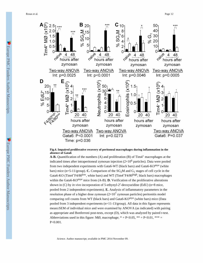
Fig.4. Impaired proliferative recovery of peritoneal macrophages during inflammation in theabsence of Gata6A-B. Quantification of the numbers (A) and proliferation (B) of Tim4+ macrophages at the
indicated times after intraperitoneal zymosan injection (2×10⁶ particles). Data were pooled
from two independent experiments with Gata6-WT (black bars) and Gata6-KOmye (white
bars) mice (n=5-11/group). C. Comparison of the SG2M and G1 stages of cell cycle in the
Gata6-KO (Tim4+F4/80low, white bars) and WT (Tim4+F4/80high, black bars) macrophages
within the Gata6-KOmye mice from (A-B). D. Verification of the proliferative alterations
shown in (C) by in vivo incorporation of 5-ethynyl-2′-deoxyuridine (EdU) (n=8 mice,
pooled from 2 independent experiments). E. Analysis of inflammatory parameters in the
resolution phase of a higher dose zymosan (2×10⁷ zymosan particles) peritonitis model
comparing cell counts from WT (black bars) and Gata6-KOmye (white bars) mice (Data
pooled from 3 independent experiments (n=11-13/group). All data in this figure represents
mean±SEM of individual mice and were examined by ANOVA (as indicated) with pairing
as appropriate and Bonferroni post-tests, except (D), which was analyzed by paired t-test.
Abbreviations used in this figure: MØ, macrophage; * = P<0.05, ** = P<0.01, *** =
P<0.001.
Rosas et al. Page 12
Science. Author manuscript; available in PMC 2014 November 09.
Europe PM
C Funders A
uthor Manuscripts
Europe PM
C Funders A
uthor Manuscripts