1. obstetrics is "bloody business." hemorrhage still remains a leading cause of maternal...
TRANSCRIPT

1
2
OBSTETRICAL HEMORRHAGE
Obstetrics is "bloody business." hemorrhage still remains a leading
cause of maternal mortality. 12 %of maternal deaths were
caused by obstetrical hemorrhage. hemorrhage is the single most
important cause of maternal death worldwide.
Obstetrical hemorrhage accounts for almost half of all postpartum deaths in developing countries
4
most maternal deaths from
hemorrhage is associated with
substandard care.
many hemorrhage-related maternal
deaths were preventable and were
associated with inadequate facilities

5
خونریزی در طی حاملگی باید بدون تاخیر د ارزیابی شوبیمارستاندر .
خونریزی مامایی نیاز به ارزیابی سریع دارد زیرا بدون توجه به منشاء
خونریزی، هر خونریزی مامایی می تواند شودتبدیل به خونریزی شدید سریعاً .
غیر وقتی خونریزی شروع می شود کرد که کی پیشگوییاست بتوان ممکن
.و چه مقدار شدید می شود

6
می اورژانسی های مراقبت چهخونریزی دچار که زنانی برای بایست
انجام اند شده آمیز مخاطره و شدیدگیرد؟
بیمار وضعیت Stable کردن
خونریزی محل تعیین
8
کمک بخواهیدبرای stable کردن بیمار Volume expansion .بدهیم
یا بزرگتر)گاهی اوقات 18ایجاد راه وریدی با سوزن شماره خونریزی بحدی است که باید چند رگ گرفته و با سرعت
مناسب مایع را جایگزین کردرینگر الکتات- انفوزیون سریع سالن
انفوزیون سریع خون
انجام آزمایشات الزمارسال نمونه برای گروه خونی و کراس مچ حداقل چهار واحد
شمارش پالکت، سطح PTT وfDP CBC، PT ،خون ،فیبرینوژن
میلی لیتر خون در داخل لوله فاقد مواد آنتی کواگوالنت 5تهیه دقیقه بعد ازنظر ایجاد لخته10- 15و مشاهده آن
کنترل حجم ادرارارزیابی سالمت جنین و سن حاملگی

9
اگر خونریزی متوقف نشد و زایمان شروع .نشدسریع ختم حاملگی داده شود
در صورتی که بیمار بتواند جراحی را تحمل کند باید سریع سزارین گردد
اگر زایمان شروع شده باشد در حالتdouble set-up بیمار را معاینه می کنیم
برای تعیین علت خونریزیاگر خونریزی متوقف شد و یا کم شد و
بیمار و ضربان قلب جنین خوبست اقدام الزم را برای تعیین محل خونریزی انجام
دهید

10
اثر جنین روی تواند می مامائی خونریزیبه باعث یا باشد جنین داشته افتادن ، مخاطره
. در بنابراین شود نوزاد بیماری و جنین مرگزایمان برای تیمحین در احیاءمناسب نوزاد
اتاقباشد آماده .زایمان
با بیمار وضعیت مورد صحبت در می فامیلش کنیم
منفی درصورت خونی بودن گروه مثبت مادروکرد توجه باید رگام تجویز به نوزاد خونی .گروه
11
ANTEPARTUM HEMORRHAGE
Placental
Abruption
Placenta Previa

12
Causes of Obstetrical Hemorrhage
Placental Abruption:
Placental separation from its
implantation site before delivery
variously called placental abruption,
abruptio placentae, accidental
hemorrhage , abruptioplacentae
13
COMPLICATIONS Shock Consumptive Coagulopathy Renal Failure Sheehan Syndrome
14
Shockshock sometimes seen with placental abruption
was disproportionate to the amount of hemorrhage.
because placental thromboplastin enters the
maternal circulation and incites intravascular
coagulation
hypovolemic shock is directly due to maternal
blood loss.
15
Consumptive Coagulopathy
Placental abruption is one of the most common causes of
clinically significant consumptive coagulopathy in
obstetrics.
In approximately a third of women with an abruption severe
enough to kill the fetus
there are measurable changes in coagulation factors.
16
Renal Failureit is more common if treatment of hypovolemia is
delayed or incomplete.
most cases of acute kidney injury are reversible, however,
acute cortical necrosis, when it occurs, is usually caused by
placental abruption.
Seriously impaired renal perfusion is the consequence of
massive hemorrhage.
Because preeclampsia frequently coexists with placental
abruption, renal vasospasm and hypoperfusion are likely
intensified.
17
Sheehan SyndromeSevere intrapartum or early postpartum
hemorrhage rarely is followed by pituitary failure
or Sheehan syndrome.
characterized by :
failure of lactation,
amenorrhea,
breast atrophy,
loss of pubic and axillary hair,
hypothyroidism,
adrenal cortical insufficiency.
exact pathogenesis is not understood,
18
DIAGNOSIS
sonography confirmes clinical diagnosis in only 25 % of women
negative findings with sonographic examination do not exclude placental abruption
19
MANAGEMENT with massive external bleeding, intensive
resuscitation with blood plus crystalloid and prompt delivery to control hemorrhage are lifesaving for mother and hopefully, for fetus.
If the diagnosis is uncertain and the fetus is alive but without evidence of compromise, then close observation can be practiced in facilities capable of immediate intervention.
for the welfare of the distressed fetus, steps should be initiated immediately to correct maternal hypovolemia, anemia, and hypoxia to restore and maintain function of any placenta that is still implanted.
20
PLACENTA PREVIA Placenta previa is used to
describe a placenta that is
implanted over or very near the
internal cervical os.
21
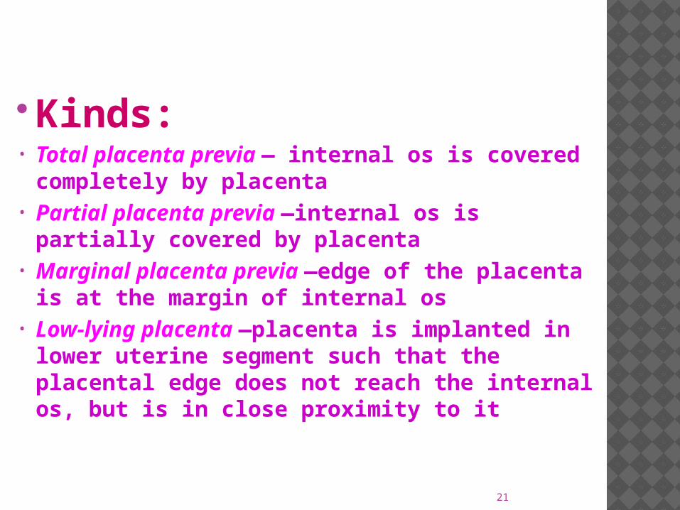
Kinds:• Total placenta previa — internal os is
covered completely by placenta • Partial placenta previa —internal os is
partially covered by placenta • Marginal placenta previa —edge of the
placenta is at the margin of internal os • Low-lying placenta —placenta is implanted
in lower uterine segment such that the placental edge does not reach the internal os, but is in close proximity to it
22
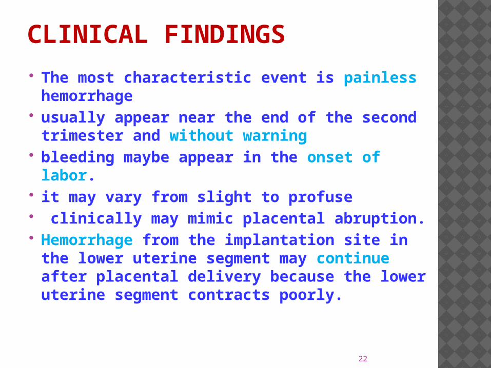
CLINICAL FINDINGS The most characteristic event is painless
hemorrhage usually appear near the end of the
second trimester and without warning bleeding maybe appear in the onset of
labor. it may vary from slight to profuse clinically may mimic placental abruption. Hemorrhage from the implantation site in
the lower uterine segment may continue after placental delivery because the lower uterine segment contracts poorly.
23
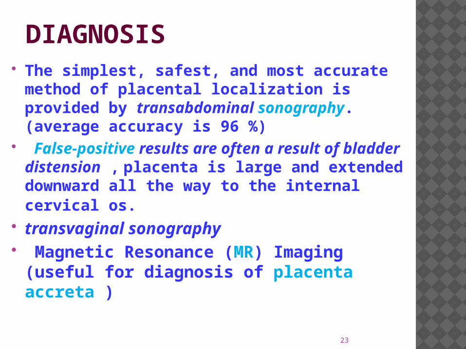
DIAGNOSIS The simplest, safest, and most accurate
method of placental localization is provided by transabdominal sonography. (average accuracy is 96 %(
False-positive results are often a result of bladder distension , placenta is large and extended downward all the way to the internal cervical os.
transvaginal sonography Magnetic Resonance (MR( Imaging
(useful for diagnosis of placenta accreta (
24
MANAGEMENT
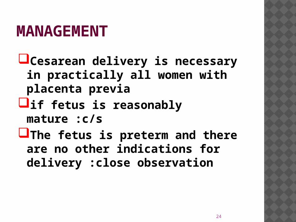
Cesarean delivery is necessary in practically all women with placenta previa
if fetus is reasonably mature :c/s The fetus is preterm and there
are no other indications for delivery :close observation
25
POSTPARTUM HEMORRHAGE
26
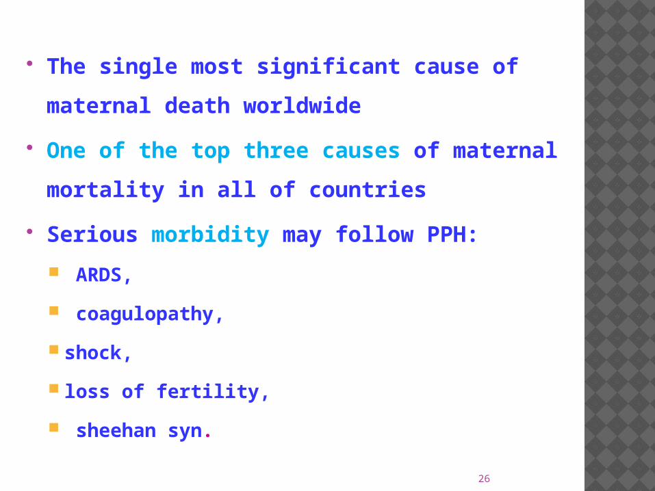
The single most significant cause of
maternal death worldwide
One of the top three causes of maternal
mortality in all of countries
Serious morbidity may follow PPH:
ARDS,
coagulopathy,
shock,
loss of fertility,
sheehan syn.
27
Incidence
The incidence varies widely:
1-5% of deliveries

28
DefinitionThere is no single, satisfactory definition of PPH.
PPH is excessive bleeding that makes the patient
symptomatic
Most common definition: Excess blood loss
(>500ml in NVD or >1000ml in C/S(
Decline in Hct of 10%(not a clinically useful
definition(
29
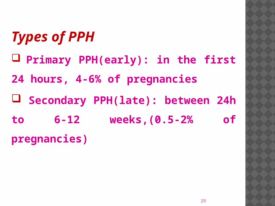
Types of PPH
Primary PPH(early(: in the first 24
hours, 4-6% of pregnancies
Secondary PPH(late(: between 24h
to 6-12 weeks,(0.5-2% of
pregnancies(
30
Atony The most common cause of PPH is uterine atony
Complicates 1 in 20 births
Responsible for at least 80 % of cases of PPH
31
only a small proportion of women
with any risk factors for PPH develop
the disorder and many women
without risk factors experience
hemorrhage after delivery; thus,
knowledge of risk factors is not
very useful clinically

32
کاهش یا کننده پیشگیری اقداماتزایمان از بعد خونریزی دهنده
یوتروتونیک داروهای از استفادهجفت مشاهده
زایمانی کانال مشاهدهزایمان از بعد اول ساعت یک در دقیق کنترل
33
Planning & prevention training team
Protocol for management of PPH
equipments of medications and
instruments are readily available in Labor
and delivery units
34
DIAGNOSIS
The differentiation between bleeding from uterine atony and genital tract lacerations is tentatively determined by predisposing risk factors and the condition of uterus.
If bleeding persists despite a firm, well-contracted uterus, the cause of the hemorrhage most likely is from lacerations
Bright red blood also suggests arterial blood from lacerations.
careful inspection of the vagina, cervix, and uterus is essential.
35
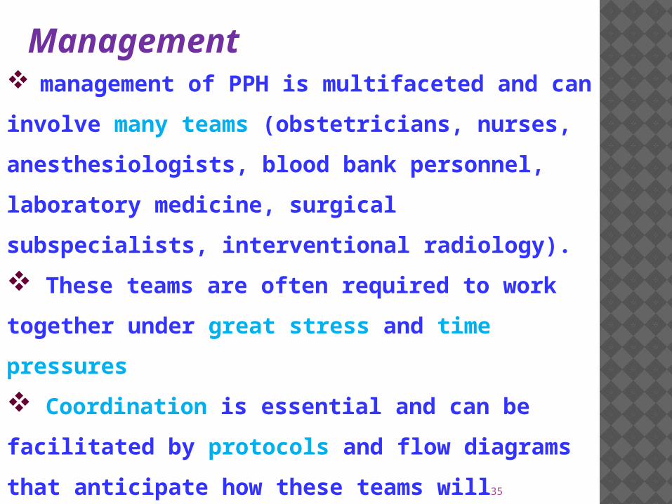
Management management of PPH is multifaceted and
can involve many teams (obstetricians,
nurses, anesthesiologists, blood bank
personnel, laboratory medicine, surgical
subspecialists, interventional radiology(.
These teams are often required to work
together under great stress and time
pressures
Coordination is essential and can be
facilitated by protocols and flow diagrams
that anticipate how these teams will
communicate and function together.
36
Management of postpartum hemorrhage at vaginal delivery
37
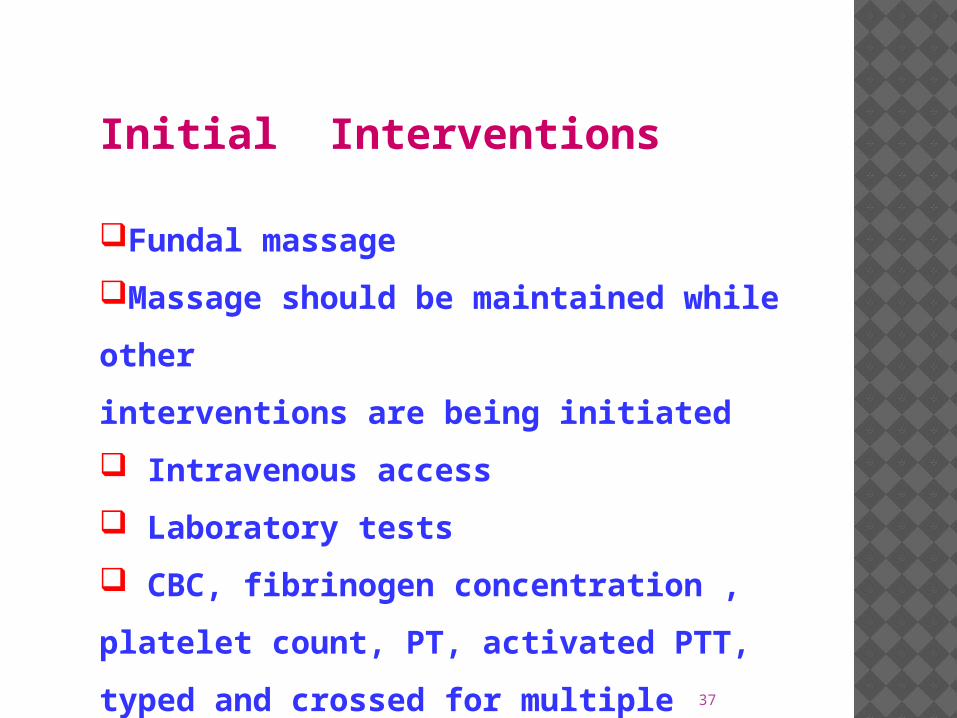
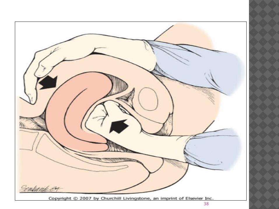
Initial Interventions
Fundal massage
Massage should be maintained
while other
interventions are being initiated
Intravenous access
Laboratory tests
CBC, fibrinogen concentration ,
platelet count, PT, activated PTT,
typed and crossed for multiple units
of packed red blood cells.
38
39
Fluid resuscitation and
transfusion
Monitoring vital signs
Bladder catheter
A large volume of crystalloid is
infused
Replacement of blood
components
40
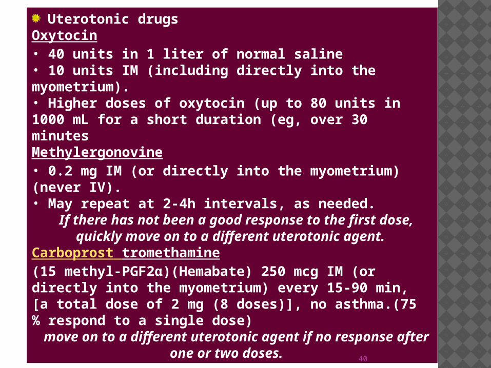
Uterotonic drugsOxytocin • 40 units in 1 liter of normal saline • 10 units IM (including directly into the myometrium(.• Higher doses of oxytocin (up to 80 units in 1000 mL for a short duration (eg, over 30 minutesMethylergonovine • 0.2 mg IM (or directly into the myometrium( (never IV(. • May repeat at 2-4h intervals, as needed. If there has not been a good response to the first
dose, quickly move on to a different uterotonic agent.
Carboprost tromethamine (15 methyl-PGF2α((Hemabate( 250 mcg IM (or directly into the myometrium( every 15-90 min, [a total dose of 2 mg (8 doses(], no asthma.(75 % respond to a single dose(
move on to a different uterotonic agent if no response after one or two doses.
41
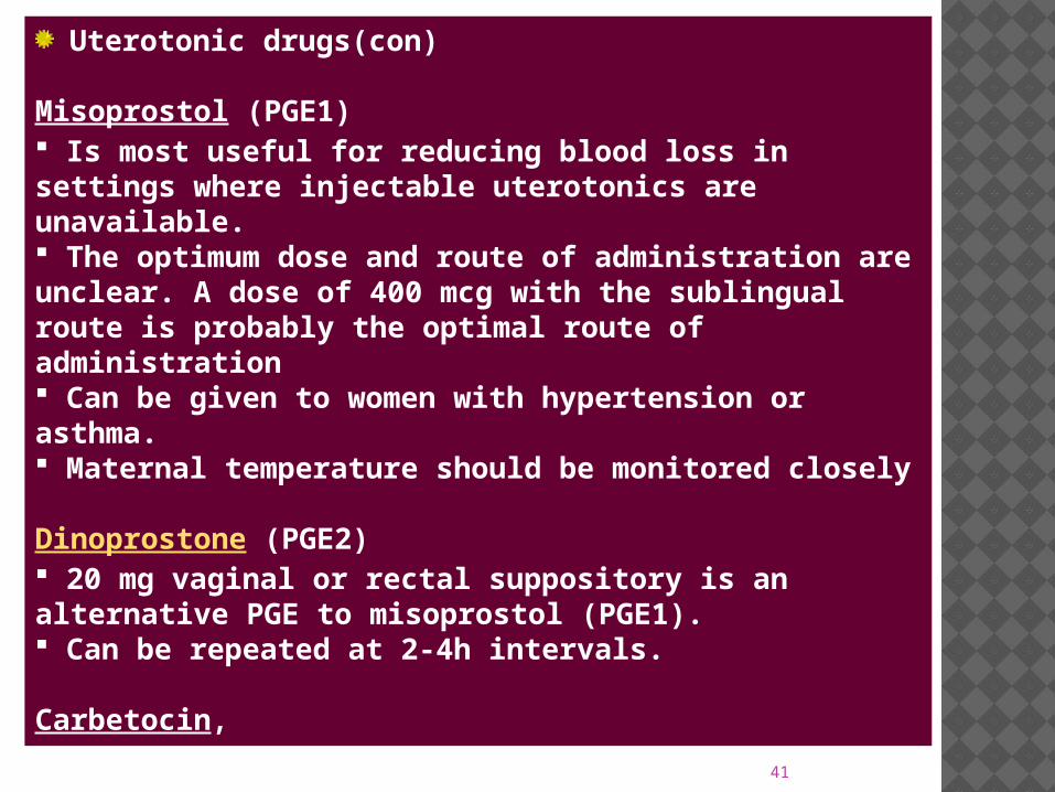
Uterotonic drugs(con(
Misoprostol (PGE1( Is most useful for reducing blood loss in settings where injectable uterotonics are unavailable. The optimum dose and route of administration are unclear. A dose of 400 mcg with the sublingual route is probably the optimal route of administration Can be given to women with hypertension or asthma. Maternal temperature should be monitored closely
Dinoprostone (PGE2( 20 mg vaginal or rectal suppository is an alternative PGE to misoprostol (PGE1(. Can be repeated at 2-4h intervals.
Carbetocin,
42
Secondary Interventions
Provide adequate anesthesia
Inspect for and repair cervical
and vaginal lacerations
Exclude uterine rupture
Remove retained products of
conception
Uterine tamponade
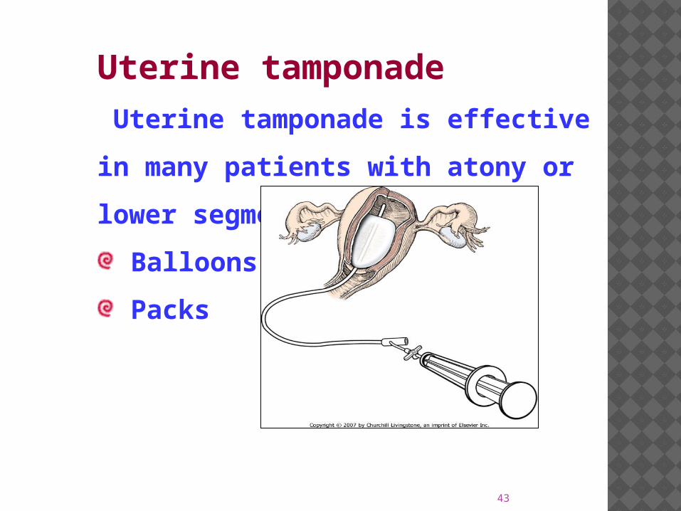
43
Uterine tamponade Uterine tamponade is effective in
many patients with atony or lower
segment bleeding
Balloons
Packs
44
INDICATIONS FOR LAPAROTOMY
If vital signs are worse than expected for
the estimated blood loss,
the possibility of internal hemorrhage
should be considered
When a vaginal laceration has extended
above the fornix
Management of uterine atony
unresponsive to the conservative
interventions described

45
ثبت دقیق مدارک پزشکی

46

47
HYPOVOLEMIC SHOCK
.
48
DEFINITIONS
Hypovolemic Shock :there is
an inadequate circulating
blood volume resulting from
hemorrhage or acute volume
depletion.

Class IV Class III Class II Class I
>2,000 1,500- 2,000 750- 1,500 <750 Blood loss (mL)
>40 30- 40 15- 30 <15 Blood volum(%)
>140 >120 >100 <100 Heart rate (beats/m)
Decreased Decreased (mean arterial pressure
<60 mm Hg)
Normal (+ tilt)
Normal or increased
Blood pressure
Decreased Decreased Decreased Normal Pulse pressure
Always delayed Usually delayed May be delayed
Normal Capillary refill
Marked tachypnea; respiratory
collapse
Moderate to marked tachypnea
Mildly increased
Normal Respirations (breaths/m)
>35 30-40 20- 30 14- 20
Essentially anuric 5- 15 20- 30 >30 Urinary output (mL/h)
Lethargic, obtunded Confused Anxious Normal or anxious
Mental status
49

Management of Shock
50

A team approach
staff trained in intensive care medicine
O R D E R
Oxygenate
Restore circulatory volume
Drug therapy
Evaluate response to therapy Remedy the underlying cause
51
52
RESTORE CIRCULATING VOLUME
Rapid volume repletion is indicated .
Delayed therapy can lead to ischemic injury
and possibly to irreversible shock and
multiorgan system failure.
1- to 2-liter fluid challenge with an isotonic
electrolyte solution, preferably Ringer's
lactate.
Fluid repletion continues at the initial rapid
rate as long as the systemic blood pressure
remains low.
53
Clinical signs, BP, urine output,
mental status, and peripheral
perfusion guide resuscitation.
54
inserting one or two large-bore (14- or 16-
gauge) angiocatheters for volume
replacement.
Trendelenburg position.
initial laboratory assessment CBC
differential and platelets, electrolytes,
BUN, Cr, calcium, magnesium, glucose,
phosphate, and, where indicated, liver
function studies, clotting profiles, serum
lactate, and blood cultures.
55
whole blood transfusion is standard .
Packed red blood cells (PRBC) have a
volume of 200 to 250 mL and a hematocrit of 70%.
normal saline+ PRBCs are the component of choice for hemorrhagic shock.
56
oxygen-carrying capacity is met in most healthy patients with a hemoglobin of 7 g/dL, then transfusion for moderate anemia (8 to 10 mg/dL) is no recommended. but, correction of anemia is important .
In critically ill patients and those with significant underlying cardiac disease, RBC transfuse to maintain hemoglobin levels between 10 and 12 g/dL

57
For avoid the risks of hemolysis,
agglutination, and clotting, all blood
products should be administered
through filtered lines with normal saline
without electrolyte or drug additives.
58
PREVENTION OF HYPOTHERMIA
Rapid transfusion of multiple units of
chilled blood may reduce the core
temperature abruptly and can lead to
cardiac arrhythmias .
hypothermia interfere with the normal
functioning of the coagulation system.
A blood warmer should be used whenever
more than three units are transfused.
59
Massive blood transfusion Massive transfusion, defined as the
replacement by transfusion of more than 50 percent of a patient's blood volume in 12 to 24 hours
Massive transfusion involves: the selection of the appropriate
amounts and types of blood componentsmanagement of bleeding and
coagulation abnormalitieschanges in ionized calcium, potassium,
and acid-base balance.
60
RED CELL AND VOLUME REPLACEMENT
Correction of the deficit in blood volume with
crystalloid volume expanders will generally maintain
hemodynamic stability, while transfusion of red cells
is used to improve and maintain tissue oxygenation .
Each unit of packed cells contains 200 mL of red cells,
will raise the hematocrit by 3-4 % unless there is
continued bleeding.
At rest, oxygen delivery is normally four times oxygen
consumption. if intravascular volume is maintained
during bleeding oxygen delivery will be adequate
until the hematocrit falls below 10 percent.
61
COAGULATION PROTEINS there will be 10 percent decrease in the
concentration of clotting proteins for each
500 mL of blood loss .
Additional bleeding based solely on
dilution can occur when the level of
coagulation proteins falls to 25 percent of
normal. This usually requires 8 to 10 units
of red cells in an adult.
Thus, the PT, aPTT, and fibrinogen should
be monitored in patients receiving
massive blood transfusions of this
magnitude.
62
Two units of FFP should be given if the
values exceed 1.5 times control.
Each unit will increase the clotting
protein levels by 10 percent.
Cryoprecipitate may be used when
fibrinogen levels are critically low (<100
mg/dL)
63
PLATELET COUNT A similar dilutional effect on the platelet
concentration can be seen with massive
transfusion .
each 10 to 12 units of transfused red cells
can produce a 50 percent fall in the platelet
count; thus, significant thrombocytopenia
can be seen after 10 to 20 units of blood.
with platelet counts below 50,000/microL.
six units of random donor platelets, or one
unit of apheresis platelets, should be given.
each unit should increase the platelet count
by 5000 to 10,000/microL.

64
THANK YOU