1. iron metabolism introductory background essential element in all living cells transports and...
TRANSCRIPT

1. IRON METABOLISM 1. IRON METABOLISM INTRODUCTORY BACKGROUNDINTRODUCTORY BACKGROUND
• Essential element in all living cells
• Transports and stores oxygen
• Integral part of many enzymes
• Usually bound to other molecules
• Quantity of body iron carefully controlled
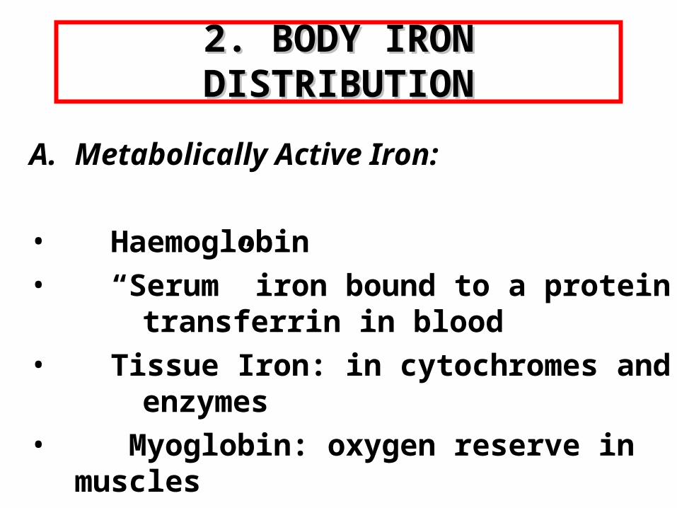
2. BODY IRON DISTRIBUTION2. BODY IRON DISTRIBUTION
A. Metabolically Active Iron:
• Haemoglobin
• “Serum” iron bound to a protein transferrin in blood
• Tissue Iron: in cytochromes and enzymes
• Myoglobin: oxygen reserve in muscles

APPROXIMATE DISTRIBUTION OF APPROXIMATE DISTRIBUTION OF BODY IRON IN A MANBODY IRON IN A MAN
Hemoglobin 2000mg Storage Iron 1000mg Myoglobin iron 130mg Labile Pool 80mg Other tissue Iron 8mg Transport Iron 3mg

2. BODY IRON DISTRIBUTION2. BODY IRON DISTRIBUTION
B. Storage Iron:
• Ferritin: found in blood, tissue fluids, and cells
• Haemosiderin: found in macrophages and assessed by staining bone marrow with Prussian Blue stain

BONE MARROW FILM STAINED FOR BONE MARROW FILM STAINED FOR HAEMOSIDERINHAEMOSIDERIN

3. 3. DIETARY SOURCES OF IRONDIETARY SOURCES OF IRON
Inorganic Iron eg lentils
Organic iron eg beef
DAILY IRON REQUIREMENT 10-15mg/day (5-10% absorbed)

4. IRON ABSORPTION4. IRON ABSORPTION
• Iron kept soluble and in ferrous state by gastric acid
• Absorbed mainly in duodenum• Quantity absorbed regulated by enterocyte• Multiple proteins involved in control of iron
transport • Haem iron enters the enterocyte through
different process than inorganic iron

ABSORPTION OF IRON
Haem
Fe+++
Fe++
Ferritin
Tf
Tf-Fe+++ Fe++
Fe++
Enterocyte GutGut

4. IRON ABSORPTION (cont)4. IRON ABSORPTION (cont)
• Transferrin bound iron in plasma delivered to body cells according to cellular iron requirements
Note:
Only 20% of plasma bound iron derived from gut. Most plasma iron is derived from breakdown of senescent red cells.

BODY IRON CYCLINGBODY IRON CYCLING

5. PROTEINS INVOLVED IN IRON METABOLISM
HEPCIDIN
FERROPORTIN
degrades
Infections and inflammatory stimuli
Upstream regulators eg. HFE
X
No cellular egress of iron
Transferrin receptors
Apoferritin
Synthesized in liver. Present in blood

Clinical RelevanceClinical Relevance• Iron balance physiologically regulated by control of
iron absorption at enterocyte. • Mutations in the gene HFE associated with most
common form of hereditary iron overload (HFE- haemochromatosis)
• Humans unable to excrete excess iron. Interventions which circumnavigate the enterocyte can result in iron loading
• Conditions such as infection and inflammation have an effect on iron metabolism

WHAT YOU NEED TO KNOWWHAT YOU NEED TO KNOW
• Daily requirements and dietary sources of iron
• Where iron is absorbed in the gut• Control of iron balance at level of enterocyte• How body stores of iron are assessed• Proteins involved in regulation of iron

IRON DEFICIENCYIRON DEFICIENCY
• Commonest cause of anaemia worldwide• Cause of chronic ill health• May indicate the presence of important
underlying disease eg. blood loss from tumour

1.EVOLUTION OF IRON 1.EVOLUTION OF IRON DEFICIENCY ANAEMIADEFICIENCY ANAEMIA
• Earliest stage : depletion of body iron stores only
• “Biochemical” iron deficiency without anaemia
• Iron deficiency anaemia

2. CLINICAL FEATURES 2. CLINICAL FEATURES IRON DEFICIENCYIRON DEFICIENCY
• Symptoms eg. fatigue, dizziness, headache • Signs eg. pallor, glossitis, angular cheilosis,
koilonychia, Plummer Vinson syndrome
Koilonychia Glossitis

Angular Cheilosis or Stomatitis
Plummer Vinson Syndrome : Oesophageal Web
CLINICAL FEATURES OF IRON DEFICIENCYCLINICAL FEATURES OF IRON DEFICIENCY

3. LABORATORY DIAGNOSIS: 3. LABORATORY DIAGNOSIS: IRON DEFICIENCYIRON DEFICIENCY
• Microcytic hypochromic anaemia • Often pencil cells and target cells on
blood film• Decreased serum ferritin• Decreased serum iron, increased TIBC,
decreased % transferrin saturation• Absent bone marrow haemosiderin :
(rarely required for diagnosis )

ABSENT IRON STORES IN BONE ABSENT IRON STORES IN BONE MARROW IN IRON DEFICIENCYMARROW IN IRON DEFICIENCY
Iron deficiencyNormal control

Things you need to know about Things you need to know about Laboratory Testing for Iron StatusLaboratory Testing for Iron Status
• Serum ferritin most useful test
• Low serum ferritin certain proof patient iron deficient
• Normal serum ferritin does not always rule out iron deficiency
• Certain conditions raise ferritin for reasons unrelated to iron status

4.DIFFERENTIAL DIAGNOSIS: IRON DEFICIENCY ANAE4.DIFFERENTIAL DIAGNOSIS: IRON DEFICIENCY ANAEMIA

LOOK FOR LOOK FOR THE CAUSE THE CAUSE
OF IRON OF IRON DEFICIENCYDEFICIENCY

6. CAUSES OF IRON DEFICIENCY6. CAUSES OF IRON DEFICIENCY
• Increased physiologic demand eg. pregnancy, lactation, rapid growth
• Blood loss from GI tract, uterus, haemoglobinuria
• Malabsorption
• Diet
colon cancer

WHAT YOU NEED TO KNOWWHAT YOU NEED TO KNOW
• Symptoms and signs of iron deficiency
• Laboratory diagnosis of iron deficiency
• Differential diagnosis of a microcytic hypochromic anaemia
• Importance of finding a cause for iron deficiency
• Principles of treatment

IRON OVERLOADIRON OVERLOAD

EFFECTS OF IRON OVERLOAD
Non-transferrin-bound iron (NTBI) circulates in the plasma
Excess iron promotes the generation of free hydroxyl radicals,
propagators of oxygen-related tissue damage
Liver cirrhosis/ fibrosis/cancer
Insoluble iron complexes are deposited in body tissues and end-organ
toxicity occurs
Diabetes mellitus
Growth failure
Capacity of serum transferrin to bind iron is exceeded
Iron overload
Cardiac failure
InfertilityHSC senescence
(Fenton Reaction)
O2- + H2O2 O2 + OH- + HO

WHEN DOES IRON BECOME A WHEN DOES IRON BECOME A PROBLEM?PROBLEM?
• Normally 2.5 – 3.5g of iron in the body.
• Tissue damage when total body iron is 7 – 15 g

LABORATORY DIAGNOSISLABORATORY DIAGNOSIS
• Elevated % transferrin saturation
• Increased serum ferritin
• Genetic testing for mutations of HFE gene
• Evidence parenchymal iron overload on liver biopsy
• Amount of iron removed by venesection

TREATMENT AND PREVENTIONTREATMENT AND PREVENTION
• Phlebotomy until ferritin <50µg/ml
• Maintenance venesection
• Screen family members
• Prevention
Cirrhosis of liver

CAUSES OF IRON OVERLOADCAUSES OF IRON OVERLOAD
• Hereditary haemochromatosis
• Multiple transfusions
• Liver disease
• Prolonged use medicinal iron
• Ineffective erythropoiesis
• African Iron Overload

HEREDITARY HEREDITARY HAEMOCHROMATOSISHAEMOCHROMATOSIS
• Most common cause of iron overload in North America
• Most cases due to mutations of the HFE gene
• Results in increased inappropriate iron absorption from gut

CLINICAL DIAGNOSISCLINICAL DIAGNOSIS
• Commonly made on basis of biochemical changes : increased serum ferritin or % transferrin saturation
• May have non-specific symptoms/signs such as fatigue or arthropathy
• Discovered as part of family screening
• Rarely fullblown picture : cirrhosis, diabetes, cardiomyopathy, skin pigmentation, gonadal dysfunction

WHAT YOU NEED TO KNOWWHAT YOU NEED TO KNOW
• Association of mutations of the HFE gene with the most common inherited iron overload disorder : HFE- hemochromatosis
• Hereditary haemochromatosis common in North America
• “Early” symptoms/signs non-specific. Have to think of it
• Severe morbidities avoidable if early diagnosis
• Genetic testing available for patient and family