1. introduction to anatomy of the eye and its adnexa - cu · 1. introduction to anatomy of the eye...
TRANSCRIPT

1. Introduction to Anatomy of the Eye and its Adnexa
Let us imagine we are traveling with a ray of light into the eye. The first structure
we will encounter is the cornea (Fig 2).This is a transparent structure which allows
light to enter inside the eye. It fits into the surrounding white opaque tissue; the
sclera like a watchglass and together they constitute the outer coat of the eye.
This outer coat protects the eye from injury and invading micro-organisms (Fig 3).
Light passing from the cornea enters a space full of clear fluid known as the
anterior chamber (Fig 4) , this space is bounded by the cornea anteriorly and by
the iris posteriorly. The iris is the part which gives the eye its color.
The Cornea is made up of 5 layers:
The epithelium, Bowman's layer, the
corneal stroma, Descemet's membrane
and the endothelium
The stroma of the cornea is made up of
collagen fibres called lamellae. The
corneal lamellae are arranged in a
specific order which helps to achieve
corneal transparency; other factors for
corneal transparency include corneal
avascularity, and the presence of active
endothelial pump which makes the
cornea dry and not misty.
Fig 1: A Cross section of the human eye.
Fig 2: The Cornea illuminated by a slit
beam.
Fig 3: The White sclera seen
underneath the transparent
conjunctiva.

The iris (Fig 5)is made up of muscles, blood vessels and pigmented cells. It has a
central circular opening through which light travels , known as the pupil (Fig 5).
The pupil is a circular opening in the iris that allows transmission of light. It
controls the amount of light reaching the retina by dilating and constricting.
The detailed anatomy of the innervation of the pupil will be discussed in chapter 9
(Connection of the eye to the brain).
The iris contains two types of smooth
muscles:
The circular constrictor pupillae and
the longitudinal dilator pupillae
The constrictor pupillae is under
parasympathetic control and on
contraction the pupil narrows
The Dilator pupillae is under
sympathetic control and on
contraction the pupil dilates
Fig 5: The iris and Pupil
Fig 4: The Anterior Chamber.

The iris is continuous posteriorly with the ciliary body (fig 6) and the choroid.
(Fig 7) These 3 structures are made up of blood vessels and contain pigment and
are collectively known as the uveal tract or the middle or vascular coat of the
eye. Its main function is to provide nutrition and support for the overlying and
underlying structures.
This middle coat of the eye would be the first structure to encounter if we were to
peel away the sclera as seen in fig 8.
The ciliary body is made up of two
parts: The pars plicata and the pars
plana.
The pars plicata is covered with
epithelium that secretes aqueous
humour and gives attachment to the
suspensory ligament or zonule that
suspends the crystalline lens in place.
It contains a smooth muscle known
as the ciliary muscle whose
contraction results in relaxation of
the zonule and bulging of the lens,
thus changing its curvature and
power. This is known as
accommodation. Fig 8: The choroid and its blood vessels with
the sclera peeled away.
Fig 7: The Choroid and its blood vessels. Fig 6: A diagram of the ciliary body (pars
plana and [pars plicata).

After light passes through the pupil it meets a transparent object suspended from
the ciliary body known as the crystalline lens (Fig 9). The lens is an important
refractive medium of the eye. The space between the crystalline lens and the iris is
known as the posterior chamber (Fig 10). The transparent fluid that fills the
anterior chamber is known as the aqueous humor and is formed by the ciliary
body and secreted into the posterior chamber, behind the iris, it then passes through
the pupil to fill the anterior chamber and be drained through the angle (Fig 10)
between the cornea and sclera.
Aqueous formation, circulation and drainage through the angle, in addition to the
structures of the angle will be discussed in more detail in chapter 5 (Aqueous
humour, IOP and glaucoma).
Fig 9: A diagramatic representation of the
crystalline lens. Fig 10: Passage of aqueous humour from the
ciliary body in the posterior chamber, through
the pupil into the anterior chamber.

Once light has been refracted by the lens it meets the transparent gel like structure
that fills the space behind the lens. This gel like structure is known as the vitreous
body (Fig 11) and (Fig 12). The vitreous is formed of 99% water in addition to
special proteins called glycosaminoglycans. No clear function is known to the
vitreous apart from transmitting light to the retina. However, it may have a shock
absorbing role due to its gel-like state and it may also act as a reservoir for certain
nutrients or chemical mediators.
The Lens:
The lens is a biconvex avascular structure present behind the
pupil. It contributes about +20 D to the refractive power of the eye.
This power can be increased during accommodation, which can be
achieved by ciliary muscle contraction resulting in relaxation of the
zonule and allowing the elastic capsule to change the shape of the
lens in order to become more convex. This allows seeing near objects
clearly.
The Lens is also a dynamic structure that is continuously growing with
human growth. The capsule which surrounds the lens is elastic and
has an epithelial layer on the inner surface of its anterior portion from
which lens proteins are formed. As it grows, the lens proteins become
more aggregated in the center and lose fluid. This hardening or
sclerosis leads to a distinction of the lens proteins into the harder
central nucleus and the softer peripheral cortex. Due to this sclerosis,
the lens becomes less pliable by age and consequently loses its ability
to increase its power by changing its shape (Accommodation).

Having traversed the vitreous body, light will reach its final destination by falling
upon the light sensitive neurosensory structure known as the retina (Fig 13). The
retina is the inner coat of the eye. The retina is a very intricate structure which is
closely connected anatomically and embryologically to the brain, and will be
discussed in more detail in chapter 8 (The Retina: Functions and diseases).
The retina performs the essential task of converting light to electrical impulses.
These impulses are then transmitted via the optic nerve (Fig 14) to the brain. The
optic nerve can be actually viewed as being a tract of the central nervous system
rather than a true nerve. The occipital cortex is the part of the brain responsible for
vision i.e. giving meaning to those electrical impulses. The route from the retina to
that part is called the visual pathway (Fig 15).
The detailed anatomy of the optic nerve and visual pathway will be discussed in
chapter 9 (Connection of the eye to the brain).
Fig 11: A diagramatic representation of the
vitreous body.
Fig 12: The vitreous body as seen after
peeling the sclera, choroid and retina away.
Fig 13: Fundus photograph showing the
appearance of a normal retina.

Due to the importance of the retina in the process of vision, all the aforementioned
ocular structures can be looked at as serving the purpose of allowing light to reach
the retina and to provide the retina with protection and nutrition.
Additional protection is provided by the ocular adnexa and orbit. The orbit (Fig
16) is a bony socket in-which the eye is placed. Within the orbit the eye is
surrounded by fat,muscles, nerves and vessels ,in addition to the lacrimal gland
(Fig 17) that secretes the main ( watery) part of the tears.The tears are then drained
through the lacrimal drainage system (Fig 17, See details in chapter 3)).
Fig 14: The Optic nerve seen passing from the
back of the eye into the cranial cavity.
Fig 15: The visual pathway.
Fig 16: The Bony orbit. Fig 17: The lacrimal gland and lacrimal drainage
system.

Protecting the surface of the eye are the eyelids (Fig 18). The eyelids and the outer
surface of the eye are covered with a transparent membrane known as the
conjunctiva (Fig 19). It lubricates the ocular surface and contributes to tear
formation aswell. The anatomy of the orbit, eyelids, lacrimal gland and lacrimal
drainage system will be discussed in further details in chapter 4 (Diseases of the
protective structures of the eye).
Fig 18: A cross-section in the eyelids. Fig 19: A cross-section in the conjunctiva of
the upper and lower lids in red.
The conjunctiva is a membrane that
lines the inner surface of the eyelids
and the outer surface of the globe. It
is formed of three parts. The
palpebral part (related to the lid),
the bulbar part (related to the globe)
and the fornix, which is the pouch
connecting both these parts. The
fornix of the lower lid is the site
where eye-drops are instilled.
The conjunctiva plays a protective
role, in addition to its important
contribution to tear formation.
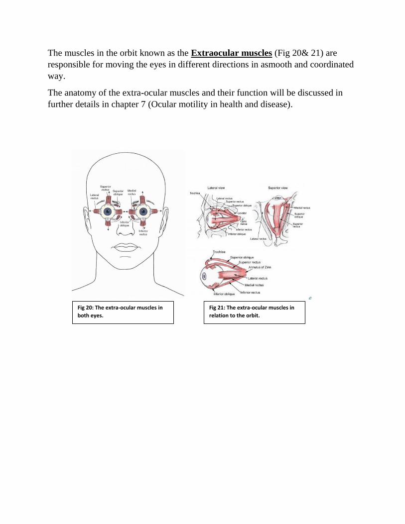
The muscles in the orbit known as the Extraocular muscles (Fig 20& 21) are
responsible for moving the eyes in different directions in asmooth and coordinated
way.
The anatomy of the extra-ocular muscles and their function will be discussed in
further details in chapter 7 (Ocular motility in health and disease).
Fig 20: The extra-ocular muscles in
both eyes.
Fig 21: The extra-ocular muscles in
relation to the orbit.