1. introduction definition: hormones are chemical messengers carried by blood to non- adjacent...
TRANSCRIPT

1. Introduction
Definition: Hormones are chemical messengers carried by blood to non-adjacent "target" cells where they regulate the metabolism.For example: adrenalin is produced in the adrenal gland and released into blood. It works in other tissues such as the liver and muscle, promoting glycogen degradation etc.
Chapter 9. Hormones

Some endocrine glands

2. Overview of hormone functions
• Regulation of growth and development.
• Homestatic control (regulation of internal environment; parameters maintained within relatively narrow limits)
• Control of reproductive system processes (ovulation, menstruation, maintenance of pregnancy)
• Effects on behavior (modification, modulation, initiation of specific patterns)

There are two main classes
1) Steroids (synthesized from cholesterol by adrenal cortex, testis, ovary and placenta)
For example: cortisol, estradiol2) Nitrogen-containing hormones- amines [epinephrine, norepinephrine]- peptides [oxytocin, ADH]- proteins [growth hormone, insulin]- glycoproteins [FSH, TSH]

Some hormones and their physiological effectsEndoc. Gland Hormone Effects
Anterior Growth hormone(GH) Stimulates growth
pituitary: Thyroid stimulating hormone Stimulates secretion of (TSH) thyroid hormones
Adrenocorticotropic hormone Stimulates secretion of (ACTH) adrenal cortex hormones
Prolactin (LTH) Stimulate milk production secretion of estrogen and progesterone by ovary
Luteinizing hormone(LH) Production of estrogen and progesterone or testosterone
Follicle-stimulating hormone Growth of ovarian follicles (FSH) or seminiferous tubules
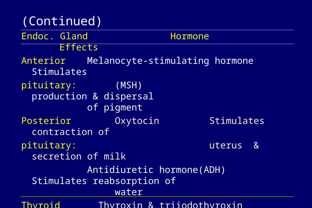
(Continued)Endoc. Gland Hormone Effects
Anterior Melanocyte-stimulating hormone Stimulates
pituitary: (MSH) production & dispersal of pigment
Posterior Oxytocin Stimulates contraction of
pituitary: uterus & secretion of milk
Antidiuretic hormone(ADH) Stimulates reabsorption of water
Thyroid Thyroxin & triiodothyroxin Stimulates metabolism,
Gland: (T4 & T3) growth and development
Calcitonin Lowers blood calcium level
Parathyroid Parathyroid hormone(PTH) Increases blood calcium
Gland:

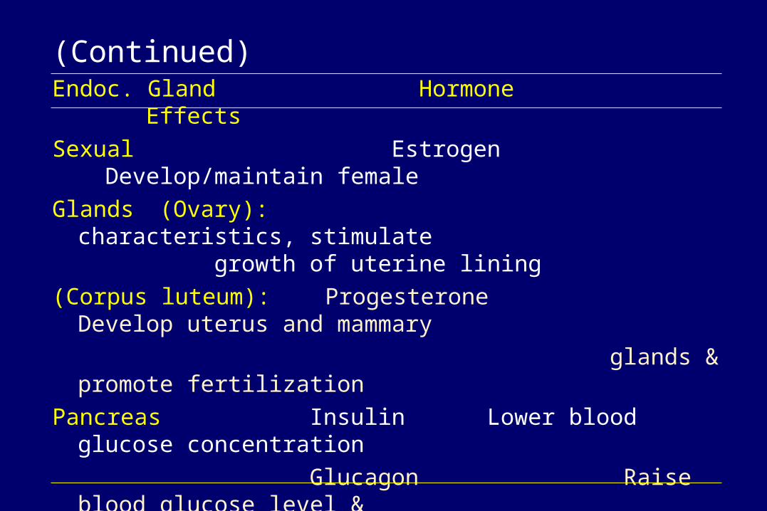
(Continued)Endoc. Gland Hormone Effects
Adrenal Glucocorticoids (cortisol) Raise blood-glucose
cortex: level
Mineralocorticoids(aldosterone) Maintain sodium and phosphate balance
Dehydroepiandrosterone(DHEA) Stimulate sex drive and induce labor
Adrenal Epinephrine(adrenalin) Stimulates glucose release
medulla: Norepinephrine Constricts blood vessels and increases heart rate
Sexual Testosterone Develops and maintains
Glands male sex characteristics,
(Testes): promotes spermatogenesis
(Continued)Endoc. Gland Hormone Effects
Sexual Estrogen Develop/maintain female
Glands (Ovary): characteristics, stimulate growth of uterine lining
(Corpus luteum): Progesterone Develop uterus and mammary
glands & promote fertilization
Pancreas Insulin Lower blood glucose concentration
Glucagon Raise blood glucose level & stimulate gluconeogenesis
Placenta Human chorionic Stimulates release of FSH & LH
gonadotropin (HCG)
Heart Atrical natriuretic factors stimulate blood vessel dilation, lowers blood pressure, inhibits ADH

3. Structures and functions of Some important hormones
1) Thyroxine: T4, T3, and rT3
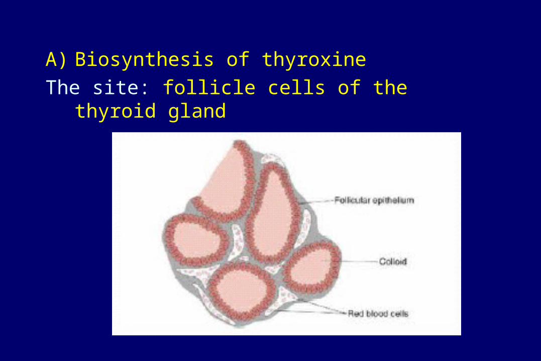
A) Biosynthesis of thyroxine
The site: follicle cells of the thyroid gland

The steps: three major steps• Accumulation of iodide (I-)
Iodide is avidly taken up from blood by thyroid epithelial cells. Once inside the cell, iodide is transported into the lumen of the follicle along with thyroglobulin, where the concentration of iodide may be as high as 30 times that in the blood.

• Synthesis of the hormones on a backbone or scaffold of precursor: iodination of tyrosines on thyroglobulin, followed by synthesis of thyroxine (T4) or triiodothyronine (T3) from two iodotyrosines.

• Release of the free hormones into blood: thyroid hormones are excised from their thyroglobulin scaffold by digestion in lysosomes of thyroid epithelial cells. Free thyroid hormones diffuse out of lysosomes and enter into blood where they quickly bind to carrier proteins for transport to target cells.
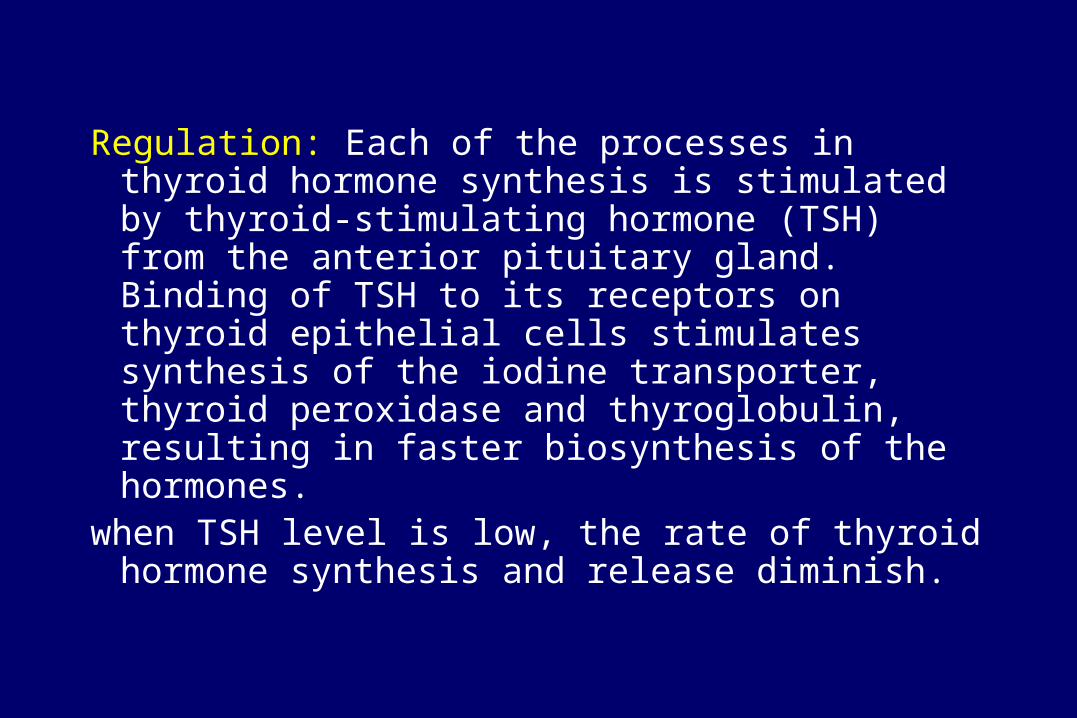
Regulation: Each of the processes in thyroid hormone synthesis is stimulated by thyroid-stimulating hormone (TSH) from the anterior pituitary gland. Binding of TSH to its receptors on thyroid epithelial cells stimulates synthesis of the iodine transporter, thyroid peroxidase and thyroglobulin, resulting in faster biosynthesis of the hormones.
when TSH level is low, the rate of thyroid hormone synthesis and release diminish.

Regulation of thyroid hormone biosynthesis
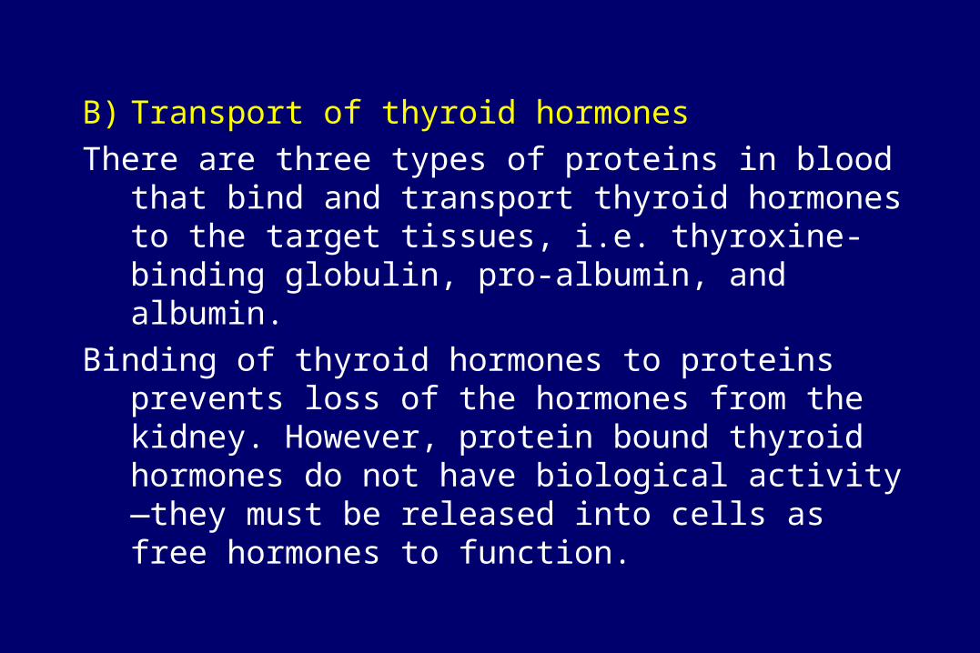
B) Transport of thyroid hormones
There are three types of proteins in blood that bind and transport thyroid hormones to the target tissues, i.e. thyroxine-binding globulin, pro-albumin, and albumin.
Binding of thyroid hormones to proteins prevents loss of the hormones from the kidney. However, protein bound thyroid hormones do not have biological activity—they must be released into cells as free hormones to function.
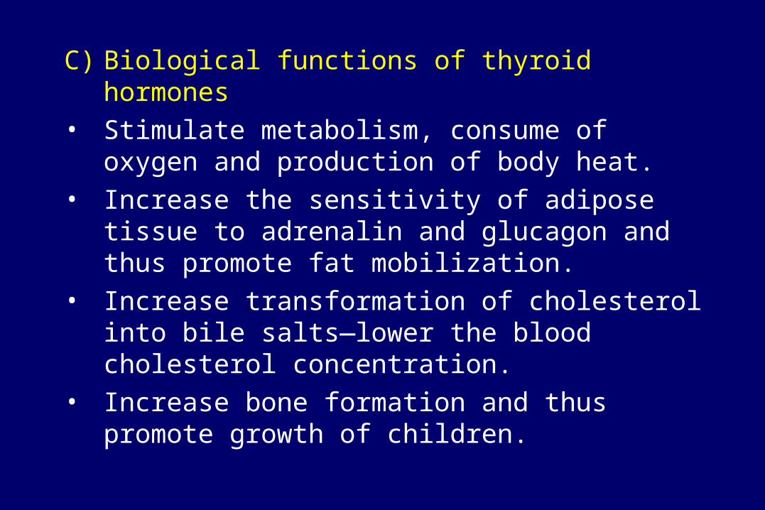
C) Biological functions of thyroid hormones
• Stimulate metabolism, consume of oxygen and production of body heat.
• Increase the sensitivity of adipose tissue to adrenalin and glucagon and thus promote fat mobilization.
• Increase transformation of cholesterol into bile salts—lower the blood cholesterol concentration.
• Increase bone formation and thus promote growth of children.

Cretinism: a disease (growth stop) caused by hypofunction of thyroid glands in children

Hyperthyroidism Hypothyroidism

2) Parathyroid hormone (parathyrin, PTH): The parathyroid glands are 4 tiny structures
embedded in the rear surface of the thyroid gland.
The sequence of the first 34 amino acid residues from the N-terminus of PTH is the key structure responsible for its biological activity.

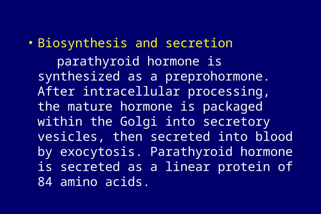
• Biosynthesis and secretion
parathyroid hormone is synthesized as a preprohormone. After intracellular processing, the mature hormone is packaged within the Golgi into secretory vesicles, then secreted into blood by exocytosis. Parathyroid hormone is secreted as a linear protein of 84 amino acids.

Functions: PTH increases the concentration of Ca2+ in the blood by three ways:
• PTH promotes release of Ca2+ from the huge reservoir in the bones.
• PTH promotes reabsorption of Ca2+ from the fluid in the tubules in the kidneys
• PTH promotes absorption of Ca2+ from the contents of the intestine (mediated by calcitriol, the active form of vitamin D.)

PTH increases the concentration of Ca2+ in the blood

3) Calcitonin Calcitonin is produced and secreted by C-cells, or
parafollicular cells that are located between the thyroid follicles. It is a polypeptide with a molecular weight of 3.6kd, consisting of 32 amino acid residues.

Biosynthesis and secretion
The C-cells have receptors that monitor extracellular fluid calcium concentration. They are stimulated to secrete calcitonin when blood calcium levels exceed 10.5 mg%. When blood calcium levels drop back below 10.5 mg%, the C-cells are inhibited and no longer secrete calcitonin.
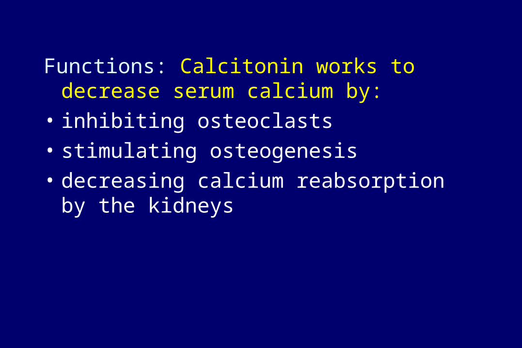
Functions: Calcitonin works to decrease serum calcium by:
• inhibiting osteoclasts
• stimulating osteogenesis
• decreasing calcium reabsorption by the kidneys

4) 1,25-dihydroxy vitamin D3 (1,25-dihydroxy- cholecalciferol)
A vitamin D derivative that acts as a hormone in the body.

Production:Vitamin D3 is first hydroxylated at its 25-carbon by
25-hydroxylase in the liver microsome and then at the 1-carbon by the 1-hydroxylase in the kidney mitochondria.

Functions:
1,25-(OH)2-D3 plays its important roles in control of calcium and phosphate metabolism in the body:
• Stimulates absorption of Ca2+ and phosphate from the intestine
• Promotes re-absorption of phosphate in kidneys
• With the help of PTH, promotes release of Ca2+ from the bone to blood and thus increases the concentration of blood Ca2+

Regulation of Ca2+ metabolismby 1,25-(OH)2-D3
PTG: parathyroid gland; PTH: parathyroid hormone

5) Insulin After the actual isolation of insulin in 1922, it was
not until 1955 that the primary structure of insulin was elucidated by Sanger and co-workers.

Production: Insulin is synthesized only in B cells in the
pancreas. The insulin mRNA is translated as a single chain precursor called preproinsulin, and removal of its signal peptide during insertion into the endoplasmic reticulum(ER) generates proinsulin. Within the ER, proinsulin is exposed to several specific endopeptidases which excise the C peptide, thereby generating the mature form of insulin. When the B cell is appropriately stimulated, insulin is secreted into the blood.
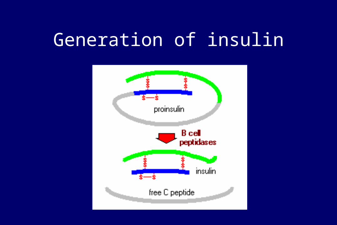
Generation of insulin

Functions:
• membrane transport of glucose, amino acids and certain ions;
• increased glycogenesis and storage of glycogen;
• decreased glycogenolysis and gluconeogenesis
• formation of triglycerides (lipogenesis);
• stimulation of DNA, RNA and protein synthesis.

Control of Insulin Secretion:
Insulin is secreted primarily in response to elevated blood concentrations of glucose.

Diabetes mellitus:
Type I: juvenile onset (insulin-dependent): Decrease in the number of beta cells insulin deficiency. Requires daily injections of insulin.
Type II: maturity onset (insulin-independent) : Insulin receptors cannot bind hormone.
Most commonly, an aberrant increase in the level of a specific hormone will cause a decrease in available receptors
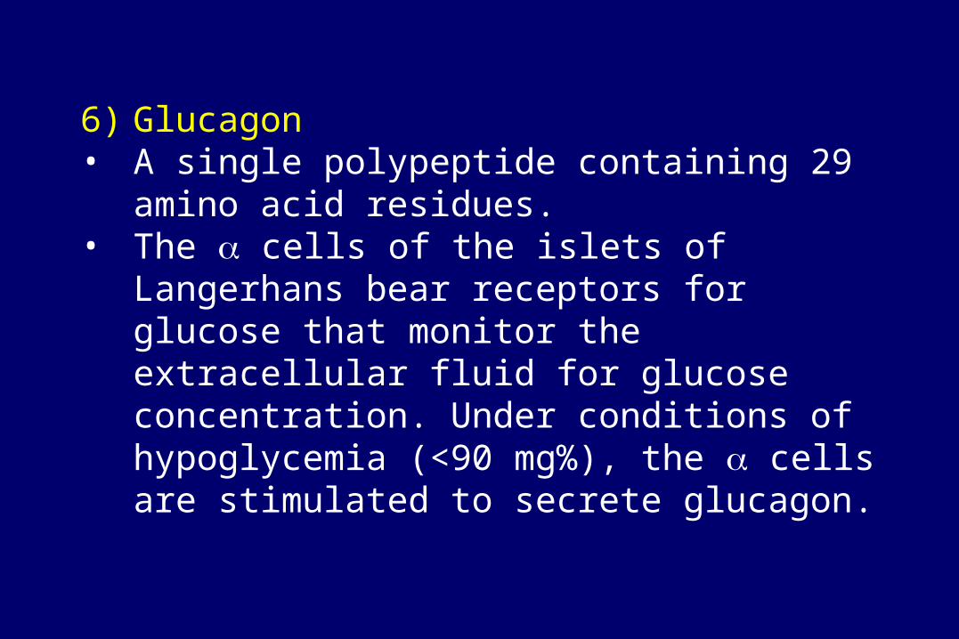
6) Glucagon• A single polypeptide containing 29 amino acid
residues. • The cells of the islets of Langerhans bear
receptors for glucose that monitor the extracellular fluid for glucose concentration. Under conditions of hypoglycemia (<90 mg%), the cells are stimulated to secrete glucagon.

Functions:
Glucagon works antagonistically with insulin to maintain blood glucose levels. It raises blood glucose level by:
• increasing glycogenolysis and gluconeogenesis
• increasing urea synthesis in the liver.
• Promoting lipolysis and increasing the free fatty acid concentration in blood.

Secretion of glucagon
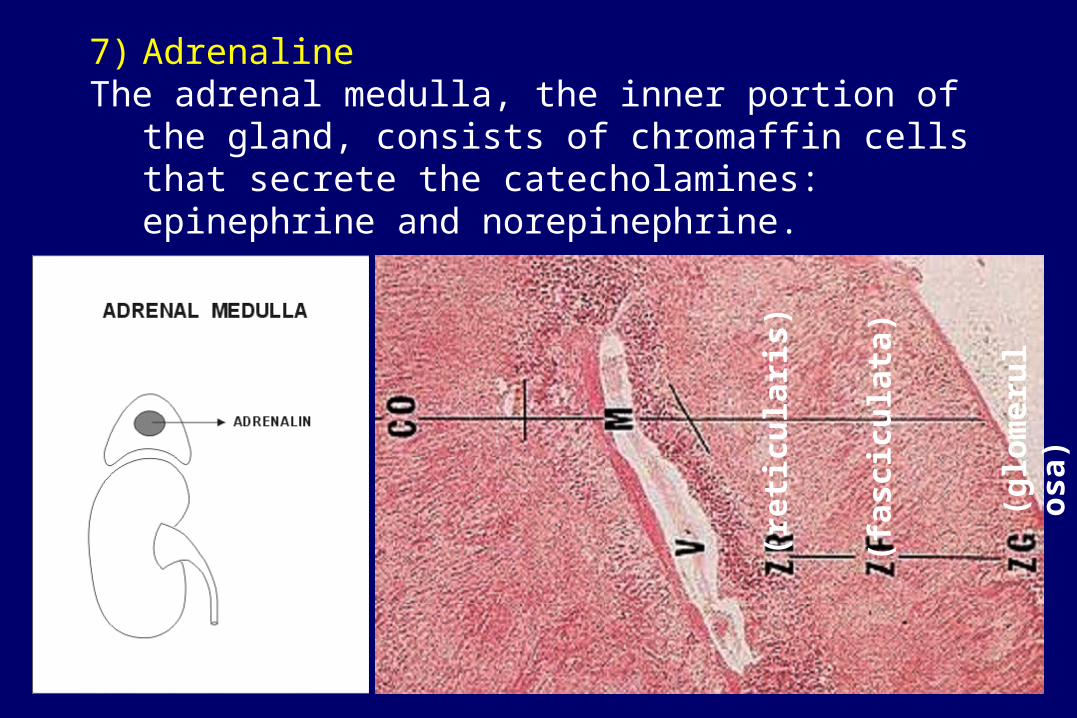
7) Adrenaline The adrenal medulla, the inner portion of the gland,
consists of chromaffin cells that secrete the catecholamines: epinephrine and norepinephrine.
(glo
mer
ulo
sa)
(fas
cicu
lata
)
(ret
icu
lari
s)
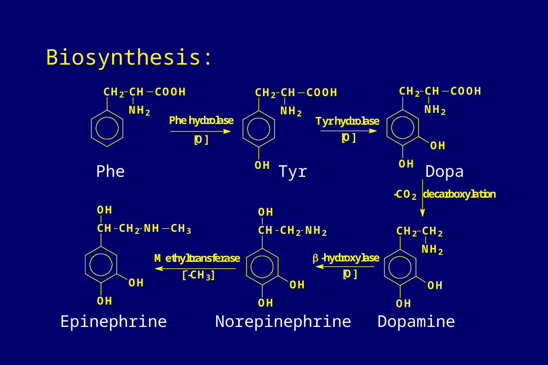
Biosynthesis:
CH2 CH COOH
NH2
CH2 CH COOH
NH2
OH
CH2 CH COOH
NH2
OH
OH
CH2 CH2
NH2
OH
OH
CH CH2
OH
OH
OH
NH2CH CH2
OH
OH
OH
NH CH3
Phe hydrolase Tyr hydrolase
decarboxylation
-hydroxylaseMethyltransferase
[O] [O]
-CO2
[O][--CH3]
Phe
NorepinephrineEpinephrine Dopamine
DopaTyr

Functions:
• vasodilation of vessels to brain, muscles, heart
• vasoconstriction of vessels to kidney, skin
• heart rate speeds up
• mental alertness increases
• fatty acid and glucose levels in blood increase (glycogenolysis and gluconeogenesis increase)
• muscles contract more strongly

8) Adrenal corticosteroids Adrenal cortex produces and secrets glucocorticoids,
mineralocorticoids and sex hormones.

Functions of aldosterone:• Aldosterone is the primary mineralocorticoid and,
work to control water and electrolyte balance, particularly by controlling sodium and potassium concentrations.
• Aldosterone secretion is in response to high potassium ion concentration in the extracellular fluid.
• Aldosterone stimulates kidney cells to lose potassium ions into the forming urine, while at the same time conserving sodium ions.

Functions of glucocorticoids: act slowly on the cell nucleus, changing the patterns of gene expression
• increased protein breakdown, lipolysis and gluconeogenesis.
• promoting sodium retention and potassium loss.
• immunosuppressive and anti-inflammatory effects.
• increased body tolerance to stress.

Cushing's syndrome (glucocorticoid excess):
• Cushing's syndrome may result from a pituitary tumor that secretes ACTH.
• Cushing's syndrome commonly results from the medical administration of immunosuppressive or anti-inflammatory doses of corticosteroid hormones, or from ectopic hormone production by neoplastic tissue elsewhere in the body.
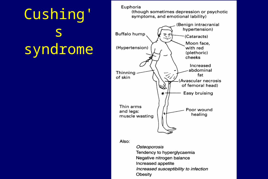
Cushing's syndrome

9) Sex hormones
A class of steroid hormones produced by ovary, testis and adrenal glands. Sex hormones include male androgens and female estrogens.
O
OH
17
O
O
HO
OH
O
C
CH3
O
Testosterone Androstenedione
Eestradiol Progesterone

Functions of androgens:• stimulates spermatogenesis. • maintain the function of the epididymis. • promotes the growth, development, and activity of
accessory sex glands and secondary sex organs. • development of male secondary sex characteristics • anabolic activity.• Stimulates production of RBC.
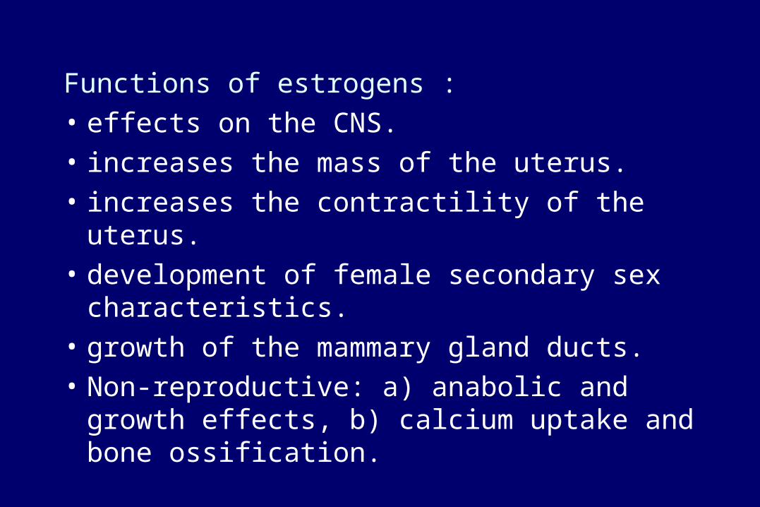
Functions of estrogens :
• effects on the CNS.
• increases the mass of the uterus.
• increases the contractility of the uterus.
• development of female secondary sex characteristics.
• growth of the mammary gland ducts.
• Non-reproductive: a) anabolic and growth effects, b) calcium uptake and bone ossification.

Functions of progesterone:
• prepares the uterus for implantation and pregnancy
• acts with estrogen to induce the behavior patterns of estrus
• develops alveoli of mammary gland
• inhibits the rise of luteinizing hormone (LH) that causes ovulation
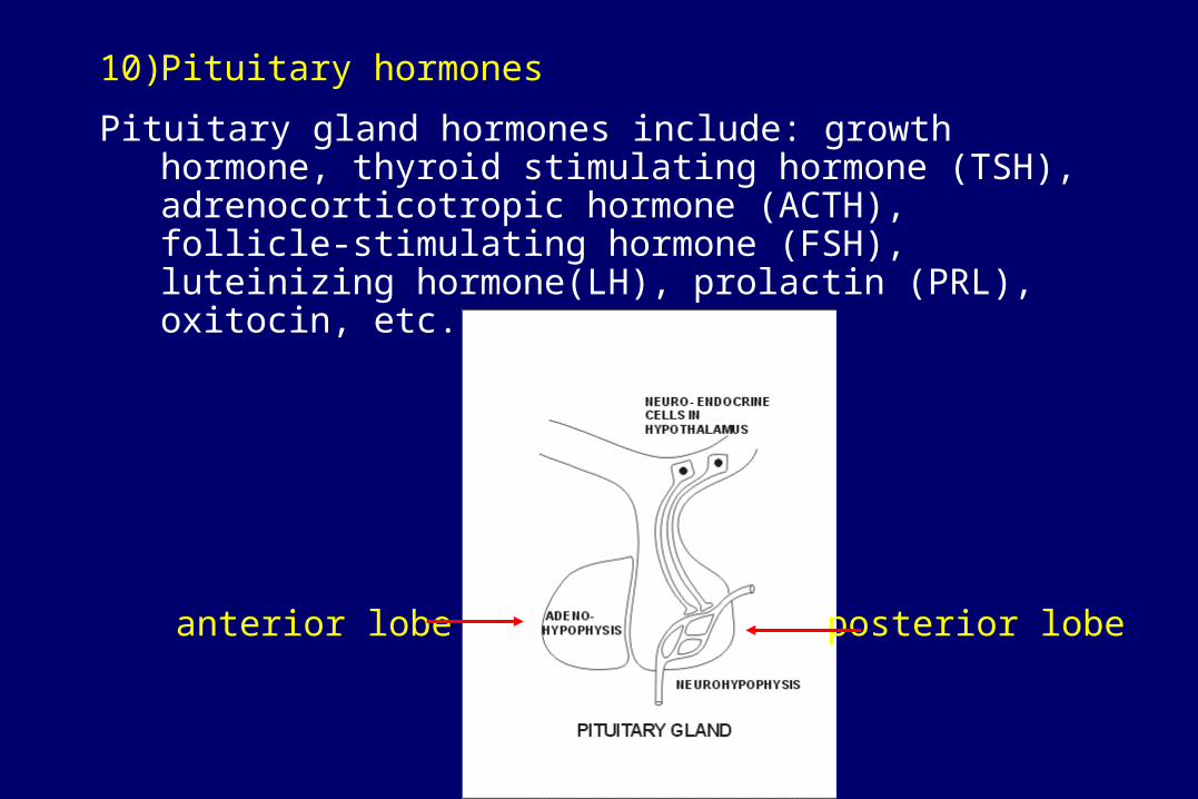
10) Pituitary hormones
Pituitary gland hormones include: growth hormone, thyroid stimulating hormone (TSH), adrenocorticotropic hormone (ACTH), follicle-stimulating hormone (FSH), luteinizing hormone(LH), prolactin (PRL), oxitocin, etc.
anterior lobe posterior lobe

Actions of growth hormone (GH):The major effect of GH is to promote the synthesis
of insulin-like growth factors (IGFs) by peripheral tissues, such as liver, skeletal muscle and bone.
GH increases the rates of amino acid uptake and protein synthesis, causes cells to grow and multiply, increases lipolysis in adipose tissue and glucose output from the liver, and decreases glucose utilization by peripheral tissues, except brain.

• Deficiency of growth hormone during development will produce dwarfism.
• Excess growth hormone in children produces gigantism; in adults excess growth hormone will produce acromegaly.

gigantism dwarfism
Average

Actions of ACTH:
ACTH is a 39 amino acid peptide derived from pituitary pro-opiomelanocortin which promotes the growth and activity of the adrenal cortex, and increases the output of glucocorticoid hormones.
Actions of prolactin:
Stimulates milk production in breast, maintains secretion of estrogen and progesterone by ovary.

Actions of TSH:
TSH is a glycoprotein consisting of two subunits, and .
Its major functions: stimulates secretion of thyroid hormones, promotes uptake of thyroid globulins by the follicle cells and uptake of iodide by thyroid glands.
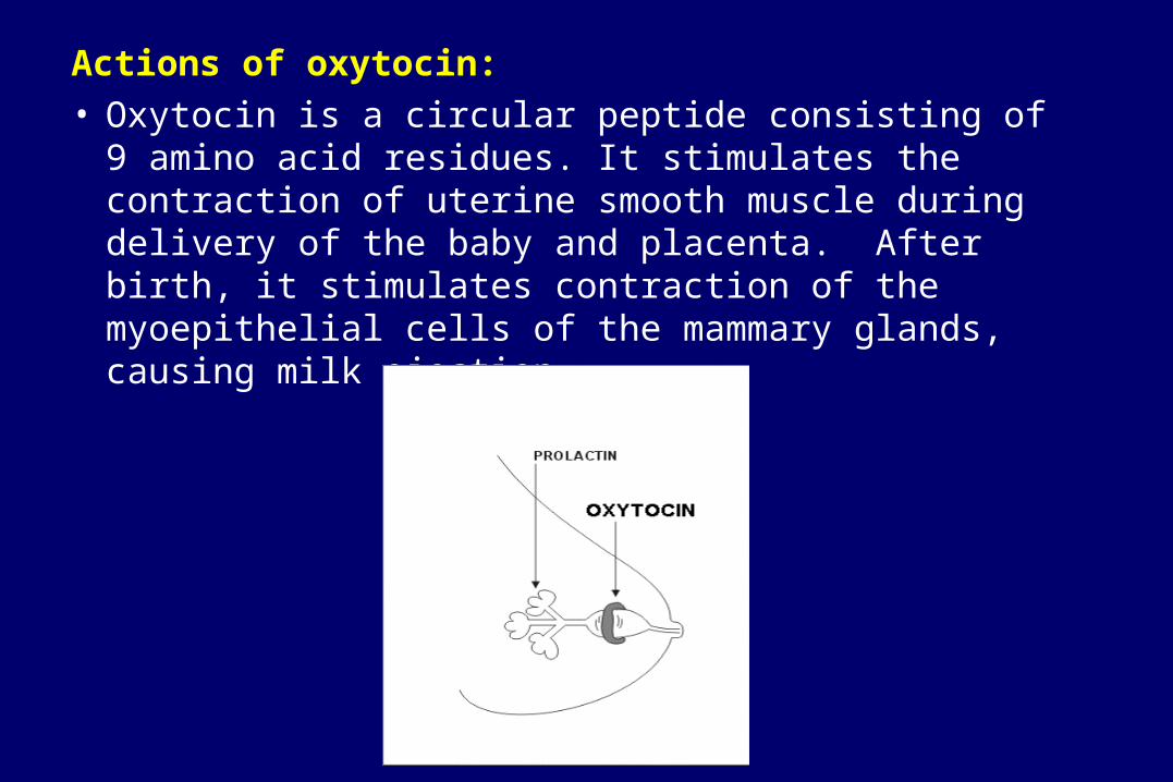
Actions of oxytocin:• Oxytocin is a circular peptide consisting of 9 amino acid
residues. It stimulates the contraction of uterine smooth muscle during delivery of the baby and placenta. After birth, it stimulates contraction of the myoepithelial cells of the mammary glands, causing milk ejection.

11) Hypothalamic hormones
• The hypothalamus serves as the master control for many of the hormones secreted by the endocrine system, and serves as the major integrator between the nervous and endocrine systems. In particular, the hypothalamus controls the secretions of the pituitary gland, also known as the hypophysis, in turn, controls activity of other endocrine glands.

• The hypothalamus also controls the autonomic nervous system, which provides direct neural regulation of blood distribution, pancreatic islet function and liver metabolism.