1 biofunctionalization of fluorescent nanoparticles · pdf file2 1 biofunctionalization of...
TRANSCRIPT

1
Biofunctionalization of Fluorescent
Nanoparticles
Michael J. Murcia and Christoph A. Naumann
1.1
Introduction
The current revolution in life sciences is strongly linked to the availability of so-
phisticated new experimental tools that enable the manipulation of biomolecules
and the study of biological processes at the molecular level using state-of-the-art
imaging techniques, such as single molecule imaging. Optical microscopy is fun-
damental to furthering our understanding of the structural, organizational, and dy-
namic properties of biological systems because a wide variety of complementary,
non-invasive optical techniques exemplified by wide-field microscopy techniques,
such as brightfield, darkfield, phase contrast, and DIC exist. Among these optical
detection techniques, fluorescence microscopy is particularly important because it
facilitates highly sensitive and specific imaging experiments. In addition, more so-
phisticated imaging approaches such as confocal and near-field imaging provide
the opportunity for 3D and sub-diffraction limit imaging, respectively.
Optical microscopy is now sensitive enough to track individual molecules if they
are conjugated to appropriate imaging probes. Traditionally, such single molecule
probes were mm-size colloidal particles and single fluorophores [1]. Colloidal
probes such as gold or fluorescently labeled polystyrene beads are typically much
larger (0.1–1 mm) than the biomolecule to be studied. However, fluorescent dyes,
though smaller, show pronounced photo-instabilities, including blinking (due to
fluorescence intensity fluctuations) and photobleaching, thus complicating single
molecule tracking experiments and other fluorescence-based long-term studies.
From the above description, further progress in the field of optical single molecule
imaging obviously depends on the availability of appropriate labels that combine
small size and high photostability with the ability to be used in multicolor studies.
Fluorescent nanoparticles fulfill these important criteria. To be used in a biological
environment, these nanoprobes need to be biofunctionalized appropriately, which
remains a significant challenge.
The main focus of this chapter is to provide an overview of recent developments
addressing the bioconjugation of fluorescent nanoparticles and their surface modi-
fication using biocompatible coatings. Section 1.2 summarizes the different types
Nanotechnologies for the Life Sciences Vol. 1Biofunctionalization of Nanomaterials. Edited by Challa S. S. R. KumarCopyright 8 2005 WILEY-VCH Verlag GmbH & Co. KGaA, WeinheimISBN: 3-527-31381-8
1
of fluorescent nanoparticles, including dye-doped nanoparticles, quantum dots
(QDs), metal nanoparticles, and mm-size hybrids comprising fluorescent nano-
probes. Section 1.3 describes recent developments concerning the conjugation of
biomolecules to fluorescent nanoparticles, and compares the different conjugation
approaches for nanoparticles consisting of polymeric, semiconductor, or metallic
materials. An overview of published design architectures of biocompatible surface
coatings as applied to fluorescent nanoparticles is given in Section 1.4. Finally, Sec-
tion 1.5 lists representative examples of how biofunctionalized nanoprobes can
be applied to problems in biosensing, as well as single cell and biological tissue
imaging.
1.2
Fluorescent Nanoparticle Probes
Gold nanoparticles that are surface-functionalized with proteins have been used in
electron microscopy (EM) applications for quite some time [2]. A prominent exam-
ple is the specific labeling of tissue by use of antibody-conjugated Au-nanoprobes
(10–40 nm diameter) and their imaging by use of transmission electron micros-
copy (TEM). Such EM studies can achieve the detection of Au-probes with a resolu-
tion of less than 10 nm [3]. Though this demonstrates a great sensitivity, EM is
limited by its inability to image living biological systems. Optical microscopy may
overcome this limitation. For example, colloidal gold of 30–40 nm diameter has
been used as an optical imaging label on multiple single molecule tracking experi-
ments at the cellular level [1, and references therein]. Here, colloidal gold is used
as a Rayleigh scatterer, for which the scatteringz d6 (d ¼ diameter), thus making
tracking experiments with probe diameters of less than 30 nm extremely challeng-
ing or even impossible.
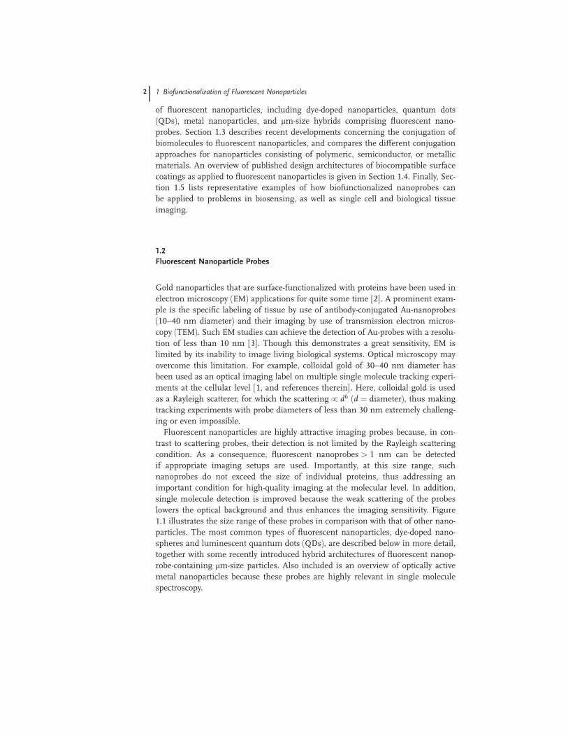
Fluorescent nanoparticles are highly attractive imaging probes because, in con-
trast to scattering probes, their detection is not limited by the Rayleigh scattering
condition. As a consequence, fluorescent nanoprobes > 1 nm can be detected
if appropriate imaging setups are used. Importantly, at this size range, such
nanoprobes do not exceed the size of individual proteins, thus addressing an
important condition for high-quality imaging at the molecular level. In addition,
single molecule detection is improved because the weak scattering of the probes
lowers the optical background and thus enhances the imaging sensitivity. Figure
1.1 illustrates the size range of these probes in comparison with that of other nano-
particles. The most common types of fluorescent nanoparticles, dye-doped nano-
spheres and luminescent quantum dots (QDs), are described below in more detail,
together with some recently introduced hybrid architectures of fluorescent nanop-
robe-containing mm-size particles. Also included is an overview of optically active
metal nanoparticles because these probes are highly relevant in single molecule
spectroscopy.
2 1 Biofunctionalization of Fluorescent Nanoparticles
1.2.1
Dye-doped Nanoparticles
Dye-doped nanoparticles are polymer or silica-based particles containing organic or
inorganic dyes [4, 5]. Dyes can be attached to the nanoparticle surface or can be
embedded inside the particles either noncovalently or covalently. For imaging ap-
plications, dye-doped nanoparticles containing embedded dyes are particularly at-
tractive because their photostability can be enhanced due to the better protection
of the dyes from oxygen. For example, incorporation of pyrene dyes into polysty-
rene particles using a normal microemulsion approach led to a 40-fold increase in
emission intensity with respect to the pure dye at the identical concentration [6].
Brightness of the fluorescence signal from such imaging probes can be controlled
by the number of dye molecules per nanoparticle, with the maximum dye density
limited only by self-quenching. Therefore, dye-doped nanoparticles can be quite
photostable without showing fluorescence intensity fluctuations (blinking).
Typical polymer-based dye-doped nanoparticles are made of hydrophobic poly-
mers. The hydrophobic dye molecules are kept within such nanoparticles through
noncovalent hydrophobic interactions, thus preventing the gradual release and
photooxidation of the dyes. Dye-doped polymeric nanoparticles have been applied
down to a size of@20 nm [7]. Although they are reasonably photostable, the hydro-
phobic nature of these probes complicates imaging applications in aqueous envi-
ronments, which are required for studies on biological systems. Common un-
wanted phenomena are clustering and non-specific binding. To overcome these
problems, the hydrophobic core particles can be surface-functionalized with hydro-
Fig. 1.1. Size ranges of commonly used fluorescent nanoprobes.
1.2 Fluorescent Nanoparticle Probes 3

philic coatings such as polymers like poly(ethylene glycol) (PEG), polysaccharides
such as dextran, or proteins such as bovine serum albumin (BSA).
Dye-doped silica nanoparticles are attractive imaging probes because their hydro-
philic nature reduces the problems of non-specific binding and clustering. Silica
shows several additional properties beneficial for optical imaging applications in
biological systems, including chemical inertness, transparency, and the ability to
act as stabilizers in protecting the embedded dyes from the outside environment
[4]. In addition, the surface hydroxyl groups can be chemically modified, allowing
for the straightforward surface modification with amines, carboxyls, or thiols. In
addition, pH changes do not lead to swelling and porosity changes, and silica par-
ticles are less prone to attack from microbes [8]. However, the incorporation of hy-
drophobic dyes into the hydrophilic silica matrix is challenging, requiring specific
modifications of either the dye molecules or the silica. For example, dyes can be
modified with a hydrophilic molecule such as dextran or a hydrophobic silica-
precursor can be used during nanoparticle synthesis [9]. Dye-doped silica particles
were first synthesized using the Stoeber method [10]. However, this method leads
to polydispersity and average particle sizes of >100 nm. More recently, these limi-
tations have been overcome by use of a reverse microemulsion method that can
create monodisperse dye-doped silica particles down to a size of 15 nm (Fig. 1.2)
[11]. Notably, this elegant approach facilitates the size-tuning via adjustment of
the microemulsion composition and does not require elevated temperatures and
pressures [11, 12]. Silica also has been used to create hollow silica nanospheres
filled with dye molecules [13].
Rare-earth-doped LaPO4 nanoparticles are another interesting class of nano-
probes that show promising properties for diagnostic and imaging applications
[14]. These systems combine small size (<10 nm), high chemical stability, very
good quantum yield, and high photostability. Furthermore, they are expected to
show low cytotoxicity. Recently, such rare-earth-doped LaPO4 nanoparticles were
successfully surface-functionalized to allow their subsequent coupling to biomole-
Fig. 1.2. Schematic of the reverse
microemulsion method for the synthesis of
dye-doped silica nanoparticles (adapted from
Bagwe et al., 2004) [11]. In this method, nm-
sized water droplets, which are stabilized by
surfactant molecules, are formed in a
continuous oil phase, thus forming a
thermodynamically stable oil–water–surfactant
microemulsion.
4 1 Biofunctionalization of Fluorescent Nanoparticles

cules [15]. The nanoparticle surface was first functionalized with aminohexanoic
acid and then linked to avidin using EDC coupling.
1.2.2
Quantum Dots (QDs)
Nanocrystals based on semiconductor materials began attracting the interest of
physicists three decades ago because of their interesting quantum properties.
These properties are the result of size-dependent band gaps, which cause the color
emitted by a semiconductor nanocrystal of a specific composition to be a function
of its diameter. Physically, quantum properties (in this case a size-dependent fluo-
rescence emission) are expected to occur if electron–hole pairs (excitons) are con-
fined to dimensions that are smaller than the electron–hole distance (exciton diam-
eter) [16–19]. As a result of this condition, the state of free charge carriers within a
nanocrystal is quantized and the spacing of the discrete energy states (emission
colors) is linked to the size of the nanoparticle. It is this quantum confinement ef-
fect that led to the term ‘‘quantum dot’’.
Quantum dots can be based on metallic or semiconductor materials. Most
widely used are CdSe and CdTe quantum dots because their quantum confinement
region spans the entire optical spectrum. More recently, there has been growing
interest in quantum dots with near-infrared emission properties, such as CdTe/
CdSe, InAs, or PbS (which are of use in animal imaging studies) [20, and refer-
ences therein]. In addition, several groups have studied silicon nanocrystals [21–
23]. Quantum dots also show other fascinating optical properties, including broad
absorption and narrow emission bands, which allows a single laser to excite dots of
a wide size-range, with each dot emitting its own specific color. This is in contrast
to organic-based fluorophores, which are characterized by narrow Stokes shifts (dif-
ference between maximum wavelengths of absorption and emission bands). To
protect their surface from photobleaching, quantum dots can be passivated by use
of a higher-bandgap semiconductor shell or an organic layer [24]. In fact, success-
fully passivated, quantum dots show dramatically enhanced photostability, en-
abling their long-term observation in optical experiments [25]. Furthermore, quan-
tum dots can be brighter than their dye counterparts at an equivalent quantum
yield [26] because of their notably higher extinction coefficients [20]. Given the
above properties, quantum dots bring fascinating possibilities for single molecule
cellular imaging studies because they combine small size, broad absorption, nar-
row size-tunable emission (covering the entire optical spectrum), and excellent
photostability. These features outperform traditional fluorescent dyes in many
respects.
The traditional approach to synthesizing quantum dots relies on heating specific
organic solvents and injection of semiconductor precursors. In a typical prepara-
tion [27], Cd(CH3)2 and elemental Se are combined with trioctylphosphine oxide
(TOPO), which acts as a solvent and stabilizing agent. This mixture is subjected
to high temperature (about 350 �C) for 24 hours at which time the mixture is
cooled and Zn(CH3)2 and S(SiMe3)2 added to form a stabilizing ZnS shell. To cre-
1.2 Fluorescent Nanoparticle Probes 5

ate nanocrystals of a narrow size distribution, an additional size-selective precipita-
tion step needs to be included. Because dialkylmetal compounds are very sensitive
to oxygen and water and become pyrophoric upon exposure to air, alternative ap-
proaches of quantum dot synthesis have been explored. Consequently, CdSe quan-
tum dots can be formed using CdO, selenium, and hexylphosphonic acid or tetra-
decylphosphonic acid [28]. This synthesis reduces the reaction times to less than
30 min, but still uses temperatures upwards of 300 �C. In another example, our
group has synthesized CdSe/ZnS quantum dots by use of sonochemistry [29].
This low-temperature approach not only produces spherical high-quality quantum
dots with quantum yields of up to 70% and emission bands of less than 50 nm
(FWHM), but also allows for straightforward control of the synthesis parameters.
Figure 1.3 shows the photoluminescence spectra of CdSe core quantum dots and
Fig. 1.3. Quantum dots show size-tunable
emission properties. The samples represent
different sizes of quantum dots, which produce
different colors upon UV light. Within the
quantum confinement region, an increase in
particle sizes produces a redshift in the
emission spectrum. Spectra and photograph
were obtained from CdSe quantum dots
synthesized sonochemically [29].
6 1 Biofunctionalization of Fluorescent Nanoparticles

the colors of these samples upon UV illumination, which are representative of this
procedure.
To be of use in biological imaging applications, quantum dots need to be water-
soluble. Indeed, synthetic approaches for CdTe [30–32] and CdS [30, 33–38] have
utilized aqueous solvent conditions. However, these nanocrystals usually lack the
quantum yield and narrow size distribution observed for TOPO-synthesized quan-
tum dots. TOPO-stabilized quantum dots, however, show hydrophobic surface
properties. To disperse TOPO-stabilized quantum dots in aqueous solution, several
surface modification strategies have been pursued (Fig. 1.4). A common approach
is to synthesize quantum dots in TOPO and replace the hydrophobic TOPO layer
with bifunctional molecules containing thiol and hydrophilic moieties separated by
a molecular spacer (Fig. 1.4: approach I) [39, 40]. The thiol groups bind to the
CdSe or ZnS surface, while the hydrophilic moieties radiate from the surface of
the corresponding semiconductor. Unfortunately, thiols bind less strongly to ZnS
than to Au, which leads to a dynamic equilibrium between bound and unbound
thiols. This behavior reduces the long-term water solubility of ZnS-capped quan-
tum dots. To shift the equilibrium towards bound moieties, monothiols have been
replaced with molecules containing more than one thiol group (Fig. 1.4: approach
II) [41–43]. Another stabilization concept is to enhance binding via surface cross-
linking of bound molecules. On the basis of this concept, ZnS-shelled quantum
dots have been made water-soluble by adding a silica shell to the nanoparticles
by using alkoxysilanes during the polycondensation (Fig. 1.4: approach III) [44–
48]. Two types of silanes have been used to stabilize quantum dots in aqueous
solution. The first includes silanes whose surface functional groups are positively
or negatively charged at neutral pH [48]. The second type includes silanes with
poly(ethylene glycol) chains [48, 49]. TOPO-coatings also can be made water-
soluble without their replacement by adding amphiphilic molecules such as lipo-
polymers or amphiphilic diblock copolymers, whose hydrophobic moiety stabilizes
the TOPO-coating via hydrophobic forces and whose hydrophilic moiety is exposed
to the solvent environment, guaranteeing water-solubility (Fig. 1.4: approach IV).
The last approach has the advantage of not exposing the sensitive surface of the
quantum dot during a surface exchange step.
1.2.3
Metal Nanoparticles
In contrast to noble metals in bulk, nanoparticular forms of such materials result
in interesting photochemical and electronic properties [50]. Three strategies have
been pursued to study the photochemical activity of metal nanoparticles: (a) direct
excitation of the metal nanoparticles; (b) indirect excitation of the metal nanopar-
ticles via surface-conjugated dye molecules; and (c) photocatalytic processes in
semiconductor–metal nanocomposites [50]. After excitation with UV or visible
light, metal nanoparticles show several interesting phenomena, including photolu-
minescence [51–53], nonlinear optical phenomena [54, 55], and surface-enhanced
1.2 Fluorescent Nanoparticle Probes 7
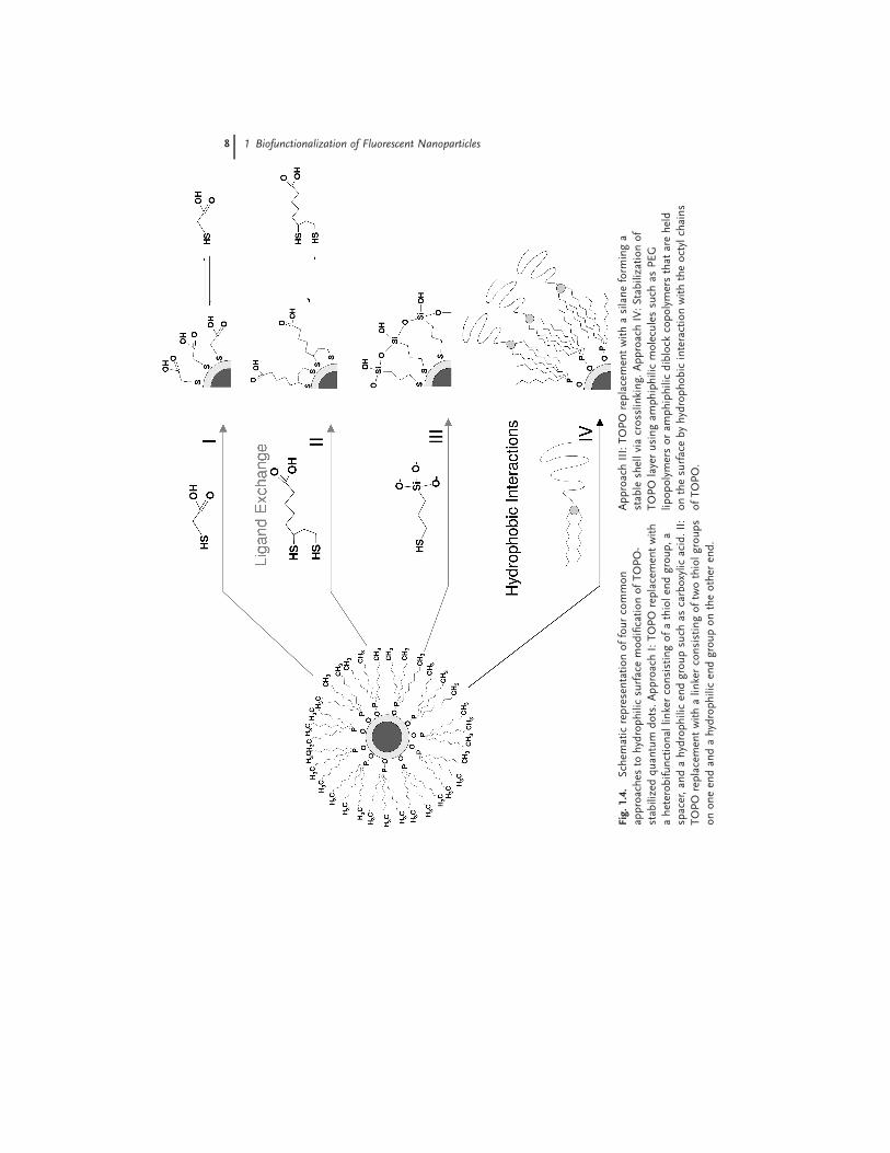
Fig.1.4.
Schem
aticrepresentationoffourcommon
approaches
tohydrophilicsurfacemodificationofTOPO-
stab
ilizedquan
tum
dots.ApproachI:TOPO
replacemen
twith
aheterobifunctional
linkerconsistingofathiolen
dgroup,a
spacer,an
dahydrophilicen
dgroupsuch
ascarboxylic
acid.II:
TOPO
replacemen
twithalin
kerconsistingoftwothiolgroups
ononeen
dan
dahydrophilicen
dgroupontheother
end.
ApproachIII:TOPO
replacemen
twithasilaneform
inga
stab
leshellviacrosslin
king.ApproachIV:Stabilizationof
TOPO
layerusingam
phiphilicmoleculessuch
asPEG
lipopolymersoram
phiphilicdiblock
copolymersthat
areheld
onthesurfacebyhydrophobicinteractionwiththeoctylchains
ofTOPO.
8 1 Biofunctionalization of Fluorescent Nanoparticles
Raman scattering (SERS) [56–61]. Due to the significant field enhancement, SERS
can be used as an extremely sensitive analytical tool, thereby exceeding the sen-
sitivity from luminescence-detecting techniques. For example, biomolecules can
be detected with 1000-fold better sensitivity if they are bound to Au nano-
particles [62]. Silver nanoparticles also are useful in this respect. The main experi-
mental challenge in SERS is to keep the surface roughness uniform and
reproducible.
There are multiple strategies for synthesizing metal nanoparticles [50]. For
example, they can be synthesized using a biphasic reduction approach [63–69]. In
this procedure, a noble metal salt such as HAuCl4 is dissolved in water and phase-
transfer extracted into an organic solvent followed by reduction with NaBH4. Metal
nanoparticles also have been synthesized using reverse micelle procedures where
the size and size distribution of nanospheres can be controlled by the micelle com-
position [12, 70–73]. Gold nanoparticles are particularly attractive for studies in a
biological environment because they show no surface oxidation and high biocom-
patibility without any surface modification. In addition, thiol chemistry can be ap-
plied to conjugate molecules to the gold surface.
1.2.4
Hybrid Architectures Involving Fluorescent Nanoprobes
1.2.4.1 Metal–Dye
Another interesting application of metal nanoparticles is their use in combina-
tion with conjugated dye molecules (Fig. 1.5). Such hybrid systems are attrac-
tive because they can be studied by use of electron microscopy in addition to
fluorescence-based techniques such as fluorescence microscopy and spectroscopy.
Importantly, if conjugated to biomolecules, metal nanoparticle–dye hybrids can be
used as very sensitive biomolecular imaging probes [74, 75]. Interestingly, dye mol-
ecules attached to metal nanoparticles can show enhanced emission. For example,
Py-CH2NH2 molecules bound to gold particles show pronounced emission that is
much stronger than that for unbound Py-CH2NH2 in THF [50]. The main disad-
vantage of this approach is that the surface-exposure of the dyes promotes their
photooxidation.
1.2.4.2 Dye-doped Silica Shells
To reduce photooxidation, dyes also can be embedded within the nanoparticle. As
illustrated in Fig. 1.5(B), dyes can be incorporated into a nanoparticle-capping sil-
ica shell. For example, up to 12 alternating fluorescent and nonfluorescent silica
shells have been added to silica core particles, where the shells were doped with
six dyes of different emission wavelengths. Dyes of multiple colors have been in-
corporated previously in silica beads throughout the matrix of the particle [76],
but not in an ordered fashion [77]. This multi-shell approach helps to reduce un-
wanted energy transfer. As a multiplexing probe, the system is not as simple to
analyze as a quantum dot-based one. Re-absorption of the fluorescence emission,
1.2 Fluorescent Nanoparticle Probes 9

which is related to the identity of the fluorophore, alters the emission intensity. As
a result, it is difficult to determine the possible number of spectral combinations in
a designed system, and a flow cytometer must be used to identify the number of
individual species in the mixture. This shelling approach has also been reported
for single dye systems for enhanced fluorescence [10].
1.2.4.3 Quantum Dot-containing Microspheres
An interesting hybrid architecture that has been developed recently is the quantum
dot embedded microsphere. Unlike a dye embedded polymeric bead, the quantum
dot microbeads utilize the narrow emission of quantum dots combined with their
broad absorption characteristics to perform multicolor detection experiments with
one excitation wavelength [78–81]. By incorporating up to six different colors of
quantum dots and ten intensities of those colors, the quantum dot microbeads
can, theoretically, optically ‘‘bar code’’ biomolecules with one million possible spec-
tral signatures. Realistically, the number is likely to be substantially lower due
to spectral overlap, fluorescence intensity variations, and signal-to-noise require-
ments. The multiplexing approach has been tried with organic dyes, but was
limited to two colors due to spectral overlapping and the inability to excite more
than two or three dyes with the same wavelength [82].
Fig. 1.5. Common hybrid architectures
containing fluorescent nanoprobes include (A)
dyes bound to a nanoparticle surface; (B) dyes
incorporated into the silica shell of a
nanoparticle; (C) fluorescent probes such
quantum dots or dyes incorporated into the
core of microparticles; and (D) a multiplexing
approach by embedding probes of different
colors.
10 1 Biofunctionalization of Fluorescent Nanoparticles

1.3
Bioconjugation of Fluorescent Nanoparticles
1.3.1
General Considerations
1.3.1.1 Overview
To apply fluorescent nanoparticles to biosensing and biomedical imaging applica-
tions, it is crucial to develop strategies towards their biofunctionalization. These in-
clude the proper linkage of biomolecules to nanoparticles (bioconjugation) and the
design of appropriate biocompatible coatings. This section outlines the different
aspects of bioconjugation, and Section 1.4 provides a corresponding discussion of
biocompatible coatings.
Bioconjugation can be described as any procedure that links a nanoparticle to a
biomolecule under mild conditions [83]. As described above, the synthesis of nano-
particles often does not render them capable of attachment to biomolecules, be-
cause their surface-chemical properties are not appropriate. Therefore, nanopar-
ticles frequently must undergo surface transformations to create the chemistry
needed for coupling to biomolecules under mild (physiological) conditions. There
are a few key requirements for successful bioconjugation reactions [4]. Crucially,
the conjugation process must avoid compromising the activity of biomolecules. In
addition, the bioconjugation ideally should not hinder the signal of the nanopar-
ticle. Another requirement is the ability to control the number of linkage sites on
the nanoparticle surface where biomolecules can bind. This requirement can be
quite challenging. In addition, the biomolecule–nanoparticle coupling should be
stable and, for crystalline particles, the surface should be covered to avoid free
valence states. Finally, the thickness of any nanoparticle shell should remain as
small as possible relative to the nanoparticle size.
Figure 1.6 illustrates several possible strategies to bioconjugate nanoparticles.
Simple adsorption of biomolecules to the nanoparticle surface via noncovalent
forces represents the least demanding approach (Fig. 1.6A). However, in this case,
the activity of bound biomolecules may be compromised and the amount of bound
biomolecules per nanoparticle is difficult to control. In addition, this bioconjuga-
tion approach does not target one protein preferentially. Another concept is based
on the physisorption (noncovalent coupling) of biomolecules to molecules (e.g.,
other biomolecules) acting as mediators between biomolecule and nanoparticle
surface (Fig. 1.6B). This design may be advantageous over the first one, because it
may help biomolecules to bind in a proper orientation. Figure 1.6(C) is based on
the chemical coupling between reactive groups of biomolecules (e.g., thiols and
primary amines) and crosslinker molecules. These crosslinkers may bind to the
nanoparticle surface via physisorption or chemisorption (covalent coupling). How-
ever, there are frequently multiple active sites on the target biomolecule to which
the probe can bind, thus preventing controlled binding. Furthermore, uncontrolled
1.3 Bioconjugation of Fluorescent Nanoparticles 11

binding may interfere with the biologically active sites of biomolecules. To over-
come this limitation, a ligand (typically an antibody) can be bound to the nanopar-
ticle via the crosslinker. This ligand then facilitates coupling to the biomolecule of
interest with high specificity. These crosslinker-based bioconjugation strategies are
best executed if appropriate heterobifunctional crosslinkers are used. A large
variety of such crosslinker molecules is now commercially available. Alternatively,
homobifunctional crosslinkers can be used where one reactive end group is pro-
tected. Here, nanoparticle and biomolecule are coupled by binding of the partially
protected linker to either nanoparticle or biomolecule and by deprotecting the
linker prior to bioconjugation. Unprotected homobifunctional crosslinkers are al-
ways problematic because they can induce the clustering of nanoparticles. Figure
1.6(D) shows a facile approach for the chemical coupling of biomolecules to nano-
particles. This approach is particularly useful for attaching oligonucleotides to
nanoparticles, e.g., via mercapto groups [84–86]. Still, control over the number of
bound biomolecules per nanoparticle remains a challenging endeavor. To achieve
such control, a separation step, such as nanoparticle separation using gel electro-
phoresis, must be added [87]. Another popular approach is to link biotinylated li-
gands or target biomolecules to streptavidin (or avidin)-functionalized nanopar-
ticles with high specificity (Fig. 1.6E). In this case, the biotin-binding proteins,
avidin or streptavidin, act as linker molecules [88, 89].
Fig. 1.6. Common strategies for the
conjugation of biomolecules to nanoparticles
include direct physisorption of biomolecules
(A), assisted physisorption using pre-bound
molecules (B), chemical linkage of biomole-
cules to crosslinkers either physisorbed or
chemisorbed on the nanoparticle surface (C),
direct chemical coupling of biomolecules to
nanoparticles (D), and the targeted binding of
biotinylated biomolecules to streptavidin-
coated nanoparticles via biotin–streptavidin
coupling (E).
12 1 Biofunctionalization of Fluorescent Nanoparticles

1.3.1.2 Common Coupling Reactions
The bioconjugation of nanoparticles is critically dependent on the availability of ef-
fective covalent coupling procedures. Figure 1.7 lists the most common coupling
procedures. The reaction between primary amine and carboxylic acid groups is a
very popular coupling procedure (Fig. 1.7A). In a first step, a carboxylic acid reacts
with 1-ethyl-3-(3-dimethylaminopropyl)carbodiimide (EDAC) and N-hydroxysulfo-succinimide (NHS) to form an acyl amino ester that subsequently reacts with the
primary amine to create a stable amide bond. This approach also is attractive be-
cause preparation of the ligands is less demanding. A widely used approach is
based on the reaction between thiol and maleimide groups (Fig. 1.7B). This affords
a stable thioether bond, typically with good yield at physiological pH. Most promi-
nently, this reaction can be used to directly link maleimide-functionalized ligands
to thio-groups of proteins. If no thiols are available, they can be created by using
heterobifunctional crosslinkers with one thiol endgroup or by reducing disulfide
bonds within the target protein. The coupling of two thiols to form a disulfide link-
age is another straightforward reaction approach (Fig. 1.7C). Unfortunately, the re-
sulting disulfide bond is relatively labile in biological fluids like serum. Another
useful reaction scheme is based on the covalent linkage between aldehyde and
hydrazide groups to form a hydrazide bond (Fig. 1.7D). Reactive aldehyde groups
can be created by oxidation of carbohydrate groups. Obviously, mild oxidation
conditions have to be used for the oxidation reaction on relatively unstable mole-
cules like antibodies. Chemical reaction between two primary amines is another
approach for bioconjugation applications (Fig. 1.7E). In this case, coupling can be
achieved using homobifunctional crosslinkers such as glutaraldehyde [90, 91].
1.3.2
Bioconjugation of Polymeric Nanoparticles
From a bioconjugation perspective, polymeric nanoparticles are attractive because
they can be prepared with various different reactive groups on their surface. The
obvious benefit is that a comparably broad spectrum of conjugation chemistry ap-
proaches can be used, including covalent and noncovalent ones (Fig. 1.6). These
different bioconjugation strategies are reviewed below.
1.3.2.1 Noncovalent Approaches
One of the most widely applied approaches in bioconjugating polymeric nanopar-
ticles is simple physisorption of the biomolecules on the particle surface (Fig. 1.6A)
[93–97]. Because biomolecule activity is compromised by uncontrolled adsorption,
spacer molecules have been introduced (Fig. 1.6B). For example, nanoparticles first
coated with Protein A (isolated from the cell wall of Staphylococcus aureus), whichattaches specifically to the Fc fragment of the IgG antibody, have proper Fab orien-
tation for antibody binding [97]. Unfortunately, such simple adsorption approaches
are only of limited use when applied in serum. This is because serum proteins
compete with antibodies for adsorption sites [93–95]. Furthermore, in vivo experi-
ments have revealed that such simply designed nanoparticles tend to accumulate
1.3 Bioconjugation of Fluorescent Nanoparticles 13

14 1 Biofunctionalization of Fluorescent Nanoparticles

in the liver and spleen. Using a similar design strategy, polystyrene nanoparticles
have been functionalized with humanized mAB HuEP5C7.g2 [98]. These delivery
systems target cells expressing E and P-selectin.
1.3.2.2 Covalent Approaches
Obviously, the activity of ligands bound to nanoparticles strongly depends on the
stability of the ligand–nanoparticle linkage and the proper orientation of the li-
gand. To address these needs, ligands have been bound covalently to polymeric
nanoparticles using several different conjugation approaches (Fig. 1.6E) [7, 99–
102]. For example, an amide linkage strategy was pursued to bind lactose to the
poly(vinylamine)-grafted nanoparticles [99]. Such lactose-functionalized carrier sys-
tems are useful because they specifically bind to lectin RCA120. The same amide-
based linkage chemistry was used to bind proteins to dye encoded latex particles
[7]. In another approach, transferrin was conjugated to PEG coated poly(cyanoacry-
late) nanocrystals, with the transferrin bound to PEG via periodate oxidation [100].
A heterobifunctional linker was used to bind antibodies to BSA-containing nano-
particles [101, 102]. In this case, the crosslinker (glutaraldehyde) was bound to
BSA to provide free aldehyde groups, which could then be linked to the primary
amines of the antibodies. Another interesting strategy is based on formation of
ligand-carrying nanoparticles via polymerization of monomers with bound ligands.
Such a strategy has been applied to create nanoparticles with biotin groups, which
then allowed for specific linkage to avidin [103, 104]. The grafting of carbohydrates
to poly(l-lysine) was also reported [105]. This carrier can bind to carbohydrate-
binding proteins on the surface of target cells.
1.3.3
Bioconjugation of Quantum Dots
Though quantum dots are very small, their surface area is large enough for linking
to multiple biomolecules [106]. There are several ways to bind biomolecules to
quantum dots. These involve either direct binding to the quantum dot surface or
attachment via a stabilizing layer acting as a crosslinker between the ligand and re-
active surface of the nanoparticle. In the first case, ligand binding can be achieved
covalently or noncovalently. Direct covalent coupling is accomplished commonly
by use of thiol coupling chemistry, although a silane-based coupling is needed if
quantum dots carry a stabilizing silica shell. Noncovalent ligand-quantum dot cou-
pling, however, is typically pursed using electrostatic or hydrophobic forces. On the
basis of these general concepts, several bioconjugation strategies have been pur-
sued. These are discussed below.
Fig. 1.7. Commonly used chemical reactions
in the bioconjugation of fluorescent
nanoparticles. Shown are the coupling
chemistries between (A) carboxylic acid and
primary amine, (B) thiol and maleimide
groups, (C) two thiols to form a disulfide
bond, (D) hydrazide and aldehyde groups, and
(E) two primary amines. (Adapted from Nobs
et al., 2004 [92].)
H————————————————————————————————————————
1.3 Bioconjugation of Fluorescent Nanoparticles 15
1.3.3.1 Noncovalent Approaches
An electrostatic coupling approach has been applied to link negatively charged
CdTe quantum dots and positively charged enzymes [107]. Electrostatic coupling
also was pursued in several applications to bind positively charged protein do-
mains (pentahistadine segment) to oppositely charged alkyl-COOH-capped quan-
tum dots [31, 42, 88, 108–112]. An alternative electrostatic-based coupling strategy
has been used to bind nanocrystals coated with trimethoxysilylpropyl-urea and ace-
tate groups with high affinity in the cell nucleus [44]. Here the binding of the
silanized nanocrystal surface is controlled with an anionic silane reagent. Nega-
tively charged quantum dots have also been electrostatically bound to positively
charged Maltose Binding Protein [113]. Quite often, adsorbed proteins show a
stabilizing effect on the quantum dot surface, thereby improving the optical prop-
erties of the probes. For example, a two-fold increase in fluorescence was observed
after adsorption of 10–15 Maltose-Binding Protein Pentahistadine (MBP-5HIS) to
the quantum dot surface [114].
1.3.3.2 Covalent Approaches
A relatively straightforward covalent bioconjugation approach is based on the re-
placement of thiol acids present on the quantum dot surface with thiolated bio-
molecules. This strategy has been used to attach oligonucleotides, DNA [41, 84]
and bovine serum albumin (BSA) [86] to quantum dots. Proteins also have been
conjugated to quantum dots by reacting carboxylic acid groups on the nanoparticle
surface with amine groups of the proteins [115]. A similar strategy was used to co-
valently attach IgG and streptavidin to quantum dots. The IgG system was applied
as a cancer marker, whereas the streptavidin system was utilized for labeling actin,
microtubules and the cell nucleus [116].
In another application, antibodies have been bound to quantum dots via sulfo-
NHS crosslinking [117]. In a different approach, reactive biotin was covalently
linked to either surface sulfhydryl or amine functionalities, thus allowing for the
biotinylation of the quantum dot surface and the subsequent binding to streptavi-
din [44]. A biotin–streptavidin coupling approach was chosen, for example, to bind
quantum dots to fibroblasts [44]. In this case, the nanocrystals were functionalized
with biotinamidocaproic acid 3-sulfo-N-hydroxysuccinimide ester and then allowed
to bind to fibroblasts previously incubated with phalloidin–biotin and streptavidin.
A combination of covalent and noncovalent coupling strategies was pursued by
conjugating quantum dots stabilized with amphiphilic PEG lipids to DNA. Here,
the DNA coupling was achieved by replacing 12 of the PEG-PE phospholipids with
amino PEG-PE. Thiol modified DNA was then covalently coupled to the amines on
the surface of the quantum dot [118].
1.3.4
Bioconjugation of Metallic Nanoprobes
Most conjugation concepts described for the linkage of ligands to quantum dots
can also be applied to metal nanoparticles, such as Au or Ag nanoprobes. Again,
16 1 Biofunctionalization of Fluorescent Nanoparticles

surface modification is accomplished using thiol-coupling chemistry. The binding
of biomolecules to these nanoparticles can be covalent or noncovalent. With re-
spect to biofunctionalization, gold nanospheres are particularly attractive because
gold shows excellent biocompatibility properties, and does not require additional
surface chemical modification prior to bioconjugation. In addition, gold nanopar-
ticles can be made with very narrow size distribution. This is advantageous if accu-
rate control of the number of bound biomolecules per nanoparticle is required.
1.3.4.1 Noncovalent Approaches
One option for bioconjugation is the direct attachment of a biomolecule to the
metallic nanoparticle surface. Short peptide chains such as tiopronin [119] and
the tripeptide glutathione [120] have been conjugated directly to a gold surface.
Globular proteins have also been directly conjugated. Miziani et al. conjugated
globular proteins directly to the surface of silver sulfide nanoparticles [121]. In an-
other application, gold nanoparticles were coated with BSA via electrostatic interac-
tions under mild conditions [122]. Due to the bioinertness of gold, biomolecules
directly bound on the gold surface can retain their activity, as shown, for example,
for redox enzymes [123] and cytochrome c [124]. Biotin–streptavidin coupling con-
cepts have also been used. For example, disulfide-modified biotin was conjugated
to gold nanoparticles and subsequently reacted with streptavidin [125].
1.3.4.2 Covalent Approaches
Direct coupling of biomolecules to the surface of metal nanoparticles represents a
very facile bioconjugation strategy. Among the different metals, this conjugation
strategy is best suited for gold because of its excellent bioinertness. For example,
thiol-modified DNA has been conjugated directly to gold particles [74, 126–128].
Direct thiol coupling was also pursued to conjugate SH modified PEG to gold-
shelled silica nanoparticles [129]. Covalent bioconjugation to the nanoparticle can
also be mediated by heterobifunctional crosslinker molecules. One example is the
surface modification of iron oxide nanoparticles with a trifluoroethylester-terminal
poly(ethylene glycol) (PEG) silane and the subsequent coupling of biomolecules via
their terminal amine or carboxyl groups [130].
1.4
Design of Biocompatible Coatings
1.4.1
General Considerations
1.4.1.1 Overview
Though multiple bioconjugation strategies have been worked out, the routine ap-
plication of fluorescent nanoparticles in biomedical imaging remains challenging.
This is largely because not only appropriate conjugation between biomolecules to
nanoparticles is required but other important criteria concerning biocompatibility
1.4 Design of Biocompatible Coatings 17

have to be fulfilled. These criteria include (a) prevention of nanoparticle aggrega-
tion in a biological environment, (b) effective suppression of non-specific adsorp-
tion of biomolecules at the nanoparticle surface or their accumulation close to the
surface, and (c) low cytotoxicity (Fig. 1.8). To address these criteria, nanoparticles
need to be capped with protective coatings that show passivating surface proper-
ties. Notably, the first criterion, good colloidal stability, does not guarantee the
biocompatibility of nanoparticles as, firstly, ions and proteins may be accumulated
on or near the nanoparticle surface, thus varying the complex molecular composi-
tion nearby. Secondly, especially with heavy metal-containing quantum dots (e.g.,
CdSe, CdTe), individual nanoparticles may remain cytotoxic if the coating does
not protect the biological environment from released heavy metals ions.
1.4.1.2 Colloidal Stability
The dispersion stability of nanoparticles is well-described by the Derjaguin–
Landau–Verwey–Overbeek (DLVO) theory, which predicts that such stability is
determined by the balance between attractive van der Waals and repulsive electro-
static forces. As a result, nanoparticles are expected to remain stable (show no par-
ticle aggregation) if the strength of the repulsive electrostatic force exceeds that of
the attractive van der Waals (vdW) force. In other words, nanoparticles show no ag-
gregation if they contain a sufficient density of surface charges. Indeed, particles
with charged surface properties do not aggregate in ion-free solutions. At the
same time, this balance can clearly be shifted in favor of vdW forces if the charges
on the particle surface are screened by counter ions in the solution. This is an
important aspect to consider during chemical surface modification, because such
Fig. 1.8. Potential problems compromising the
biocompatibility of fluorescent nanoparticles, including
nanoparticle (NP) clustering, non-specific adsorption of
proteins (P), and release of cytotoxic ions (CT).
18 1 Biofunctionalization of Fluorescent Nanoparticles

modifications may result in surface charge neutralization, thereby compromising
dispersion stability. More importantly, nanoparticles with charged surfaces tend to
become unstable (show aggregation) in a biological environment. Figure 1.9 shows
the loss of colloidal stability due to charge screening. Here comparative fluores-
cence correlation spectroscopy (FCS) autocorrelation curves from our laboratory
are presented for CdSe/ZnS quantum dots with negatively charged carboxylic acid
surface groups in Millipore water and in 10 mm MES buffer. As illustrated, the
nanoparticles in MES buffer show notably higher diffusion times, indicating their
increasing aggregation. If quantum dots are surface-functionalized with a layer of
hydrophilic polymers such as poly(ethylene glycol) (PEG), colloidal stability can be
achieved even in the presence of ions in solution. Such particle stabilization via
polymer coatings is attributed to the flexibility of polymer chains and their repul-
sive ‘‘entropic spring’’ effect during particle–particle interaction. Currently, the
concept of stabilization of nanoparticles due to the addition of polymeric coatings
is widely accepted. Indeed, several experimental results seem to support the
concept [131, 132]. Peptides adsorbed to the quantum dot surface can also have a
similar stabilizing effect [133]. Conversely, it has been theoretically predicted that
homopolymers, if end-grafted to nanoparticles in good solvents, may lead to nano-
particle attraction [134].
1.4.1.3 Biocompatible Surfaces
To make fluorescent nanoparticles ‘bioinert’, their surfaces need to be designed so
as to prevent the non-specific adsorption of all relevant molecules in the biological
medium. It is challenging to improve the design of surface coatings to a level of
sophistication that fulfills this requirement. Fortunately, promising strategies for
Fig. 1.9. Fluorescence correlation spectros-
copy autocorrelation curves of ZnS-capped
CdSe quantum dots surface-functionalized
with carboxylic acid in Millipore water (dark
curve) and 10 mm MES buffer (lighter curve).
The notably higher diffusion time obtained
for the buffer-based system indicates nano-
particle clustering.
1.4 Design of Biocompatible Coatings 19

the creation of bioinert surfaces are known from biomaterials research in the bio-
medical engineering community. As a result, our understanding about the host re-
sponse of living tissue with respect to biomaterials has improved significantly and
numerous sophisticated biomaterials have been developed [135]. A key aspect for
the creation of colloidal solids with sufficient stealth properties in a cellular envi-
ronment is the understanding of protein adsorption. Like surfactants, proteins
have a high tendency to adsorb at interfaces. Interactions between proteins and
surfaces are primarily noncovalent and include electrostatic, hydrophobic, and
hydrogen-bonding interactions [136]. The different interactions result from the
‘‘surface inhomogeneity’’ of proteins, which typically are characterized by surface
patches that may be charged, neutral, hydrophilic, or hydrophobic in character
[137]. Consequently, surface properties of biomaterials affect the mechanism, rate,
and extent of protein adsorption. Protein adsorption can be suppressed most effi-
ciently on biomaterials whose surfaces are neutral, hydrophilic, and highly dy-
namic [138]. A paradigm of a protein-resistant surface is a solid substrate surface-
functionalized with grafted PEG chains [139]. This is so because PEG is electrically
neutral, thus minimizing electrostatic interactions, and highly hydrophilic, thus
minimizing hydrophobic interactions. As the PEG chains are highly dynamic in
an aqueous environment, the formation of strong hydrogen-bonding between pro-
tein and polymer is effectively suppressed. Besides those enthalpic effects, the high
dynamics of the PEG chains results in a high entropy, which is also unfavorable for
protein adsorption. It has been generally assumed that the surface resistance of
PEG functionalized substrates is a result of the steric repulsion grafted PEG chains
show for adsorbing proteins [138, 140, 141]. However, this assumption is valid only
if the grafted PEG chains are of very high molecular weight and show large
enough grafting densities to form polymer brushes [142]. Interestingly, Prime
and Whitesides found that protein adsorption is prevented effectively even if the
PEG chains have only two ethylene oxide segments, thus indicating that surface
coverage is the most important parameter to prevent protein adsorption [143,
144]. In the same context, Single-chain Mean Field Theory (SCMF) calculations
and experimental studies showed that protein adsorption on hydrophobic surfaces
is prevented because PEG chains bind to the hydrophobic substrate, thereby block-
ing possible protein binding sites [145]. A later SCMF study by Szleifer and co-
workers found that polymers with a low substrate affinity are more effective for
kinetic control, whereas those with a higher affinity lead to a lower equilibrium
concentration of adsorbed proteins [146]. Obviously, protein adsorption on sub-
strates surface grafted with synthetic polymers is dependent on both the steric
hindrance effect of the grafted polymer layer and the affinity of the underlying sub-
strate for proteins and other molecules.
1.4.1.4 Cytotoxicity
Several potential processes lead to cytotoxicity. One source of nanoparticle-induced
cytotoxicity, rather independent of particle composition, occurs if nanoparticles
adsorb to cell surfaces [147, 148] or if they get ingested by cells [149, 150]. The cy-
totoxicity of CdSe/ZnS quantum dots is suggested to be due mostly to their inter-
20 1 Biofunctionalization of Fluorescent Nanoparticles
action with cells [151]. Another source of nanoparticle-induced cytotoxicity occurs
when the nanoparticle is composed of toxic materials that can be gradually re-
leased. A prominent example concerning fluorescent nanoparticles is the gradual
release of heavy metal ions such as Cd2þ from CdSe or CdSe/ZnS quantum dots
[152, 153]. Heavy metal ions are cytotoxic and often show several pathways of cyto-
toxicity. Indeed, Cd2þ may induce hepatotoxicity, immunotoxicity, and nephrotoxic-
ity; apoptosis being a critical part of each toxicity type [154]. Studies concerning
Cd-induced hepatotoxicity show, for example, the relevance of direct and indirect
cytotoxic pathways [155]. The direct pathway is caused by Cd2þ binding to sulf-
hydryl groups on key mitochondrial molecules, thus damaging the mitochondria.
The indirect pathway, though, is assumed to occur via activation of Kupffer cells.
However, not all metals show such pronounced cytotoxicity. For example, gold
nanoparticles have good biocompatibility properties [156]. From the above infor-
mation, nanoparticles only show very low cytotoxicity if all potential sources of
cytotoxicity are prevented efficiently.
1.4.2
Nanoparticle-stabilizing Coatings
To enhance the biocompatibility of nanoparticles, one popular strategy has been to
cap the nanoparticles with polymeric coatings of high biocompatibility. In particu-
lar, poly(ethylene glycol) (PEG) has been applied as a coating material because of
its excellent biocompatibility properties. Originally studied on flat macroscopic
surfaces for biomaterials applications, PEG coatings have been widely applied to
sub-micron sized delivery systems and imaging probes [48, 49, 116, 118, 131, 157,
158]. Delivery systems coated with PEG have notably longer circulation times in
the blood, thus exceeding traditional non-nanoparticle-based delivery methods for
the prolonged delivery of drugs, diagnostics, genes, and vaccines [159–162]. Based
on the success of PEG coatings in drug delivery systems, similar coating strategies
have been applied to the design of biocompatible coatings of nm-size imaging
probes in biological and biomedical imaging. A facile approach for adding a PEG
layer to the quantum dot surface is via physisorption of amphiphilic molecules
[116, 118]. Here one interesting strategy has been to use PEG-containing lipopoly-
mers, which result in excellent colloidal stability in Xenopus embryo cells over
several days [118]. In an alternative amphiphilic passivation approach, block co-
polymers have been utilized to enable colloidal stability of quantum dots [116,
163, 164].
Chemically coupled PEG coatings have even greater stability. Wuelfing et al. re-
ported the surface functionalization of gold nanoparticles using CH3O-PEG-SH
[131]. Covalent PEG coatings were also used to increase the colloidal stability of
CdSe [49], CdS, [158] and SiO2-capped CdS [48] quantum dots. However, in these
cases, the PEG coatings lack reactive surface groups, thus preventing the further
immobilization of ligands on the PEG layer. To overcome this limitation, nanopar-
ticles need to be surface-functionalized via mixtures of mono-functional PEGs and
other linker molecules [48] or via heterobifunctional PEG-linkers. Heterobifunc-
1.4 Design of Biocompatible Coatings 21

tional PEG-linkers can be synthesized via monoprotected symmetric PEG linkers,
such as monoprotected diamines derived from tetraethylene glycol [165–167].
Another strategy for the synthesis of heterobifunctional PEG linkers is based on
the ring-opening polymerization of ethylene oxide using a metal alkoxide initiator
[168–171]. For example, a-acetal-PEG-SH has been synthesized using this strategy
[172]. Here the facile modification of the acetal group into a reactive aldehyde al-
lowed the immobilization of ligand molecules on the surface of PEGylated gold
particles [173]. Meanwhile, various heterobifunctional PEG linkers are also com-
mercially available. Figure 1.10 shows the design of a biocompatible surface coat-
ing where a few linkers support the targeted immobilization of ligand molecules.
Such a design can be accomplished by using a mixture of homofunctional and het-
erobifunctional PEGs. An excess of homofunctional PEG molecules, which have
no reactive surface groups after surface coating, suppresses the non-specific bind-
ing of proteins, whereas the heterobifunctional linkers facilitate the targeted cou-
pling to ligands. Notably, however, a PEG layer does not prevent cytotoxic side re-
actions due to the leaching of toxic heavy metal ions from quantum dots. In those
cases, an additional protective layer preventing the egress of harmful ions is essen-
tial [153].
PEG-based coatings are not the only biocompatible polymeric coatings. For ex-
ample, poly(acrylic acid) has been used to stabilize luminescent silicon nanopar-
ticles [174]. Because this anionic polyelectrolyte has a high density of carboxylic
acid moieties, the immobilization of amine-containing molecules is straightfor-
ward. A similar concept has been applied to conjugate CdS quantum dots with
aminodextran [175]. Dendrimers have also been used as stabilizing coating mate-
rial [176]. Quantum dots have been embedded into larger polymeric spheres via a
facile procedure using block copolymers [163, 177], and CdSe-ZnS quantum dots
have been incorporated into glyconanospheres via electrostatic coupling with car-
boxymethyldextran and polylysine [178]. Several other procedures have afforded
gold glyconanospheres [173, 179, 180]. Quantum dots capped with specific peptide
coatings have recently been shown to be quite promising biocompatible imaging
Fig. 1.10. Design of nanoparticles with
stabilizing, biocompatible coatings, including
(A) adsorption of amphiphilic molecules such
as amphiphilic diblock copolymers; (B)
chemical coupling of hydrophilic polymeric
systems; and (C) linkage of spacer molecules
with hydrophilic surface groups.
22 1 Biofunctionalization of Fluorescent Nanoparticles

probes that can be designed for targeted labeling of biomolecules [20, and refer-
ences therein].
1.4.3
Low Cytotoxicity Coatings
As stated before, successful application of fluorescent nanoparticles in a biological
environment requires not only high dispersion stability and the suppression of
non-specific biomolecule adsorption, but also should show low cytotoxicity. This
topic is particularly important for applications involving quantum dots where toxic
heavy metal ions can be released [152]. If properly passivated, quantum dots seem
to show no significant cytotoxicity. For example, a study on Xenopus embryos did
not reveal significant cytotoxicity after injection of 2� 109 and 5� 109 CdSe/ZnS
quantum dots per cell [118].
Several strategies have been pursued to suppress this oxidation process. One
approach is simply to passivate the quantum dot surface with binding ligands
[181]. Importantly, such a passivation not only lowers the cytotoxicity of nano-
probes, but also enhances their quantum yields. Quantum dots can be further pas-
sivated by adding a protecting semiconductor shell. Such coatings also significantly
lower the cytotoxicity of CdSe quantum dots, but do not completely eliminate the
problem [152]. Surface silanization is another promising approach to suppress-
ing surface oxidation [44, 46, 182–184]. The stability of the coating is provided by
crosslinks within the siloxane shell. Importantly, such shells are quite stable in a
biological environment [46]. Furthermore, CdSe/ZnS quantum dots with a protec-
tive shell of crosslinked silica significantly reduce the release of Cd2þ, thus lower-
ing the cytotoxicity of fluorescent nanoparticles [153]. Interestingly, the addition of
a bovine serum albumin (BSA) layer further reduced the cytotoxicity of CdSe/ZnS
quantum dots [152]. The BSA, added to the mercaptoacetic acid-functionalized
quantum dot surface using EDC coupling, may act as a diffusion barrier for O2
molecules.
Based on the above discussion, the appropriate biofunctionalization of nanopar-
ticles clearly must address the features of colloidal stability, bioinertness, specificity
with respect to target biomolecules, and low cytotoxicity. Figure 1.11 shows a
biofunctionalization architecture where these crucial features are considered in its
design.
1.5
Applications
Our growing ability to synthesize and surface-functionalize nanoparticles has
provided new opportunities in bioanalytical and biological imaging applica-
tions. Metallic nanoparticles have been used primarily in bioanalytical projects.
Nanoparticle-based imaging applications, however, rely more on dye-doped poly-
meric nanoparticles and quantum dots due to their excellent fluorescence proper-
1.5 Applications 23

ties and photostability. While dye-doped systems have been used initially in such
imaging applications, quantum dots have recently increasingly become the nano-
particle systems of choice. It is fair to say that further progress in bioanalytical
and imaging applications is strongly linked to the availability of novel biofunction-
alized nanoparticles of improved designs. Though developing rapidly, this highly
interdisciplinary field is still in its infancy. The rapidly growing number of publica-
tions over the last decade reflect its relevance.
Though this chapter focuses primarily on the relevant concepts of biofunctional-ization, this section describes recent applications using biofunctionalized fluores-
cent nanoparticles. We do not claim to review all published applications where
such particles have been used because this is clearly beyond the scope of the
chapter. Instead, the intent is to provide a representative overview of recent
nanoparticle-related applications in the fields of diagnostics and live cell and invivo imaging.
1.5.1
Biosensing
Fluorescent nanoparticles are highly promising probes for bioanalytical appli-
cations. In particular, their use in the field of biosensors is attractive because
fluorescence-based techniques are extremely sensitive and nanoparticle probes
show high photostability, thus allowing for long-term observations.
Fig. 1.11. Schematic of an appropriately biofunctionalized
quantum dot. The inner coating protects from surface oxidation
whereas the outer coating guarantees dispersion stability and
biocompatibility. Immobilized ligands mediate the specific
binding to biomolecules.
24 1 Biofunctionalization of Fluorescent Nanoparticles

1.5.1.1 Polymeric Sensors
Bioanalytical applications based on polymeric nanoparticles are now well estab-
lished. In one application, the combination of two silica precursors, tetraethyl-
orthosilicate and phenyltriethoxysilane, was used to synthesize organic dye-doped
silica nanoparticles. The nanoparticles were coated with avidin to determine bio-
tinylated bovine serum albumin. In addition, a glutamate sensor was designed by
immobilizing glutamate dehydrogenase on the nanoparticle surfaces [185]. In an-
other application, a BODIPY-doped polymeric nanoparticle was designed for the
detection of Cu2þ. Here the metal-chelating receptor, cyclam, was covalently bound
to the surface of the nanoparticle. In the presence of Cu2þ the fluorescence emis-
sion of the BODIPY is quenched by the overlapping adsorption band of the metal–
chelator complex [186]. Fluorescent polymeric nanoparticles have also been used
for the intracellular monitoring of key biological components. Using three differ-
ent nanoparticle fabrication technologies, sensors based on polyacrylamide, cross-
linked decyl methacrylate, and silica-based sol–gel have been characterized in
aqueous solution and intracellular surroundings. Each matrix can be combined
with specific ‘‘free dyes’’, ionophores, or enzymes to produce sensors selective for
the biological component of interest. These nano-optodes are termed PEBBLEs
(probes encapsulated by biol. localized embedding). Spherical sensors less than
600 nm in diam. (and reducible to below 100 nm) have been made from all three
matrices. Acrylamide-based sensors have been used to monitor intracellular pH
and calcium (with proven selectivity over Mg2þ). Decyl methacrylate has been suc-
cessfully applied to intracellular potassium monitoring with probes 1000� more
selective for potassium than sodium. Such a sol–gel-based approach has also
proven useful for monitoring intracellular oxygen at physiologically interesting
concentrations. PEBBLEs, with a wide range of both simple and complex sensing
schemes, provide a unique tool for minimally invasive intracellular monitoring,
with many significant advantages over free dyes as well as over fiber-optic sensors
[187–189].
1.5.1.2 Quantum Dot Sensors
The great potential of quantum dots for sensing applications has been demon-
strated in several instances. In one application, quantum dots have been utilized
as maltose sensors [114]. Here quantum dots were surface-functionalized with a
maltose-binding protein (MBP) containing b-cyclodextrin-QSY-9 attached to the
binding site to quench the quantum dot emission. The senor concept is that malt-
ose displaces the b-cyclodextrin-QSY-9, thus inducing quantum dot emission. An-
other reported quantum dot-based maltose sensor using MBP acts as a double
FRET sensor [114]. Here the quantum dot emission excites Cy3 that is adsorbed
on the surface of the MBP. The Cy3 emission then excites the b-cyclodextrin–
Cy3.5 whose emission is observed. Once maltose is introduced to the sensor, the
Cy3 emission is no longer quenched by the Cy3.5, because the b-cyclodextrin–
Cy3.5 has been displaced by maltose [114].
Quantum dots also show great promise in immunoassays. For example, an im-
munoassay has been performed by binding an antibody covalently to a glass chip,
1.5 Applications 25

followed by binding of the antigen. The antigen was then detected with the addi-
tion of antibody labeled quantum dots, which bound selectively to the captured an-
tigen [190]. In another application, Tran et al. prepared quantum dot conjugates
with the immunoglobin G (IgG) binding domain of streptococcal protein G (PG)
appended with a basic leucine zipper attachment domain (PG-zb). They also dem-
onstrated that the quantum dot/PG conjugates retain their ability to bind IgG anti-
bodies, and that a specific antibody coupled to quantum dots via the PG functional
domain efficiently binds its antigen. Preliminary results indicated that electrostati-
cally self-assembled quantum dot/PG-zb/IgG bioconjugates can be used in fluoro-
immunoassays [191]. Quantum dot–antibody conjugates were again successfully
used in fluoro-immunoassays to detect both a protein toxin (staphylococcal enter-
otoxin B) and a small molecule (2,4,6-trinitrotoluene) [108]. Goldman et al. de-
scribed a conjugation strategy employing an engineered molecular adaptor protein,
attached to the quantum dots via electrostatic/hydrophobic self-assembly, to link
quantum dots with antibodies. Quantum dots can also be used as sensors for sug-
ars; a quantum dot FRET application using adsorbed MBP has been mentioned
[114].
Powerful sensor concepts can also be designed using the multiplexing capa-
bility of quantum dots. This results from the broad excitation and narrow, size-
dependent emission bands of quantum dots. This concept has been used, for ex-
ample, to embed quantum dots of six different colors and ten intensities per color
in nanoparticles composed of styrene, divinylbenzene, and acrylic acid. This com-
bination can, theoretically, code one million nucleic acids or protein sequences
[80]. In another application, a high-throughput assay has been developed for the
parallel detection of antibodies using quantum dot microbeads. In this case, a cus-
tom designed microfluidic chip with multiple micro-wells was utilized for captur-
ing of microbeads, antibody injection into each micro-well, QD injection, and fluo-
rescence detection [192]. These beads could be identified with a standard flow
cytometer at a rate of 1000 beads s�1 [193].
1.5.1.3 Metallic Sensors
Gold nanoparticles (2.5 nm) have been used to recognize and detect specific DNA
sequences and single-base mutations. Detection is based on fluorescently tagged,
thiol modified oligonucleotides bound to the surface of the gold nanoparticle.
Upon self-assembly, the fluorophores arch toward the gold surface, quenching the
fluorescence. Upon binding the appropriate target sequence, the oligonucleotide
undergoes a conformational change, moving it away from the gold nanoparticle
surface, and restoring the fluorescence [194]. Another interesting approach to
ultrasensitive detection of DNA hybridization is based on Au nanoparticle-
amplified surface plasmon resonance (SPR). Interestingly, even without further op-
timization, the sensitivity of this technique begins to approach that of traditional
fluorescence-based methods for DNA hybridization and oligonucleotide detection
[62]. Gold nanoparticles have also been used in the detection of glucose and glu-
cose oxidase. The growth of the Au nanoparticles requires H2O2 and converts it
into O2. In the presence of glucose oxidase and glucose, O2 is converted into
26 1 Biofunctionalization of Fluorescent Nanoparticles

H2O2, allowing the Au nanoparticles to grow, thus giving a shift in absorbance
over time [195]. Finally, Ag has also been employed for surface plasmon-based
nanosensors concerning the detection of Alzheimer’s disease. Autopsied brain
samples of humans with Alzheimer’s disease show elevated levels of amyloid-
derived diffusible ligands (ADDLs), which suggest that a definitive chemical diag-
nosis of Alzheimer’s could be achieved with a sensitive method to detect ADDL or
anti-ADDL antibodies. The lmax of silver nanoparticles conjugated with anti-ADDL
was monitored before and after the incubation with ADDL. The resulting Dlmax
indicated that it is possible to monitor the interaction between ADDL and anti-
ADDL using Ag nanoparticles. While undergoing a promising initial trial, the
practicality of the system as a sensor is limited because its specificity becomes low
at nm concentrations of anti-ADDLs [196].
1.5.2
Fluorescent Nanoparticles as Labels in Biological Imaging
1.5.2.1 Dye-doped Nanoparticles
Though very small, fluorescent dyes are only of limited applicability in biological
imaging because of their poor photostability and limited brightness. To improve
the photostability, initially, dye-doped nanoparticles have been used as photostable
imaging probes. For example, 20 nm dye-doped latex nanoparticles have been
linked to DNA-binding proteins (restriction enzyme EcoR1) to probe specific se-
quences on stretched DNA [7]. In addition, ruthenium bipyridyl-doped silica nano-
particles can be used to stain and image leukemia cells [4]. More recently, Ru(bpy)-
doped silica nanoparticles have been applied successfully in several fluorescent
imaging applications, including immunocytochemistry, immunohistochemistry,
and DNA and protein microarrays [197]. Here the nanoparticles were surface-
functionalized with avidin, thus allowing straightforward coupling to biotinylated
antibodies. In one case, avidin-capped, dye-doped silica nanoparticles were conju-
gated with biotinylated mouse anti-hIgM to stain human peripheral blood mono-
nuclear cells. In another case, these photostable nanoprobes were coupled to
biotinylated goat anti-ChAT antibodies to label choline acetyltransferase to image
mouse brain tissue. Interestingly, these dye-doped silica nanoparticles are not out-
performed by quantum dots with respect to photostability.
1.5.2.2 Quantum Dots
Among the different types of fluorescent nanoparticles, quantum dots seem to
show the greatest promise as labels in biological imaging applications. Since their
introduction as labels in cellular imaging in 1998 [44, 115], multiple reports have
shown the strengths of these nanoprobes for tagging and imaging applications
in biological systems. This strength is based on the impressive photostability of
quantum dots and on their broad excitation and narrow size-dependent emission
properties. Most importantly, the high photostability guarantees the observation of
biomolecules over longer time periods. Dubertret et al. have reported a very elegant
example of such a long-term observation of biological processes [118]. Here, quan-
1.5 Applications 27

tum dots with biocompatible coatings were injected into Xenopus embryos and
their movement into different cells during tadpole development was monitored
over several days.
Quantum dot multi-color imaging in cells is another fascinating imaging
approach. For example, quantum dots biofunctionalized with streptavidin and IgG
have been used to label the breast cancer marker Her2 on fixed and live cancer
cells, to tag actin and microtubules, and to stain nuclear antigens inside the nu-
cleus [116]. Quantum dots have also been shown to be useful in studying ATP
driven biological processes. The in vitro sliding of quantum dot labeled actin fila-
ments was observed over periods of 10–12 s [198]. The chaperonin proteins GroEL
(from Escherichia coli) and T.th (‘T.th cpn’, from Thermus thermophilus HB8) typi-
cally encapsulate denatured proteins within a cavity and release them in the pres-
ence of ATP. This encapsulation and release technique was used to give quantum
dots high thermal and chemical stability in various aqueous mediums, while also
allowing the controlled release of quantum dots [199].
Quantum dot labels are particularly advantageous over traditional organic dyes
in single-molecule tracking applications because dye-based tracking experiments
are limited by very short observation times [200, 201]. Quantum dot-based single
molecule tracking in biological systems represents a fascinating emerging research
area. The first results indicate great promise. Dahan et al. have successfully tracked
individual quantum dot-tagged glycine receptors in the neuronal membrane of live
cells and analyzed the diffusion properties [202]. Furthermore, this technique al-
lowed them to observe the entry of individual glycine receptors into the synapse,
which was confirmed by electron microscopy experiments. The ability to detect
quantum dots not only optically but also by electron microscopy is clearly a very
powerful feature. Another recent single-molecule application was related to the
monitoring of quantum dot-labeled epidermal growth factor. In this case, a new
transport process was discovered [203].
Fluorescent nanoparticles have also been applied in animal imaging experi-
ments; again, quantum dots seem to show great promise. Proper biocompatible
surface coating is essential for long-term stability of the probes in a biological envi-
ronment [204]. In another application, quantum dots have been targeted in vivo viapeptides immobilized on the quantum dot surface [205]. Here, several types of
peptides were used to label different regions of the mice tissue. Thus, quantum
dots with lung-targeting peptides were shown to accumulate in the lungs of mice
whereas other peptides targeted quantum dots to blood or lymphatic vessels in tu-
mors. Targeted quantum dot transport in live animals was recently reported in
mice [164]. In this case, PEG-coated nanoprobes were used that carried conjugated
antibodies directed against prostate-specific membrane antigens. A clear boost in
animal imaging should be expected from the recent development of quantum
dots with emission bands in the near-infrared (NIR) because this wavelength range
offers an optical window for tissue imaging. Indeed, NIR quantum dots have been
applied successfully to conduct imaging experiments on rat and porcine tissue
[206, 207]. In one case, rat coronary vasculature was studied. In the other case, por-
cine sentinel lymph nodes were imaged. Interestingly, when injected interdermally
28 1 Biofunctionalization of Fluorescent Nanoparticles

on the thigh of a 35 kg pig, doctors could visualize the nearby sentinel lymph
nodes within 3–4 min and use the infrared signal as a guide during surgery. De-
spite the relatively low power NIR excitation used (5 mW cm�2) the sentinel lymph
nodes were observed as deep as 1 cm below the skin surface.
References
1 Saxton, M. J., Jacobson, K., Single
particle tracking: Applications to
membrane dynamics. Annu. Rev.Biophys. Biomol. Struct. 1997, 26, 373–399.
2 Kreuter, J., in Microcapsules andNanoparticles in Medicine andPharmacy (Ed.: N. Dombrow), CRC,Boca Raton, FL, 1992.
3 Niemeyer, C. M., Nanoparticles,
proteins, nucleic acids: Biotechnology
meets Materials Science. Angew. Chem.Int. Ed. 2001, 40, 4128–4158.
4 Bagwe, R. P., Zhao, X., Tan, W.,
Bioconjugated luminescent nano-
particles for biological applications.
J. Dispers. Sci. Technol. 2003, 3&4,453–464.
5 Santra, S., Wang, K., Tapec, R., Tan,
W. H., Development of novel dye
doped silica nanoparticles for
biomarker applications. J. Biomed.Opt. 2001, 6, 160–166.
6 Gao, H., Zhao, Y., Fu, S., Li, B., Li,
M., Preparation of novel polymeric
fluorescent nanoparticles. ColloidPolym. Sci. 2002, 280, 653–660.
7 Taylor, J. R., Fang, M. M., Nie, S.,
Probing specific sequences on single
DNA molecules with bioconjugated
fluorescent nanoparticles. Anal. Chem.2000, 72, 1979–1986.
8 Jain, T. K., Roy, I., De, T. K., Maitra,
A. N., Nanometer silica particles
encapsulating active compounds: a
novel ceramic drug carrier. J. Am.Chem. Soc. 1998, 120, 11092–11095.
9 Zhao, X. J., Tapec-Dytioco, R.,
Wang, K. M., Tan, W. H., Collection
of trace amounts of DNA/mRNA
molecules using genomagnetic
nanocapturers. Anal. Chem. 2003, 75,3476–3483.
10 Van Blaaderen, A., Vrji, A.,
Synthesis and characterization of
colloidal dispersions of fluorescent,
monodisperse silica spheres. Langmuir1992, 8, 2921–2931.
11 Bagwe, R. P., Yang, C., Hillard,
L. R., Tan, W., Optimization of dye-
doped silica nanoparticles prepared
using a reverse microemulsion
method. Langmuir 2004, 20, 8336–8342.
12 Bagwe, R. P., Khilar, K. C., Effects of
intermicellar exchange rate on the
formation of silver nanoparticles in
reverse microemulsions of AOT.
Langmuir 2000, 16, 906–910.13 Makarova, A. V., Ostafin, A. E.,
Miyoshi, H., Norris, J. R., Meisel,
D., Adsorption and encapsulation of
fluorescent probes in nanoparticles. J.Phys. Chem. B 1999, 103, 9080–9084.
14 Meyssamy, H., Riwotzki, R.,
Kornowski, A., Naused, S., Haase,
M., Wet-chemical synthesis of doped
colloidal nanomaterials: Particles and
fibers of LaPO4:Eu, LaPO4:Ce, and
LaPO4:Ce,Tb. Adv. Mater. 1999, 11,840–844.
15 Meiser, F., Cortez, C., Caruso, F.,
Biofunctionalization of fluorescent
rare-earth-doped lanthanum
phosphate colloidal nanoparticles.
Angew. Chem. Int. Ed. 2004, 43, 5954–5957.
16 Brus, L. E., A simple model for the
ionization potential, electron affinity,
and aqueous redox potentials of small
semiconductor crystallites. J. Chem.Phys. 1983, 79, 5566–5571.
17 Brus, L. E., Electron-electron and
electron-hole interactions in small
semiconductor crystallites: the size
dependence of the lowest excited
References 29

electronic state. J. Chem. Phys. 1983,80, 4403–4409.
18 Chestnoy, N., Harris, T. D., Hull,
R., Brus, L. E., Luminescence and
photophysics of cadmium sulfide
semiconductor clusters: the nature of
the emitting electronic state. J. Phys.Chem. 1986, 90, 2555–2560.
19 Bawendi, M. G., Steigerwald,
M. W., Brus, L. E., The quantum
mechanics of larger semiconductor
clusters (‘‘quantum dots’’). Annu. Rev.Phys. Chem. 1990, 41, 477–496.
20 Michalet, X., Pinaud, F. F.,
Bentolila, L. A., Tsay, J. M., Doose,
S., Li, J. J., Sundaresan, G., Wu,
A. M., Gambhir, S. S., Weiss, S.,
Quantum dots for live cells, in vivo
imaging, and diagnostics. Science2005, 307, 538–544.
21 Wilson, W. L., Szajowski, P. F.,
Brus, L. E., Quantum confinement in
size-selected surface-oxidised silicon
nanocrystals. Science 1993, 262, 1242–1244.
22 Bley, R. A., Kauzlarich, S. M., A
Low-temperature solution phase route
for the synthesis of silicon nano-
clusters. J. Am. Chem. Soc. 1996, 118,12461–12462.
23 Holmes, J. D., Ziegler, K. J., Doty,
R. C., Pell, L. E., Johnston, K. P.,
Korgel, B. A., Highly luminescent
silicon nanocrystals with discrete
optical transitions. J. Am. Chem. Soc.2001, 123, 3743–3748.
24 Hines, M. A., Guyot-Sionnest, P.,
Synthesis and characterization of
strongly luminescing ZnS-capped
CdSe nanocrystals. J. Phys. Chem.1996, 100, 468–471.
25 Stsiapura, V., Sukhanova, A.,
Artemyev, M., Pluot, M., Cohen, J.
H. M., Baranov, A. V., Oleinikov, V.,
Nabiev, I., Functionalized nanocrystal-
tagged fluorescent polymer beads:
synthesis, physicochemical charac-
terization, and immunolabeling
application. Anal. Biochem. 2004, 334,257–265.
26 Reiss, P., Bleuse, J., Pron, A., Highly
luminescent CdSe/ZnSe core/shell
nanocrystals of low size dispersion.
Nano Lett. 2002, 2, 781–784.
27 Peng, X., Wickham, J., Alivisatos,
A. P., Epitaxial growth of highly
luminescent CdSe/CdS core/shell
nanocrystals with photostability and
electronic accessibility. J. Am. Chem.Soc. 1998, 120, 5343–5344.
28 Peng, Z. A., Peng, X., Kinetics of II–
VI and III–V colloidal semiconductor
nanocrystal growth: ‘‘Focusing’’ of size
distributions. J. Am. Chem. Soc. 2001,123, 183–184.
29 Murcia, M. J., Shaw, D. L.,
Woodruff, H., Naumann, C. A.,
Young, B. A., Long, E. C., Facile
sonochemical synthesis of highly-
luminescent ZnS-shelled CdSe
quantum dots. Submitted to Chem.Mater.
30 Rogach, A. L., Katsikas, L.,
Kornowski, A., Su, D., Eychmueller,
A., Weller, H., Bunsenges, B.,
Synthesis, morphology, and optical
properties of thiol-stabilized CdTe
nanoclusters in aqueous solution.
Berich. Bunsen Gesell. 1997, 101(11),1668–1670.
31 Mamedova, N. N., Kotov, N. A.,
Rogach, A. L., Studer, J., Albumin-
CdTe nanoparticle bioconjugates:
Preparation, structure, and interunit
energy transfer with antenna effect.
Nano Lett. 2001, 1, 281–286.32 Lakowicz, J. R., Gryczynski, I.,
Gryczynski, Z., Murphy, C. J.,
Luminescence spectral properties of
CdS nanoparticles. J. Phys. Chem. B1999, 103, 7613–7620.
33 Torimoto, T., Yamashita, M.,
Kuwabata, S., Sakata, T., Mori, H.,
Yoneyama, H., Fabrication of CdS
nanoparticle chains along DNA double
strands. J. Phys. Chem. B 1999, 103,8799–8803.
34 Lakowicz, J. R., Gryczynski, I.,
Gryczynski, Z., Nowaczyk, K.,
Murphy, C. J., Luminescence spectral
properties of CdS nanoparticles. Anal.Biochem. 2000, 280, 128–136.
35 Bigham, S. R., Coffer, J. L.,
Thermochemical passivation of DNA-
stabilized Q-cadmium sulfide
nanoparticles. J. Cluster Sci. 2000, 11,359–372.
36 Li, X., Coffer, J. L., Effect of pressure
30 1 Biofunctionalization of Fluorescent Nanoparticles

on the photoluminescence of
polynucleotide-stabilized cadmium
sulfide nanocrystals. Chem. Mater.1999, 11, 2326–2330.
37 Willner, I., Patolsky, F.,
Wasserman, J., Photoelectrochemistry
with controlled DNA-cross-linked CdS
nanoparticle arrays. Angew. Chem. Int.Ed. 2001, 40, 1861–1864.
38 Chen, H. M., Huang, X. F., Xu, L.,
Xu, J., Chen, K. J., Feng, D., Self-
assembly and photoluminescence of
CdS-mercaptoacetic clusters with
internal structures. Superlatt.,Microstruct. 2000, 27, 1–5.
39 Peng, X., Schlamp, M. C.,
Kadavanich, A. V., Alivisatos, A. P.,
Epitaxial growth of highly luminescent
CdSe/CdS core/shell nanocrystals with
photostability and electronic
accessibility. J. Am. Chem. Soc. 1997,119, 7019–7029.
40 Passow, T., Leonardi, K., Hommel,
D., Optical and structural properties of
CdSe/Zn(S)Se quantum dot stacks.
Phys. Stat. Sol. B 2001, 224, 143–146.41 Pathak, S., Choi, S. K., Arnheim,
N., Thompson, M. E., Hydroxylated
quantum dots as luminescent probes
for in situ hybridization. J. Am. Chem.Soc. 2001, 123(17), 4103–4104.
42 Mattoussi, H., Mauro, J. M.,
Goldman, E. R., Anderson, G. P.,
Sundar, V. C., Mikulec, F. V.,
Bawendi, M. G., Bioconjugation of
highly luminescent colloidal CdSe-
ZnS quantum dots with an
engineered two-domain recombinant
protein. J. Am. Chem. Soc. 2000, 122,12142–12150.
43 Mattoussi, H., Mauro, J. M.,
Goldman, E. R., Green, T. M.,
Anderson, G. P., Sundar, V. C.,
Bawendi, M. G., Bioconjugation of
highly luminescent colloidal CdSe-
ZnS quantum dots with an
engineered two-domain recombinant
protein. Phys. Stat. Sol. B 2001, 224(1),277–283.
44 Brunchez, M. J., Moronne, M., Gin,
P., Weiss, S., Alivisatos, A. P.,
Semiconductor nanocrystals as
fluorescent labels. Science 1998, 281,2013–2016.
45 Liz-Marzan, L. M., Giersig, M.,
Mulvaney, P., Synthesis of nanosized
gold-silica core-shell particles.
Langmuir 1996, 12, 4329–4335.46 Gerion, D., Pinaud, F., Williams,
S. C., Parak, W. J., Zanchet, D.,
Weiss, S., Alivisatos, A. P., Synthesis
and properties of biocompatible water-
soluble silica-coated CdSe/ZnS
semiconductor quantum dots. J. Phys.Chem. B 2001, 105, 8861–8871.
47 Schroedter, A., Weller, H., Ligand
design and bioconjugation of colloidal
gold nanoparticles. Angew. Chem. Int.Ed. 2002, 41, 3218–3221.
48 Parak, W. J., Gerion, D., Zanchet,
D., Woerz, A. S., Pellegrino, T.,
Micheel, C., Williams, S. C., Seitz,
M., Bruehl, R. E., Bryant, Z.,
Bustamante, C., Bertozzi, C. R.,
Alivisatos, A. P., Conjugation of
DNA to silanized colloidal semicon-
ductor nanocrystalline quantum dots.
Chem. Mater. 2002, 14, 2113–2119.49 Skaff, H., Emrick, T., The use of
4-substituted pyridines to afford
amphiphilic, pegylated cadmium
selenide. Chem. Commun. 2003, 52–53.
50 Kamat, P. V., Photophysical,
photochemical and photocatalytic
aspects of metal nanoparticles. J. Phys.Chem. B 2002, 106, 7729–7744.
51 Wilcoxon, J. P., Martin, J. E.,
Parsapour, F., Wiedenman, B.,
Kelley, D. F., Photoluminescence
from nanosize gold clusters. J. Chem.Phys. 1998, 108(21), 9137–9143.
52 Moffitt, M., Vali, H., Eisenberg, A.,
Spherical assemblies of semiconductor
nanoparticles in water-soluble block
copolymer aggregates. Chem. Mater.1998, 10, 1021–1028.
53 Huang, T., Murray, R. W., Visible
Luminescence of water-soluble
monolayer-protected gold clusters. J.Phys. Chem. B 2001, 105, 12498–12502.
54 Sato, T., Ichikawa, T., Ito, T.,
Yonezawa, Y., Kadono, K.,
Sakaguchi, T., Miya, M., Nonlinear
optical properties of silver sols
prepared by photoreduction method.
Chem. Phys. Lett. 1995, 242, 310–314.
References 31

55 Francois, L., Mostafavi, M.,
Belloni, J., Delouis, J. F., Delaire,
J., Feneyrou, P., Optical limitation
induced by gold clusters: 1. Size effect.
J. Phys. Chem. B 2000, 104, 6133–6137.
56 Keating, C. D., Kovaleski, K. K.,
Natan, M. J., Heightened electro-
magnetic fields between metal nano-
particles: Surface enhanced raman
scattering from metal-cytochrome
c-metal sandwiches. J. Phys. Chem. B1998, 102, 9414–9425.
57 Hulteen, J. C., Treichel, D. A.,
Smith, M. T., Duval, M. L., Jensen,
T. R., Van, R. P., Nanosphere
lithography: Size-tunable silver
nanoparticle and surface cluster
arrays. J. Phys. Chem. B 1999, 103,3854–3863.
58 Michaels, A. M., Jiang, J., Brus, L.,
Ag nanocrystal junctions as the site
for surface-enhanced Raman
scattering of single rhodamine 6G
molecules. J. Phys. Chem. B 2000, 104,11965–11971.
59 Oldenburg, S. J., Westcott, S. L.,
Averitt, R. D., Halas, N. J., Surface
enhanced Raman scattering in the
near infrared using metal nanoshell
substrates. J. Chem. Phys. 1999, 111,4729–4735.
60 Lee, P. C., Meisel, D., Adsorption and
surface-enhanced Raman of dyes on
silver and gold sols. J. Phys. Chem.1982, 86(17), 3391–3395.
61 Freeman, R. G., Hommer, M. B.,
Grabar, K. C., Jackson, M. A.,
Natan, M. J., Ag-clad Au nanopar-
ticles: Novel aggregation, optical, and
surface-enhanced Raman scattering
Properties. J. Phys. Chem. 1996, 100,718–724.
62 He, L., Musick, M. D., Nicewarner,
S. R., Salias, F. G., Benkovic, S. J.,
Natan, N. J., Keating, C. D.,
Colloidal Au-enhanced surface
plasmon resonance for ultrasensitive
detection of DNA hybridization. J. Am.Chem. Soc. 2000, 122, 9071–9077.
63 Brust, M., Walker, M., Bethell, D.,
Schiffrin, D. J., Whymann, R. J.,
Synthesis of thiol-derivatised gold
nanoparticles in a two-phase liquid–
liquid system. J. Chem. Soc., Chem.Commun. 1994, 7, 801–802.
64 Brust, M., Fink, J., Bethell, D.,
Schiffrin, D. J., Kiely, C. J.,
Synthesis and reactions of
functionalised gold nanoparticles. J.Chem. Soc., Chem. Commun. 1995, 16,1655–1656.
65 Kolb, U., Quasier, S. A., Winter, M.,
Reetz, M. T., Investigation of tetra-
alkylammonium bromide stabilized
palladium/platinum bimetallic
clusters using extended X-ray absorp-
tion fine structure. Chem. Mater. 1996,8, 1889–1894.
66 Fink, J., Kiely, C., Bethell, D.,
Schiffrin, D. J., Self-organization of
nanosized gold particles. Chem. Mater.1998, 10, 922–926.
67 Perez, M., Pradeau, J. P., Albouy,
P. A., Perez-Omil, J., Synthesis and
characterization of functionalized
platinum nanoparticles. Chem. Mater.1999, 11, 3460–3463.
68 Subramanian, V., Wolf, E., Kamat,
P. V., Semiconductor-metal composite
nanostructures. To what extent do
metal nanoparticles improve the photo-
catalytic activity of TiO2 films. J. Phys.Chem. B 2001, 105, 11439–11446.
69 Niidome, Y., Hori, A., Sato, T.,
Yamada, S., Enormous size growth of
thiol-passivated gold nanoparticles
induced by near-IR laser light. Chem.Lett. 2000, 310–311.
70 Bagwe, R. P., Mishra, B. K., Khilar,
K. C., Effect of chain length of
oxyethylene group on particle size and
absorption spectra of silver nanopar-
ticles prepared in non-ionic water-in-
oil microemulsions. J. Disp. Sci. Technol.1999, 20(6), 1569–1579.
71 Bagwe, R. P., Khilar, K. C., Effects of
intermicellar exchange rate on the
formation of silver nanoparticles in
reverse microemulsions of AOT.
Langmuir 1997, 13(24), 6432–6438.72 Shah, D. O., Bagwe, R. P., Parmar,
B. S., The effects of interfacial
viscosity on the kinetics of formation
of silver nanparticles using water-in-oil
microemulsions as nanoreactors.
Mater. Res. Soc. Symp. Proc. 2002, 704,W10.1.1–W10.1.8.
32 1 Biofunctionalization of Fluorescent Nanoparticles

73 Schlupen, J., Haegel, F. H.,
Kuhlmann, J., Geisler, H.,
Schwuger, M. J., Synthesis of nano-
sized gold-silica core-shell particles.
Colloid Surf. A 1999, 156, 335–347.74 Elghanian, R., Storhoff, J. J.,
Mucic, R. C., Letsinger, R. L.,
Mirkin, C. A., Selective colorimetric
detection of polynucleotides based on
the distance-dependent optical
properties of gold nanoparticles.
Science 1997, 277, 1078–1080.75 Hainfeld, J. F., Furuya, F. R., A 1.4
nm gold cluster covalently attached to
antibodies improves immunolabeling.
J. Histochem. Cytochem. 1992, 40, 177–184.
76 Matthews, D. C., Grondahl, L.,
Battersby, B. J., Trau, M., Multi-
fluorescent silica colloids for encoding
large combinatorial libraries. Aust. J.Chem. 2001, 54(9 & 10), 649–656.
77 Lawrie, G. A., Battersby, B. J., Trau,
M., Synthesis of optically complex
core-shell colloidal suspensions:
Pathways to multiplexed biological
screening. Adv. Funct. Mater. 2003,13(11), 887–896.
78 Goldman, E. R., Clapp, A. R.,
Anderson, G. P., Uyeda, H. T.,
Mauro, J. M., Mednitz, I. L.,
Matoussi, H., Multiplexed toxin
analysis using four colors of quantum
dot fluororeagents. Anal. Chem. 2004,76, 684–688.
79 Rosenthal, S. J., Bar-coding
biomolecules with fluorescent
nanocrystals., Nat. Biotechnol. 2001,19(7), 621–622.
80 Han, M., Gao, X., Su, J. Z., Nie, S.,
Quantum-dot-tagged microbeads for
multiplexed optical coding of
biomolecules. Nat. Biotechnol. 2001,19(7), 631–635.
81 Xu, H., Sha, M. Y., Wong, E. Y.,
Upho, J., Xu, Y., Treadway, J. A.,
Truong, A., O’Brien, E., Asquith,
S., Stubbins, M., Spurr, N. K., Lai,
E. H., Mahoney, W., Multiplexed SNP
genotyping using the Qbead system: a
quantum dot-encoded microsphere-
based assay. Nucleic Acids Res. 2003,31, e43/1–e43/10.
82 Fulton, R. J., McDade, R. L., Smith,
P. L., Kienker, L. J., Kettman Jr.,
L. R., Advanced multiplexed analysis
with the FlowMetrixTM system. Clin.Chem. 1997, 43, 1749–1756.
83 Veronese, F. M., Morpurgo, M.,
Bioconjugation in pharmaceutical
chemistry. Farmaco 1999, 54(8), 497–516.
84 Mitchell, G. P., Mirkin, C. A.,
Letsinger, R. L., Programmed
assembly of DNA functionalized
quantum dots. J. Am. Chem. Soc. 1999,121(35), 8122–8123.
85 Zhang, C. Y., Ma, H., Nie, S. M.,
Ding, Y., Jin, L., Chen, D. Y.,
Quantum dot-labeled trichosanthin.
Analyst 2000, 125, 1029–1031.86 Willard, D. M., Carillo, L. L., Jung,
J., Orden, A. V., CdSe-ZnS quantum
dots as resonance energy transfer
donors in a model protein-protein
binding assay. Nano Lett. 2001, 1(9),469–474.
87 Parak, W. J., Gerion, D.,
Pellegrino, T., Zanchet, D.,
Micheel, C., Williams, S. C.,
Boudreau, R., Le Gros, M. A.,
Larabell, C. A., Alivisatos, A. P.,
Biological applications of colloidal
nanocrystals. Nanotechnology 2003, 14,R15–R27.
88 Goldman, E. R., Balighaian, E. D.,
Mattoussi, H., Kuno, M. K., Mauro,
J. M., Tran, P. T., Anderson, G. P.,
Avidin: A natural bridge for quantum
dot-antibody conjugates. J. Am. Chem.Soc. 2002, 124, 6378–6382.
89 Baeumle, M., Stamou, D., Segura,
J. M., Hovius, R., Vogel, H., Vitro
sliding of actin filaments labelled with
single quantum dots. Langmuir 2004,314(2), 529–534.
90 Torchilin, V. P., Khaw, B. A.,
Smirnov, V. N., Haber, E., Preserva-
tion of antimyosin antibody activity
after covalent coupling to liposomes.
Biochem. Biophys. Res. Commun. 1979,85, 1114–1119.
91 Magnani, P., Paganelli, G.,
Modorati, G., Zito, F., Songini, C.,
Sudati, F., Koch, P., Maecke, H. R.,
Brancato, R., Siccardi, A. G., Fazio,
F., Quantitative comparison of direct
antibody labeling and tumor
References 33

pretargeting in uveal melanoma. J.Nucleic Med. 1996, 37, 967–971.
92 Nobs, L., Buchegger, F., Gurny, R.,
Allemann, E., Current methods for
attaching targeting ligands to
liposomes and nanoparticles. J.Pharm. Sci. 2004, 93, 1980–1992.
93 Illum, L., Jones, P. D. E., Kreuter, J.,
Baldwin, R. W., Davis, S. S.,
Adsorption of monoclonal antibodies
to polyhexylcyanoacrylate nanoparti-
cles and subsequent immunospecific
binding to tumour cells in vitro. Int. J.Pharm. 1983, 17, 65–76.
94 Illum, L., Jones, P. D., Baldwin,
R. W., Davis, S. S., Tissue distribution
of poly(hexyl 2-cyanoacrylate)
nanoparticles coated with monoclonal
antibodies in mice bearing human
tumor xenografts. J. Pharmacol. Exp.Ther. 1984, 230, 733–736.
95 Kubiak, C., Manil, L., Couvreur, P.,
Sorptive properties of antibodies onto
cyanoacrylic nanoparticles. Int. J.Pharm. 1988, 41, 181–187.
96 Manil, L., Roblot-Treupel, L.,
Couvreur, P., Isobutyl cyanoacrylate
nanoparticles as a solid phase for an
efficient immunoradiometric assay.
Biomaterials 1986, 7, 212–216.97 Couvreur, P., Aubry, J., Breimer,
D. D., Speises, P., Monoclonal anti-
bodies for the targeting of drugs:
Application to nanoparticles. Top.Pharmaceut. Sci. 1983, 305–316.
98 Blackwell, J. E., Dagia, N. M.,
Dickerson, J. B., Berg, E. L., Goetz,
D. J., Ligand coated nanosphere
adhesion to E- and P-selectin under
static and flow conditions. Ann.Biomed. Eng. 2001, 29, 523–533.
99 Serizawa, T., Uchida, T., Akashi,
M., Synthesis of polystyrene nano-
spheres having lactose-conjugated
hydrophilic polymers on their surfaces
and carbohydrate recognition by
proteins. Biomater. Sci. Polym. Ed.1999, 10, 391–401.
100 Li, Y., Ogris, M., Wagner, E.,
Pelisek, J., Ruffer, M., Nanoparticles
bearing polyethylenglycol-coupled
transferrin as gene carriers: prepara-
tion and in vitro evaluation. Int. J.Pharm. 2003, 259, 93–101.
101 Akasaka, Y., Ueda, H., Takayama, K.,
Machida, Y., Nagai, T., Preparation
and evaluation of bovine serum
albumin nanospheres coated with
monoclonal antibodies. Drug Des. Disc.1988, 3, 85–97.
102 Rolland, A., Bourel, D., Genetet,
B., Le Verge, R., Monoclonal anti-
bodies covalently coupled to polymetha-
crylic nanoparticles: in vitro specific
targeting to human T lymphocytes.
Int. J. Pharm. 1987, 39, 173–180.103 Gautier, S. N., Grudzielski, N.,
Goffinet, G., de Hassonville, S. H.,
Delattre, L., Jerome, R., Preparation
of poly(D,L-lactide) nanoparticles
assisted by amphiphilic poly(methyl
methacrylate-co-methacrylic acid)
copolymers. J. Biomater. Sci. Polym.2001, 12, 429–450.
104 Gref, R., Couvreur, P., Barratt, G.,
Mysiakine, E., Surface-engineered
nanoparticles for multiple ligand
coupling. Biomaterials 2003, 24, 4529–4537.
105 Maruyama, A., Ishihara, T., Kim,
J. S., Kim, S. W., Akaike, T., Nano-
particle DNA carrier with poly(L-lysine)
grafted polysaccharide copolymer
and poly(D,L-lactic acid). Bioconjug.Chem. 1997, 8, 735–742.
106 Chan, W. C. W., Maxwell, D. J., Gao,
X., Bailey, R. E., Han, M., Nie, S.,
Luminescent quantum dots for
multiplexed biological detection and
imaging. Curr. Opin. Biotechnol. 2002,13(1), 40–46.
107 Lin, Z., Cui, S., Zhang, H., Chen,
Q., Yang, B., Su, X., Zhang, J., Jin,
Q., Studies on quantum dots
synthesized in aqueous solution for
biological labeling via electrostatic
interaction. Anal. Biochem. 2003,319(2), 239–243.
108 Goldman, E. R., et al., Conjugation
of luminescent quantum dots with
antibodies using an engineered
adaptor protein to provide new
reagents for fluoroimmunoassays.
Anal. Chem. 2002, 274, 841–847.109 Mattoussi, H., et al. in Optical
Biosensors: Present and Future (Ed.:F. S. Ligler, C. A. Rowe Taitt),
Elsevier, The Netherlands, 2002.
34 1 Biofunctionalization of Fluorescent Nanoparticles

110 Kagan, C. R., Murray, C. B.,
Bawendi, M. G., Long-range
resonance transfer of electronic
excitations in close-packed CdSe
quantum-dot solids. Phys. Rev. B 1996,
54, 8633–8643.111 Wang, L. Y., Kan, X. W., Zhang,
M. C., Zhu, C. Q., Wang, L., Fluo-
rescence for the determination of
protein with functionalized nano-ZnS.
Analyst 2002, 127, 1531–1534.112 Hanaki, K., Momo, A., Oku, T.,
Komoto, A., Maenosono, S.,
Yamaguchi, Y., Yamamoto, K.,
Semiconductor quantum dot/albumin
complex is a long-life and highly
photostable endosome marker.
Biochem. Biophys. Res. Commun. 2003,302, 496–501.
113 Tran, P. T., Goldman, E. R.,
Anderson, G. P., Mauro, J. M.,
Mattoussi, H., Use of luminescent
CdSe-ZnS nanocrystal bioconjugates
in quantum dot-based nanosensors.
Phys. Stat. Sol. B 2002, 229(1), 427–432.
114 Medintz, I. L., Clapp, A. R.,
Mattoussi, H., Goldman, E. R.,
Fisher, B., Mauro, J. M., Self-
assembled nanoscale biosensors based
on quantum dot FRET donors. Nat.Mater. 2003, 2(9), 630–638.
115 Chan, W. C. W., Nie, S., Quantum
dot bioconjugates for ultrasensitive
nonisotopic detection. Science 1998,281(5385), 2016–2018.
116 Wu, X., Liu, J., Liu, H., Haley, K. N.,
Treadway, J. A., Larson, J. P., Ge, N.,
Peale, F., Bruchez, M. P., Immuno-
fluorescent labeling of cancer marker
Her2 and other cellular targets with
semiconductor quantum dots. Nat.Biotechnol. 2003, 21, 41–46.
117 Wang, S. P., Mamedova, N., Kotov,
N. A., Chen, W., Studer, J., Antigen/
antibody immunocomplex from CdTe
nanoparticle bioconjugates. Nano Lett.2002, 2, 817–822.
118 Dubertret, B., Skourides, P.,
Norris, D. J., Noireaux, V.,
Brivanlou, A. H., In vivo imaging
of quantum dots encapsuled in
phospholipid micelles. Science 2002,298, 1759–1762.
119 Templeton, A. C., Chen, S., Gross,
S. M., Murray, R. W., Water-soluble,
isolable gold clusters protected by
tiopronin and coenzyme A mono-
layers. Langmuir 1999, 15, 66–76.120 Schaaff, T. G., Knight, G.,
Shafigullin, M. N., Borkman, R. F.,
Whetten, R. L., Isolation and selected
properties of a 10.4 kDa gold:
Glutathione cluster compound. J.Phys. Chem. B 1998, 102, 10643–10646.
121 Miziani, M. J., Sun, Y. P., Protein-
conjugated nanoparticles from rapid
expansion of supercritical fluid
solution into aqueous solution. J. Am.Chem. Soc. 2003, 125, 8015–8018.
122 Burt, J. L., Gutierrez-Wing, C.,
Miki-Yoshida, M., Jose-Yacaman, M.,
Noble-metal nanoparticles directly
conjugated to globular proteins.
Langmuir 2004, 20(26), 11778–11783.123 Crumbliss, A. L., Perine, S. C.,
Stonehuerner, J., Tubergen, K. R.,
Zhao, Junguo, Henkens, R. W.,
O’Daly, J. P., Colloidal gold as an
enzyme immobilization matrix for
electrochemical biosensors. Biotechnol.Bioeng. 1992, 40, 483–490.
124 Brown, K. R., Fox, A. P., Natan,
M. J., Morphology-dependent
electrochemistry of cytochrome c at
Au colloid-modified SnO2 electrodes.
J. Am. Chem. Soc. 1996, 118, 1154–1157.
125 Sastry, M., Lala, N., Patil, V.,
Chavan, S. P., Chittiboyina, A. G.,
Optical absorption study of the biotin-
avidin interaction on colloidal silver
and gold particles. Langmuir 1998,14(15), 4138–4142.
126 Alivisatos, A. P., Johnsson, K. P.,
Peng, X., Wilson, T. E., Loweth,
C. J., Bruchez Jr., M. P., Schultz,
P. G., Organization of ‘nanocrystal
molecules’ using DNA. Nature 1996,382, 609–611.
127 Mucic, R. C., Storhoff, J. J.,
Mirkin, C. A., Letsinger, R. L.,
DNA-directed synthesis of binary
nanoparticle network materials. J. Am.Chem. Soc. 1998, 120, 12674–12675.
128 Zanchet, D., Micheel, C. M., Paral,
W. J., Gerion, D., Alivisatos, A. P.,
References 35

Electrophoretic isolation of discrete Au
nanocrystal/DNA conjugates. NanoLett. 2001, 1, 32–35.
129 Hirsch, L. R., Stafford, R. J.,
Bankson, J. A., Sershen, S. R.,
Rivera, B., Price, R. E., Hazle, J. D.,
Halas, N. J., West, J. L., Nanoshell-
mediated near-infrared thermal
therapy of tumors under magnetic
resonance guidance. Proc. Natl. Acad.Sci. U.S.A. 2003, 100(23), 13549–13554.
130 Kohler, N., Fryxell, G. E., Zhang,
M., A bifunctional poly(ethylene
glycol) silane immobilized on metallic
oxide-based nanoparticles for
conjugation with cell targeting agents.
J. Am. Chem. Soc. 2004, 126(23),7206–7211.
131 Wuelfing, W. P., Gross, S. M.,
Miles, D. T., Murray, R. W.,
Nanometer gold clusters protected by
surface-bound monolayers of thiolated
poly(ethylene glycol) polymer
electrolyte. J. Am. Chem. Soc. 1998,120, 12696–12697.
132 Quaroni, L., Chumanov, G.,
Preparation of polymer-coated
functionalized silver nanoparticles. J.Am. Chem. Soc. 1999, 121, 10642–10643.
133 Pinaud, F., King, D., Moore, H. P.,
Weiss, S., Bioactivation and cell
targeting of semiconductor CdSe/ZnS
nanoscrystals with phytochelation-
related peptides. J. Am. Chem. Soc.2004, 126(19), 6115–6123.
134 Roan, J. R., Attraction between nano-
particles by end-grafted homopolymers
in good solvent. Phys. Rev. Lett. 2001,86, 1027–1030.
135 Okano, T., Biorelated Polymers andGels, Academic Press, Boston, 1998.
136 Horbett, T. A., Brash, T. J. L.,
Proteins at Interfaces II., ACSSymposium Series 602, Washington,
DC, 1995.
137 Andrade, J. D., Hlady, V., Wei, A. P.,
Adsorption of complex proteins at
interfaces. Pure Appl. Chem. 1992, 64,1777–1781.
138 Jeon, S. I., Lee, J. H., Andrade, J. D.,
de Gennes, P. G., Protein-surface
interactions in the presence of
polyethylene oxide. I. Simplified
theory. J. Colloid Interface Sci. 1991,142, 149–158.
139 Harris, J. M., Poly(ethylene glycol)Chemistry, Biotechnical and BiomedicalApplications, Plenum Press, New York,
1992.
140 Ikada, Y., Blood-compatible polymers.
Adv. Polym. Sci. 1984, 57, 104–140.141 Amiji, M., Park, K., Surface modifica-
tion of polymeric biomaterials with
poly(ethylene oxide), albumin, and
heparin for reduced thrombogenicity.
J. Biomater. Sci. Polym. Ed. 1993, 4,217–234.
142 Szleifer, I., Polymers and proteins:
interactions at interfaces. Curr. Opin.Solid State Mater. Sci. 1997, 2, 337–344.
143 Prime, K. L., Whitesides, G. M., Self-
assembled organic monolayers: model
systems for studying adsorption of
proteins at surfaces. Science 1991, 252,1164–1167.
144 Ostuni, E., Chapman, R. G.,
Holmlin, R. E., Takayama, S.,
Whitesides, G. M., A survey of
structure-property relationships of
surfaces that resist the adsorption of
protein. Langmuir 2001, 17, 5605–5620.
145 McPearson, T., Kidane, A., Szleifer,
I., Park, K., Prevention of protein
adsorption by tethered poly(ethylene
oxide) layers: experiments and single-
chain mean-field analysis. Langmuir1998, 14, 176–186.
146 Satulovsky, J., Carigano, M. A.,
Szleifer, I., Kinetic and thermo-
dynamic control of protein adsorption.
Proc. Natl. Acad. Sci. U.S.A. 2000, 10,9037–9041.
147 Ghitescu, L., Fixman, A., Surface
charge distribution on the endothelial
cell of liver sinusoids. J. Cell Biol.1984, 99, 639–647.
148 Jordan, A., Scholz, R., Wust, P.,
Schirra, H., Schiestel, T., Schmidt,
H., Felix, R., Visualization and
registration of three-dimensional E-
field distributions in annual-phased-
array applicators. J. Magn. Magn.Mater. 1999, 194, 185–196.
149 Wilhelm, C., Billotey, C., Roger, J.,
36 1 Biofunctionalization of Fluorescent Nanoparticles

Bittoun, J., Pons, J. N., Bacri, J. C.,
Gazeau, F., Intracellular uptake of
anionic superparamagnetic
nanoparticles as a function of their
surface coating. Biomaterials 2003, 24,1001–1011.
150 Kloepfer, J. A., Mielke, R. E., Wong,
M. S., Nealson, K. H., Stucky, G.,
Nedeau, J. L., Quantum dots as
strain- and metabolism-specific
microbiological labels. Appl. Environ.Microbiol. 2003, 69, 4205–4213.
151 Hoshino, A., Fujioka, K., Oku, T.,
Suga, M., Sasaki, Y. F., Ohta, T.,
Yasuhara, M., Suzuki, K.,
Yamamoto, K., Physicochemical
properties and cellular toxicity of
nanocrystal quantum dots depend on
their surface modification. Nano Lett.2004, 4, 2163–2169.
152 Derfus, A. M., Chan, W. C. W.,
Bhatia, S. N., Probing the cytotoxicity
of semiconductor quantum dots. NanoLett. 2004, 4, 11–18.
153 Kirchner, C., Liedl, T., Kudera, S.,
Pellegrino, T., Javier, A. M., Gaub,
H. E., Stoelzle, S., Fertig, N.,
Parak, W. J., Cytotoxicity of colloidal
CdSe and CdSe/ZnS nanoparticles.
Nano Lett. 2005, 5(2), 331–338.154 Kondoh, M., Araragi, S., Sato, K.,
Higashimoto, M., Takiguchi, M.,
Sato, M., Cadmium induces apoptosis
partly via caspapase-9 activation in
HL-60 cells. Toxicology 2002, 170, 111–117.
155 Rikans, L. E., Yamano, T., Mecha-
nisms of cadmium-induced acute
hepatotoxicity. J. Biochem. Mol. Toxicol.2000, 14, 110–117.
156 Hayat, M., Colloidal Gold: Principles,Methods and Applications, Academic,
San Diego, 1989.
157 Otsuka, H., Nagasaki, Y., Kataoka,
K., PEGylated nanoparticles for
biological and pharmaceutical
applications. Adv. Drug Deliv. 2003, 55,403–419.
158 Qi, L., Colfen, H., Antonietti, M.,
Synthesis and characterization of CdS
nanoparticles stabilized by double-
hydrophilic block copolymers. NanoLett. 2001, 1, 61–65.
159 Mumper, R. J., Ledebur, H. C.,
Dendritic cell delivery of plasmid
DNA: applications for genetic
vaccines. Mol. Biotechnol. 2001, 19,79–95.
160 MacLaughlin, F. C., Mumper, R. J.,
Wang, J., Tagliaferri, J. M.,
Roland, A. P., Nanotemplate engi-
neering of cell specific nanoparticles.
J. Controlled Rel. 1998, 4(56), 259–272.161 Kreuter, J., Nanoparticles as
adjuvants for vaccines. Pharm.Biotechnol. 1995, 6, 463–472.
162 Douglas, S. J., Davis, S. S., Illum, L.,
Nanoparticles in drug delivery. Crit.Rev. Ther. Drug Carrier Syst. 1987, 3,233–261.
163 Wang, C. W., Moffitt, M. G.,
Surface-tunable photoluminescence
from block copolymer-stabilized
cadmium sulfide quantum dots.
Langmuir 2004, 20(26), 11784–96.164 Gao, X., Cui, Y., Levenson, R. M.,
Chung, L. W. K., Nie, S., In vivo
cancer targeting and imaging with
semiconductor quantum dots. Nat.Biotechnol. 2004, 22(8), 969–976.
165 Bertozzi, C. R., Bednarski, M. D.,
The synthesis of heterobifunctional
linkers for the conjugation of ligands
to molecular probes. J. Org. Chem.1991, 56, 4326–4329.
166 Cook, R. M., Adams, J. H., Hudson,
D., The preparation and synthetic
application of heterobifunctional
biocompatible spacer arms. Tetra-hedron Lett. 1994, 35, 6777–6780.
167 Schwabacher, A. W., Lane, J. W.,
Schiesher, M. W., Leigh, K. M.,
Johnson, C. W., Desymmetrization
reactions: Efficient preparation of
unsymmetrically substituted linker
molecules. J. Org. Chem. 1998, 63,1727–1729.
168 Nagasaki, Y., Kutsuna, T., Iijima,
M., Kato, M., Kataoka, K., Formyl-
ended heterobifunctional
poly(ethylene oxide): synthesis of
poly(ethylene oxide) with a formyl
group at one end and a hydroxyl
group at the other. Bioconj. Chem.1995, 6, 231–233.
169 Nagasaki, Y., Iijima, M., Kato, M.,
Kataoka, K., Primary amino-terminal
heterobifunctional poly(ethylene
References 37

oxide). Facile synthesis of
poly(ethylene oxide) with a primary
amino group at one end and a
hydroxylgroup at the other. Bioconj.Chem. 1995, 6, 702–704.
170 Nagasaki, Y., Ogawa, R., Yamamoto,
S., Kato, M., Kataoka, K., Synthesis
of heterotelechelic poly(ethylene
glycol) macromonomers. Preparation
of poly(ethylene glycol) possessing a
methacryloyl group at one end a
formyl group at the other end.
Macromolecules 1997, 30, 6489–6493.171 Nakamura, T., Nagasaki, Y.,
Kataoka, K., Synthesis of hetero-
bifunctional poly(ethylene glycol) with
a reducing monosaccharide residue at
one end. Bioconj. Chem. 1998, 9, 300–303.
172 Akiyama, Y., Otsuka, H., Nagasaki,
Y., Kataoka, K., Selective synthesis of
heterobifunctional poly(ethylene
glycol) derivatives containing both
mercapto and acetal terminals.
Bioconj. Chem. 2000, 11, 947–950.173 Otsuka, H., Akiyama, Y., Nagasaki,
Y., Kataoka, K., Quantitative and
reversible lectin-induced association
of gold nanoparticles modified with
a-lactosyl-o-mercapto-poly(ethylene
glycol). J. Am. Chem. Soc. 2001, 123,8226–8230.
174 Li, Z. F., Ruckenstein, E., Water-
soluble poly(acrylic acid) grafted
silicon nanoparticles and their use as
fluorescent biological staining labels.
Nano Lett. 2004, 4, 1463–1467.175 Sondi, I., Siiman, O., Koester, S.,
Matijevic, E., Preparation of
aminodextran-CdS nanoparticle
complexes and biologically active
antibody-aminodextran-CdS
nanoparticle conjugates. Langmuir2000, 16, 3107–3118.
176 Wang, Y. A., Li, J. J., Chen, H. Y.,
Peng, X. G., Stabilization of inorganic
nanocrystals by organic dendrons. J.Am. Chem. Soc. 2002, 124, 2293–2298.
177 Moffitt, M., Vali, H., Eisenberg, A.,
Spherical assemblies of semiconductor
nanoparticles in water-soluble block
copolymer aggregates. Chem. Mater.1998, 10, 1021–1028.
178 Chen, Y., Ji, T., Rosenzweig, Z.,
Synthesis of glyconanospheres
containing luminescent CdSe-ZnS
quantum dots. Nano Lett. 2003, 3,581–584.
179 de la Fuente, J. M., Barrientos,
A. G., Rojas, T. C., Rojo, J., Canada,
J., Fernandez, A., Penades, S., Gold
glyconanoparticles as water-soluble
polyvalent models to study carbo-
hydrate interactions. Angew. Chem. Int.Ed. 2001, 40, 2258–2261.
180 Rojas, T. C., de la Fuente, J. M.,
Barrientos, A. G., Penades, S., Gold
glyconanoparticles as binding blocks
for nanomaterials design. Adv. Mater.2002, 14, 585–588.
181 Aldana, J., Wang, Y. A., Peng, X. G.,
Photochemical instability of CdSe
nanocrystals coated by hydrophilic
thiols. J. Am. Chem. Soc. 2001, 123,8844–8850.
182 Correa-Duarte, M. A., Giersig, M.,
Liz-Marzan, L. M., Stabilization of
CdS semiconductor nanoparticles
against photodegradation by a silica
coating procedure. Chem. Phys. Lett.1998, 286, 497–501.
183 Mulvaney, P., Liz-Marzan, L. M.,
Giersig, M., Ung, T., Silica encap-
sulation of quantum dots and metal
clusters. J. Mater. Chem. 2000, 10,1259–1270.
184 Rogach, A. L., Nagesha, D.,
Ostrander, J. W., Giersig, M.,
Kotov, N. A., ‘‘Raisin bun’’-type
composite spheres of silica and
semiconductor nanocrystals. Chem.Mater. 2000, 12, 2676–2685.
185 Tapec, R., Zhao, X. J., Tan, W.,
Development of organic dye-doped
silica nanoparticles for bioanalysis and
biosensors. J. Nanosci. Nanotech. 2002,2(3–4), 405–409.
186 Meallet-Renault, R., Pansu, R.,
Amigoni-Gerbier, S., Larpent, C.,
Metal-chelating nanoparticles as
selective fluorescent sensor for Cu2þ.
Chem. Commun. 2004, (20), 2344–2345.
187 Brasuel, M., Kopelman, R., Aylott,
J. W., Clark, H., Xu, H., Hoyer, M.,
Miller, T. J., Tjalkens, R., Philbert,
M. A., Production, characteristics and
applications of fluorescent PEBBLE
38 1 Biofunctionalization of Fluorescent Nanoparticles

nanosensors: potassium, oxygen,
calcium and pH imaging inside live
cells. Sensors Mater. 2002, 14(6), 309–338.
188 Park, E. J., Brasuel, M., Behrend,
C., Philbert, M. A., Kopelman, R.,
Ratiometric optical PEBBLE nano-
sensors for real-time magnesium ion
concentrations inside viable cells.
Anal. Chem. 2003, 75(15), 3784–3791.189 Buck, S. M., Xu, H., Brasuel, M.,
Philbert, M. A., Kopelman, R.,
Nanoscale probes encapsulated by
biologically localized embedding
(PEBBLEs) for ion sensing and
imaging in live cells. Talanta 2004,
63(1), 41–59.190 Sun, B., Xie, W., Yi, G., Chen, D.,
Zhou, Y., Cheng, J., Microminia-
turized immunoassays using quan-
tum dots as fluorescent label by laser
confocal scanning fluorescence
detection. J. Immunol. Methods 2001,249(1–2), 85–89.
191 Tran, P. T., Goldman, E. R.,
Mattoussi, H., Anderson, G. P.,
Mauro, J. M., Bioconjugates of
luminescent CdSe-ZnS quantum dots
with an engineered two-domain
protein G for use in fluoroimmuno-
assays. Int. Soc. Opt. Eng. 2001,4258, 1–7.
192 Yun, K. S., Lee, D., Kim, M. S., Kim,
H. S., Lee, G. M., Yoon, E., High-
throughput bio-molecule detection
using microbead-based assay with
quantum dot fluorescence in a
microfluidic chip, in Micro TotalAnalysis Systems 2004, Vol. 2,(Ed.: Royal Society of Chemistry),
Cambridge, UK, 2004, pp. 222–224.
193 Gao, X., Nie, S., Quantum dot-
encoded mesoporous beads with high
brightness and uniformity: Rapid
readout using flow cytometry. Anal.Chem. 2004, 76(8), 2406–2410.
194 Maxwell, D. J., Taylor, J. R., Nie, S.,
Self-assembled nanoparticle probes
for recognition and detection of
biomolecules. J. Am. Chem. Soc. 2002,124(32), 9606–9612.
195 Zayats, M., Baron, R., Popov, I.,
Willner, I., Biocatalytic growth of Au
nanoparticles: From mechanistic
aspects to biosensors design. NanoLett. 2005, 5(1), 21–25.
196 Haes, A. J., Hall, W. P., Chang, L.,
Klein, W. L., Van Duyne, R. P., A
localized surface plasmon resonance
biosensor: First steps toward an assay
for Alzheimer’s disease. Nano Lett.2004, 4(6), 1029–1034.
197 Lian, W., Litherland, S. A.,
Badrane, H., Tan, W., Wu, D.,
Baker, H. V., Gulig, P. A., Lim,
D. V., Jin, S., Ultrasensitive detection
of biomolecules with fluorescent dye-
doped nanoparticles. Anal. Biochem.2004, 334, 135–144.
198 Mansson, A., Sundberg, M., Balaz,
M., Bunk, R., Nicholls, I. A.,
Omling, P., Tagerud, S., Montelius,
L., In vitro sliding of actin filaments
labeled with single quantum dots.
Biochem. Biophys. Res. Commun. 2004,314(2), 529–534.
199 Daisuke, I., Kazushi, K., Yasuhiro,
I., Noriyuki, I., Mina, O., Masafumi,
Y., Takuzo, A., Chaperonin-mediated
stabilization and ATP-triggered release
of semiconductor nanoparticles.
Nature 2003, 423, 628–632.200 Schmidt, T., Schuetz, G. J.,
Baumgartner, W., Gruber, H. J.,
Schindler, H., Characterization of
photophysics and mobility of single
molecules in a fluid lipid membrane.
J. Phys. Chem. 1995, 99, 17662–17668.201 Deverall, M. A., Gindl, E., Sinner,
E. K., Ruehe, J., Saxton, M. J.,
Naumann, C. A., Membrane lateral
mobility obstructed by polymer-
tethered lipids studied at the single
molecule level. Biophys. J. 2005, 88,1875–1886.
202 Dahan, M., Levi, S., Luccardini, C.,
Rostaing, P., Riveau, B., Triller, A.,
Diffusion dynamics of glycine recep-
tors revealed by single-quantum dot
tracking. Science 2003, 302, 442–445.203 Lidke, D. S., Nagy, P., Heintzmann,
R., Arndt-Jovin, D. J., Post, J. N.,
Greeco, H. E., Jares-Erijman, E. A.,
Jovin, T. M., Quantum dot ligands
provide new insights into erbB/HER
receptor-mediated signal transduction.
Nat. Biotechnol. 2004, 22, 198–203.204 Ballou, B., Lagerholm, B. C., Ernst,
References 39

L. A., Brunchez, M. P., Waggoner,
A. S., Noninvasive imaging of
quantum dots in mice. Bioconj. Chem.2004, 15, 79–86.
205 A˚kerman, M. E., Chan, W. C. W.,
Laakonen, P., Bhatia, S. N.,
Ruoslahti, E., Nanocrystal targeting
in vivo. Proc. Natl. Acad. Sci. U.S.A.2002, 99, 12617–12621.
206 Kershaw, S. V., Harrison, M.,
Rogach, A. L., Kornowski, A.,
Development of IR-emitting colloidal
II–VI quantum-dot materials. IEEE J.Sel. Top. Quantum Electron. 2000, 6,534–543.
207 Kim, S., Lim, Y. T., Soltesz, E. G., De
Grand, A. M., Lee, J., Nakayama, A.,
Parker, J. A., Mihaljevic, T.,
Laurence, R. G., Dor, D. M., Cohn,
L. H., Bawendi, M. G., Frangioni,
J. V., Near-infrared fluorescent type II
quantum dots for sentinel lymph node
mapping. Nat. Biotechnol. 2004, 22,93–97.
40 1 Biofunctionalization of Fluorescent Nanoparticles