1 1 2 3 4 5 6 chip-chip analysis suggests functional cooperation between kaposi's sarcoma- 7
TRANSCRIPT

Chromatin Immunoprecipitation and Microarray Analysis SuggestFunctional Cooperation between Kaposi’s Sarcoma-AssociatedHerpesvirus ORF57 and K-bZIP
Olga V. Hunter, Emi Sei, R. Blake Richardson, Nicholas K. Conrad
UT Southwestern Medical Center, Department of Microbiology, Dallas, Texas, USA
The Kaposi’s sarcoma-associated herpesvirus (KSHV) open reading frame 57 (ORF57)-encoded protein (Mta) is a multifunc-tional regulator of viral gene expression. ORF57 is essential for viral replication, so elucidation of its molecular mechanisms isimportant for understanding KSHV infection. ORF57 has been implicated in nearly every aspect of viral gene expression, includ-ing transcription, RNA stability, splicing, export, and translation. Here we demonstrate that ORF57 interacts with the KSHVK-bZIP protein in vitro and in cell extracts from lytically reactivated infected cells. To further test the biological relevance of theinteraction, we performed a chromatin immunoprecipitation and microarray (ChIP-chip) analysis using anti-ORF57 antibodiesand a KSHV tiling array. The results revealed four specific areas of enrichment, including the ORF4 and K8 (K-bZIP) promoters,as well as oriLyt, all of which interact with K-bZIP. In addition, ORF57 associated with DNA corresponding to the PAN RNAtranscribed region, a known posttranscriptional target of ORF57. All of the peaks were RNase insensitive, demonstrating thatORF57 association with the viral genome is unlikely to be mediated exclusively by an RNA tether. Our data demonstrate thatORF57 associates with the viral genome by using at least two modes of recruitment, and they suggest that ORF57 and K-bZIPcoregulate viral gene expression during lytic infection.
Kaposi’s sarcoma (KS)-associated herpesvirus (KSHV) is anoncogenic double-stranded DNA virus that causes KS, as well
as the lymphoproliferative disorders primary effusion lymphoma(PEL) and multicentric Castleman’s disease (1–4). Like the lifecycle of all herpesviruses, that of KSHV includes both latent andlytic phases. During latency, viral replication is absent and fewviral genes are expressed (5–7). Entry into the lytic phase launchesa complex, temporally regulated pattern of viral gene expressionthat ultimately produces infectious virions. While the mechanismof viral transformation remains obscure, the expression of bothlatent- and lytic-phase genes has been implicated in viral patho-genesis and oncogenesis (1–4, 8). Thus, an understanding of themechanisms that control KSHV gene expression is essential tounderstanding both the life cycle and the pathogenesis of KSHV.
In order to achieve the intricate regulation of gene expressionnecessary for viral replication and survival in its host, KSHV en-codes factors that modulate gene expression at nearly every level(9–14). One such regulator of gene expression is the multifunc-tional 51-kDa open reading frame 57 (ORF57)-encoded proteinMta (KS-SM). ORF57 encodes a member of a family of proteinsconserved throughout the Herpesviridae, and it is essential forKSHV replication (15–21). ORF57 has been reported to affectnearly every posttranscriptional stage of gene expression, includ-ing RNA stability, pre-mRNA splicing, RNA export, and transla-tion (11, 13, 16, 22). Consistent with its role as a posttranscrip-tional regulator, ORF57 binds RNA directly (23–27). ORF57binds specific RNA sequences preferentially, and this interactionpromotes ORF57-mediated regulation of the transcript. However,ORF57 also binds and stimulates the expression of many reportertranscripts with no biological relevance for the host or virus, so itlikely functions in a nonspecific fashion as well.
In addition to its posttranscriptional activities, several obser-vations suggest that ORF57 regulates viral transcription. ORF57interacts with the KSHV lytic transactivator Rta (replication and
transcription activator; ORF50) (28–30), a transcription factorthat is both necessary and sufficient to drive lytic reactivation ofthe virus (10, 31). Furthermore, viral gene expression from Rta-responsive promoters is synergistically upregulated in the pres-ence of ORF57, and the effect is cell and promoter specific (28, 29,32). ORF57 contains putative AT-hook and leucine zipper do-mains (16, 28, 33), both of which are commonly found in tran-scription factors. Indeed, ORF57 associates with DNA, as deter-mined by gel shift and chromatin immunoprecipitation (ChIP)assays (28, 29). However, how these activities relate to ORF57function during viral reactivation is unknown, as are the mecha-nistic details of ORF57 interactions with genes and transcriptionfactors.
K-bZIP is a leucine zipper-containing protein that is a posi-tional homolog of the Epstein-Barr virus (EBV) Zta (ZEBRA,BZLF1) protein (14). While Zta is involved in EBV DNA replica-tion and the transcriptional activation of specific viral genes, therole of K-bZIP in KSHV lytic reactivation is less clear (14). Twoindependent studies showed that K-bZIP is not essential for lyticreactivation in KSHV BACmid systems (34, 35), but it was re-ported to be essential for virus production in infected PEL cells(36). K-bZIP associates with oriLyt (37–40) and is essential fororiLyt-dependent DNA replication in a plasmid-based system(41), although its absence can be complemented by high levels ofRta expression (38). In addition, K-bZIP appears to act at an early
Received 17 December 2012 Accepted 22 January 2013
Published ahead of print 30 January 2013
Address correspondence to Nicholas K. Conrad,[email protected].
Copyright © 2013, American Society for Microbiology. All Rights Reserved.
doi:10.1128/JVI.03459-12
April 2013 Volume 87 Number 7 Journal of Virology p. 4005–4016 jvi.asm.org 4005
Dow
nloa
ded
from
http
s://j
ourn
als.
asm
.org
/jour
nal/j
vi o
n 23
Nov
embe
r 20
21 b
y 59
.13.
189.
217.

stage of viral replication, perhaps by modulating the activities ofthe latency protein LANA (38) or by promoting DNA replicationearly in latent viral infection (35). Additionally, K-bZIP interactswith Rta and represses transcription from some Rta-dependentviral promoters (42, 43) and from the cellular beta interferon pro-moter, suggesting a role as an immune response modulator (44).In addition to its role as a transcription repressor, K-bZIP acti-vates the transcription of specific KSHV (45) and cellular (40)promoters. Thus, K-bZIP is a multifunctional KSHV protein in-volved in viral DNA replication and transcriptional control.
A high-throughput approach identified K-bZIP as an ORF57-interacting protein (30), but no validation of this putative inter-action has been reported. Here we show that ORF57 and K-bZIPinteract in cell extracts from both transfected cells and lyticallyreactivated PEL cells. Furthermore, we show that ORF57 and K-bZIP interact in vitro and identify a minimal region in ORF57necessary for this binding. Using a ChIP and tiling microarrayapproach (ChIP-chip), we demonstrate that ORF57 binds to theKSHV genome at several of the same sites as K-bZIP during lyticreactivation. In addition to these regions, the ORF57 ChIP-chipexperiments show interactions with the PAN RNA transcribedregion, which encodes a noncoding RNA that binds ORF57. In-terestingly, all of the interactions between ORF57 and the KSHVgenome are insensitive to RNase, demonstrating that while RNAmay recruit ORF57 to the PAN RNA gene, ORF57 makes addi-tional contacts with protein factors at the genome. Taken to-gether, our data support the conclusions that ORF57 interactswith K-bZIP and that ORF57 is recruited to the viral genomeduring lytic reactivation by at least two distinct mechanisms. Wepropose that one mechanism results from ORF57 binding to nas-cent transcripts and association with the transcription machinery.In the alternative mechanism, the viral transcription factor K-bZIP recruits ORF57 to specific viral promoters.
MATERIALS AND METHODSCell culture, transfection, and Northern blotting. TREx BCBL1-Rta cells(46) were passaged in RPMI 1640 medium (Sigma) supplemented with10% tetracycline-free fetal bovine serum (Clontech), penicillin-strepto-mycin (Sigma), 2 mM L-glutamate, and 100 �g/ml hygromycin (Sigma).Lytic reactivation was achieved by adding 1 �g/ml doxycycline to themedium. HEK293 cells were grown and transfected as previously de-scribed (27).
Plasmids. For bacterial expression of glutathione S-transferase (GST)and GST–K-bZIP, the parallel series of vectors was used (47). The K-bZIPcoding region was transferred from pcK8 (gift of Pinghui Feng, Universityof Southern California) into a GST-coupled parallel expression vector byusing EcoRI and XhoI with standard molecular biology techniques. Togenerate an intronless version of ORF57, we amplified a cDNA version ofthe 5= end of the ORF57 mRNA by using primers NC497 (5= TGGGATTCCCTGGGCCGACT 3=) and NC495 (5= ATTAGCGGATTCATGGTACAAGCAATGATAGAC 3=). This fragment was inserted into pcFl-ORF57II (27) by using BamHI and KpnI digestion to generate pcFl-ORF57II�i. The first 52 amino acids of ORF57 were deleted from pcFl-ORF57 to generate pcFl-ORF57II�1-52 by using gene splicing by overlapextension PCR (48).
Microarray design. With Roche NimbleGen, we developed a tilingmicroarray that covers the 138-kbp KSHV unique sequence using U75698as a reference. The mean and median probe spacing was 14 bp, and theprobe sizes ranged from 50 to 70 bp with a mean size of 51 bp. Repeatsequences were excluded, which generated a few apparent gaps in the data.For example, inspection of the ChIP-chip data around the oriLyt/PANRNA locus shows three gaps, two in the oriLyt region and one immedi-
ately following the PAN RNA gene. All probes were unique to the KSHVgenome and were checked against the human genome to minimize thepossibility of cross-hybridization with human DNA. After these exclu-sions, 133,780 bp of the 137,508-bp KSHV genome was covered by 8,838probes, all of which were present in both the forward and reverse orien-tations. The array had the probe set printed three times, which controls forrandom variation during the hybridization and washing steps. While onlyone replicate for each experiment is shown, complete data sets for theexperiments, including biological and independent printing replicates,were evaluated during the manuscript review process.
ChIP. For each sample, 5 � 106 TREx BCBL1-Rta cells in 20 ml me-dium were induced by adding 1 �g/ml doxycycline to the medium for 18to 22 h. ChIP was performed as previously described (27), except that 12�g of an affinity-purified polyclonal ORF57 antibody or Ig purified fromserum from the same rabbit taken prior to inoculation (prebleeding) wasused for immunoprecipitation (24). For the RNase experiments, the pel-lets were washed once with radioimmunoprecipitation assay buffer, equil-ibrated in TE buffer (10 mM Tris [pH 8.0], 1 mM EDTA), and incubatedfor 30 min at room temperature with TE plus 0.1 mg/ml RNase A. Subse-quently, the standard ChIP wash steps were performed (27). We alsoperformed an experiment in which the RNase was added to the lysatesduring immunoprecipitation and obtained similar results (data notshown). To verify the RNase activity, the samples were removed at thesteps in the procedure described in the legend to Fig. 5D. To harvest theRNA, we treated the samples with proteinase K for 60 min at 37°C andreversed the cross-links at 65°C for only 45 min to minimize hydrolysis.Northern blot assays were performed by using standard protocols, and5=-end labeling was performed with T4 polynucleotide kinase (PNK) byusing standard protocols. We note that the RNA was not decapped priorto PNK labeling, but we reasoned that this is suitable because our goal wasto monitor RNA decay and not specifically examine mRNAs.
For the ChIP-chip assays, the DNA was amplified using the Genome-Plex Complete WGA kit (WGA2; Sigma). In each case, we verified that theamplification was in the linear range by removing aliquots in the final twocycles of the PCR and visually inspecting the amplified DNA by agarose gelelectrophoresis. If the amount of DNA did not change, we repeated theprocedure with fewer cycles. However, if the DNA content doubled, weconcluded that the PCR was still in the linear range. Generally, we used 13or 14 cycles of amplification. WGA2-amplified input and pellet DNA washybridized to the KSHV microarray, detected, and quantified by techni-cians at Roche-NimbleGen. For our confirmatory studies, we used real-time PCR for detection and quantification.
Real-time PCR. Real-time PCR was performed by using iTAQ FastSYBR green Supermix with ROX (Bio-Rad) with a final primer concen-tration of 100 nM. The conditions were 40 cycles of 95°C for 3 s and 60°Cfor 30 s with a 7500 Fast real-time PCR system (Applied Biosystems).Primers and amplification efficiencies (49) are given in Table 1.
Recombinant and in vitro translated proteins. Recombinant GSTand GST–K-bZIP were expressed in Rosetta (DE3) pLysS cells (Novagen).Transformed bacteria were grown to mid-log phase in Luria broth con-taining 15 �g/ml chloramphenicol and 100 �g/ml ampicillin. Isopropyl-�-D-1-thiogalactopyranoside (IPTG) was added to 1 mM, and inductionwas allowed to proceed for 2 h at 30°C. Bacteria were pelleted, frozen at�80°C, and lysed in lysis buffer (150 mM NaCl, 50 mM Tris HCl [pH 7.5],1% Triton X-100) supplemented with 1 mM phenylmethylsulfonyl fluo-ride (PMSF), 1� protease inhibitors (cocktail V; Calbiochem), and 1mg/ml lysozyme for 30 min on ice. Subsequently, the lysate was sonicatedand clarified by centrifugation for 15 min at 10,000 � g at 4°C. The clar-ified extract was bound to glutathione Sepharose beads (Pierce) in batch,and the beads were washed extensively with ice-cold phosphate-bufferedsaline (PBS) supplemented with 1% Triton X-100. Bound proteins wereeluted in 50 mM Tris HCl (pH 8.0)–10 mM glutathione–10% glycerol.Elution buffer was replaced with exchange buffer (20 mM Tris HCl [pH8.0], 1 mM dithiothreitol [DTT], 10% glycerol, 50 mM NaCl) by using an
Hunter et al.
4006 jvi.asm.org Journal of Virology
Dow
nloa
ded
from
http
s://j
ourn
als.
asm
.org
/jour
nal/j
vi o
n 23
Nov
embe
r 20
21 b
y 59
.13.
189.
217.

Amicon Ultra-4 centrifugal filter unit (Millipore) with a molecular masscutoff of 3,000 Da.
In vitro-translated proteins were generated by using the TNT T7 Cou-pled Reticulocyte Lysate System (Promega) according to the manufactur-er’s instructions. The products were radiolabeled by the incorporation ofradiolabeled [35S]methionine. DNA fragments used for coupled tran-scription-translation reactions were generated by PCR using pcFl-ORF57II�i as a PCR template, except for fragments 96-260�181-208 and53-455, which used pcFl-ORF57II�ReBD (50) and pcFl-ORF57�1-52 asPCR templates, respectively. The sequences of the primers used are givenin Table 1. All of the proteins generated that include the natural transla-tion start site were generated by using primer NC487, and those with thenatural termination codon used NC496 as a reverse primer. Each of pro-teins produced contains an N-terminal Flag tag and Kozak consensussequences, which were derived from pcFl-ORF57�i or from the primersused for amplification (NC958, NC873).
GST–K-bZIP pull-down reactions. For binding reactions, 12 �g ofGST or GST-K8 (final concentrations, �10 and 5 �M, respectively) inexchange buffer was mixed with 10 �l in vitro-translated ORF57 fragmentin binding buffer (20 mM Tris HCl [pH 7.5], 100 mM NaCl, 0.1% Igepal,1 mM DTT) supplemented with 1 mM PMSF, 1� protease inhibitors(cocktail V; Calbiochem), and 0.1 mg/ml RNase A in a total volume of 50�l. The reaction mixtures were incubated for 15 min at 30°C and thenadded to glutathione beads for 1 h of incubation at room temperature.The beads were washed five times in 1 ml binding buffer. Input and boundfractions were subjected to SDS-PAGE and detected with a Phosphor-Imager (Molecular Dynamics). For quantification, the signals from thebound samples were divided by the signals from the inputs and expressedrelative to a full-length ORF57 sample, which was repeated in every bind-ing experiment to control for experimental variation.
Antibodies. The ORF57 antibodies used were previously described(24). Prebleed antibodies were derived from serum from the same rabbit
TABLE 1 Primers used in this study
Primer use and amplicon orconstruct Name Orientation Efficiency Sequence (5= to 3=)Quantitative PCR
K1 NC710 Forward 1.89 CAGAGTTCGCCAAGCTCTACGTK1 NC711 Reverse 1.89 TTTGGTCAAGTACACCGAACACTTAAK2 NC712 Forward 1.89 ACCGGTCTACCTTCCGAGGATK2 NC713 Reverse 1.89 GGCGAGCTGACCCTTGGTK3 NC714 Forward 1.90 AGGTGTGCCGTGTAGAGATTCAAK3 NC715 Reverse 1.90 TCTGCTTTCGTTTGGGTGTTGK4 NC716 Forward 1.90 CGAATCTGGTTGATTGTGACTATTTGK4 NC717 Reverse 1.90 TTGGCAGGGTTACACGTTTACACK5 NC718 Forward 1.92 GCTTCCAACTCGCAGATCCAK5 NC719 Reverse 1.92 CCCTGTTTGGCCTTAGTGCATK6 NC720 Forward 1.91 GGCGAGATTGGAGGCACATAK6 NC721 Reverse 1.91 GCTGGCTGCTGGCACATTP1 NC698 Forward 1.86 CACGTCTTCGGCCAATGCP1 NC699 Reverse 1.86 TTGTCGGACTCGTCTACCTATCAGTP2 NC700 Forward 1.90 CCGACTGGTTGCGGAAGTATTP2 NC701 Reverse 1.90 ACCCCCCGCCCAGTTATTP3 NC722 Forward 1.65 TTGAAGGATGATGTTAATGACATAAAGGP3 NC723 Reverse 1.65 ATCTCCAGTGTTCCCATATTTTGGP4 NC702 Forward 1.93 GCTCGCTGCTTGCCTTCTTP4 NC703 Reverse 1.93 CCAAAAGCGACGCAATCAAP5 NC704 Forward 1.92 CTTGCGGGTTATTGCATTGGP5 NC705 Reverse 1.92 GACACGTTAAGTATCCTCGCATATCAP6 NC706 Forward 1.81 TTTTCCAGTGTAAGCAAGTCGATTTP6 NC707 Reverse 1.81 TGTTCTTACACGACTTTGAAACTTCTGP7 NC708 Forward 1.85 TTAACGTGCCTAGAGCTCAAATTAAACP7 NC709 Reverse 1.85 TTGACCTTTATTTATGTTGTAAGTTGCATTA
Generation of in vitro translationtemplates
FL NC487 Forward ATATAAGCAGAGCTCTCTGGFL NC496 Reverse AAAAGGCTCGAGTTAAGAAAGTGGATAAAAGAATAAACCC1-370 NC861 Reverse TTAGAAGTTAAGCTTGACCTCGCC1-341 NC862 Reverse TTATGCTAGCTCGCTCTGCTTGTC1-300 NC956 Reverse TTACCCGTGGCGCACAACCGTCTG1-260 NC957 Reverse TTAGCGGCTGGTCACAAAAGACAG1-220 NC863 Reverse TTACCTGTCCGTAAACACCTCCGG1-157 NC864 Reverse TTACTGGCAATTGACCTGCGG96-455 NC958 Forward TAATACGACTCACTATAGGGAGACCCAAGCTGCCACCATGG
ACTACAAGGACGACGATGACAAGTCACCAGTAAACAGGTACGGT
152-455 NC873 Forward TAATACGACTCACTATAGGGAGACCCAAGCTGCCACCATGGACTACAAGGACGACGATGACAAGCCGCAGGTCAATTGCCAG
ORF57 Interacts with Viral Promoters
April 2013 Volume 87 Number 7 jvi.asm.org 4007
Dow
nloa
ded
from
http
s://j
ourn
als.
asm
.org
/jour
nal/j
vi o
n 23
Nov
embe
r 20
21 b
y 59
.13.
189.
217.

that generated the ORF57 antibodies but were taken prior to inoculation.The prebleed antibodies were purified from serum by using protein Aagarose (Pierce). Anti-Flag antibodies and agarose were obtained fromSigma. K-bZIP polyclonal antibodies were a generous gift from PinghuiFeng (University of Southern California).
Coimmunoprecipitation experiments. For coimmunoprecipitationexperiments, HEK293 cell transfections were conducted with 60-mmdishes and a total of 5 �g of plasmid DNA. For the BCBL1 experiments, atotal of 5 � 106 TREx BCBL1-Rta cells per condition were collected at 18to 20 h postinduction and washed in 10 ml of ice-cold PBS (Sigma) priorto use.
Cells were collected by centrifugation at 700 � g for 3 min at 4°C andthen lysed by incubation on ice for 5 min in RSB100-I buffer (100 mMNaCl, 2.5 mM MgCl2, 0.5% Igepal, 10 mM Tris [pH 7.5]) plus 0.1 mg/mlRNase A, protease inhibitors (cocktail V; Calbiochem), and 1 mM PMSF(Sigma). Lysate was clarified by centrifugation at 16,000 � g for 10 min at4°C. For HEK293 experiments, the soluble protein extracts were incu-bated with 20 �l of washed anti-Flag M2 affinity beads (Sigma) for a totalof 2 h at 4°C. Immunoprecipitation from TREx BCBL1-Rta cell lysates wascarried out by nutation for 1 h with antibody and then for an additionalhour with washed protein A agarose. Beads were washed five times with500 �l RSB100-I buffer. Proteins were eluted by boiling in SDS gel loadingbuffer and analyzed by 10 or 12% SDS-PAGE.
Western blot assays. Western blot assays were performed by usingstandard procedures, except for the TREx BCBL1-Rta cell immunopre-cipitations, which utilized the Clean Blot IP detection reagent in accor-dance with the manufacturer’s instructions.
RESULTSORF57 coimmunoprecipitates with K-bZIP. High-throughputscreening showed that ORF57 interacts with K-bZIP in a yeasttwo-hybrid assay (30). To validate this interaction in a biologicallyrelevant context, we performed coimmunoprecipitation experi-ments. Initially, we transfected HEK293 cells with Flag-tagged
ORF57 (Fl-ORF57) and a K-bZIP expression vector. We immu-noprecipitated Fl-ORF57 with immobilized anti-Flag antibodiesand observed coimmunoprecipitation of K-bZIP (Fig. 1A). As ex-pected, no coimmunoprecipitation was observed when Fl-ORF57was not coexpressed. In addition, we performed the reciprocalcoimmunoprecipitation using an anti-K-bZIP antibody (Fig. 1B).Fl-ORF57 was immunoprecipitated by these antibodies but onlywhen K-bZIP was expressed, further supporting an interactionbetween K-bZIP and ORF57. We extended the coimmunoprecipi-tation experiments by assaying coimmunoprecipitation from ex-tracts generated from a lytically reactivated PEL cell line (46).Immunoprecipitation with affinity-purified anti-ORF57 poly-clonal antibodies led to coimmunoprecipitation of K-bZIP(Fig. 1C, left). Conversely, ORF57 coimmunoprecipitated withK-bZIP antibodies (Fig. 1C, right). Importantly, minimal coim-munoprecipitation was observed with prebleed control antibod-ies. These data demonstrate that ORF57 and K-bZIP interact incell lysate and imply an in vivo interaction between these tworegulators of KSHV gene expression.
Amino acids 96 to 260 are sufficient for ORF57–K-bZIP in-teraction in vitro. We next used an in vitro pulldown assay todefine the amino acids in ORF57 necessary for interaction withK-bZIP. Figure 2A highlights several ORF57 protein domains, in-cluding three nuclear localization signals (NLS) (51), the REF/Alyinteraction domain (ReBD) (52), an Arg-rich motif (Arg-R), aputative leucine zipper (LZ), and the C-terminal UL69 domain, amotif of unknown function conserved among ORF57 homologs.We expressed and purified the GST and GST–K-bZIP proteinsand determined their ability to pull down radiolabeled, in vitro-translated fragments of ORF57. Representative results are shownin Fig. 2B. The top panel shows the proteins prior to binding (20%
FIG 1 ORF57 and K-bZIP coimmunoprecipitate. (A) Cell extracts from HEK293 cells transfected with Fl-ORF57 and/or K-bZIP were precipitated with anti-Flagantibody. The immunoprecipitated proteins were assayed by Western blotting with either K-bZIP (top) or ORF57 (bottom) antibodies. As negative controls, weimmunoprecipitated by using extracts from cells not expressing Fl-ORF57 or K-bZIP, as indicated. The panels on the left are Western blot assays of 1% of the totallysate used for immunoprecipitation. (B) Immunoprecipitations were performed as in panel A, except that anti-K-bZIP antibodies were used. Proteins weredetected by Western blot (WB) assay with anti-Flag (top) or anti-K-bZIP (bottom) antibodies. We substituted mouse IgG for the anti-K-bZIP antibody in theno-antibody control. The asterisk marks cross-reaction with the antibody heavy chain. (C) Immunoprecipitations (IP) were performed with extracts fromlytically reactivated TREx BCBL1-Rta cells. Immunoprecipitation was performed with anti-ORF57 (left), anti-K-bZIP (right), or prebleed negative-controlantibodies, as indicated. The total cell lysate used in each experiment is also shown (1% Input). Immunoprecipitated proteins were detected by anti-K-bZIP oranti-ORF57 antibodies, as indicated.
Hunter et al.
4008 jvi.asm.org Journal of Virology
Dow
nloa
ded
from
http
s://j
ourn
als.
asm
.org
/jour
nal/j
vi o
n 23
Nov
embe
r 20
21 b
y 59
.13.
189.
217.

input), the middle and bottom panels show the bound fractionsfrom GST alone and GST–K-bZIP, respectively. To quantify theresults across several different experiments, each replicate in-cluded a full-length ORF57 control and the bound fraction of eachfragment was determined relative to this control (Fig. 2C). Be-cause both ORF57 and K-bZIP have putative leucine zipper mo-tifs, we hypothesized that they interacted in this domain. To testthis, we first compared two C-terminal deletions, 1 to 370 and 1 to341, that maintain and delete the putative leucine zipper, respec-tively. Deletion of the leucine zipper motif did not decrease thebinding of ORF57 to K-bZIP. In fact, both of these truncationsresulted in a statistically significant increase in ORF57 binding toK-bZIP compared to full-length ORF57 (Fig. 2C). The reason for
this increase is unknown, but it may result from more efficientprotein folding when this region is lost or it may be due to aregulatory domain at the C terminus of the protein. We concludethat the interaction between ORF57 and K-bZIP is not the resultof a direct interaction between the putative leucine zipper regions.
To determine which region is necessary for the interaction, wetested additional truncations of the C terminus of ORF57(Fig. 2A). The first 300 amino acids of ORF57 bound to GST–K-bZIP slightly more efficiently than the wild type, and the first 260amino acids were indistinguishable from those of the wild type (P� 0.5) (Fig. 2C). In contrast, proteins consisting of the N-terminal220 or the N-terminal 157 amino acids bound significantly lessefficiently to GST–K-bZIP (Fig. 2C). Three truncations of the N
FIG 2 ORF57 amino acids 96 to 260 are sufficient for K-bZIP interaction in vitro. (A, top) Diagram of ORF57 highlighting several domains, including an acidicN terminus, NLS (black boxes), an arginine-rich domain (Arg-R, overlaps two NLS), the REF/Aly binding domain (ReBD), a predicted leucine zipper (LZ), anda C-terminal UL-69 domain. (A, bottom) ORF57 protein derivatives used in GST–K-bZIP pulldown experiments. (B) Representative data from a pulldownexperiment. The top panel shows 20% of the input in vitro-translated 35S-labeled ORF57 fragments. The middle panel shows fragments bound to GST, while thebottom panel shows the fragments bound to GST–K-bZIP. (C) Quantification of pull-down data for GST–K-bZIP (black) and GST alone (gray). First,pellet-to-input ratios were determined and then each value was normalized to the full-length control from the same experiment. Bar graphs show average values,and error bars represent standard deviations. Each fragment was tested a minimum of three times but up to seven times. P values are shown for each data setrelative to the full-length ORF57. P values were determined with a two-tailed unpaired Student t test.
ORF57 Interacts with Viral Promoters
April 2013 Volume 87 Number 7 jvi.asm.org 4009
Dow
nloa
ded
from
http
s://j
ourn
als.
asm
.org
/jour
nal/j
vi o
n 23
Nov
embe
r 20
21 b
y 59
.13.
189.
217.

terminus that delete the first 52, 95, and 151 amino acids were alsotested. Among these truncations, only deletion of the first 151amino acids produced a significant decrease in association withK-bZIP. We then tested the binding of minimal ORF57 aminoacids 96 to 260, and this region binds K-bZIP with an efficiencysimilar to that of the full-length protein. Finally, we tested anORF57 fragment that deletes the REF/Aly interaction, �ReBD(50), in the context of the minimal binding fragment (Fig. 2A,96-260�181-208). This deletion reduced binding to near back-ground levels, demonstrating that the amino acids between 181and 208 contribute to the ORF57–K-bZIP interaction. In addi-tion, this observation demonstrates that the Arg-R region is notsufficient for binding, thereby ruling out the hypothesis that theORF57 interaction with K-bZIP is due exclusively to a highlycharged basic patch in the protein. Interestingly, coimmunopre-cipitation assays showed that Flag-ORF57�ReBD maintained itsinteraction with K-bZIP (data not shown). Therefore, regions offull-length ORF57 outside amino acids 96 to 260 may contributeto its interaction with K-bZIP in vivo. Alternatively, cellular pro-teins not present in the in vitro assay may contribute to K-bZIP-ORF57 binding in vivo. Regardless, we can conclude that ORF57amino acids 96 to 260 are sufficient for the interaction with K-bZIP in vitro. Taken with the coimmunoprecipitation experi-ments, our results strongly support the idea that ORF57 and K-bZIP bind each other directly and specifically and they suggest thatthe proteins interact in the context of viral reactivation.
ChIP-chip analysis reveals ORF57 association with tran-scribed regions and promoters. A ChIP-chip assay with K-bZIPantibodies revealed that the genomic regions with the highest oc-cupancy of K-bZIP were promoters for ORF4, K8, ORF57,ORF75, and K15 (45). In addition, K-bZIP binds oriLyt (37–40). IfORF57 interacts with K-bZIP in the context of viral replication,we reasoned that the interaction may occur at one or more of thesepromoters. To test this idea, we performed ChIP-chip analysiswith ORF57 antibodies. We induced TREx BCBL1-Rta cells (46)with doxycycline for �20 h. This cell line is latently infected byKSHV but has a copy of the gene for Rta under the control of atetracycline-responsive promoter stably integrated into the hostgenome. Addition of doxycycline to the medium induces Rta ex-pression, resulting in KSHV lytic reactivation. We avoided the useof sodium butyrate for reactivation, as its deacetylase inhibitionactivity could alter epigenetic marks and thereby alter transcrip-tion factor association with the viral genome. We performedChIP-chip with affinity-purified anti-ORF57 antibodies or withprebleed control antibodies (24). Input DNA and pellet sampleswere used for hybridization to a tiling array that covers the 138-kbKSHV genome, excluding the terminal repeats. The probes are�50- to 70-base overlapping oligonucleotides specific for theKSHV genome with an average offset of 14 bp (see Materials andMethods). A trace of the pellet/input ratios for one such experi-ment is shown in Fig. 3A. While the prebleed control showed noconsistent peaks, four peaks were reproducibly observed with the
FIG 3 ChIP-chip analysis with ORF57. (A) Traces from a ChIP-chip analysis with antibodies against ORF57 or a prebleed control. TREx BCBL1-Rta cells wereinduced with doxycycline for 20 h, and ChIP-chip was performed with DNA immunoprecipitated by anti-ORF57 antibodies (bottom) or a prebleed control(top). The red blocks below the data represent the ORFs in the KSHV genome using the GenBank entry with accession no. U75698 as a reference. The upper ORFsare transcribed in the forward orientation (left to right as drawn), and the lower ORFs are in the reverse orientation. The positions of the oriLyt and PAN RNAgene are colored orange and yellow, respectively. Three independent experiments for the anti-ORF57 antibodies and two replicates of the prebleed antibodiesyielded similar results (data not shown). (B to D) Enlargements of ChIP-chip peaks found at the ORF4 promoter, oriLyt, PAN RNA, and K8 promoter; the regionsshown are boxed in panel A. Selected ORF numbers are shown as markers. The broken spaces in the data near the oriLyt region are due to repeated sequencesexcluded from the microarray probe set. Vertical dashed lines demonstrate the alignment between the peaks and the promoter regions of ORF4 (B) and K8 (D)or highlight the breadth of the peaks (C).
Hunter et al.
4010 jvi.asm.org Journal of Virology
Dow
nloa
ded
from
http
s://j
ourn
als.
asm
.org
/jour
nal/j
vi o
n 23
Nov
embe
r 20
21 b
y 59
.13.
189.
217.

ORF57 antibodies in three biological replicates (Fig. 3A and datanot shown). Proceeding from left to right on the genome, the firstpeak was observed near the genes encoding ORF K1 and ORF4.The center of the peak aligned very closely with the small pro-moter region immediately downstream of the K1 gene that drivesORF4 expression (Fig. 3B, dotted line). The second observed peakis disrupted by a repeat sequence that was absent from the tilingarray, generating the observed gaps in the histogram. There are nocoding genes in this region, but the lytic origin of replication is inthis area (orange, Fig. 3C). The third peak was distributed over thetranscribed region of PAN RNA (yellow, Fig. 3C), and the fourthpeak was centered at the K8 promoter immediately downstreamof the ORF50 coding region (Fig. 3D). Thus, three of our observedORF57 peaks, the ORF4 promoter, the K8 promoter, and oriLyt,correlate with K-bZIP binding sites. These data support the hy-pothesis that ORF57 interacts with K-bZIP in specific regions ofthe KSHV genome in lytically reactivated cells.
The observation that ORF57 binds to the PAN RNA tran-scribed region is consistent with data showing that ORF57 bindsdirectly to PAN RNA and stabilizes the transcript (23–25, 27, 32).It is important to note that RNA-binding proteins that associatewith nascent transcripts can often be detected by ChIP analysis(for example, see reference 53). Such interactions are observedbecause formaldehyde efficiently cross-links proteins with otherproteins or nucleic acids (54). As a result, cross-linking networksbetween the nascent RNA, the RNA-binding protein, RNA poly-merase II (pol II), and DNA may be detected by ChIP. Thus, ourdata show that ORF57 binds to PAN RNA prior to its release fromthe transcription site, most likely as the nascent transcript emergesfrom pol II.
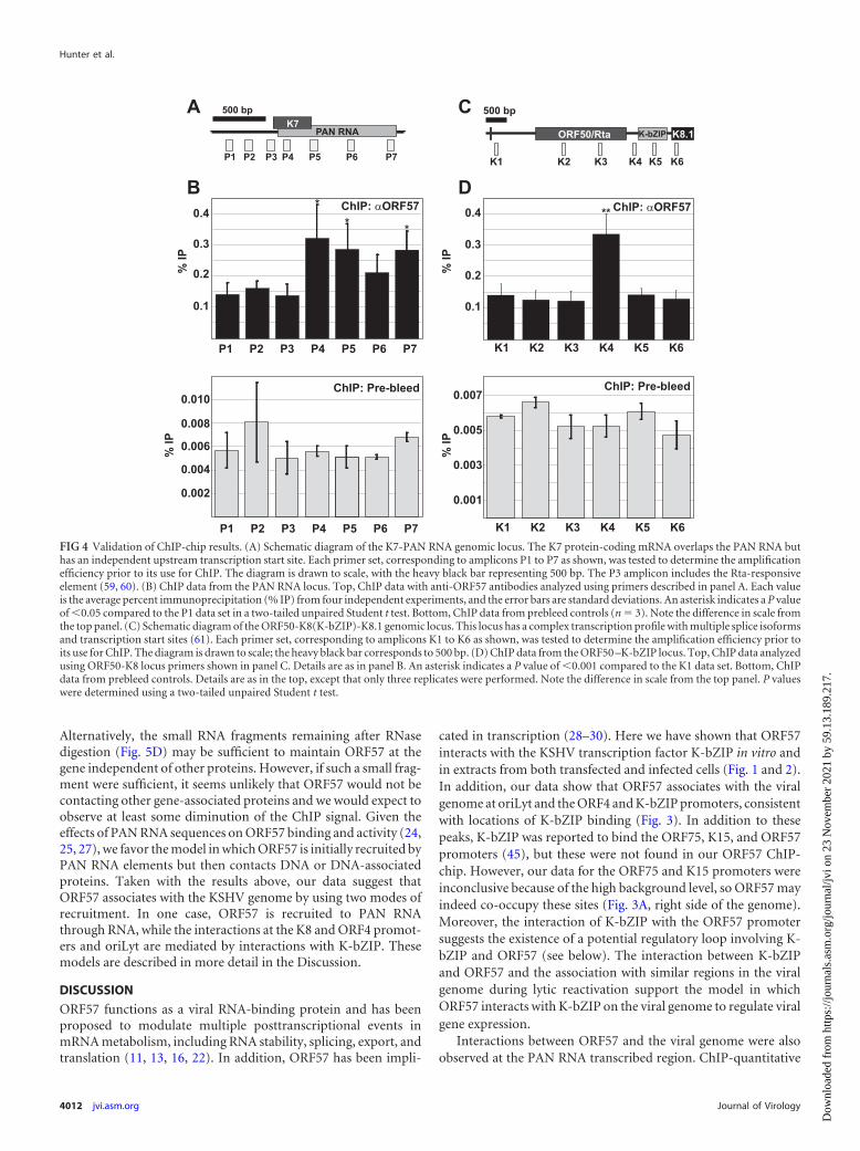
Validation of ChIP-chip results with the PAN RNA tran-scribed region and K8 promoter. We next confirmed the ChIP-chip results by using a conventional ChIP assay with real-timePCR as the readout. To do this, we designed real-time PCR oligo-nucleotide sets across the PAN RNA and ORF50-K8 regions. Wechose these two genes because they show different patterns ofORF57 binding to the locus. For PAN RNA, the ChIP peak wasseen broadly across the transcribed region whereas the K8 peakwas sharp and restricted to the K8 promoter region (Fig. 3). To testthese ChIP patterns, we designed multiple primer pairs that wereupstream or downstream of or overlapped the expected ChIP peak(Fig. 4A and C). The PAN RNA transcribed region was precipi-tated with anti-ORF57 antibodies, but the promoter region wasnot (compare the P1 to P3 signals with the P4, P5, and P7 signals).This change occurs relatively sharply in the transcribed region, asthe P3 and P4 amplicons (Fig. 4A) were separated by only 78 bp ofintervening sequence. This result confirms the data observed inthe ChIP-chip assay, and it also is consistent with reports demon-strating that ORF57 binds to a 5= element in PAN RNA (24, 25,27). Interestingly, we saw a consistent decrease in the ChIP signalwhen using the P6 primer pair and a subtle M-shaped pattern inthe PAN RNA ChIP-chip peak (Fig. 3C). While the relevance ofthis is uncertain, we speculate that this may be the result of gene orRNA looping during transcription bringing the 5= and 3= endstogether. Next, we verified the ORF57 ChIP results at theORF50-K8 locus. Once again, the data from the ChIP-chip wereconfirmed. The primer set at the K8 promoter showed a signifi-cant enhancement of ORF57, while all five other amplicons werenot enhanced (Fig. 4C and D). We also tested the same ampliconswith prebleed antibodies and saw no enhancement of the ChIP
signal at any of the primer sets (Fig. 4B and D, bottom). Interest-ingly, the percent immunoprecipitation was over 10-fold lowerwith the prebleed sample at each of the loci, which could be due tononspecific interactions of ORF57 with the viral genome-associ-ated proteins or nonspecific interactions between the antibodiesand the genome. In either case, these results validate our ChIP-chip approach and demonstrate that ORF57 associates with thetranscribed region of the PAN RNA gene, as well as the K8 pro-moter region.
RNase treatment does not alter the ORF57 ChIP pattern. Be-cause ORF57 binds to RNA, its association with genes may be duesolely to interaction with nascent transcripts that, in turn, cross-link pol II and the gene. If this were true, removal of the RNAtether should eliminate the association of ORF57 with DNA. Incontrast, ORF57 genome association mediated by direct DNAcontacts or by direct interactions with DNA-binding proteins willbe unaffected by RNase treatment. To test this, we determined theeffects of RNase treatment on the association of ORF57 withKSHV genes. After immunoprecipitation but prior to washes, weresuspended the beads in buffer containing high levels of RNase Aand incubated the mixture at room temperature to degrade RNA.We then washed away complexes under stringent conditions.First, we used the samples for a ChIP-chip experiment. As shownin Fig. 5A, the pattern of the ORF57 ChIP-chip was unaffected bythe addition of RNase. The ORF4 promoter, PAN RNA, oriLyt,and K8 promoter peaks were all present after this treatment. Toverify these results, we next performed confirmatory ChIP exper-iments at the PAN RNA and ORF50/K8 loci (Fig. 5B and C).Consistent with the ChIP-chip results, we saw no significant dif-ference between the samples with RNase and those withoutRNase.
Given that ORF57 binds directly to PAN RNA, we were initiallysurprised that RNase treatment had no effect on the ChIP signalacross the PAN RNA transcribed region. To verify that the RNasetreatment worked, we followed PAN RNA (Fig. 5D, top) or bulkRNA (Fig. 5D, bottom) through the ChIP protocol (Fig. 5D). Weharvested RNA from samples prior to immunoprecipitation (lane1), after immunoprecipitation but prior to RNase treatment (lane2), immediately following RNase treatment (lane 3), and afterwashes (lane 4). The ChIP protocols were not optimized to main-tain RNA integrity (e.g., we added no RNase inhibitor to theseassays), so some RNA degradation was observed in the input sam-ples and this was exacerbated during the long immunoprecipita-tion step (lanes 1 and 2, top). However, PAN RNA was detectablein both samples and it remained relatively long (�100 to 400nucleotides [nt]), even after immunoprecipitation. Most impor-tantly, the PAN RNA signal was completely lost after RNase treat-ment. We further examined bulk RNA with 5=-end labeling andurea-PAGE. After RNase treatment, the average RNA sizes were�30 nt (lane 3, bottom). Moreover, these fragments were nearlycompletely lost from the beads after washing (lane 4, bottom).Thus, the RNase treatment step degraded PAN RNA and the over-whelming majority of viral and cellular RNA.
One interpretation of these results is that ORF57 associationwith the viral genome is exclusively driven by ORF57-protein orORF57-DNA interactions. However, the RNase insensitivity ofthe ChIP signal does not necessarily indicate that ORF57 is re-cruited by DNA or proteins. It is formally possible that the smallamount of ORF57-associated nascent PAN RNA is resistant toRNase but is not abundant enough to be detected in our assays.
ORF57 Interacts with Viral Promoters
April 2013 Volume 87 Number 7 jvi.asm.org 4011
Dow
nloa
ded
from
http
s://j
ourn
als.
asm
.org
/jour
nal/j
vi o
n 23
Nov
embe
r 20
21 b
y 59
.13.
189.
217.
Alternatively, the small RNA fragments remaining after RNasedigestion (Fig. 5D) may be sufficient to maintain ORF57 at thegene independent of other proteins. However, if such a small frag-ment were sufficient, it seems unlikely that ORF57 would not becontacting other gene-associated proteins and we would expect toobserve at least some diminution of the ChIP signal. Given theeffects of PAN RNA sequences on ORF57 binding and activity (24,25, 27), we favor the model in which ORF57 is initially recruited byPAN RNA elements but then contacts DNA or DNA-associatedproteins. Taken with the results above, our data suggest thatORF57 associates with the KSHV genome by using two modes ofrecruitment. In one case, ORF57 is recruited to PAN RNAthrough RNA, while the interactions at the K8 and ORF4 promot-ers and oriLyt are mediated by interactions with K-bZIP. Thesemodels are described in more detail in the Discussion.
DISCUSSION
ORF57 functions as a viral RNA-binding protein and has beenproposed to modulate multiple posttranscriptional events inmRNA metabolism, including RNA stability, splicing, export, andtranslation (11, 13, 16, 22). In addition, ORF57 has been impli-
cated in transcription (28–30). Here we have shown that ORF57interacts with the KSHV transcription factor K-bZIP in vitro andin extracts from both transfected and infected cells (Fig. 1 and 2).In addition, our data show that ORF57 associates with the viralgenome at oriLyt and the ORF4 and K-bZIP promoters, consistentwith locations of K-bZIP binding (Fig. 3). In addition to thesepeaks, K-bZIP was reported to bind the ORF75, K15, and ORF57promoters (45), but these were not found in our ORF57 ChIP-chip. However, our data for the ORF75 and K15 promoters wereinconclusive because of the high background level, so ORF57 mayindeed co-occupy these sites (Fig. 3A, right side of the genome).Moreover, the interaction of K-bZIP with the ORF57 promotersuggests the existence of a potential regulatory loop involving K-bZIP and ORF57 (see below). The interaction between K-bZIPand ORF57 and the association with similar regions in the viralgenome during lytic reactivation support the model in whichORF57 interacts with K-bZIP on the viral genome to regulate viralgene expression.
Interactions between ORF57 and the viral genome were alsoobserved at the PAN RNA transcribed region. ChIP-quantitative
FIG 4 Validation of ChIP-chip results. (A) Schematic diagram of the K7-PAN RNA genomic locus. The K7 protein-coding mRNA overlaps the PAN RNA buthas an independent upstream transcription start site. Each primer set, corresponding to amplicons P1 to P7 as shown, was tested to determine the amplificationefficiency prior to its use for ChIP. The diagram is drawn to scale, with the heavy black bar representing 500 bp. The P3 amplicon includes the Rta-responsiveelement (59, 60). (B) ChIP data from the PAN RNA locus. Top, ChIP data with anti-ORF57 antibodies analyzed using primers described in panel A. Each valueis the average percent immunoprecipitation (% IP) from four independent experiments, and the error bars are standard deviations. An asterisk indicates a P valueof �0.05 compared to the P1 data set in a two-tailed unpaired Student t test. Bottom, ChIP data from prebleed controls (n 3). Note the difference in scale fromthe top panel. (C) Schematic diagram of the ORF50-K8(K-bZIP)-K8.1 genomic locus. This locus has a complex transcription profile with multiple splice isoformsand transcription start sites (61). Each primer set, corresponding to amplicons K1 to K6 as shown, was tested to determine the amplification efficiency prior toits use for ChIP. The diagram is drawn to scale; the heavy black bar corresponds to 500 bp. (D) ChIP data from the ORF50 –K-bZIP locus. Top, ChIP data analyzedusing ORF50-K8 locus primers shown in panel C. Details are as in panel B. An asterisk indicates a P value of �0.001 compared to the K1 data set. Bottom, ChIPdata from prebleed controls. Details are as in the top, except that only three replicates were performed. Note the difference in scale from the top panel. P valueswere determined using a two-tailed unpaired Student t test.
Hunter et al.
4012 jvi.asm.org Journal of Virology
Dow
nloa
ded
from
http
s://j
ourn
als.
asm
.org
/jour
nal/j
vi o
n 23
Nov
embe
r 20
21 b
y 59
.13.
189.
217.

PCR analysis confirmed the existence of a broad association acrossthe PAN RNA transcribed region, as predicted if it associates withthe nascent RNA. Even so, extensive RNase treatment does notdiminish the ORF57 association with KSHV DNA, demonstratingthat ORF57 does not exclusively contact the viral genome throughan RNA tether. Thus, it seems likely that ORF57 contacts compo-nents of the transcriptional machinery cotranscriptionally, but inthis case, the RNA is likely responsible for the initial interaction(24, 25, 27) (see below). Given the broad role of ORF57 as a post-transcriptional modulator of gene expression, we were surprisedthat other transcribed regions were not enriched in the ORF57ChIP-chip experiments. We suspect that we can detect a signal forthe PAN RNA transcribed region because it is, by far, the mosthighly expressed transcript in reactivated cells. Even so, the ChIPsignal is only about as strong as the promoter peaks, even for thishighly transcribed transcript. Therefore, we think it is likely thatother KSHV nascent transcripts associate with ORF57 but the in-teractions do not exceed the limits of detection of our assay.
We propose that ORF57 associates with the KSHV genome byusing at least two distinct molecular mechanisms (Fig. 6). In oneof these mechanisms, the K-bZIP protein, perhaps in concert with
other viral or host factors, recruits ORF57 to the viral promoters(Fig. 6A). Upon the recruitment of ORF57, the complex may beinvolved in the negative or positive regulation of transcription(Fig. 6A, transcription model). Alternatively, it is possible that it isrecruited to these promoters and is subsequently transferred tothe emerging nascent transcript (Fig. 6A, hand-off model) to en-hance ORF57-mediated posttranscriptional activity. The secondmode of ORF57 association with the genome is observed withPAN RNA (Fig. 6B). After pol II initiates transcription elongation,the 5=-proximal ORF57-responsive element (ORE) emerges fromthe transcript (24, 25, 27) and is bound by ORF57. Next, ORF57multimerization and nonspecific RNA binding activity drive theassociation of ORF57 to lower-affinity sites on the RNA. The non-specific binding of ORF57 may be strongest at the sites adjacent tothe ORE and to the 3= end of the transcript (Fig. 3 and 4) (24, 25),suggesting a potential RNA looping mechanism (Fig. 6B). Whilebound to the RNA, ORF57 makes additional contacts with thepolymerase, with DNA-bound factors, or directly with the DNA(dashed arrows, Fig. 6B). Indeed, the ORF57 homolog in herpessimplex virus, ICP27, binds pol II (55). While our data stronglysupport the existence of two modes of ORF57 genome recruit-
FIG 5 ORF57 association with KSHV genes is RNase insensitive. (A) ChIP-chip data from RNase-treated samples. Details are as described in the legend to Fig.3. An independent biological replicate yielded the same results (B, C) ChIP analysis of the RNase-treated samples; the details are as described in the legend to Fig.4. The untreated samples (black) are the same as those in Fig. 2 and are displayed here for comparison to the RNase-treated samples (gray). (D) Verification ofefficient RNase treatment. RNA was harvested from a chip experiment before immunoprecipitation (Input, 1%), after immunoprecipitation (IP), immediatelyfollowing RNase treatment, or after RNase treatment and bead washing. The samples were analyzed by Northern blot assay for PAN RNA (top) or by 5=-endradiolabeling and urea-PAGE (bottom). The mobility of the DNA markers (in nucleotides) is shown on the left. The distinct bands in the input lanes observedin the urea-PAGE are likely abundant small RNAs (e.g., tRNAs, snRNAs, etc.).
ORF57 Interacts with Viral Promoters
April 2013 Volume 87 Number 7 jvi.asm.org 4013
Dow
nloa
ded
from
http
s://j
ourn
als.
asm
.org
/jour
nal/j
vi o
n 23
Nov
embe
r 20
21 b
y 59
.13.
189.
217.

ment, significant gaps remain in the definition of the molecularmechanisms of ORF57-mediated gene regulation. Future studieswill focus on the validation of these models and the testing of theirmolecular details.
ORF57 interacts with the transcription factor Rta, the primarylatency-to-lytic switch protein (28–30), but we do not see a corre-lation between ORF57 ChIP and Rta-responsive promoters. Forexample, the Rta-responsive PAN RNA promoter does not inter-act with ORF57 in our ChIP-chip or conventional ChIP assays(Fig. 3 and 4). We interpret these negative results cautiously andcannot conclude that ORF57 does not interact with Rta at viralpromoters for several reasons. First, our ChIP assays were all per-formed at approximately the same time following lytic reactiva-tion (�18 to 22 h after doxycycline addition), so it remains pos-sible that the interaction between ORF57 and Rta occurs at adifferent stage of viral reactivation. Second, the ORF57 epitopesmay be buried in the context of an ORF57-Rta complex but re-main exposed in other ORF57 complexes. Finally, it is importantto note that the K8 promoter is Rta responsive (56–58), so it ispossible that the ORF57 association at the K8 promoter reflectsthe importance of both the ORF57-Rta and ORF57–K-bZIP inter-actions. Thus, while our data do not lend direct support to theexistence of a functional interaction between ORF57 and Rta atviral promoters, they do not exclude the model in which ORF57and Rta functionally interact at promoters during viral reactiva-tion.
Three viral gene regulatory proteins, Rta, ORF57, and K-bZIP,appear to function interdependently to regulate KSHV gene ex-
pression. ORF57 binds to K-bZIP and to Rta (Fig. 1 and 2) (28–30)and binds to the same promoters as K-bZIP (Fig. 3 and 4) (45).K-bZIP binds Rta and inhibits its transcriptional activation func-tion, but in some cases, it coactivates Rta-dependent promoters(42, 43, 45). Rta activates the ORF57 promoter, and K-bZIP alsobinds the ORF57 promoter, presumably to regulate ORF57 ex-pression (45). We do not know how these interactions affectKSHV gene regulation. Unfortunately, attempts to use reporterconstructs in HeLa or HEK293 cell lines to assess the functionalconsequences of ORF57–K-bZIP interaction have failed thus far(unpublished observations). In addition, the dissection of the mo-lecular mechanism is complicated by several factors. First, tran-scriptional activation is cell line dependent and the K8 promoterspecifically behaves differently in distinct cell lines (57). Second,other viral components could be necessary for regulation. Third,distinguishing a mechanism that entails synergistic posttranscrip-tional and transcriptional activities from a mechanism in whichthe transcriptional and posttranscriptional activities are indepen-dent can be difficult. For example, we can imagine that a particulargene depends on Rta for transcription activation and on ORF57for posttranscriptional activation. In this case, ORF57 posttran-scriptional stimulation strictly depends on the presence of Rta,giving the appearance of synergy. However, the mechanism is in-direct and not the result of a bona fide mechanistic coupling be-tween ORF57 and Rta. Empirical distinction between this type ofactivation and true mechanistic coupling is challenging. Fourth,ORF57 acts nonspecifically, so care must be taken to discernwhether stimulation of Rta or K-bZIP activity is due to molecularinteractions or simply due to ORF57 upregulation of Rta or K-bZIP. Indeed, �2-fold differences in the expression of any of thesefactors can alter the gene expression of transiently expressed K8promoter-driven constructs (unpublished observations). Theseexperimental challenges require rigorous approaches to addressthe complex regulatory interplay among ORF57, K-bZIP, and Rta.However, an understanding of these mechanisms is necessary toelucidate the molecular mechanisms of KSHV gene regulation.
ACKNOWLEDGMENTS
We thank Pinghui Feng (University of Southern California) for reagentsand Ivan D’Orso and Julie K. Pfeiffer for critical reviews of the manu-script.
This work was funded by NIH-NIAID grant AI081710. N.K.C. is aSouthwestern Medical Foundation Scholar in Biomedical Research. E.S. issupported by NIH-NIAID grant AI081710-02S1.
REFERENCES1. Ganem D. 2006. KSHV infection and the pathogenesis of Kaposi’s sar-
coma. Annu. Rev. Pathol. 1:273–296.2. Nicholas J. 2007. Human herpesvirus 8-encoded proteins with potential
roles in virus-associated neoplasia. Front. Biosci. 12:265–281.3. Dourmishev LA, Dourmishev AL, Palmeri D, Schwartz RA, Lukac DM.
2003. Molecular genetics of Kaposi’s sarcoma-associated herpesvirus (hu-man herpesvirus-8) epidemiology and pathogenesis. Microbiol. Mol. Biol.Rev. 67:175–212, table of contents.
4. Greene W, Kuhne K, Ye F, Chen J, Zhou F, Lei X, Gao SJ. 2007.Molecular biology of KSHV in relation to AIDS-associated oncogenesis.Cancer Treat. Res. 133:69 –127.
5. Dittmer D, Lagunoff M, Renne R, Staskus K, Haase A, Ganem D. 1998.A cluster of latently expressed genes in Kaposi’s sarcoma-associated her-pesvirus. J. Virol. 72:8309 – 8315.
6. Sarid R, Flore O, Bohenzky RA, Chang Y, Moore PS. 1998. Transcrip-tion mapping of the Kaposi’s sarcoma-associated herpesvirus (humanherpesvirus 8) genome in a body cavity-based lymphoma cell line (BC-1).J. Virol. 72:1005–1012.
FIG 6 Two modes of ORF57 recruitment to the KSHV genome. (A) K-bZIPrecruitment models. The details are discussed in the text. HF/VF ovals repre-sent undefined host or viral factors, respectively (e.g., Rta or RBPJ). Note thatwe are not proposing any specific molecular architecture of the complex, ex-cept for the existence of an ORF57–K-bZIP interaction. For example, we donot know which component(s) directly contacts DNA. In the transcriptionmodel (top), recruitment of ORF57 to the promoter regulates transcriptionefficiency. In the “hand-off” model, ORF57 recruitment at gene promotersleads to higher local concentrations that promote ORF57 binding to nascenttranscripts. (B) ORF57 recruitment to the PAN RNA gene. See the text fordetails.
Hunter et al.
4014 jvi.asm.org Journal of Virology
Dow
nloa
ded
from
http
s://j
ourn
als.
asm
.org
/jour
nal/j
vi o
n 23
Nov
embe
r 20
21 b
y 59
.13.
189.
217.

7. Sun R, Lin SF, Staskus K, Gradoville L, Grogan E, Haase A, Miller G.1999. Kinetics of Kaposi’s sarcoma-associated herpesvirus gene expres-sion. J. Virol. 73:2232–2242.
8. Staudt MR, Dittmer DP. 2003. Viral latent proteins as targets for Kaposi’ssarcoma and Kaposi’s sarcoma-associated herpesvirus (KSHV/HHV-8)induced lymphoma. Curr. Drug Targets Infect. Disord. 3:129 –135.
9. Deng H, Liang Y, Sun R. 2007. Regulation of KSHV lytic gene expression.Curr. Top. Microbiol. Immunol. 312:157–183.
10. Staudt MR, Dittmer DP. 2007. The Rta/Orf50 transactivator proteins ofthe gamma-herpesviridae. Curr. Top. Microbiol. Immunol. 312:71–100.
11. Conrad NK. 2009. Posttranscriptional gene regulation in Kaposi’s sarco-ma-associated herpesvirus. Adv. Appl. Microbiol. 68:241–261.
12. Glaunsinger BA, Ganem DE. 2006. Messenger RNA turnover and itsregulation in herpesviral infection. Adv. Virus Res. 66:337–394.
13. Swaminathan S. 2005. Post-transcriptional gene regulation by gammaherpesviruses. J. Cell. Biochem. 95:698 –711.
14. Sinclair AJ. 2003. bZIP proteins of human gammaherpesviruses. J. Gen.Virol. 84:1941–1949.
15. Boyne JR, Whitehouse A. 2006. Gamma-2 herpes virus post-transcriptional gene regulation. Clin. Microbiol. Infect. 12:110 –117.
16. Majerciak V, Zheng ZM. 2009. Kaposi’s sarcoma-associated herpesvirusORF57 in viral RNA processing. Front. Biosci. 14:1516 –1528.
17. Han Z, Swaminathan S. 2006. Kaposi’s sarcoma-associated herpesviruslytic gene ORF57 is essential for infectious virion production. J. Virol.80:5251–5260.
18. Majerciak V, Pripuzova N, McCoy JP, Gao SJ, Zheng ZM. 2007.Targeted disruption of Kaposi’s sarcoma-associated herpesvirus ORF57 inthe viral genome is detrimental for the expression of ORF59, K8alpha, andK8.1 and the production of infectious virus. J. Virol. 81:1062–1071.
19. Ote I, Piette J, Sadzot-Delvaux C. 2010. The varicella-zoster virus IE4protein: a conserved member of the herpesviral mRNA export factorsfamily and a potential alternative target in antiherpetic therapies.Biochem. Pharmacol. 80:1973–1980.
20. Sandri-Goldin RM. 2008. The many roles of the regulatory protein ICP27during herpes simplex virus infection. Front. Biosci. 13:5241–5256.
21. Toth Z, Stamminger T. 2008. The human cytomegalovirus regulatoryprotein UL69 and its effect on mRNA export. Front. Biosci. 13:2939 –2949.
22. Boyne JR, Jackson BR, Whitehouse A. 2010. ORF57: master regulator ofKSHV mRNA biogenesis. Cell Cycle 9:2702–2703.
23. Nekorchuk M, Han Z, Hsieh TT, Swaminathan S. 2007. Kaposi’s sar-coma-associated herpesvirus ORF57 protein enhances mRNA accumula-tion independently of effects on nuclear RNA export. J. Virol. 81:9990 –9998.
24. Sei E, Conrad NK. 2011. Delineation of a core RNA element required forKaposi’s sarcoma-associated herpesvirus ORF57 binding and activity. Vi-rology 419:107–116.
25. Massimelli MJ, Kang JG, Majerciak V, Le SY, Liewehr DJ, Steinberg SM,Zheng ZM. 2011. Stability of a long noncoding viral RNA depends on a9-nt core element at the RNA 5= end to interact with viral ORF57 andcellular PABPC1. Int. J. Biol. Sci. 7:1145–1160.
26. Boyne JR, Colgan KJ, Whitehouse A. 2008. Recruitment of the completehTREX complex is required for Kaposi’s sarcoma-associated herpesvirusintronless mRNA nuclear export and virus replication. PLoS Pathog.4:e1000194. doi:10.1371/journal.ppat.1000194.
27. Sahin BB, Patel D, Conrad NK. 2010. Kaposi’s sarcoma-associated her-pesvirus ORF57 protein binds and protects a nuclear noncoding RNAfrom cellular RNA decay pathways. PLoS Pathog. 6:e1000799. doi:10.1371/journal.ppat.1000799.
28. Palmeri D, Spadavecchia S, Carroll KD, Lukac DM. 2007. Promoter-and cell-specific transcriptional transactivation by the Kaposi’s sarcoma-associated herpesvirus ORF57/Mta protein. J. Virol. 81:13299 –13314.
29. Malik P, Blackbourn DJ, Cheng MF, Hayward GS, Clements JB. 2004.Functional co-operation between the Kaposi’s sarcoma-associated her-pesvirus ORF57 and ORF50 regulatory proteins. J. Gen. Virol. 85:2155–2166.
30. Uetz P, Dong YA, Zeretzke C, Atzler C, Baiker A, Berger B, RajagopalaSV, Roupelieva M, Rose D, Fossum E, Haas J. 2006. Herpesviral proteinnetworks and their interaction with the human proteome. Science 311:239 –242.
31. West JT, Wood C. 2003. The role of Kaposi’s sarcoma-associated herpes-virus/human herpesvirus-8 regulator of transcription activation (RTA) incontrol of gene expression. Oncogene 22:5150 –5163.
32. Kirshner JR, Lukac DM, Chang J, Ganem D. 2000. Kaposi’s sarcoma-associated herpesvirus open reading frame 57 encodes a posttranscrip-tional regulator with multiple distinct activities. J. Virol. 74:3586 –3597.
33. Aravind L, Landsman D. 1998. AT-hook motifs identified in a widevariety of DNA-binding proteins. Nucleic Acids Res. 26:4413– 4421.
34. Kato-Noah T, Xu Y, Rossetto CC, Colletti K, Papousková I, Pari GS.2007. Overexpression of the Kaposi’s sarcoma-associated herpesvirustransactivator K-Rta can complement a K-bZIP deletion BACmid andyields an enhanced growth phenotype. J. Virol. 81:13519 –13532.
35. Wang Y, Sathish N, Hollow C, Yuan Y. 2011. Functional characterization ofKaposi’s sarcoma-associated herpesvirus open reading frame K8 by bacterialartificial chromosome-based mutagenesis. J. Virol. 85:1943–1957.
36. Lefort S, Flamand L. 2009. Kaposi’s sarcoma-associated herpesvirus K-bZIP protein is necessary for lytic viral gene expression, DNA replication,and virion production in primary effusion lymphoma cell lines. J. Virol.83:5869 –5880.
37. Lin CL, Li H, Wang Y, Zhu FX, Kudchodkar S, Yuan Y. 2003. Kaposi’ssarcoma-associated herpesvirus lytic origin (ori-Lyt)-dependent DNAreplication: identification of the ori-Lyt and association of K8 bZip pro-tein with the origin. J. Virol. 77:5578 –5588.
38. Rossetto C, Yamboliev I, Pari GS. 2009. Kaposi’s sarcoma-associatedherpesvirus/human herpesvirus 8 K-bZIP modulates latency-associatednuclear protein-mediated suppression of lytic origin-dependent DNAsynthesis. J. Virol. 83:8492– 8501.
39. Izumiya Y, Ellison TJ, Yeh ET, Jung JU, Luciw PA, Kung HJ. 2005.Kaposi’s sarcoma-associated herpesvirus K-bZIP represses gene transcrip-tion via SUMO modification. J. Virol. 79:9912–9925.
40. Wu FY, Wang SE, Tang QQ, Fujimuro M, Chiou CJ, Zheng Q, Chen H,Hayward SD, Lane MD, Hayward GS. 2003. Cell cycle arrest by Kaposi’ssarcoma-associated herpesvirus replication-associated protein is medi-ated at both the transcriptional and posttranslational levels by binding toCCAAT/enhancer-binding protein alpha and p21(CIP-1). J. Virol. 77:8893– 8914.
41. AuCoin DP, Colletti KS, Cei SA, Papousková I, Tarrant M, Pari GS.2004. Amplification of the Kaposi’s sarcoma-associated herpesvirus/human herpesvirus 8 lytic origin of DNA replication is dependent upon acis-acting AT-rich region and an ORF50 response element and the trans-acting factors ORF50 (K-Rta) and K8 (K-bZIP). Virology 318:542–555.
42. Izumiya Y, Lin SF, Ellison T, Chen LY, Izumiya C, Luciw P, Kung HJ.2003. Kaposi’s sarcoma-associated herpesvirus K-bZIP is a coregulator ofK-Rta: physical association and promoter-dependent transcriptional re-pression. J. Virol. 77:1441–1451.
43. Liao W, Tang Y, Lin SF, Kung HJ, Giam CZ. 2003. K-bZIP of Kaposi’ssarcoma-associated herpesvirus/human herpesvirus 8 (KSHV/HHV-8)binds KSHV/HHV-8 Rta and represses Rta-mediated transactivation. J.Virol. 77:3809 –3815.
44. Lefort S, Soucy-Faulkner A, Grandvaux N, Flamand L. 2007. Binding ofKaposi’s sarcoma-associated herpesvirus K-bZIP to interferon-responsivefactor 3 elements modulates antiviral gene expression. J. Virol. 81:10950–10960.
45. Ellison TJ, Izumiya Y, Izumiya C, Luciw PA, Kung HJ. 2009. A com-prehensive analysis of recruitment and transactivation potential of K-Rtaand K-bZIP during reactivation of Kaposi’s sarcoma-associated herpesvi-rus. Virology 387:76 – 88.
46. Nakamura H, Lu M, Gwack Y, Souvlis J, Zeichner SL, Jung JU. 2003.Global changes in Kaposi’s sarcoma-associated virus gene expression pat-terns following expression of a tetracycline-inducible Rta transactivator. J.Virol. 77:4205– 4220.
47. Sheffield P, Garrard S, Derewenda Z. 1999. Overcoming expression andpurification problems of RhoGDI using a family of “parallel” expressionvectors. Protein Expr. Purif. 15:34 –39.
48. Horton RM. 1995. PCR-mediated recombination and mutagenesis. SOE-ing together tailor-made genes. Mol. Biotechnol. 3:93–99.
49. Pfaffl MW. 2001. A new mathematical model for relative quantification inreal-time RT-PCR. Nucleic Acids Res. 29:e45.
50. Stubbs SH, Hunter OV, Hoover A, Conrad NK. 2012. Viral factorsreveal a role for REF/Aly in nuclear RNA stability. Mol. Cell. Biol. 32:1260 –1270.
51. Majerciak V, Yamanegi K, Nie SH, Zheng ZM. 2006. Structural andfunctional analyses of Kaposi sarcoma-associated herpesvirus ORF57 nu-clear localization signals in living cells. J. Biol. Chem. 281:28365–28378.
52. Malik P, Blackbourn DJ, Clements JB. 2004. The evolutionarily con-served Kaposi’s sarcoma-associated herpesvirus ORF57 protein interacts
ORF57 Interacts with Viral Promoters
April 2013 Volume 87 Number 7 jvi.asm.org 4015
Dow
nloa
ded
from
http
s://j
ourn
als.
asm
.org
/jour
nal/j
vi o
n 23
Nov
embe
r 20
21 b
y 59
.13.
189.
217.

with REF protein and acts as an RNA export factor. J. Biol. Chem. 279:33001–33011.
53. Sapra AK, Ankö ML, Grishina I, Lorenz M, Pabis M, Poser I, Rollins J,Weiland EM, Neugebauer KM. 2009. SR protein family members displaydiverse activities in the formation of nascent and mature mRNPs in vivo.Mol. Cell 34:179 –190.
54. Conrad NK. 2008. Chapter 15. Co-immunoprecipitation techniques for as-sessing RNA-protein interactions in vivo. Methods Enzymol. 449:317–342.
55. Dai-Ju JQ, Li L, Johnson LA, Sandri-Goldin RM. 2006. ICP27 interactswith the C-terminal domain of RNA polymerase II and facilitates its re-cruitment to herpes simplex virus 1 transcription sites, where it undergoesproteasomal degradation during infection. J. Virol. 80:3567–3581.
56. Lukac DM, Garibyan L, Kirshner JR, Palmeri D, Ganem D. 2001. DNAbinding by Kaposi’s sarcoma-associated herpesvirus lytic switch protein isnecessary for transcriptional activation of two viral delayed early promot-ers. J. Virol. 75:6786 – 6799.
57. Wang Y, Chong OT, Yuan Y. 2004. Differential regulation of K8 geneexpression in immediate-early and delayed-early stages of Kaposi’ssarcoma-associated herpesvirus. Virology 325:149 –163.
58. Seaman WT, Quinlivan EB. 2003. Lytic switch protein (ORF50) responseelement in the Kaposi’s sarcoma-associated herpesvirus K8 promoter islocated within but does not require a palindromic structure. Virology310:72– 84.
59. Song MJ, Brown HJ, Wu TT, Sun R. 2001. Transcription activation ofpolyadenylated nuclear rna by rta in human herpesvirus 8/Kaposi’s sarco-ma-associated herpesvirus. J. Virol. 75:3129 –3140.
60. Chang PJ, Shedd D, Gradoville L, Cho MS, Chen LW, Chang J, MillerG. 2002. Open reading frame 50 protein of Kaposi’s sarcoma-associatedherpesvirus directly activates the viral PAN and K12 genes by binding torelated response elements. J. Virol. 76:3168 –3178.
61. Zheng ZM. 2003. Split genes and their expression in Kaposi’s sarcoma-associated herpesvirus. Rev. Med. Virol. 13:173–184.
Hunter et al.
4016 jvi.asm.org Journal of Virology
Dow
nloa
ded
from
http
s://j
ourn
als.
asm
.org
/jour
nal/j
vi o
n 23
Nov
embe
r 20
21 b
y 59
.13.
189.
217.