download.lww.comdownload.lww.com/wolterskluwer_vitalstream_com/permalink/... · web viewa...
TRANSCRIPT
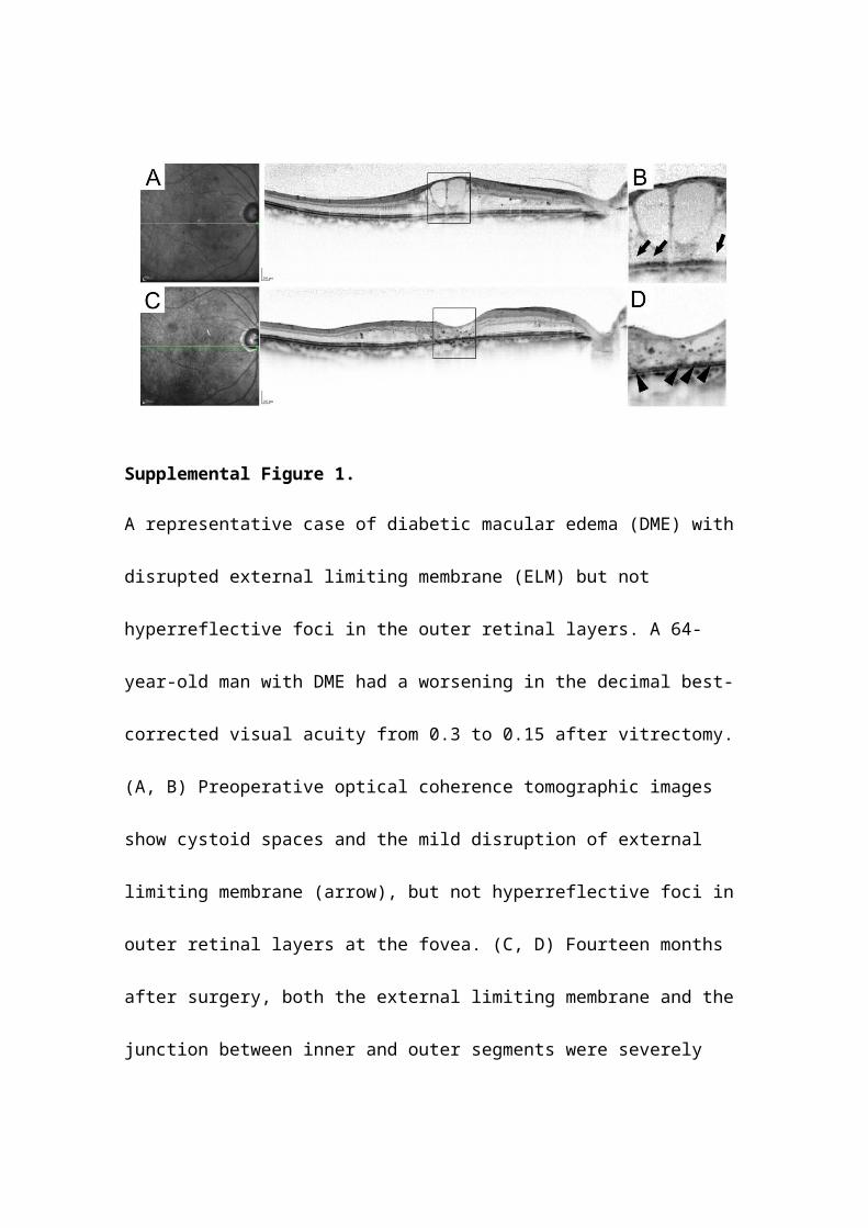
Supplemental Figure 1.
A representative case of diabetic macular edema (DME) with disrupted external limiting
membrane (ELM) but not hyperreflective foci in the outer retinal layers. A 64-year-old
man with DME had a worsening in the decimal best-corrected visual acuity from 0.3 to
0.15 after vitrectomy. (A, B) Preoperative optical coherence tomographic images show
cystoid spaces and the mild disruption of external limiting membrane (arrow), but not
hyperreflective foci in outer retinal layers at the fovea. (C, D) Fourteen months after
surgery, both the external limiting membrane and the junction between inner and outer
segments were severely damaged, and hyperreflective foci there were present
(arrowhead). B, D are magnified versions of the inset in A, C.
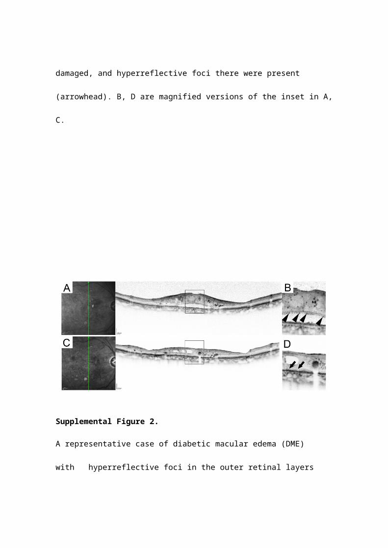
Supplemental Figure 2.
A representative case of diabetic macular edema (DME) with hyperreflective foci in
the outer retinal layers than intact external limiting membrane (ELM). A 74-year-old
man with DME did not have an improvement in the decimal best-corrected visual acuity
(from 0.3 to 0.2). (A, B) Preoperative optical coherence tomographic images show both
cystoid spaces and serous retinal detachment, and hyperreflective foci around the outer
segments. (C, D) Fifteen months after surgery, both the external limiting membrane and
the junction between inner and outer segments were disrupted (arrowhead). B, D are
magnified versions of the inset in A, C.
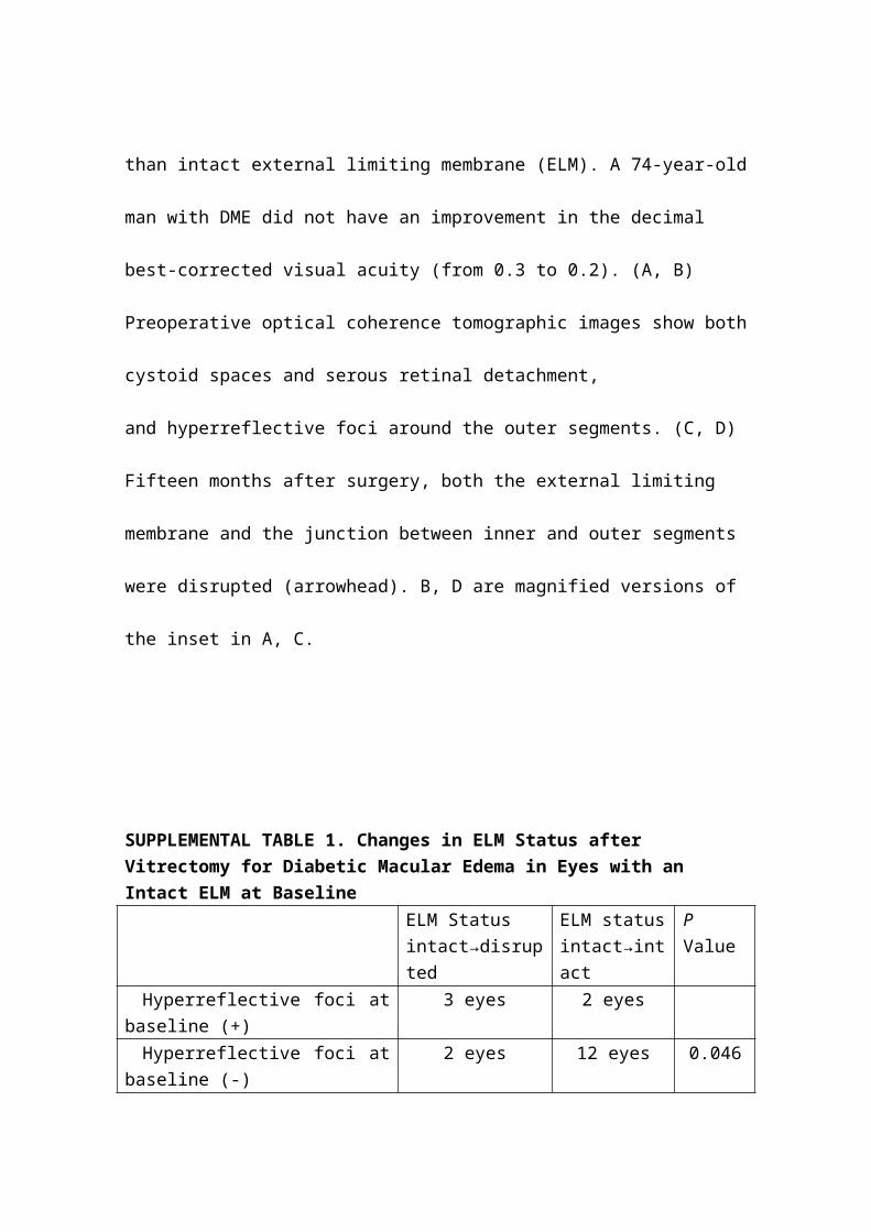
SUPPLEMENTAL TABLE 1. Changes in ELM Status after Vitrectomy for Diabetic Macular Edema in Eyes with an Intact ELM at Baseline
ELM Statusintact→disrupted
ELM statusintact→intact
P Value
Hyperreflective foci at baseline (+) 3 eyes 2 eyesHyperreflective foci at baseline (-) 2 eyes 12 eyes 0.046
ELM = external limiting membrane; hyperreflective foci = hyperreflective foci in the outer retinal layers; intact→disrupted = eyes in which the ELM status changed from intact at baseline to disrupted at the final visit; intact→intact = eyes in which the ELM status remained intact from baseline to the final visit.

SUPPLEMENTAL TABLE 2. The changes in hyperreflective foci in the outer retinal layers after vitrectomy for diabetic macular edema.
hyperreflective foci (-)→(+)
hyperreflective foci (-)→(-)
P Value
disrupted ELM at baseline 6 eyes 2 eyesintact ELM at baseline 4 eyes 10 eyes 0.035ELM = external limiting membrane; hyperreflective foci (-)→(+) = eyes in which hyperreflective foci in the outer retinal layers were absent at baseline, whereas they were present at final visit; hyperreflective foci (-)→(-) = eyes in which hyperreflective foci in the outer retinal layers were kept absent from baseline to final visit.