) the sazanami pathology network: a pioneering service
TRANSCRIPT
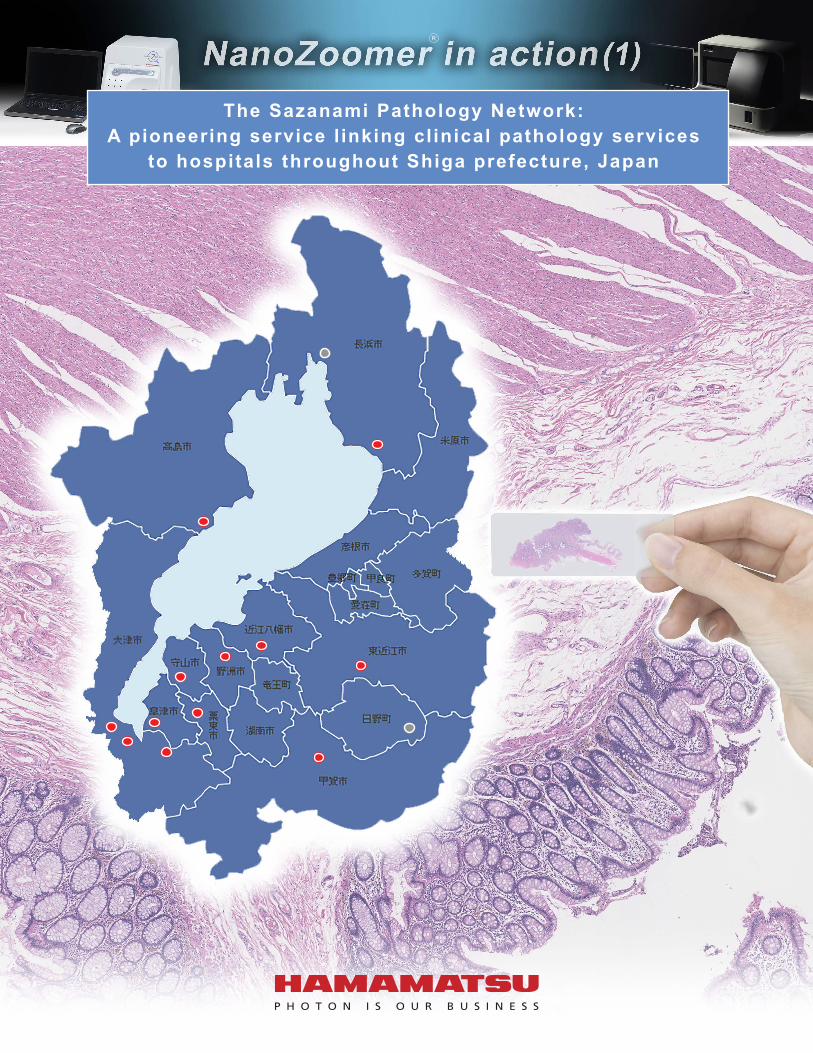
The Sazanami Pathology Network: A pioneering service l inking cl inical pathology services
to hospitals throughout Shiga prefecture, Japan
R

What is the "Sazanami Pathology Network"?
The Lake Biwa
Medical Network
Clinic
Reference Laboratory
Full-time pathologist
Remote intraoperative
diagnosisPrimary
diagnosis support Consultation Education Companion diagnostics
Consultation Ordering
Consultation Ordering
Consultation Ordering
Accepting consultation
Accepting consultationNo
pathologist No pathologist
Diagnostic request flow chart: Shiga Medical Center for Adults
Consultation Ordering
Ordering of diagnosis Result of diagnosis
Shiga Medical Center for Adults
Pathologist CytotechnologistDuties
Tissue diagnosisCyto-diagnosis
DutiesCyto-diagnosisManagement of systemManagement of specimen
Shiga Medical Center for Adults
* WebPath made by Seiko-tec is used as pathology information system.
Seven NanoZoomer units arecurrently deployed as of July 2015.
Mainapplication
A unique tele-pathology network catering to the greater healthcare community in Shiga prefecture, Japan.Harnessing the power of whole slide imaging (WSI) and Information and Communication Technology (ICT), the network
serves as a veritable "Virtual Pathology Department" accessible to all healthcare facilities under the same network umbrella.
Receive tissue diagnosis orders from clinics(skin and gastrointestinal biopsies)

Why did you start thinking about Sazanami Pathology Network?
It was when I returned to Japan from the US in 1977. I can say I think the situation surrounding Japanese pathologists was in a crisis. Unlike the US, there were nowhere near enough pathologists in Japan, with many hospitals not staffing them. There also wasn't much thought given to managing diagnosis precision, something which also surprised me compared to the US. In the mid 80s it was demonstrated that sending images was a way to enable intraoperative diagnoses at hospitals with no pathologists on site, and to enable remote pathology as a ways for pathologists to support each others' diagnoses. So images began to be sent. However, this initially meant sending numerous static images, which posed various problems. It wasn't long before remote pathologists were able to send videos, which allowed pathologists to search the entire specimen but depending on the Internet connection took a lot of time to get the equipment up and running.I was transferred to Kyoto University Hospital in 2002, where there was now the need to support many related hospitals. I had to send pathologists to facilities with only one pathologist and to hospitals with none, but because there were so few pathologists that could be sent anyway it was difficult to find someone to send. In such an environment I thought about creating a system that could provide support through various means. Instead of creating a lot of hospitals with only one pathologist, we would gather many pathologists that were specialists in the various organs who would then handle the pathological diagnoses of multiple hospitals — a sort of existential collection of hospital pathology departments. One way was through remote pathology, but due to the disadvantages of the medical treatment provided by healthcare providers and the need to have pathologists present in hospitals designated for training, designated cancer hospitals, and so on, it was difficult to realize this method. Around that time remote pathology implemented the whole slide imaging system, where entire histopathology sections were photographed at high power under a microscope and the sections where combined one by one by a computer for viewing. If a system was created to link whole slide imaging and other ICT technology together, a virtual collection of hospital pathology departments, then it could support diagnoses made by fellow pathologists, train other pathologists, and handle the pathological diagnoses of hospitals with no pathologists even if the pathologists were scattered around various hospitals. Halfway through 2010, when this concept was crystallizing, I retired from Kyoto University Hospital. The chief of the Shiga Medical Center for Adults at the time approached me about whether it would be possible to realize this virtual collection of hospital pathology departments in Shiga, so I moved to Shiga and got to work realizing operation of the Sazanami Pathology Network. Shiga prefecture wanted to create a medical treatment for the whole prefecture, and so searched for a way to alleviate the adverse effects of this lack of pathologists. If a cancer pathology diagnosis is not properly performed, then the patient cannot receive the proper cancer treatment. And the patient may become emotionally distraught if it takes a lot of time until the pathology diagnosis results are in. So it was very encouraging to get support in realizing the creation of a virtual collection of hospital pathology departments (the current Sazanami Pathology Network) as a means for the prefecture to solve these problems.*ICT stands for Information and Communication Technology.
Interview with Dr. Toshiaki Manabe, Shiga Medical Center for Adults
What are the benefits of using this?In operating the Sazanami Pathology Network, we provide support between hospitals, pathologists, and cytoscreeners who have joined the network in remote intraoperative rapid diagnoses and normal pathological diagnoses, we provide consultations (official and opinions), training, and companion diagnoses. If there is collaboration between regions at the prefectural level then everyone knows each other so it is easy to ask for or provide information, which creates an extremely good environment.The biggest benefit of the Sazanami Pathology Network is that it uses ICT, which allows you to virtually eliminate much of the work that a person would traditionally do. For example, when asked for consultation you would normally take the glass slide specimen and request out of the bag that was sent, refer to the specimen while confirming the information, and then set the slide into the microscope. After examining the specimen under a microscope a medical certificate would either be handwritten or printed out by computer, and then the contents of the certificate and the patient's ID would be confirmed. Finally the glass slide and certificate would be bagged together, the sender's information would be confirmed, and then the bag would be posted to the user. So the non-examination part of the process took a lot of time. The Sazanami Pathology Network uses ICT so all of this work is handled and managed on the system side. The person making the request uses a barcode to accurately identify what materials they are required to send, and the pathology image is sent electronically together with the request. The person receiving it changes and displays the request as a medical certificate, and they can see the attached images for confirmation. Once they complete the medical certificate they click the save button to instantly return the certificate to the person making the request. This greatly reduces the above mentioned labor on both the requesting and diagnosing sides. This is the heart and soul of the network system operation.There is also not a lot of work, which means diagnoses can be completed in a short time, which reduces the load on the pathologist commissioned to do the diagnosis. Best of all the diagnosis can be quickly returned to the clinician, and the patient. The diagnosis in a consultation can be sent at a time that suits you, which makes for a pathologist-friendly system. You can also confirm any uncertainties using chat. You don't need to create a new request when getting the opinion of a third party pathologist either — you can just instantly send what you have and make the request.
▲ Dr. Toshiaki Manabe, Chief of Shiga Medical Center for Adults
What difficulties did you have in realizing the Sazanami Pathology Network?
There were two major difficulties. The first was awareness that there was a system in place. It's important that people involved know about it first, so we explained the system at medical and hospital associations throughout Shiga. We also looked for hospitals in Shiga with pathological examination rooms but no pathologists and went to explain the system to them directly. At the time there weren't a lot of exchanges between pathologists in Shiga, so we created a mailing list to allow them to communicate. I'm aware that the presence and cooperation of professors from the Shiga University of Medical Science Pathology Department and various other doctors in establishing this network was a huge help.Right now we are holding an annual regional collaboration symposium using community ICT. This symposium initially played a major role and getting people to understand the goal and details of the Sazanami Pathology Network. Today we hold the symposium to popularize the network not only within Shiga and the Kinki Region, but also around the country. The symposium is also to hold discussions as a means to solve the problems of a lack of pathologists and their uneven distribution. Second is the budget necessary to get the equipment. Although all of the facilities agreed to run the system, the actual problem was what to do about a budget for general and equipment side improvements. So we got the support of the government (Ministry of Internal Affairs and Communications and Ministry of Health, Labour and Welfare) and the prefecture. We petitioned both ministries several times and since our concept meshed with their move as a government toward focusing on ICT use, all the hospitals participating in the network were able to get financial backing.
Do you have any advice for others out there who are considering using a local pathology network?
Well, it comes down to whether to purchase a device or not. To be honest, it's a difficult question to answer. First, I think it's important to investigate other networks that are slowly taking shape elsewhere, and to consider what networks are convenient and whether it's necessary for the hospital to join that network. When making these considerations, it's very important for the hospital to thoroughly understand that there are limits to

What do you hope for in the future?In the future we plan to promote operation of the Sazanami Pathology Network nationwide. There are many regions just like Shiga that are facing problems with insufficient pathologists and their uneven distribution. It's of course OK if they participate in the network directly, but each region or block could also make a similar network of their own and tie it in with networks all over Japan. We are already reaching out to pathologists around the country. Our goal is to have female pathologists who are currently out on maternity or childcare leave, or retired pathologists, join the network, and to create a consultation network of specialized panels for each organ and disease. If we can use this system to manage the precision of pathological diagnoses, then I think we can not only increase the performance of these diagnoses but also increase the quality of writing in the diagnosis reports and other areas across the board.
What do you need to expand use of this?It's important to widely communicate information in order to popularize this, and for that it's important to communicate information not only to pathologists but to companies involved in pathology and the clinicians, hospitals, and general public that are using the system. I think you also have to work together with companies when communicating this information, which is why I agreed to this interview.
What do you expect from a whole slide imaging system?
First, to reduce the amount of time it takes to capture an image. We use the system right now for intraoperative diagnoses, but when I looked into the time needed for each of the various processes it turns out that you need a lot of time to excise, create the specimen, and create the image. This correlated with the large amount of time needed overall, but the time needed for diagnosis was short, only a few minutes. Therefore I hope that companies can revolutionize the technology to enable even quicker image capture.Next, I hope for the development of a compact, low cost whole slide imaging system that can be used by individual pathologists and cytoscreeners. I hope we are entering an age where individuals will have their own whole slide imaging system connected to one network thanks to the creation of a virtual collection of hospital pathology departments that I mentioned in the introduction. It's necessary to create an environment where someone can quickly ask a question about a difficult case in a virtual space. Especially when diagnosing difficult cases, you can ask someone nearby (supervisor) about the necessity of an immunohistochemical staining, but because there isn't an actual environment where you can consult someone one big benefit of using a whole slide imaging system is that you can create just such an environment. If this happens, I think we will have wiped away that crisis that I felt when I returned to Japan. It's important to reduce the image capture time in order to make this consultation possible during day to day work.When used to manage the precision before making a cytodiagnosis, you need something where you can easily identify just the problem region and take a photo, as opposed to taking a photo of the entire image. To accomplish this, it would be nice if you could create equipment and systems where live mode and scan mode are linked together well. It would be extremely useful if you could insert a specimen, identify the areas you want to take a photo of while viewing it in live mode, then quickly switch to scan mode to take and send an image to get real-time support during screenings.Elsewhere, it's important for the pathologists and cytoscreeners, who are the long-term users, and the vendors providing the whole slide imaging, to discuss what it is that pathologists want and where development of the whole slide imaging system should lead in the future.
what just one pathologist can do in making accurate pathological diagnoses, and that essentially a specialized pathologist needs to exchange opinions with other pathologists. After they understand this I would want them to ask the hospital for funding to join the network.The situation regarding remote pathological diagnosis networks varies by region. It might be better to join an existing network or a network that is just getting off the ground instead of creating a new network. Either way, I would want them to look for and investigate a network that is closest to what they need.I want the government and prefecture to understand the importance of precision management. It's important for everyone to let them know just how big a role an ICT network that uses whole slide imaging plays in this, and to explain the importance of national/prefectural subsidies in spreading a remote pathological diagnosis network.
In the end, what was the reaction?It's been extremely well received. The clinical side has told me that it allows them to receive intraoperative rapid diagnoses to let them finish their surgeries safely, and that they were surprised they could get a second opinion so easily and quickly (even getting a reply the same day), while on the pathologist side they say that the lightning speed of the system has been a huge help. They were shocked at all of the observations that they did not themselves see.
© 2017 Hamamatsu Photonics K.K.
HAMAMATSU PHOTONICS K.K.HAMAMATSU PHOTONICS K.K., Systems Division812 Joko-cho, Higashi-ku, Hamamatsu City, 431-3196, Japan, Telephone: (81)53-431-0124, Fax: (81)53-435-1574, E-mail: [email protected].: Hamamatsu Corporation: 360 Foothill Road, Bridgewater, NJ 08807, U.S.A., Telephone: (1)908-231-0960, Fax: (1)908-231-1218 E-mail: [email protected]: Hamamatsu Photonics Deutschland GmbH.: Arzbergerstr. 10, D-82211 Herrsching am Ammersee, Germany, Telephone: (49)8152-375-0, Fax: (49)8152-265-8 E-mail: [email protected]: Hamamatsu Photonics France S.A.R.L.: 19, Rue du Saule Trapu, Parc du Moulin de Massy, 91882 Massy Cedex, France, Telephone: (33)1 69 53 71 00, Fax: (33)1 69 53 71 10 E-mail: [email protected] Kingdom: Hamamatsu Photonics UK Limited: 2 Howard Court,10 Tewin Road, Welwyn Garden City, Hertfordshire AL7 1BW, UK, Telephone: (44)1707-294888, Fax: (44)1707-325777 E-mail: [email protected] Europe: Hamamatsu Photonics Norden AB: Torshamnsgatan 35 16440 Kista, Sweden, Telephone: (46)8-509-031-00, Fax: (46)8-509-031-01 E-mail: [email protected]: Hamamatsu Photonics Italia S.r.l.: Strada della Moia, 1 int. 6, 20020 Arese (Milano), Italy, Telephone: (39)02-935-81-733, Fax: (39)02-935-81-741 E-mail: [email protected]: Hamamatsu Photonics (China) Co., Ltd.: 1201 Tower B, Jiaming Center, 27 Dongsanhuan Beilu, Chaoyang District, 100020 Beijing, China, Telephone: (86)10-6586-6006, Fax: (86)10-6586-2866 E-mail: [email protected]: Hamamatsu Photonics Taiwan Co., Ltd.: 8F-3, No.158, Section2, Gongdao 5th Road, East District, Hsinchu, 300, Taiwan R.O.C. Telephone: (886)03-659-0080, Fax: (886)07-811-7238 E-mail: [email protected]
www.hamamatsu.com
Cat.No.SBIS0113E02JUL/2017 CRCreated in Japan
NanoZoomer or NDP is a registered trademark of Hamamatsu Photonics K.K. (EU, Japan, U.S.A)Product and software package names noted in this documentation are trademarks or registered trademarks of their respective manufacturers.NanoZoomer is for research use only and is not permitted to use for clinical diagnostic purposes.Subject to local technical requirements and regulations. Availability of products included in this promotional material may vary. Please consult with your local sales representative.Information furnished by HAMAMATSU is believed to be reliable. However, no responsibility is assumed for possible inaccuracies or omissions.Specifications and external appearance are subject to change without notice.