reproduction one property of a living thing great variety of methods sexual reproduction each...
TRANSCRIPT
Reproduction one property of a living thing great variety of methods
Sexual reproduction each offspring has 2 parents and receives genetic
material from both provides genetic diversity foundation for survival and evolution of species
27-1

Male and female gametes (sex cells) combine their genes to form a fertilized egg (zygote) one gamete has motility (sperm)
parent producing sperm considered male has Y chromosome in most mammals
other gamete (egg or ovum) contains nutrients for developing zygote parent producing eggs considered female in mammals the female also provides shelter for the
developing fetus (uterus and placenta)
27-2

Primary sex organs produce gametes (testes or ovaries)
Secondary sex organs male - ducts, glands, penis deliver sperm cells female - uterine tubes, uterus and vagina receive sperm
and nourish developing fetus Secondary sex characteristics
develop at puberty to attract a mate pubic, axillary and facial hair, scent glands, body morphology
and low-pitched voice in males
27-3

Our cells contain 23 pairs of chromosomes 22 pairs of autosomes 1 pair of sex chromosomes (XY males: XX females)
males produce 50% Y carrying sperm and 50% X carrying all eggs carry the X chromosome
Sex of child determined by type of sperm that fertilizes mother’s egg
27-4

Gonads begin to develop at 6 weeks
The male and female reproductive systems have different embryological origins mesonephric ducts
develop into male reproductive system
paramesonephric ducts (müllerian ducts) develop into female reproductive tract
27-5
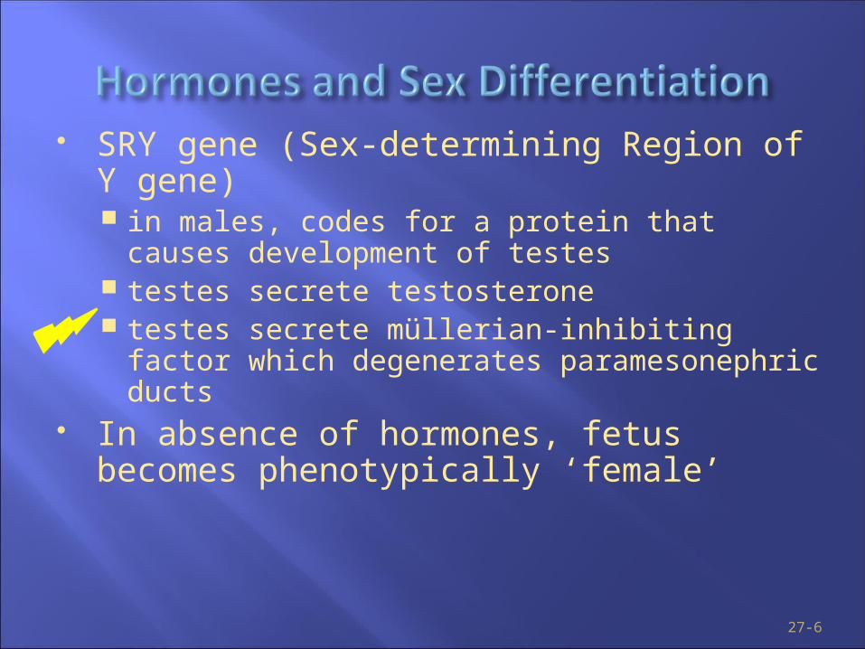
SRY gene (Sex-determining Region of Y gene) in males, codes for a protein that causes development of
testes testes secrete testosterone testes secrete müllerian-inhibiting factor which
degenerates paramesonephric ducts In absence of hormones, fetus becomes
phenotypically ‘female’
27-6
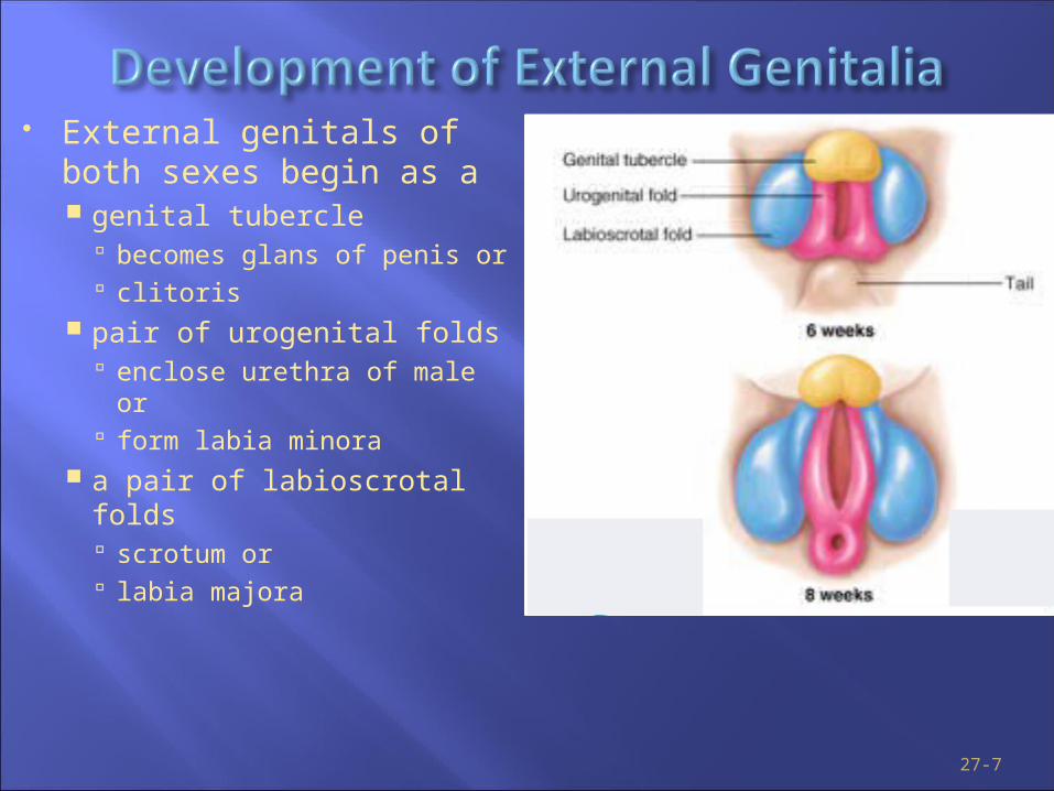
External genitals of both sexes begin as a genital tubercle
becomes glans of penis or clitoris
pair of urogenital folds enclose urethra of male or form labia minora
a pair of labioscrotal folds scrotum or labia majora
27-7

All 8 week old fetuses have same 3 structures by end of week 9, begin to
show sexual differentiation distinctly male or female by
end of week 12
27-8

Begin development near kidney gubernaculum (cordlike structure containing muscle) extends
from gonad to abdominopelvic floor it shortens, guides testes to scrotum
Descent begins in weeks 6-10, finished by 28 3% born with undescended testes (cryptorchidism)
Location outside pelvic cavity essential for low temperatures needed for sperm production
27-9

27-10

Impotence – Inability to sustain an erection sufficient for sexual intercourse, or inability to ejaculate
Male Sterility – Infertility caused by disorders of the male reproductive system
BPH – Benign Prostatic Hypertrophy Cryptorchidism – Undescended testicle(s)
27-11
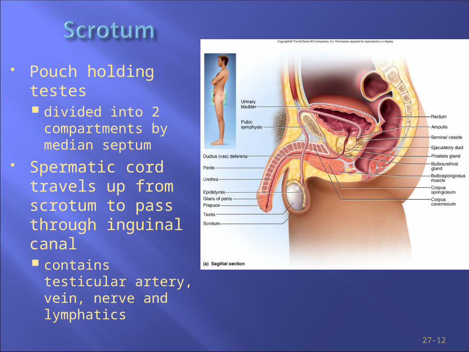
Pouch holding testes divided into 2
compartments by median septum
Spermatic cord travels up from scrotum to pass through inguinal canal contains testicular
artery, vein, nerve and lymphatics
27-12

27-13
Circumcision
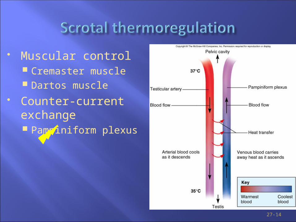
Muscular control Cremaster muscle Dartos muscle
Counter-current exchange Pampiniform plexus
27-14
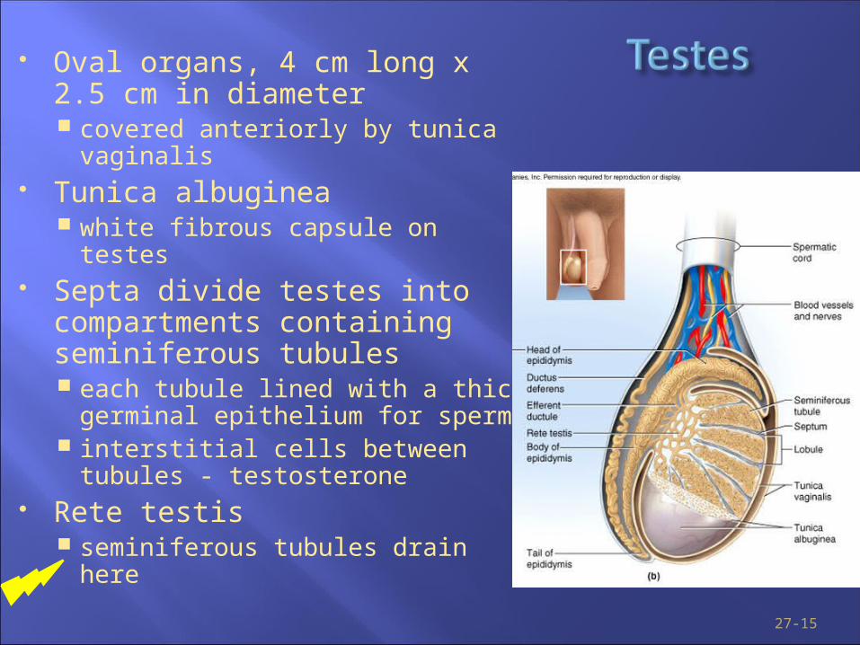
Oval organs, 4 cm long x 2.5 cm in diameter covered anteriorly by tunica vaginalis
Tunica albuginea white fibrous capsule on testes
Septa divide testes into compartments containing seminiferous tubules each tubule lined with a thick
germinal epithelium for sperm interstitial cells between tubules -
testosterone Rete testis
seminiferous tubules drain here
27-15

Efferent ductules 12 small ciliated ducts collecting sperm
from rete testes and transporting it to epididymis
Epididymis (head, body and tail) 6 m long coiled duct adhering to
posterior of testis site of sperm maturation and storage
(fertile for 60 days) Ductus deferens (peristalsis during
orgasm) muscular tube 45 cm long passing up
from scrotum through inguinal canal to posterior surface of bladder
Ejaculatory duct 2 cm duct formed from ductus
deferens and seminal vesicle and passing through prostate to empty into urethra
27-16
Vasectomy

Regions: prostatic, membranous and penile --- totals 20 cm long
27-17
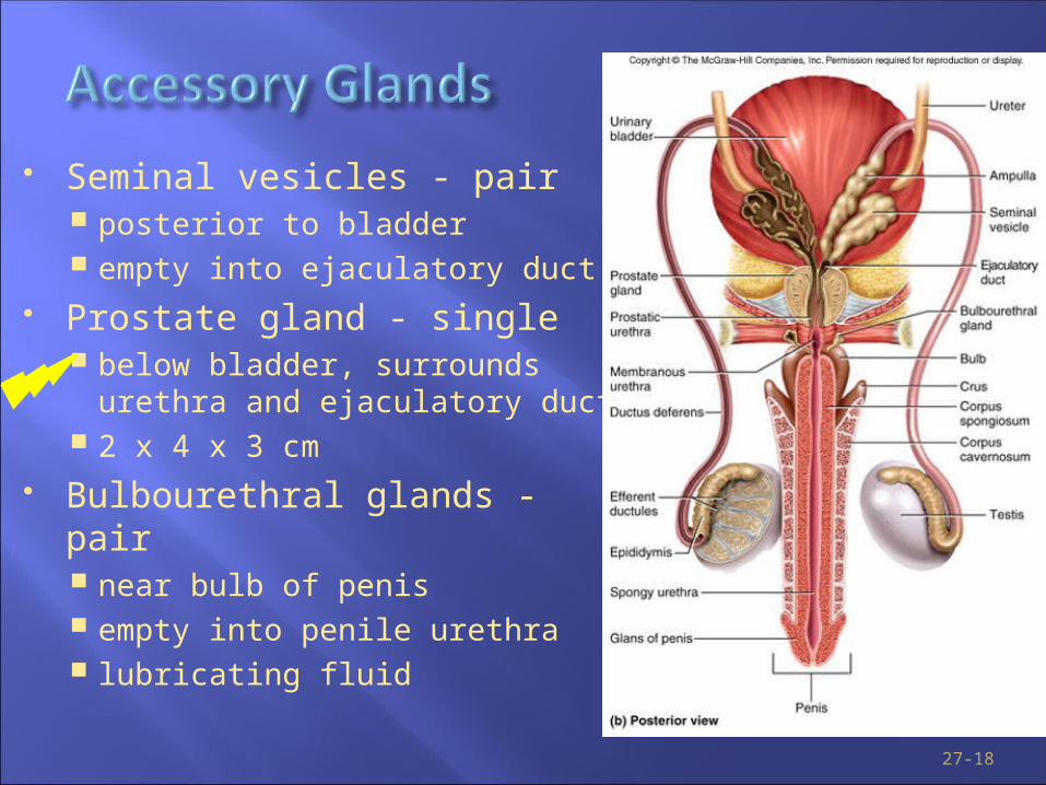
Seminal vesicles - pair posterior to bladder empty into ejaculatory duct
Prostate gland - single below bladder, surrounds urethra
and ejaculatory duct 2 x 4 x 3 cm
Bulbourethral glands - pair near bulb of penis empty into penile urethra lubricating fluid
27-18

Internal root, shaft, and glans external portion 4 in. long when flaccid
The foreskin is termed the prepuce 3 cylindrical bodies of erectile tissue
corpus spongiosum along ventral side of penis encloses penile urethra
corpora cavernosa diverge like arms of a Y Erection
27-19

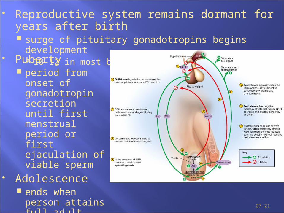
Hypothalamus produces GnRH Stimulates anterior pituitary (gonadotrope
cells) to secrete LH
stimulates interstitial cells to produce testosterone FSH
stimulates sustentacular cells (Sertoli cells) to secrete androgen-binding protein (ABP) that interacts with testosterone to stimulate spermatogenesis
27-20
Reproductive system remains dormant for years after birth surge of pituitary gonadotropins begins development
10-12 in most boys; 8-10 in most girls
27-21
Puberty period from onset of
gonadotropin secretion until first menstrual period or first ejaculation of viable sperm
Adolescence ends when person
attains full adult height

Enlargement of secondary sexual organs penis, testes, scrotum, ducts, glands
Development of secondary sexual characteristics hair, scent and sebaceous glands develop muscle mass, vocal quality stimulates erythropoiesis and libido
During adulthood, testosterone sustains libido, spermatogenesis and reproductive tract
27-22
Enlargement of secondary sexual organs penis, testes, scrotum, ducts, glands
Development of secondary sexual characteristics hair, scent and sebaceous glands develop muscle mass, vocal quality stimulates erythropoiesis and libido
During adulthood, testosterone sustains libido, spermatogenesis and reproductive tract
27-23

Mitosis produces two genetically identical daughter cells (for tissue repair, embryonic growth)
Meiosis produces gametes for sexual reproduction 2 cell divisions (only one
replication of DNA) meiosis I separates
homologous chromosome pairs into 2 haploid cells
meiosis II separates duplicated sister chromatids into 4 haploid cells
27-24

Blood-testis barrier is formed by tight junctions between and basement membrane under sustentacular cells (Sertoli cells)
27-25
1 basal lamina, 2 spermatogonia, 3 spermatocyte 1st order, 4 spermatocyte 2nd order, 5 spermatid, 6 mature spermatid, 7 Sertoli cell, 8 tight junction (blood testis barrier)

Spermatogonia produce 2 kinds of daughter cells type A remain outside blood-testis
barrier and produce more daughter cells until death
type B differentiate into primary spermatocytes cells must pass through
BTB to move inward toward lumen - new tight junctions form behind these cells
meiosis I 2 secondary spermatocytes
meiosis II 4 spermatids
27-26

Changes that transform spermatids into spermatozoa discarding excess cytoplasm and growing tails
27-27

Head is pear-shaped front end 4 to 5 microns long structure
containing the nucleus, acrosome and basal body of the tail flagella nucleus contains haploid set of
chromosomes acrosome contains enzymes that
penetrate the egg basal body
27-28

Tail is divided into 3 regions midpiece contains mitochondria
around axoneme of the flagella (produce ATP for flagellar movement)
principal piece is axoneme surrounded by fibers
endpiece is very narrow tip of flagella
27-29

2-5 mL of fluid expelled during orgasm 60% seminal vesicle fluid, 30% prostatic, 10% sperm normal sperm count 50-120 million/mL
Other components of semen fructose - energy for sperm motility fibrinogen causes clotting
enzymes convert fibrinogen to fibrin fibrinolysin liquefies semen within 30 minutes prostaglandins stimulate female peristaltic contractions spermine is a base stabilizing sperm pH at 7.2 to 7.6
27-30

27-31
Arteries of penis dorsal and deep arteries (branches of internal pudendal) deep artery supplies lacunae of corpora cavernosa
dilation fills lacunae causing an erection normal penile blood supply comes from dorsal artery
Nerves of penis abundance of tactile, pressure and temperature receptors dorsal nerve of penis and internal pudendal nerves lead to
integrating center in sacral spinal cord both autonomic and somatic motor fibers carry impulses
from integrating center to penis
27-32
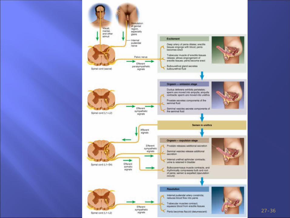
Excitement is characterized by vasocongestion of genitals, myotonia, and increases in heart rate, blood pressure and pulmonary ventilation
Initiated by many different erotic stimuli Erection of penis is due to parasympathetic triggering
of nitric oxide (NO) secretion dilation of deep arteries and filling of lacunae with blood
Erection is maintained during plateau phase
27-33
Climax (orgasm) is 15 second reaction that typically includes the discharge of semen (ejaculation)
Ejaculation has two stages emission = sympathetic nervous system propels sperm
through ducts as glandular secretions are added expulsion = semen in urethra activates muscular
contractions that lead to expulsion
27-34
Sympathetic signals constrict internal pudendal artery and reduce blood flow to penis penis becomes soft and flaccid (detumescence)
Cardiovascular and respiratory responses return to normal
Refractory period (10 minutes to few hours)

27-35
27-36