#-protein folding: discrete molecular dynamics study
TRANSCRIPT
Subscriber access provided by DREXEL UNIV
Journal of the American Chemical Society is published by the American ChemicalSociety. 1155 Sixteenth Street N.W., Washington, DC 20036
Article
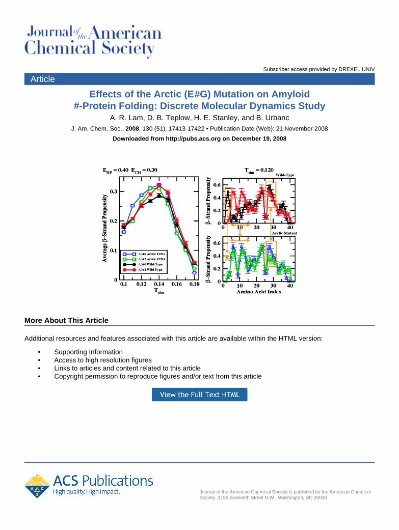
Effects of the Arctic (E22
#G) Mutation on Amyloid#-Protein Folding: Discrete Molecular Dynamics Study
A. R. Lam, D. B. Teplow, H. E. Stanley, and B. UrbancJ. Am. Chem. Soc., 2008, 130 (51), 17413-17422 • Publication Date (Web): 21 November 2008
Downloaded from http://pubs.acs.org on December 19, 2008
More About This Article
Additional resources and features associated with this article are available within the HTML version:
• Supporting Information• Access to high resolution figures• Links to articles and content related to this article• Copyright permission to reproduce figures and/or text from this article
Effects of the Arctic (E22fG) Mutation on Amyloid �-ProteinFolding: Discrete Molecular Dynamics Study
A. R. Lam,*,§,# D. B. Teplow,† H. E. Stanley,§ and B. Urbanc‡,§
Center for Polymer Studies, Physics Department, Boston UniVersity, Boston, Massachusetts 02215,Department of Neurology, DaVid Geffen School of Medicine, and Molecular Biology Institute and
Brain Research Institute, UniVersity of California, Los Angeles, California 90095, and Department ofPhysics, Drexel UniVersity, Philadelphia, PennsylVania 19104
Received July 9, 2008; E-mail: [email protected]; [email protected]
Abstract: The 40–42 residue amyloid �-protein (A�) plays a central role in the pathogenesis of Alzheimer’sdisease (AD). Of the two main alloforms, A�40 and A�42, the longer A�42 is linked particularly strongly toAD. Despite the relatively small two amino acid length difference in primary structure, in vitro studiesdemonstrate that A�40 and A�42 oligomerize through distinct pathways. Recently, a discrete moleculardynamics (DMD) approach combined with a four-bead protein model recapitulated the differences in A�40and A�42 oligomerization and led to structural predictions amenable to in vitro testing. Here, the sameDMD approach is applied to elucidate folding of A�40, A�42, and two mutants, [G22]A�40 and [G22]A�42,which cause a familial (“Arctic”) form of AD. The implicit solvent in the DMD approach is modeled by aminoacid-specific hydropathic and electrostatic interactions. The strengths of these effective interactions arechosen to best fit the temperature dependence of the average �-strand content in A�42 monomer, asdetermined using circular dichroism (CD) spectroscopy. In agreement with these CD data, we show thatat physiological temperatures, the average �-strand content in both alloforms increases with temperature.Our results predict that the average �-strand propensity should decrease in both alloforms at temperatureshigher than ∼370 K. At physiological temperatures, both A�40 and A�42 adopt a collapsed-coil conformationwith several short �-strands and a small (<1%) amount of R-helical structure. At slightly above physiologicaltemperature, folded A�42 monomers display larger amounts of �-strand than do A�40 monomers. Atincreased temperatures, more extended conformations with a higher amount of �-strand (j30%) structureare observed. In both alloforms, a �-hairpin at A21-A30 is a central folding region. We observe threeadditional folded regions: structure 1, a �-hairpin at V36-A42 that exists in A�42 but not in A�40; structure2, a �-hairpin at R5-H13 in A�42 but not in A�40; and structure 3, a �-strand A2-F4 in A�40 but not A�42.At physiological temperatures, the Arctic mutation, E22G, disrupts contacts in the A21-A30 region of both[G22]A� peptides, resulting in a less stable main folding region relative to the wild type peptides. TheArctic mutation induces a significant structural change at the N-terminus of [G22]A�40 by preventing theformation of structure 3 observed in A�40 but not A�42, thereby reducing the structural differences between[G22]A�40 and [G22]A�42 at the N-terminus. [G22]A�40 is characterized by a significantly increased amountof average �-strand relative to the other three peptides due to an induced �-hairpin structure at R5-H13,similar to structure 2. Consequently, the N-terminal folded structure of the Arctic mutants closely resemblesthe N-terminal structure of A�42, suggesting that both Arctic A� peptides might assemble into structuressimilar to toxic A�42 oligomers.
1. Introduction
Alzheimer’s disease (AD) is a progressive neurodegenerativedisorder that is characterized pathologically by extensiveneuronal loss and the accumulation of extracellular senileplaques and intracellular neurofibrillary tangles. Senile plaquescontain fibrillar aggregates of the amyloid �-protein (A�). A�is produced through cleavage of the amyloid precursor protein(APP) and is normally present in the body predominantly intwo alloforms, A�40 and A�42, that differ structurally by the
absenceorpresenceof twoC-terminalaminoacids, respectively.1,2
An important hypothesis of disease causation, strongly supportedby genetic and experimental evidence, posits that A� oligomers,rather than fibrils, are the proximate neurotoxic agents in AD.3
In particular, A�42 oligomers appear to be the most toxic A�assemblies.4 The linkage of A� oligomerization to AD makesimperative the detailed elucidation of the oligomerizationprocess. Unfortunately, the A� system is remarkably complex
# Also affiliated with Department of Chemistry, 420 Chemical Sciences,University of California, Riverside, CA 92521.
§ Boston University.† University of California, Los Angeles.‡ Drexel University.
(1) Hardy, J.; Selkoe, D. J. Science 2002, 297, 353–356.(2) Hardy, J. Neurobiol. Aging 2002, 23, 1073–1074.(3) Roychaudhuri, R.; Yang, M.; Hoshi, M. M.; Teplow, D. B. J. Biol.
Chem. 2008, doi: 10.1074/jbc.R800036200.(4) Dahlgren, K. N.; Manelli, A. M.; Stine, W. B.; Baker, L. K.; Krafft,
G. A.; LaDu, M. J. J. Biol. Chem. 2002, 277 (35), 32046–32053.
Published on Web 11/21/2008
10.1021/ja804984h CCC: $40.75 2008 American Chemical Society J. AM. CHEM. SOC. 2008, 130, 17413–17422 9 17413
in its conformational and assembly dynamics.3,5 This hascomplicated the application of classical structure determinationmethods such as X-ray crystallography and solution state NMRto the oligomerization question. One approach that has providedinformation on the initial self-association of A� has been insitu chemical cross-linking (for a review, see ref 6). Thisapproach allowed Bitan et al.7 to determine quantitatively theA� oligomer size frequency distribution, which demonstratedthat A�40 and A�42 exhibit different oligomerization pathways.A�42 assembled into pentamer/hexamer units (paranuclei) andmultiples of paranuclei, while A�40 only formed dimers throughtetramers in equilibrium with monomers. However, the resolu-tion of the method was insufficient to reveal the interatomicinteractions controlling the oligomerization processes.
In Vitro studies showed that A�40 and A�42 monomersadopted a predominantly R-helical structure in a membrane-mimicking environment,8,9 while a collapsed coil structure wasreported for A�(10–35) in an aqueous solution.10 A� foldedstructure clearly depends on the solvent. Earlier studies of A�40using a mixture of trifluoroethanol and water demonstrated asubstantial amount of R-helical structure.11,12 Initial studies oftemperature dependence of the secondary structure of A�40 inaqueous solution demonstrated that �-strand propensity increasedwith temperature.13 Using CD spectroscopy on both A�40 andA�42 monomers in aqueous solution, Lim et al. recentlydemonstrated14 that the average �-strand structure increased withtemperature, in agreement with Gursky and Aleshkov,13 withA�42 monomers having a slightly higher amount of average�-strand structure than A�40 monomers, suggesting that the twoalloforms are characterized by differences in folded structures.
Knowledge-based therapeutic drug design requires the defini-tion of target structures at atomic resolution. In silico approachesprovide a powerful means to achieve this goal. Several in silicostudies addressed folding of A�42,15,16 A�39,17 A�(10–35),18,19
A�(25–35),20 A�(1–28),21 and A�(21–30) decapeptide.22-27 The
latter was hypothesized to nucleate monomer folding.28,29
Replica-exchange all-atom molecular dynamics (MD) study ofA�42 monomer in implicit water by Baumketner et al. showedthree distinct families of folded structures, all dominated by turnsand loops with a small amount of R-helical structure at theC-terminus.16 Using similar all-atom MD approach, Anand etal. found that at room temperature, the A�(1–39) monomer didnot have a unique structure but rather three distinct families ofmostly collapsed coil-like structures existed.17 Using all-atomMD in explicit water, Massi et al. demonstrated that althoughA�(10–35) is somewhat disordered in water, the centralhydrophobic cluster, L17-A21, and the turn region, V24-N27,were particularly stable.18,19 Wei et al.20 used replica-exchangeMD in pure water and HFIP/water cosolvent to demonstratethat A�(25–35) preferentially populated an R-helical structurein apolar organic solvent, while in water, a collapsed coil, andto a lesser extent �-hairpin conformations, were observed. Donget al.21 explored the energy landscape of A� (1–28) monomersand concluded that the monomer was predominantly in acollapsed coil conformation with a non-negligible �-strandstructure at the N-terminus.
An ab initio DMD approach using a four-bead protein modelwith backbone hydrogen bonding in implicit solvent recentlydemonstrated that despite relatively small differences in theprimary structure, A�40 and A�42 not only followed differentoligomerization pathways but also folded differently, with A�42displaying a turn structure centered at G37-G38 that was notpresent in A�40 monomer.30 This structural difference betweenA�40 and A�42 was corroborated by several independent inVitro studies.28,31,32 In addition, a combined MD/NMR studyconfirmed that A�42 monomer was more structured at theC-terminus than A�40.33 Using the same DMD approach asthe initial study by Urbanc et al.,30 Lam et al. showed that onlyselected regions of A�42 had a well-defined folded structureand that the average amount of �-strand increased withtemperature,34 consistent with in Vitro findings.13 Because A�40and A�42 were shown to oligomerize through distinct pathwaysin Vitro7 and in silico,30 the present study is based on ahypothesis that different oligomerization pathways leading todistinct effects in ViVo are a consequence of folding differencesbetween A�40 and A�42. We employ the DMD approach with
(5) Teplow, D. B. Methods Enzymol. 2006, 413, 20–33.(6) Bitan, G.; Teplow, D. B. Acc. Chem. Res. 2004, 37, 357–364.(7) Bitan, G.; Kirkitadze, M. D.; Lomakin, A.; Vollers, S. S.; Benedek,
G. B.; Teplow, D. B. Proc. Natl. Acad. Sci. USA 2003, 100, 330–335.
(8) Coles, M.; Bicknell, W.; Watson, A. A.; Fairlie, D. P.; Craik, D. J.Biochemistry 1998, 37, 11064–11077.
(9) Crescenzi, O.; Tomaselli, S.; Guerrini, R.; Salvatori, S.; D’Ursi, A. M.;Temussi, P. A.; Picone, D. Eur. J. Biochem. 2002, 269, 5642–5648.
(10) Zhang, S.; Iwata, K.; Lachenmann, M. J.; Peng, J. W.; Li, S.; Stimson,E. R.; Lu, Y. A.; Felix, A. M.; Maggio, J. E.; Lee, J. P. J. Struct.Biol. 2000, 130 (1), 130–141.
(11) Sticht, H.; Bayer, P.; Willbold, D.; Dames, S.; Hilbich, C.; Beyreuther,K.; Frank, R.; Rosch, P. Eur. J. Biochem. 1995, 233, 293–298.
(12) Fezoui, Y.; Teplow, D. B. J. Biol. Chem. 2002, 277, 36948–36954.(13) Gursky, O.; Aleshkov, S. Biochim. Biophys. Acta 2000, 1476, 93–
102.(14) Lim, K. H.; Collver, H. H.; Le, Y. T. H.; Nagchowdhuri, P.; Kenney,
J. M. Biochem. Biophys. Res. Commun. 2006, 353, 443–449.(15) Bernstein, S. L.; Wyttenbach, T.; Baumketner, A.; Shea, J.-E.; Bitan,
G.; Teplow, D. B.; Bowers, M. T. J. Am. Chem. Soc. 2005, 127, 2075–2084.
(16) Baumketner, A.; Bernstein, S. L.; Wyttenbach, T.; Bitan, G.; Teplow,D. B.; Bowers, M. T.; Shea, J.-E. Protein Sci. 2006, 15, 420–428.
(17) Anand, P.; Nadel, F. S.; Hansmann, U. H. E. J. Chem. Phys. 2008,128 (1–5), 165102.
(18) Massi, F.; Peng, J. W.; Lee, J. P.; Straub, J. E. Biophys. J. 2001, 80,31–44.
(19) Massi, F.; Straub, J. E. Biophys. J. 2001, 81, 697–709.(20) Wei, G. H.; Shea, J. E. Biophys. J. 2006, 91 (5), 1638–1647.(21) Dong, X.; Chen, W.; Mousseau, N.; Derreumaux, P. J. Chem. Phys.
2008, 128 (1–10), 125108.(22) Borreguero, J. M.; Urbanc, B.; Lazo, N.; Buldyrev, S. V.; Teplow,
D. B.; Stanley, H. E. Proc. Natl. Acad. Sci. USA 2005, 102, 6015–6020.
(23) Cruz, L.; Urbanc, B.; Borreguero, J. M.; Lazo, N. D.; Teplow, D. B.;Stanley, H. E. Proc. Natl. Acad. Sci. USA 2005, 102, 18258–18263.
(24) W. Chen, N. M.; Derreumaux, P. J. Chem. Phys. 2006, 125 (8),084911.
(25) Baumketner, A.; Bernstein, S. L.; Wyttenbach, T.; Lazo, N. D.;Teplow, D. B.; Bowers, M. T.; Shea, J.-E. Protein Sci 2006, 15, 1239–1247.
(26) Krone, M. G.; Baumketner, A.; Bernstein, S. L.; Wyttenbach, T.; Lazo,N. D.; Teplow, D. B.; Bowers, M. T.; Shea, J.-E. J. Mol. Biol. 2008,
(27) Tarus, B.; Straub, J. E.; Thirumalai, D. J. Mol. Biol. 2008, 379 (4),815–829.
(28) Lazo, N. D.; Grant, M. A.; Condron, M. C.; Rigby, A. C.; Teplow,D. B. Protein Sci. 2005, 14 (6), 1581–1596.
(29) Grant, M. A.; Lazo, N. D.; Lomakin, A.; Condron, M. M.; Arai, H.;Yamin, G.; Rigby, A. C.; Teplow, D. B. Proc. Natl. Acad. Sci. USA2007, 104 (42), 16522–16527.
(30) Urbanc, B.; Cruz, L.; Yun, S.; Buldyrev, S. V.; Bitan, G.; Teplow,D. B.; Stanley, H. E. Proc. Natl. Acad. Sci. USA 2004, 101, 17345–17350.
(31) Murakami, K.; Irie, K.; Ohigashi, H.; Hara, H.; Nagao, M.; Shimizu,T.; Shirasawa, T. J. Am. Chem. Soc. 2005, 127 (43), 15168–15174.
(32) Yan, Y.; Wang, C. J. Mol. Biol. 2006, 364 (5), 853–862.(33) Sgourakis, N. G.; Yan, Y.; McCallum, S. A.; Wang, C.; Garcia, A. E.
J. Mol. Biol. 2007, 368, 1448–1457.(34) Lam, A.; Urbanc, B.; Borreguero, J.; Lazo, N.; Teplow, D.; Stanley,
H. E. Proceedings of 2006 International Conference on Bioinformatics& Computational Biology 2006, 1, 322–328.
17414 J. AM. CHEM. SOC. 9 VOL. 130, NO. 51, 2008
A R T I C L E S Lam et al.
implicit solvent parameters mimicking the in Vitro aqueoussolution14 to elucidate all structural differences between A�40and A�42 monomers at different temperatures. Because theArctic mutation, E22fG, is associated with the familiar formof AD with distinct pathology relative to the sporadic AD, weexamine also the effects of the Arctic mutation on foldedstructures of both isoforms and discuss the implications of ourfindings for understanding A� isoform-specific folding and itsrelationship to AD.
2. Methods
2.1. Discrete Molecular Dynamics. Zhou et al. proposed theidea of applying discrete molecular dynamics (DMD) combinedwith a simplified protein model to study protein folding.35 Sincethen, many groups have implemented this approach to investigateprotein folding mechanisms.36-40 In DMD, all interparticle interac-tions are modeled by square-well and step-like potentials. Particlesmove with constant speeds along straight lines. When two particlesreach a distance at which the potential is discontinuous, a collisionoccurs. The pair of particles with the shortest collision time ischosen as the next collision event and the new positions andvelocities of the two particles involved are calculated based onconservation laws for the linear momentum, angular momentum,and total energy. The advantage of DMD is that the numericalintegration of Newton’s second law equations is avoided, resultingin a substantial decrease in computational burden. This makes theDMD approach much faster than all-atom MD with continuousinterparticle potentials.
2.2. Four-Bead Protein Model and Interactions. We use afour-bead protein model,41-43 in which up to four beads are usedto represent an amino acid. Three beads are used to model thebackbone groups N, CR, and C ′. The fourth bead represents theside chain centered at the C� group. Only glycine lacks the C� beadand is thus modeled by three beads only. Adjacent beads areconnected to each other through covalent or peptide bonds, whichare modeled as square well potentials with infinite walls but offinite width corresponding to ∼2% variability in covalent/peptidebond lengths. In addition to covalent and peptide bonds, constraintsare implemented to ensure the proper geometry of the proteinbackbone. These constraints are modeled in the same ways as thebonds. All lengths of bonds and constraints are based on statisticalproperties derived from the protein database of known proteinstructures.44
The backbone hydrogen bond was introduced into the four beadmodel to account for the R-helical and �-strand secondary struc-ture.43 The bond is introduced between the Ni bead of amino acidi and C′ j bead of amino acid j. For a hydrogen bond between Ni
and C′ j to form, these two beads need to be at a distance <4.2 Å.In addition, auxiliary bonds between the two amino acids involvedare introduced to account for the particular backbone geometryallowing hydrogen bond formation. The absolute value of the
potential energy associated with formation of a single hydrogenbond, EHB, represents a unit of energy in our approach. Thesimulation temperature Tsim is expressed in units of EHB/kB, wherekB is the Boltzmann constant.
The model implements amino acid-specific interactions betweentwo side chain beads due to effective hydropathy30 and charge.34,44
The side chain bead of each amino acid is characterized by aneffective hydropathy following the Kyte and Dolittle scale.45
Because the solvent is not explicitly present in the model, effectiveattractive interactions between two hydrophobic, and repulsiveinteractions between two hydrophilic, side chain beads areintroduced.30,44 The strength of the effective hydropathic interac-tions as given by the absolute value of the potential energy betweentwo isoleucines EHP (relative to the energy unit EHB) is the firstinteraction parameter. A double square-well potential is applied tomodel the effective electrostatic interactions between two chargedside chain beads.44 The maximal absolute value of the potentialenergy between two charged side chain beads ECH (relative to theenergy unit EHB) is the second interaction parameter. Both interac-tion parameters EHP and ECH strongly depend on and need to beadjusted to the particular solvent.
2.3. Secondary Structure Analysis. The secondary structurepropensities of each amino acid were calculated using the STRIDEprogram46 within the Visual Molecular Dynamics (VMD) softwarepackage.47 The secondary structure propensities included R-helical,�-strand, turn, and random coil per amino acid. We calculated theaverage �-strand and R-helix propensities, ⟨�⟩ and ⟨R⟩, by averagingthe �-strand/R-helix propensity over all amino acids at a giventemperature and interaction parameters (EHP, ECH).
2.4. Intramolecular Contact Map. We determined the averageintramolecular contact frequency for each temperature and interac-tion parameter set (EHP, ECH). Two amino acids, i and j, wereconsidered to be in contact when the distance between them dij
e7.5 Å. The contact was counted with variable Cij that was definedas the average number of contact pairs between amino acids i andj from different trajectories. Because each amino acid had up tofour beads, the maximum number of contacts Cij (between any twoamino acids) was 16. We normalized the contact maps to the samemaximum value.
3. Results
The DMD approach employed here has been described indetail by Urbanc et al.44 In earlier studies, DMD combined witha four-bead amino acid model, and considering backbonehydrogen bonding only, resulted in �-hairpin monomer andplanar dimer conformations.48 Introducing amino acid-specificinteractions due to hydropathy into the four-bead model enabledthe successful in silico reproduction of experimentally observed7
oligomerization differences between A�40 and A�42 andyielded new structural predictions amenable to in Vitro testing.30
This same study indicated that alloform-specific differencesalready existed at the stage of monomer folding. In particular,the turn structure centered at G37-G38 was present in a foldedA�42 monomer but not in a folded A�40 monomer and wasassociated with the first contacts that formed during monomerfolding. Yun et al.,49 using the same DMD approach, showedthat electrostatic interactions promote formation of largeroligomers in both A�40 and A�42 while preserving the
(35) Zhou, Y.; Hall, C. K.; Karplus, M. Phys. ReV. Lett. 1996, 77, 2822–2825.
(36) Zhou, Y. Q.; Karplus, M. Proc. Natl. Acad. Sci. USA 1997, 94, 14429–14432.
(37) Zhou, Y. Q.; Karplus, M.; Wichert, J. M.; Hall, C. K. J. Chem. Phys.1997, 107, 10691–10708.
(38) Dokholyan, N. V.; Buldyrev, S. V.; Stanley, H. E.; Shakhnovich, E. I.Folding Design 1998, 3, 577–587.
(39) Zhou, Y. Q.; Karplus, M. J. Mol. Biol. 1999, 293, 917–951.(40) Dokholyan, N. V.; Buldyrev, S. V.; Stanley, H. E.; Shakhnovich, E. I.
J. Mol. Biol. 2000, 296, 1183–1188.(41) Smith, A. V.; Hall, C. K. Proteins: Struct. Funct. Genet. 2001, 4,
344–360.(42) Smith, A. V.; Hall, C. K. J. Mol. Biol. 2001, 312, 187–202.(43) Ding, F.; Borreguero, J. M.; Buldyrev, S. V.; Stanley, H. E.;
Dokholyan, N. V. Proteins: Struct. Funct. Genet. 2003, 53, 220–228.(44) Urbanc, B.; Borreguero, J. M.; Cruz, L.; Stanley, H. E. Methods
Enzymol. 2006, 412, 314–338.
(45) Kyte, J.; Doolittle, R. F. J. Mol. Biol. 1982, 157, 105–132.(46) Heinig, M.; Frishman, D. Nucleic Acids Res. 2004, 32, W500–W502.(47) Humphrey, W.; Dalke, A.; Schulten, K. J. Mol. Graphics 1996, 14,
33–38.(48) Urbanc, B.; Cruz, L.; Ding, F.; Sammond, D.; Khare, S.; Buldyrev,
S.; Stanley, H. E.; Dokholyan, N. V. Biophys. J. 2004, 87 (4), 2310–2321.
(49) Yun, S. J.; Urbanc, B.; Cruz, L.; Bitan, G.; Teplow, D. B.; Stanley,H. E. Biophys. J. 2007, 92 (11), 4064–4077.
J. AM. CHEM. SOC. 9 VOL. 130, NO. 51, 2008 17415
Amyloid �-Protein Folding A R T I C L E S
differences between the two alloforms at both the folding andoligomerization stages of assembly. Lam et al. applied thisapproach in studies of the temperature dependence of A�42folding, showing that a collapsed coil conformation at lowtemperatures converts to a more extended, �-strand-rich con-formation at higher temperatures.34 Interestingly, while severalregions of A�42 consistently exhibit a temperature-dependentfold, significant variability of folded structures was found ateach temperature. The coexistence of a multitude of monomerconformations is characteristic of naturally unfolded proteins.Here we explore more deeply the differences between the full-length A�40 and A�42 peptides. We first describe a techniquethat allows us to determine the implicit solvent parameters thatbest match experimental conditions. We then use these param-eters to explore not only the folding of wild type A�40 andA�42 but also of two clinically relevant mutant alloforms, Arctic[G22]A�40 and [G22]A�42.
The primary structure of A�42 differs from A�40 by twoadditional amino acids of A�42, I and A, at the C-terminus.The sequence of A�42 is DAEFRHDSGYEVHHQK16LVF-FAE22DVGSNKGA30IIGLMV36GGVV40IA. We refer to thesegments (L17-A21) and (I31-V36) as the central hydrophobiccluster (CHC) and the mid-hydrophobic region (MHR), respec-tively. We define the C-terminal region (CTR) as the segmentV39-V40 in A�40 or V39-V42 in A�42.
The energy unit is set to the absolute value of the hydrogenbond potential energy, EHB ) 1.0 and the simulation temperatureTsim is expressed in units of EHB/kB. We explore folding at fourdifferentstrengthsofhydropathicinteractions,EHP)0.1, 0.2, 0.3, 0.4,and three different strengths of EIs, ECH ) 0, 0.15, 0.3. For eachset of these effective interaction parameters (EHP, ECH), we firstperform DMD simulations of a monomer at a high temperature(Tsim ) 4) to obtain 100 distinct random coil-like initialconformations to be used in production runs. For giveninteraction parameters (EHP, ECH), we simulate 100 trajectoriesspanning a temperature range of [0.10, 0.18].
3.1. Interaction Parameters for Aqueous A� Folding. In anaqueous environment, hydrophobic and hydrophilic effects playkey roles in protein folding and assembly. In the DMD approach,
we model hydropathic effects by a single-well attractive/repulsive potential between two side-chain beads. The strengthof the effective hydropathic interactions, EHP, is by definitionequal to the absolute value of the potential energy between twoI residues at a distance of <7.5 Å. Similarly, the strength ofeffective electrostatic interactions, ECH, is defined as the absolutevalue of the potential energy between two oppositely chargedside-chain atoms at a distance of <6 Å.
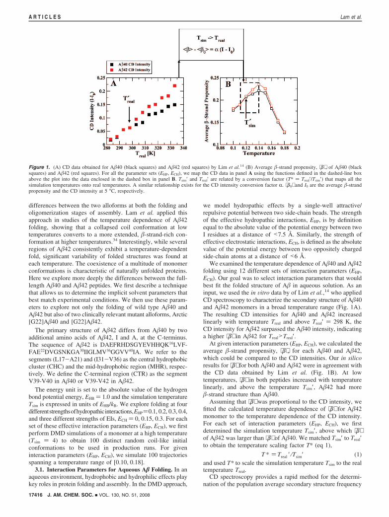
We examined the temperature dependence of A�40 and A�42folding using 12 different sets of interaction parameters (EHP,ECH). Our goal was to select interaction parameters that wouldbest fit the folded structure of A� in aqueous solution. As aninput, we used the in Vitro data by of Lim et al.,14 who appliedCD spectroscopy to characterize the secondary structure of A�40and A�42 monomers in a broad temperature range (Fig. 1A).The resulting CD intensities for A�40 and A�42 increasedlinearly with temperature Treal and above Treal′ ) 298 K, theCD intensity for A�42 surpassed the A�40 intensity, indicatinga higher ⟨�⟩ in A�42 for Treal>Treal′.
At given interaction parameters (EHP, ECH), we calculated theaverage �-strand propensity, ⟨�⟩ , for each A�40 and A�42,which could be compared to the CD intensities. Our in silicoresults for ⟨�⟩ for both A�40 and A�42 were in agreement withthe CD data obtained by Lim et al. (Fig. 1B). At lowtemperatures, ⟨�⟩ in both peptides increased with temperaturelinearly, and above the temperature Tsim′, A�42 had more�-strand structure than A�40.
Assuming that ⟨�⟩ was proportional to the CD intensity, wefitted the calculated temperature dependence of ⟨�⟩ for A�42monomer to the temperature dependence of the CD intensity.For each set of interaction parameters (EHP, ECH), we firstdetermined the simulation temperature Tsim′, above which ⟨�⟩of A�42 was larger than ⟨�⟩ of A�40. We matched Tsim′ to Treal′to obtain the temperature scaling factor T* (eq 1),
T * ) Treal ′ ⁄Tsim′ (1)and used T* to scale the simulation temperature Tsim to the realtemperature Treal.
CD spectroscopy provides a rapid method for the determi-nation of the population average secondary structure frequency
Figure 1. (A) CD data obtained for A�40 (black squares) and A�42 (red squares) by Lim et al.14 (B) Average �-strand propensity, ⟨�⟩ , of A�40 (blacksquares) and A�42 (red squares). For all the parameter sets (EHP, ECH), we map the CD data in panel A using the functions defined in the dashed-line boxabove the plot into the data enclosed in the dashed box in panel B. Tsim′ and Treal′ are related by a conversion factor (T* ) Treal′/Tsim′) that maps all thesimulation temperatures onto real temperatures. A similar relationship exists for the CD intensity conversion factor R. ⟨�0⟩ and I0 are the average �-strandpropensity and the CD intensity at 5 °C, respectively.
17416 J. AM. CHEM. SOC. 9 VOL. 130, NO. 51, 2008
A R T I C L E S Lam et al.
distribution. In a recent paper, Greenfield suggested that a linearrelationship exists between CD intensity and the sum of thesecondary structure elements contributing to it.50 To relate theaverage �-strand propensity ⟨�⟩ derived from our simulationsto CD intensities ICD determined experimentally, we used thedata of Lim et al.14 These data were obtained at 222 nm, not at∼218 nm, where the minimum in �-strand ellipticity occurs.Nevertheless, because the �-strand is the dominant secondarystructure element in our A� monomer conformations andbecause θ218 ∝ θ 222, ⟨�⟩ will be proportional to CD intensity at222 nm. We thus relate the CD intensity data, ICD-I0 (the y-axisof Fig. 2C in ref 14), to the calculated ⟨�⟩ using the followingequation:
⟨� ⟩ -⟨�0 ⟩ )R(ICD - I0) (2)where ⟨�0⟩ was ⟨�⟩ at 5 °C (278 K) and I0 was the correspondingCD intensity at 5 °C. The scaling factor R was obtained byfitting ICD to ⟨�⟩ .
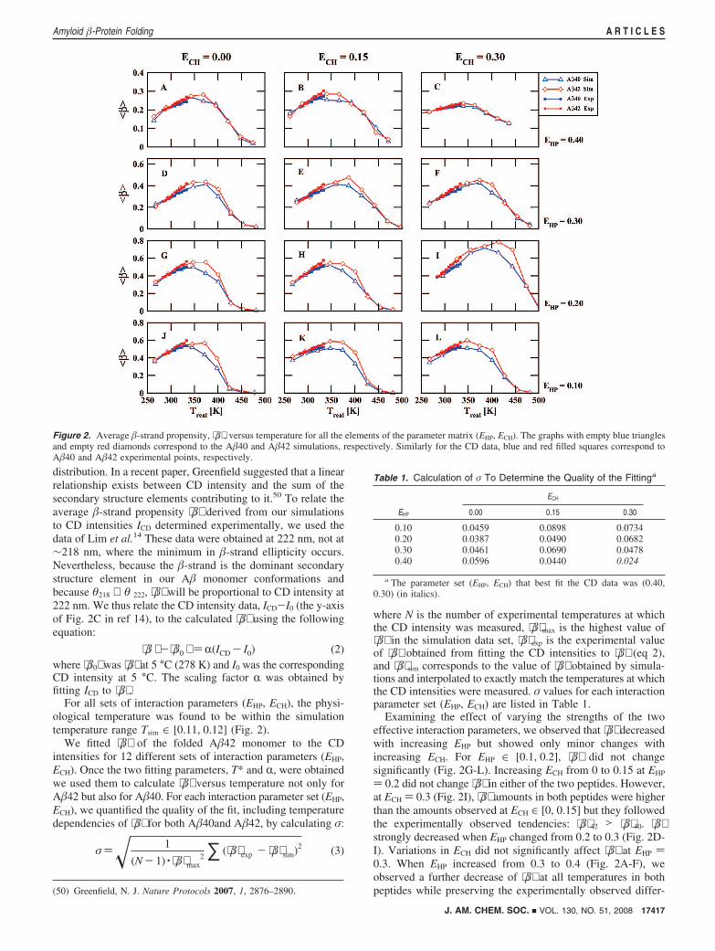
For all sets of interaction parameters (EHP, ECH), the physi-ological temperature was found to be within the simulationtemperature range Tsim ∈ [0.11, 0.12] (Fig. 2).
We fitted ⟨�⟩ of the folded A�42 monomer to the CDintensities for 12 different sets of interaction parameters (EHP,ECH). Once the two fitting parameters, T* and R, were obtainedwe used them to calculate ⟨�⟩ versus temperature not only forA�42 but also for A�40. For each interaction parameter set (EHP,ECH), we quantified the quality of the fit, including temperaturedependencies of ⟨�⟩ for both A�40and A�42, by calculating σ:
σ)� 1
(N- 1) · ⟨�⟩max2 ∑ (⟨�⟩exp - ⟨�⟩ sim)2 (3)
where N is the number of experimental temperatures at whichthe CD intensity was measured, ⟨�⟩max is the highest value of⟨�⟩ in the simulation data set, ⟨�⟩exp is the experimental valueof ⟨�⟩ obtained from fitting the CD intensities to ⟨�⟩ (eq 2),and ⟨�⟩ sim corresponds to the value of ⟨�⟩ obtained by simula-tions and interpolated to exactly match the temperatures at whichthe CD intensities were measured. σ values for each interactionparameter set (EHP, ECH) are listed in Table 1.
Examining the effect of varying the strengths of the twoeffective interaction parameters, we observed that ⟨�⟩ decreasedwith increasing EHP but showed only minor changes withincreasing ECH. For EHP ∈ [0.1, 0.2], ⟨�⟩ did not changesignificantly (Fig. 2G-L). Increasing ECH from 0 to 0.15 at EHP
) 0.2 did not change ⟨�⟩ in either of the two peptides. However,at ECH ) 0.3 (Fig. 2I), ⟨�⟩ amounts in both peptides were higherthan the amounts observed at ECH ∈ [0, 0.15] but they followedthe experimentally observed tendencies: ⟨�⟩42 > ⟨�⟩40. ⟨�⟩strongly decreased when EHP changed from 0.2 to 0.3 (Fig. 2D-I). Variations in ECH did not significantly affect ⟨�⟩ at EHP )0.3. When EHP increased from 0.3 to 0.4 (Fig. 2A-F), weobserved a further decrease of ⟨�⟩ at all temperatures in bothpeptides while preserving the experimentally observed differ-(50) Greenfield, N. J. Nature Protocols 2007, 1, 2876–2890.
Figure 2. Average �-strand propensity, ⟨�⟩ , versus temperature for all the elements of the parameter matrix (EHP, ECH). The graphs with empty blue trianglesand empty red diamonds correspond to the A�40 and A�42 simulations, respectively. Similarly for the CD data, blue and red filled squares correspond toA�40 and A�42 experimental points, respectively.
Table 1. Calculation of σ To Determine the Quality of the Fittinga
ECH
EHP 0.00 0.15 0.30
0.10 0.0459 0.0898 0.07340.20 0.0387 0.0490 0.06820.30 0.0461 0.0690 0.04780.40 0.0596 0.0440 0.024
a The parameter set (EHP, ECH) that best fit the CD data was (0.40,0.30) (in italics).
J. AM. CHEM. SOC. 9 VOL. 130, NO. 51, 2008 17417
Amyloid �-Protein Folding A R T I C L E S
ences in ⟨�⟩ between the two alloforms. At EHP ) 0.4 (Fig.2A-C), increase in ECH from 0 to 0.3 resulted in a decrease of⟨�⟩ at all temperatures in both peptides.
In the following, we used the interaction parameters that bestmatched the experimental data, EHP ) 0.4 and ECH ) 0.3, tocharacterize the structural differences in A�40 and A�42monomer folding at different temperatures. The physiologicaltemperature Treal ) 310 K was found to correspond to thesimulation temperature Tsim ) 0.124.
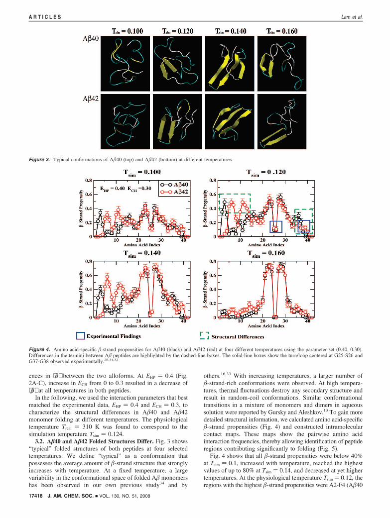
3.2. A�40 and A�42 Folded Structures Differ. Fig. 3 shows“typical” folded structures of both peptides at four selectedtemperatures. We define “typical” as a conformation thatpossesses the average amount of �-strand structure that stronglyincreases with temperature. At a fixed temperature, a largevariability in the conformational space of folded A� monomershas been observed in our own previous study34 and by
others.16,33 With increasing temperatures, a larger number of�-strand-rich conformations were observed. At high tempera-tures, thermal fluctuations destroy any secondary structure andresult in random-coil conformations. Similar conformationaltransitions in a mixture of monomers and dimers in aqueoussolution were reported by Gursky and Aleshkov.13 To gain moredetailed structural information, we calculated amino acid-specific�-strand propensities (Fig. 4) and constructed intramolecularcontact maps. These maps show the pairwise amino acidinteraction frequencies, thereby allowing identification of peptideregions contributing significantly to folding (Fig. 5).
Fig. 4 shows that all �-strand propensities were below 40%at Tsim ) 0.1, increased with temperature, reached the highestvalues of up to 80% at Tsim ) 0.14, and decreased at yet highertemperatures. At the physiological temperature Tsim ) 0.12, theregions with the highest �-strand propensities were A2-F4 (A�40
Figure 3. Typical conformations of A�40 (top) and A�42 (bottom) at different temperatures.
Figure 4. Amino acid-specific �-strand propensities for A�40 (black) and A�42 (red) at four different temperatures using the parameter set (0.40, 0.30).Differences in the termini between A� peptides are highlighted by the dashed-line boxes. The solid-line boxes show the turn/loop centered at G25-S26 andG37-G38 observed experimentally.28,31,32
17418 J. AM. CHEM. SOC. 9 VOL. 130, NO. 51, 2008
A R T I C L E S Lam et al.
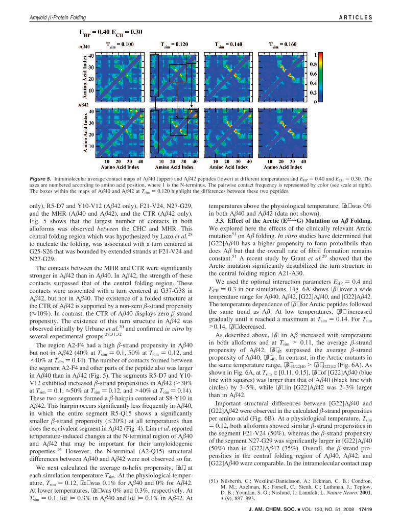
only), R5-D7 and Y10-V12 (A�42 only), F21-V24, N27-G29,and the MHR (A�40 and A�42), and the CTR (A�42 only).Fig. 5 shows that the largest number of contacts in bothalloforms was observed between the CHC and MHR. Thiscentral folding region which was hypothesized by Lazo et al.28
to nucleate the folding, was associated with a turn centered atG25-S26 that was bounded by extended strands at F21-V24 andN27-G29.
The contacts between the MHR and CTR were significantlystronger in A�42 than in A�40. In A�42, the strength of thesecontacts surpassed that of the central folding region. Thesecontacts were associated with a turn centered at G37-G38 inA�42, but not in A�40. The existence of a folded structure atthe CTR of A�42 is supported by a non-zero �-strand propensity(≈10%). In contrast, the CTR of A�40 displays zero �-strandpropensity. The existence of this turn structure in A�42 wasobserved initially by Urbanc et al.30 and confirmed in Vitro byseveral experimental groups.28,31,32
The region A2-F4 had a high �-strand propensity in A�40but not in A�42 (40% at Tsim ) 0.1, 50% at Tsim ) 0.12, and>40% at Tsim ) 0.14). The number of contacts formed betweenthe segment A2-F4 and other parts of the peptide also was largerin A�40 than in A�42 (Fig. 5). The segments R5-D7 and Y10-V12 exhibited increased �-strand propensities in A�42 (>30%at Tsim ) 0.1, ≈50% at Tsim ) 0.12, and >40% at Tsim ) 0.14).These two segments formed a �-hairpin centered at S8-Y10 inA�42. This hairpin occurs significantly less frequently in A�40,in which the entire segment R5-Q15 shows a significantlysmaller �-strand propensity (e20%) at all temperatures thandoes the equivalent segment in A�42 (Fig. 4). Lim et al. reportedtemperature-induced changes at the N-terminal region of A�40and A�42 that may be important for their amyloidogenicproperties.14 However, the N-terminal (A2-Q15) structuraldifferences between A�40 and A�42 were not observed so far.
We next calculated the average R-helix propensity, ⟨R⟩ , ateach simulation temperature Tsim. At the physiological temper-ature, Tsim ) 0.12, ⟨R⟩ was 0.1% for A�40 and 0% for A�42.At lower temperatures, ⟨R⟩ was 0% and 0.3%, respectively. AtTsim ) 0.1, ⟨R⟩ ) 0.3% in A�40 and ⟨R⟩ ) 0.1% in A�42. At
temperatures above the physiological temperature, ⟨R⟩ was 0%in both A�40 and A�42 (data not shown).
3.3. Effect of the Arctic (E22fG) Mutation on A� Folding.We explored here the effects of the clinically relevant Arcticmutation51 on A� folding. In Vitro studies have determined that[G22]A�40 has a higher propensity to form protofibrils thandoes A� but that the overall rate of fibril formation remainsconstant.51 A recent study by Grant et al.29 showed that theArctic mutation significantly destabilized the turn structure inthe central folding region A21-A30.
We used the optimal interaction parameters EHP ) 0.4 andECH ) 0.3 in our simulations. Fig. 6A shows ⟨�⟩ over a widetemperature range for A�40, A�42, [G22]A�40, and [G22]A�42.The temperature dependence of ⟨�⟩ for Arctic peptides followedthe same trend as A�. At low temperatures, ⟨�⟩ increasedgradually until it reached a maximum at Tsim ) 0.14. For Tsim
>0.14, ⟨�⟩ decreased.As described above, ⟨�⟩ in A� increased with temperature
in both alloforms and at Tsim > 0.11, the average �-strandpropensity of A�42, ⟨�⟩42 surpassed the average �-strandpropensity of A�40, ⟨�⟩40. In contrast, in the Arctic mutants inthe same temperature range, ⟨�⟩ [G22]40 > ⟨�⟩ [G22]42 (Fig. 6A). Asshown in Fig. 6A, at Tsim ∈ [0.11, 0.15], ⟨�⟩ of [G22]A�40 (blueline with squares) was larger than that of A�40 (black line withcircles) by 3–5%, while ⟨�⟩ in [G22]A�42 was 2–3% largerthan in A�42.
Important structural differences between [G22]A�40 and[G22]A�42 were observed in the calculated �-strand propensitiesper amino acid (Fig. 6B). At a physiological temperature, Tsim
) 0.12, both alloforms showed similar �-strand propensities inthe segment F21-V24 (50%), whereas the �-strand propensityof the segment N27-G29 was significantly larger in [G22]A�40(50%) than in [G22]A�42 (35%). Overall, the �-strand pro-pensities in the central folding region of A�40, A�42, and[G22]A�40 were comparable. In the intramolecular contact map
(51) Nilsberth, C.; Westlind-Danielsson, A.; Eckman, C. B.; Condron,M. M.; Axelman, K.; Forsell, C.; Stenh, C.; Luthman, J.; Teplow,D. B.; Younkin, S. G.; Naslund, J.; Lannfelt, L. Nature Neuro. 2001,4 (9), 887–893.
Figure 5. Intramolecular average contact maps of A�40 (upper) and A�42 peptides (lower) at different temperatures and EHP ) 0.40 and ECH ) 0.30. Theaxes are numbered according to amino acid position, where 1 is the N-terminus. The pairwise contact frequency is represented by color (see scale at right).The boxes within the maps of A�40 and A�42 at Tsim ) 0.120 highlight the differences between these two peptides.
J. AM. CHEM. SOC. 9 VOL. 130, NO. 51, 2008 17419
Amyloid �-Protein Folding A R T I C L E S

corresponding to the Arctic mutants, decreased numbers ofcontacts were observed in the central folding region of both[G22]A�40 and [G22]A�42 relative to A�40 and A�42 (Fig.7), suggesting that the Arctic mutation destabilizes the centralfolding region as also observed in Vitro for the decapeptide A�(21–30) by Grant et al.29
At the N-terminus, the �-strand propensity of the segmentR5-Q15 (Fig. 6B, top and bottom) was similar in [G22]A�40,[G22]A�42, and A�42, while in A�40, no significant �-strandpropensity was associated with this region. The �-strand
propensity of the segment A2-F4 was small (10%) and similarin both Arctic peptides, similar to A�42 but in contrast tothat same region in A�40, where the �-strand propensity was40–50%. The segments R5-D7 and Y10-V12 in [G22]A�40had �-strand propensities >50%, compared to only slightlyreduced propensity (≈47%) in [G22]A�42. In both Arcticpeptides, as well as in A�42, these two segments form a�-hairpin centered at S8-Y10.
In [G22]A�42, the �-strand propensity of the CTR was 20%higher than that in A�42, however, the intramolecular contactmap shows a slightly decreased number of contacts in thisregion, suggesting that the Arctic mutation induced a confor-(52) Han, W.; Wu, Y. D. J. Am. Chem. Soc. 2005, 127 (44), 15408–15416.
Figure 6. (A) The average �-strand propensity of [G22]A� (solid lines with empty squares; blue for [G22]A�40 and green for [G22]A�42) and A� (solidlines with filled circles; black for A�40 and red for A�42) at different temperatures. (B) Amino acid-specific �-strand propensity at Tsim ) 0.120 for A� (top)and [G22]A� (bottom) peptides with parameters EHP ) 0.40 and ECH ) 0.30. The boxes indicate the structural differences between wild type and Arcticpeptides. The yellow X indicates the substantial differences between the N-termini of the two peptides.
Figure 7. Intramolecular contact maps for A� (top) and [G22]A� (bottom) peptides with parameters EHP ) 0.40 and ECH ) 0.30 at Tsim ) 0.120. The blackboxes highlight differences in contact frequency between segments of wild type (top) and Arctic mutant (bottom) peptides.
17420 J. AM. CHEM. SOC. 9 VOL. 130, NO. 51, 2008
A R T I C L E S Lam et al.

mational change in the CTR from a more collapsed into a moreextended structure. The turn structure centered at G37-G38 wasobserved in [G22]A�42 but not in [G22]A�40 (data not shown).[G22]A�40 did not show any �-strand propensity in the CTRregion (Fig. 6B, bottom). Consistent with this result, the CTRof [G22]A�42 had a larger number of contacts with the MHR(Fig. 7, right column), relative to A�42. In the MHR, both Arcticpeptides exhibited similar �-strand propensity (>20%). Here,we observed that the MHR of [G22]A�42 had higher �-strandpropensity than A�42. Comparing the intramolecular contactmaps of [G22]A� and A� at a physiological temperature Tsim
) 0.120, we found an increased number of contacts betweenthe MHR and CTR in [G22]A�42 relative to [G22]A�40.[G22]A�40 had less contacts between the N-terminus and theCHC relative to A�40 (Fig. 7).
3.4. The Role of Hydrogen Bonding in A� Folding. Weexamined the backbone hydrogen bonds formed in A� and[G22]A� at the physiological temperature Tsim ) 0.120. Wedetermined the percentage of hydrogen bonds present in threeimportant segments: F20-I31 (central folding region TR1), V36-V39 (C-terminal folding region TR2), and R5-K16 (N-terminalfolding region, NTR). The hydrogen bond formation propensityin these segments was lower than suggested by the �-strandpropensities per amino acid (Fig. 6B). The most frequentbackbone hydrogen bonds are listed in Table 2.
In all four peptides, the central folding region TR1 wasassociated with the highest hydrogen bond propensity. In A�40,the hydrogen bonds F20:D23 and E22:G29 appeared in 5% and9% conformations, respectively, and the hydrogen bond V24:N27 appeared in 31% conformations. In [G22]A�40, thehydrogen bond F20:D23 appeared in 10% of conformationswhile the propensity of the hydrogen bond E22:G29 decreasedto 3%. The hydrogen bond V24:N27 in [G22]A�40 had anincreased frequency of 33% with respect to the wild type (21%).In A�42, the hydrogen bond F20:D23 had low hydrogen bondpropensities of 4% while E22:G29 and V24:N27 had propensi-ties of 10% and 31%, respectively. These propensities decreasedin [G22]A�42 to 7%, 5%, and 21%.
The hydrogen bond in the TR2 region V36:V39 was formedwith 11% propensity in A�40 and with 5% in A�42 conforma-tions. In [G22]A�40 and [G22]A�42, the TR2 showed anincrease 12% and 6%, respectively.
In the N-terminal folding region NTR, there were five relevanthydrogen bonds: R5:S8, R5:V12, D7:Y10, S8:E11, and H13:K16. In A�40, the backbone hydrogen bonds R5:S8 and H13:K16 were present with the highest propensities at 11% and 14%,respectively, while R5:V12 (1%), D7:Y10 (3%), and S8:E11
(1%) were present with low propensities. In A�42 the hydrogenbonds with the highest propensities D7:Y10 with 14% and S8:E11 with 19%. R5:S8, R5:V12, and H13:K16 had low propen-sities of 2%, 4%, and 8%, respectively. The Arctic peptide[G22]A�40 was characterized by hydrogen bonds D7:Y10 with20%, S8:E11 with 13%, and H13:K16 with 15%, while R5:S8and R5:V12 ended with 3% and 4%, respectively. In [G22]A�42only the hydrogen bonds R5:V12, D7:Y10, and S8:E11 werepresent, with 10%, 20%, and 13% propensities, respectively.Here, R5:S8 was absent and H13:K16 remained with propensityof 8%. These results indicate that the Arctic mutation increasedthe propensity for backbone hydrogen bond formation withrespect to wild type peptides. However, the pattern of thebackbone hydrogen bonds in [G22]A�40 and [G22]A�42 wasconsistent with a �-hairpin structure at R5:Q15 similar to theone in A�42.
4. Conclusions
In this paper we examined folding of full-length A�40 andA�42, and their Arctic mutants, using DMD combined with afour-bead protein model and implicit solvent interactions. Thetemperature-induced conformational transitions obtained in silicowere consistent with in Vitro experiments that showed confor-mational transitions from a collapsed coil at low temperaturesto a �-strand-rich extended conformations at higher tempera-tures.13 Consistent with the CD measurements by Lim et al.,14
we observed a faster increase of the average amount of �-strandin A�42 relative to A�40. Our model predicted the centralfolding region centered at G25-S26 in both A�40 and A�42,and the C-terminal folded structure centered at G37-G38 in onlyA�42, in agreement with in Vitro findings of several groups.28,31,32
Existing experimental10 and all-atom MD18,52,53 studies onthe fragment A�(10–35) are consistent with our observation ofthe collapsed coil monomer structure dominated by loops, bends,and turns at low temperatures. Our results demonstrate that smallchanges in the primary structure can have significant impacton folding, suggesting that full-length A�40 and A�42 and theirmutants need to be examined to gain insights into pathologicaldifferences between the alloforms. The present study extendsour understanding of how the additional amino acids I41 andA42 at the CTR of A�42 significantly impact full-length A�folding. The more hydrophobic CTR of A�42 is known tofacilitate structural changes resulting in different oligomerizationpathways and pathologies of A�40 and A�42. Bitan et al.7
reported that A�40 forms smaller oligomers (from dimers totetramers) while A�42 forms larger oligomers (pentamers/hexamers) and their multiples. Our studies demonstrate thatstructural differences between A�40 and A�42 that mediate thisdistinct oligomerization behavior already exist in the isolatedpeptide monomers. The structural difference between the twoalloforms at the C-terminus, a turn centered at G37-G38 in A�42but not in A�40, seems to be a direct consequence of twoadditional hydrophobic amino acids at the C-terminus of A�42.However, the folding differences between A�40 and A�42 atthe N-termini, the �-strand at A2-F4 in A�40 but not in A�42as well as a �-hairpin centered at S8-Y10 in A�42 but not inA�40, were surprising. This structural difference at the N-terminus of A�40 versus A�42 has not been reported experi-mentally, to our knowledge. Hou et al.54 studied A� withreduced and oxidized M35 and showed that a turn or bend-likestructure at D7-E11 in oxidized peptides was less frequent than
(53) Baumketner, A.; Shea, J. E. J. Mol. Biol. 2007, 366 (1), 275–285.
Table 2. Hydrogen Bonds Appearing Most Frequently in A� and[G22]A� peptidesa
segment HB pair A�40 [G22]A�40 A�42 [G22]A�42
TR1 F20:D23 5 ( 0.62 10 ( 1.27 4 ( 0.49 7 ( 0.88E22:G29 9 ( 1.14 3 ( 0.35 10 ( 1.27 5 ( 0.62V24:N27 31 ( 3.69 33 ( 3.89 31 ( 3.69 21 ( 2.60
TR2 V36:V39 11 ( 1.40 12 ( 1.52 5 ( 0.62 6 ( 0.75
NTR R5:S8 11 ( 1.40 3 ( 0.35 2 ( 0.22 0R5:V12 1 ( 0.10 4 ( 0.49 4 ( 0.49 10 ( 1.27D7:Y10 3 ( 0.35 20 ( 2.48 14 ( 1.77 20 ( 2.48S8:E11 1 ( 0.10 13 ( 1.65 19 ( 2.37 13 ( 1.65H13:K16 14 ( 1.77 15 ( 1.89 8 ( 1.02 8 ( 1.02
a The numbers are percent (%) occurrence ( the standard errors.
J. AM. CHEM. SOC. 9 VOL. 130, NO. 51, 2008 17421
Amyloid �-Protein Folding A R T I C L E S
in the redox peptides. As the structure at the N-terminal regionwas suggested to impact the amyloidogenic properties of A�,14
the structural difference between the two alloforms reported heremight provide a new clue to understanding oligomerizationdifferences between A�40 and A�42.
Examining folding of the two Arctic mutants, [G22]A�40and [G22]A�42, we showed that the presence of Gly22 disruptscontacts close to position 22, and importantly, also at theN-terminus of A�40, resulting in a [G22]A�40 conformer thatis structurally similar to A�42 in this region. The averageamount of �-strand formed at a physiological temperature in[G22]A�40 is higher than in [G22]A�42. Our observation thatthe substitution E22G increases the propensity for �-strandformation is not surprising. This substitution not only reducesthe overall negative charge of the Arctic peptides but also,through the G22 substituent, increases the local backboneflexibility needed for a collective hydrogen bond ordering intoa �-strand. In our study, the Arctic mutation did not significantlyalter the structure of A�42. Instead, the major effect appearedto be on the secondary structure of A�40, which was more“A�42-like”. The increased level of regular secondary structurein A�40 is likely to affect its oligomerization pathway, asobserved in Vitro and in ViVo.4,55,56
Several studies have reported that the Arctic mutationsignificantly increases the protofibril formation rate relative tothe wild type.51,57 Our simulation result for [G22]A�40 showsan increase in the average �-strand propensity when compared
to the wild type, which is consistent with these experimentalfindings. Dahlgren et al.4 developed two aggregation protocolsfor the production of stable oligomeric or fibrillar preparationsof A�42 and its Dutch (E22fQ) and Arctic mutants. In termsof neurotoxicity, the wild type and the mutants were notsignificantly different, but they observed extensive protofibriland fibril formation by the mutant peptides. Experimental studiesby Murakami et al. demonstrated that the mutations at positions22 and 23 played a significant role in A� assembly.58 Specif-ically, the Arctic mutant showed a 50% increase in the average�-strand content in A� oligomers. Whalen et al. found thatArctic A� had an increased rate of assembly into oligomersand that these oligomers were more toxic to neurons in culturethan were wild type oligomers.59 These experimental findingson Arctic peptides are consistent with the increased �-strandpropensity in folded Arctic monomers relative to their wild typecounterparts. Take together with other data extant, our resultssuggest that small changes in the primary structure of A� notonly may affect peptide monomer folding itself but also therate of formation, structure, and neurotoxic properties of higherorder assemblies.
Acknowledgment. This work was supported by the NIHprogram project grant AG023661. The work at Boston Universitywas also supported by the Alzheimer Association through the awardof a Zenith Award, and by a gift from Stephen Bechtel. D.B.T.acknowledges support from NIH grant AG027818, as well as theJim Easton Consortium for Alzheimer’s Drug Discovery andBiomarkers at UCLA and the State of California Alzheimer’sDisease Research Fund (#07-65806).
JA804984H
(54) Hou, L. M.; Shao, H. Y.; Zhang, Y. B.; Li, H.; Menon, N. K.; Neuhaus,E. B.; Brewer, J. M.; Byeon, I. J. L.; Ray, D. G.; Vitek, M. P.; Iwashita,T.; Makula, R. A.; Przybyla, A. B.; Zagorski, M. G. J. Am. Chem.Soc. 2004, 126 (7), 1992–2005.
(55) Cheng, I. H.; Palop, J. J.; Esposito, L. A.; Bien-Ly, N.; Yan, F. G.;Mucke, L. Nature Med. 2004, 10 (11), 1190–1192.
(56) Cheng, I. H.; Scearce-Levie, K.; Legleiter, J.; Palop, J. J.; Gerstein,H.; Bien-Ly, N.; Puolivali, J.; Lesne, S.; Ashe, K. H.; Muchowski,P. J.; Mucke, L. J. Biol. Chem. 2007, 282 (33), 23818–23828.
(57) Johansson, A. S.; Berglind-Dehlin, F.; Karlsson, G.; Edwards, K.;Gellerfors, P.; Lannfelt, L. FEBS 2006, 273 (12), 2618–2630.
(58) Murakami, K.; Irie, K.; Morimoto, A.; Ohigashi, H.; Shindo, M.;Nagao, M.; Shimizu, T.; Shirasawad, T. Biochem. Biophys. Res.Commun. 2002, 294, 5–10.
(59) Whalen, B. M.; Selkoe, D. J.; Hartley, D. M. Neurobiology of Disease2005, 20, 254–266.
17422 J. AM. CHEM. SOC. 9 VOL. 130, NO. 51, 2008
A R T I C L E S Lam et al.