-{promacetin in treatment of leprosy …ila.ilsl.br/pdfs/v18n2a09.pdf · although anyone of these...
TRANSCRIPT
-{PROMACETIN IN TREATMENT OF LEPROSY 1
P.ROGRESS REPORT .,
FREDERICK A. JOHANSEN, M. D. • Med~cal Officer in Charge
PAUL T. ERICKSON, M. D.; ROLLA R. WOLCOTT, M. D. WILLIAM H. MEYER, M. D.; ' 'HERMAN H. GRAY, M. D.
B. M. PREJEAN, D. D. S. and SISTER HILARY Ross, B. S. United States Marine Hospital (National Leprosarium)
Carville, Louisiana
The value of promin, diasone~ and sulphetrone in the treatment of leprosy has been reported previously in the medical literature (1,2,3). Although anyone of these drugs at present might be termed the treatment of choice, the need for more rapidly acting remedies is demonstrated by their exceedingly slow action in effecting a disappearance of the leprosy bacillus from active lesions (4). Furthermore, although these sulfone .de~ivatives are · of comparatively low toxicity, they cannot be considered entirely innocuous. Constant medical supervision and periodic laboratory tests are required because of certain toxic manifestations.
Search for a drug of the lowest possible degree of toxicity and of the highest possible therapeutic efficiency in leprosy led to the present clinical evaluation of promacetin. 2 This drug, at one time referred to as Internal Antiseptic 307, is a sulfone closely related chemically to promin, diasone, and sulphetrone, and has been reported to be of relatively low toxicity (5). The explanation for its low toxicity, perhaps, lies in the evidence that it does not break down into diaminodiphenyl sulfone. Diaminodiphenyl sulfone, the parent substance of promin, diasone, sulphetrone, and promacetin, in comparable doses is a much more toxic product than anyone of these derivatives. It is believed, therefore, that the degree of toxicity of the sulfone drugs depends upon the extent to which they break down in the human body into diaminodiphenyl sulfone. Also, for promacetin, studies indi-
1 Reprinted with permission of the Surgeon General, U. S. Public Health Service conveyed via the senior author, from Public Health Reports 65 (1950) 195-207.
2 The drug for this experimental study was supplied by Parke, Davis, and Company, the manufacturers, through Dr. Eugene H. Payne, whose interest stimulated this further investigation.
221
222 Intern~.t.i0nal Journal of L eprosy 1950
cated that even w,ith massive doses by mouth the blood level of the drug will seldom attain dangerous proportions. These features of prom acetin together with evidence .(1) collected at Carville in 1943 that it has antileprotic properties on oral administration, even in comparatively small doses, made a further clinical trial of its possibilities in the treatment of leprosy particularly inviting.
Promacetin is sodium 4, 4'-diaminodiphenyl sulfone-2 acetylsulfonamide. It · is a white crystalline compound soluble up to 3 per cent in water at room temperature. Its structural formula is as follows:
NB. NB2
Q () -So,f-COCH,
so. ';) Na
The therapeutic effectiveness of promacetin compares favorably with sulfapyridine, sulfathiazole, and sulfadiazine in Strep-
, toccus hemolyticus, S. viridansand S. pneumoniae septicemia in mice. Favorable clinical response has been reported in its .use for pneumococcus pneumonia in man. Oddly enough, in view of its apparent effectivenes in the acid-fast infection of leprosy in humans, it gives no protection against tuberculosIs in guinea pigs (6). Adequate· clinical trial in human tuberculosis has not been reported.
CLINICAL MATERIAL
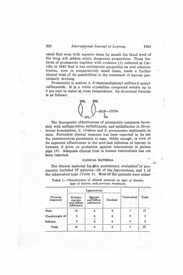
The clinical material fo.rAihis preliMinary evaluation "of promacetin included 27 patients-26 of the -lepromatous, and 1 of the tuberculoid type (Table I) '. Most of-the patients were either
TABLE 1.-Classification of clinical material on type of diseas~, type of lesions, and previous treatment. -
" Lepromatous
Previous Nodules, ~cules Tuberculoid Total' treatment macules . an ' tiiffuse Residual
and diffuse infiltration ~
infiltration -------None 10 t> 0 1' .. 17
" Chaulmoogra oil 2 0 0 0 2
Sulfones 2 0 6 0 8
Total 14 6 6 1 , 27 . ,

18,2 Johansen, et al: Promacetin P:r;ogress Report 223 ~p
moderately or far advanced with unfavorable prognoses and, therefore, exceptionally desirable material for treatment evaluation.
. '-Seventeen. patients had received no previous treatment for
leprosy, two had received chaulmoogra oil, and eight, sulfones. Of the two patients previously treated with chaulmoogra oil, o,ne had not received any treatment 'for a period of 11 years and presented nodules and heavy diffuse infiltration; the other had not received treatment for 3 years and likewise showed far advanced manifestations, including exteJ;l.sive confluent. nodulation with ulceration. Six patients previously, treated with sulfones (promin or diasone) showed only. residu~l lesions and were .i,ncluded 'n the study because, in spite of marked ' clinical improvement." on sulfones, al( continued to show after several years ~(;{ t~e~tinent a significant number of leprosy bacilli in the skin scrapings. The ' intention was to determine the effect ' promacetin might have in the further reduction of the number of bacilli in the skin of these patients. The remaining two sulfonetreated patients showed active leprous skin lesions consisting of nodules, macules, and diffuse infiltration. One of these represented a clinical relapse; the other, who in addition showed extensive nodular infiltration of the mucous membrane of the palate, tongue and pharynx, represented a clinical progression of the disease several years after the patient had left the hospit~1 and discontinued all treatment against medical advice. ,.... Table 1 further sets forth the type of lesions the patients
.;.' presented before treatment. Extensive heavy diffuse infiltration, nodules and macules of' the skin e.f,~.the lepromatous type with numerous (4+) or a moderate (3+) , Ilumber of bacilli present occurred in 13 patients. Moderate diffuse infiltration with associated infiltrated macules showing either numerous, a moderate In~mber, or few (2+) bacilli ipresent occurred in 7 patients.
j'Residual)esions presenting a variable (4 + to 1 + rare) number of j oaciHi "were seen in 6 of the patients previously treated with slJliones, while 2 showed active fresh lesions as already described. The tlJberculoid type patient showed ' plaques and patches of various sizes. dev.<?~d of bacilli, and a positive lepromin test'. All
', patien,t s" . except those showing only residual lesions and the tuberculoid type patient, had extensive involvement of the nasal mucous mejnbrane, and one had involvement of the entire mouth, ,nasopharynx, and larynx.
' ... ~. -~ ;.
! 4 ,- ,4""'" ......
. -. .. ... . ' ..

224 International Journal of L eprosy 1950
METHODS
Since it is known that the drugs so far found ,to be effective in leprosy must be administered over a long period of time, the dosage of promacetin was kept comparatively low du"ring the early stages of treatment. Although promacetin has been reported to be of low toxicity when given for short periods ofJ iIne,- it was-felt that in this group of patients, where treatment' of long duration was contemplated, chances could not be taken with initial large doses. Conseqllently, the initial daily oral dose of promacetin varied from 0.3" to 0.5 gm. except-for a small number of male patients. of good physique who were given an initial daily oral dose of 1.5 gm. Where the initial dose was 0.3 to 0.5 gm. the dosage was increased by 0.3 to 0.5 g~. every 2 weeks until a total daily dose of 1.5 gm. was reached. "Then the dosage was increased by either 1.0 gm. or 1.5 gm. up to a maximum of from 3.0 to 4.0 gm. daily. For those patients who were begun on 1.5 gm. daily the dosage was increased by either 1.0 gm. or 1~5. gm. every 2 weeks until the maximum of from 3.0 to 4.0 gm. was reached.
Promacetin was administered at meal time as has been customary at Carville with all · other orally administered sulfones. Similarly, rest periods, during which the drug "was withheld, were observed at the same intervals by the promacetin group of patients as those observed routinely by all other patients taking orally administered sulfones. The rest periods were observed for 15 days during the latter part of March, July, September, and December.
Careful initial clinical "and laboratory examinations were conducted and clmical photographs taken. For follow-up, a weekly clinical examination was done and clinical photographs taken whenever sufficient improvement had occurred in" the skin lesions to make it possible to record such improvement photographically. An erythrocyte and a leucocyte count, hemoglobin determination, and a urinalysis were done on each patient every 3 weeks. Blood and urine levels for promacetin, and skin and nasal scrapings were performed periodically. •
Among patients treated with promin, diasone, and sulphetrone at this institution, it is routine practice to administer iron and vitamin therapy when there is o'nly a slight drop in the erythrocyte count, since it is known that there is . .-a tendency on the part of these drugs to produce anemia. In the promacetin group of patients, iron and vitamin therapy, as well as liver and gastric mucosal preparations, were withheld unless the patient
18,2 Johansen, et al: P1'omacetin Progress Report 225
presented an initial anemia or showed a persistent decrease in the erythrocyte count during treatment, Such a procedure was necessary in order to determine the actual effect of promacetin on the peripheral blood elements, Three patients inadvertently, however, were given blood boosting preparations during insigni:(icaI\j ; p.ecreases in their erythrocyte counts which were well within the realm of laboratory variations.
This drug evaluation began on March 1, 1948, and data were collected for the purpose of this report up to July 31, 194'9.
TOXICITY AND OTHER COMPLICATIONS
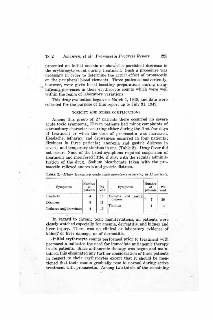
Among this group of 27 patients there occurred no severe acute toxic symptoms .. Eleven patients had mino~ complaints of a transitory character occurring either during the first few days of treatment or when the dose of promacetin was increased. Headache, lethargy, and drowsiness occurred in four patients; dizziness in three patients; anorexia and gastric distress in seven; and temporary tinnitus in one (Table 2). Drug fever did not occur. None of the listed symptoms required suspension of treatment and interfered little, if any, with the regular administration of the drug. Sodium bicarbonate taken with the promacetin relieved anorexia and gastric distress.
TABLE 2.-Minor transitory acute toxic symptoms occurring in 11 patients.
Number Number Symptoms of Per Symptoms of Per
patients cent . patients cent
Headache 4 15 Anorexia and gastric distress .. 7 26
Dizziness 3 11 Tinnitus 1 4
Lethargy anq drowsiness 4 15
In regard to chronic toxic manifestations, all patients were closely watched especially for anemia, dermatitis, and kidney and liver injury. There was no clinical or laboratory evidence of kidne1 or liver damage, or of dermatitis .
. Initial erythrocyte counts performed prior to treatment with promacetin indicated tl1e need for immediate antianemic therapy in six patients. Since antianemic therapy was begun and maintained, this eliminated any further consideration of these patients in respect to their erythrocytes except that it should be mentioned that their counts gradually rose to normal during active treatment with promacetin. Among two-thirds of the remaining
226 International Journal of L eprosy 1950
patients (14 out of 21), a slight depression of erythrocyte counts was noted in the first 2 to 4 weeks of treatment. Antianemic therapy in the form of iron and vitamins was given to four of these patients, but withheld from the remainder. With but three exceptions, the patients who received no antianemic therapy returned as rapidly to their original level of erythrocytes as those who were given antianemic therapy. The clinical course of the disease in these three patients was altered by the development of lepra reactions . associated with high fever. These reactions occurred approximately 3 months after inception of treatment, and at the time when 'a secondary rise in the erythrocyte level had begun. Two of these patients gave a history of similar lepra reactions with severe anemia prior to treatment, making it quite certain that these episodes in at least two of the patients were unrelated to the administration of promacetin. Erythrocyte counts of these patients returned to normal after the subsidence of the reactions with the administration of iron salts, vitamins, and liver extract. A summ·ary of the erythrocyte count behavior follows with the symbol "AT" denoting the administration of antianemic therapy:
Decrease less than 0.5 million per cu. mm ............. 5 patients (3 AT) Decrease between 0.5 and 1.0 million per cu. mm. 5 patients (1 AT) Decrease between 1.0 and 1.5 million per cu. rmn. 4 patients (3 AT) No change in erythrocytes .......................................... 2 patients Increase less than 0.5 million per cu. mm . ............. . 4 patients Increase between 0.5 and 1.0 million per cu. mm. ,1 patient
Leucopenia was not observed in any of the patients treated. Leucocytosis occurred with the same frequency ' as would be normally expected in moderately or far advanced leprosy. Mild erythema nodosum occurred in 11 patients. Six of these patients had experienced such reactions prior to treatment with promacetin. Three patients developed lepra reactions during which there was a temporary exacerbation of specific leprous lesions. Two of these patients had experienced similar reactions prior to treatment with promacetin.
One patient who had had frequent previous attacks of iridocyclitis had repeated exacerbations of tIiis condition. Pain from leprous neuritis, although not a common complaint in this group of patients, did persist in a mild form in some patients.
There were six patients altogether in whom treatment was discontinued, but in none of these could·· the reason for discontinuation of treatment be wholly attributed to promacetin. The reasons were as follows: refusal of patient to cooperate because

18,2 Johansen, et al: Promacetin Progress Report 227
of psychotic episode unrelated to the drug, 1 ; irregular discharges (two improved nodular cases, one showing no change), 3; and acute lepra reactions, 2. .
THERAPEUTIC EFFECT
The evaluation of the therapeutic effect of promacetin on leprous skin and mucous membrane lesions was primarily con-
. 'fined to 'the .21 'patients listed in Table 1 as having clinically active lesions when treatment was begun:' The patients are referred to as Group i. Those patients: 6 in number, who presented clinically inactive or residual lesions are utilized mainly in this report for a demonstration of bacteriologic response. These patients are referred to as' Group II. '
Group I p'atients were treated for periods varying from 4 to 16 months and received a 'total of 13,811 gm. of promacetin. The total amount for each patient averaged 658 gm. The total amount of the drug taken by each patient may be found in the summary of cases (Table 3).
Early objective improvement in specific skin lesions, as well as in secondary infected skin ulcerations, occurred in all of these patients. The duration of treatment before objective clinical improvement could be detected by careful physical examination varied from 2 to 8 weeks. The earliest change noted was. a change in color in the form of a fading or tanning of macules arid of the , diffusely infiltrated areas of skin. This change usually became evident after about 2 to 4 weeks of treatment. At the same time there was a decrease in the edema of the dermal tissues associated with softening of the skin over the areas of diffuse infiltration and a decrease in the turgidity of the nodules. Definite shrinking of the nodules and decrease in infiltration of the skin did not occur until after at least 1 to 2 months of treatment. Necrosis of nodules before actual decrease in size took place was observed in some patients. Necrosis was followed by ulceration and crusting which rapidly healed. If swelling of the hands and feet were present, this manifestation also responded early and readily.
Mucous membrane lesions of the nose, mouth, and throat showed the effects of early healing in relief obtained from nasal obstruction and hoarseness during the first few weeks of treatment. Objective evidence of healing was observed by the workers in the eye, ear, nose, and throat, and dental departments. The dental officer gave the opinion that promacetin accomplished
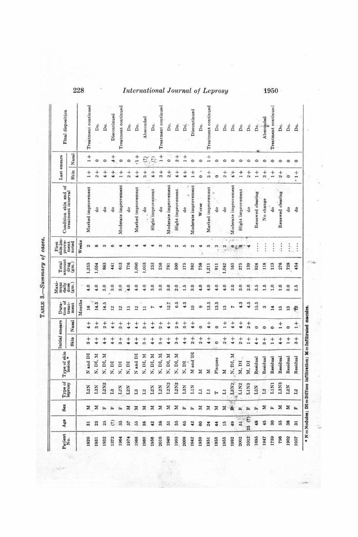
TABLE 3.--Summary of cases.
Maxi- First . Initial smears Dura- mum Total 9kin~m-
Patient Age Sex Type of Type of skin tion of daily amount prove-No. leprosy lesion * treat- dose drug ment
Skin Nasal ment (gm.) (gm.) noted --------- --- - --------Months Weeks
1920 31 M L2N Nand 01 3+ 4+ 16 4.0 1,255 2
1931 23 M L3N N, Di, M 4+ 3+ 14.5 4.0 1,054 8
1932 25 F L2N2 N, 01, M 4+ 2+ 14.5 3.0 883 3 \
1372 (1) F L3 N,DI 4+ 4+ 12 3.0 441 6
1964 35 F L2N N,DI 3+ 3+ 12 3.0 613 4
1974 57 M L2N N,DI 4+ 4+ 11 '4.0 776 4
1966 55 M L3 Nand 01 4+ 4 + 12 4.0 1,090 4 .
1960 38 M L2 N, 01, M 3+ 3+ 11 4.0 1,053 4
1956 42 M L2N N, 01, M 4+ 4+ 7 3.0 253 4
2010 38 M L2N N, 01, M 3+ 2+ 4 3.0 256 3
1949 51 M L2N2 N, 01, M 4+ 4+ 12.7 3.0 781 2
1993 . 35 M L2N2 N, 01, M 3+ 2+ 6.5 2.0 300 2
2009 65 F L3N N,DI 4+ 3+ 4.3 1.5 175 5 . 1942 42 F LIN M and 'DI 2+. 4+ 10 3.0 382 2
1959 60 M Ll M 2+ 0 9 4.0 758 "
4
1951 24 M L 1 M 4 + 4+ 13.5 4.0 1,211 3
1953 44 M ;
T Plaques 0 0 13.5 4 .0 911 . 3
1955 15 M L1 M 2+ 1+ 13 4.0 1,042 . 2 -~ .,
'. 1992 49 . L3N2 , N, 'DI, M 4+ 4+ 7 2.0 163 .6 • ,It J ' J<. 2002 51 " F , L1N2 M,DI 2+ 4+ 4.3 3.0 275 "3. .. 2012 25 (T) F L1N3 M,D I 1+ 2+ 4.5 2.0 139' ·4
1695 48 F L3N Residual 4+ 0 15.5 3.5 924 .. . . 1947 45 M L2 Residual 3+ 0 3 1.5 ll8 . ...
-1759 30 F LtNt Residual 1+ 0 14 1,0 tI3 .. .. 796 55 M L3N3 Residual 4 + 1+ 15 1'.0 276 ... .
1902 38 M L2N Residual 1+ 0 13 3.0 728
1957 31 F L2 Residual 3+ 1+ 12 2.5 454 . . ..
• N=Nodu)88; DI=Dlffuse Infiltration; M=Inflltrated macules.
Last smears Condition skin end of
treatment interval Skin Nasal
Marked improvement 1+ 1+
do 2+ 0
do 4+ 0
do 4+ .4+
Moderate improvement 1+ 0
do · 2+ 0
Marked improvement 4+ · 1+
do 3+ q .. Slight improve,ment 4+ ' (1) . ,
do 3+ 1+
Moderate improvement 2.+ 0
Slight improvement 4+ 2+
do 4+ 1+ , .' Moderate improvement 1+ 0
Worse o • 0
Marked improvement 3+ 1+ , do 0 0
do 2+ 0
Moderate improvement 4+ 0
Slight 'improvement I+. 0
do 2+ 0 Renewed clearing 1+ 0
No change 3+ 0
do 1+ 0
Renewed c1~aring · 2'+ 0
do 0 0
do · 1+ 0
Final disposition
Tre~tment continued
Do.
Do.
D iscontinued
Treatment continued
Do.
Do.
Absconded
Do.
Treatment continued
D o.
Do.
Do.
D iscontinued
Do.
Treatment continued
Do.
Do .
Do.
Do.
Do.
Do. ~
AbSCOfded
Treatment continued
D o.
Do.
D o. -- ---
I:\:) I:\:) 00
~ .,.... ~ .... ~ .,.... ..,. C ;:s !;:l <-0
'" C
~ <-0
c ........ t-< ~
't:3 .... c ~ ~
~ ~ 01 o
18,2 Johansen, et al: Promacetin Progress Report 229
the healing of active mucous membt:~ne .lesions of the mouth and throat more rapidly than that observed from other sulfones.
_.ObJectively, clinical improv.emEmt was progressive throughout the interval of treatment except in oile patient.
, -This patient (No. 1959), whose ,major skin manifestation was a num
ber of 'n'filtrated lepromatous patches, showe'd initial improvement which was followed by a temporary outburst of. many small erythematous patches. After these lesions subsided, another exacerbittion .'of lesions occurred after 6 months, of treatment overJarger areas of the body asso~iated with fever, hoarseness, and nasal obstruction typical of ' fl.¥ acute lepra reaction. Promacelin was discontinued. After remaining in a reactive stage for about 4 months this patient impro~ed slowly and the skin and mucous membrane became negative for leprosy bacilli. Although there was eventual improvement in this patient he is classified in the summary ' as in worse condition at the end of the treatment interval than he was when treatment was begun.
Another patient (No. 1942) developed a -severe lepra reaction during which there was a temporary increase in , the diffuse infiltration associated with high fever, neuritis, and secondary anemia. Promacetin was discontinued .because of , a continued reaction state and a refractory type of secondary auemia.
Among the ' ~atients 'treated was a psychotic one (No. 1372) who for a period of about 10 months had a remission from the mental disturbance. This patient presented far advanced confluent nodular skin lesions with vffi:y heavy diffuse infiltration and, during the remission of the mental state, promacetin was given and taken orally without much difficulty. Improvements in the specific lesions were rapid. By the time the nodules had flattened there was an exace~bation of the mental , ~onditi.on and all medication was refused. Four months later there was a definite return of infiltration and nodulation.
The patient having leprosy of the tuberculoid type (No. 1953) improved markedly and rapidly. This improvement cannot be attributed to promacetin entirely as tuberculoid leprosy generally improves spontaneously. The rapidity with which the lesions regressed in this cas'e, however, suggests an added factor. '
Another observation , of interest denoting clinical improvement in the skin of some patients was regrowth of hair after about 6 months of treatment. The regrowth was scanty but nevertheless pre.sent. It appeared in 'eyebrow regions where eyebrows has been absent prior to treatment, and on the extremities and chest. Subjective improvement in the sensation of the extremities took place in some patients.
Reduction ,In the number of leprosy bacilli in the skin did not become noticeable in skin smears until after one year's treatment. A few heavy nodular cases continued to show the same number of organisms in the skin smears althougll the absolute number of organisms must have been significantly reduced by the decrease in size ' of the lesions. The mucous membranes

230 International J ournal of Leprosy 1950
showed a reduction in bacilli at an earlier stage and many patients showed negative nasal smears at the end of one year's treatment.
~
Group II patients, presenting only residual lesions, were treated for periods varying from 3 to 15 months. They received a total of 2,613 gm. of promacetin or an average of 435 gm. per patient. Actual amounts given to each patient may be found in the summary of cases (Table 3).
Group II patients were expected to show little if any change in their skin manifestations. Four of these patients surprisingly,
. however, began to show renewed clearing of their skin 'after 3 to 6 months' treatment. One patient left the hospital without permission after 3 months' treatment, and the sixth patient (No. 1759), because of repeated lepra reactions which she also had prior to promacetin treatment, received only interrupted treatment in small doses to the extent of 113 gm. in 14 months. These two patients showed no change in the skin, but the repeated lepra reaction experienced by one of them gradually. abated.
There was a reduction in the number of leprosy bacilli in the skin of all of these patients who showed renewed clearing of the skin, and one became negative. While two had positive nasal smears at the inception of treatment, all of them had negative nasal smears at the end of the treatment interval.
The clinical condition of Group I patients at the end of their treatment interval is classified in Table 4. Status in regard to the bacterioscopic condition of skin and mucous membranes of both groups of patients is g:iven in Table 5.
TABLE 4.-Clinical status of patients (Group J) at end of treatment interval.
Clinical status
Treatment Period Marked Moderate Slight improve- i~prove- improve- Worse
ment )ment ment
Number patients treated 12 months or more 8 2 ------ ----
Number patients treated 6 months-12 months 1 3 2 1
Number patients treated 3 months-6 months ------ ------ 4 ----
TOTAL 9 (43% ) 5 (24% ) (} (28% ) 1 (5% )
Total
10
7
4
21
18,2 Johansen, et al: Promacetin Progress Report 231
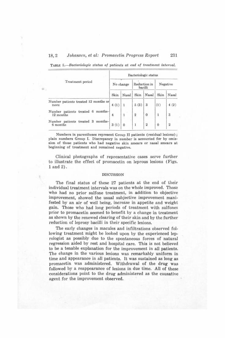
TABLE 5.-Bacteriologic status of patients at end of treatment interval.
Bacteriologic status
Treatment period -No change I Reduction in I Negative
bacilli
Skin Nasal Skin Nasal Skin Nasal ----------
Number patients treated 12 months or more 4 (1) 1 5 (3) 3 (1 ) 4 (2 )
Number patients treated 6 months-12 months 4 1 2 0 1 3
Number patients treated 3 months-6 months 3 (1) 0 1 2 0 2
Numbers in parentheses represent Group II patients (residual lesions) ; plain numbers Group 1. Discrepancy in number is accounted flY!.' by omission of those patients who had negative skin smears or nasal smears at beginning of treatment and remained negative.
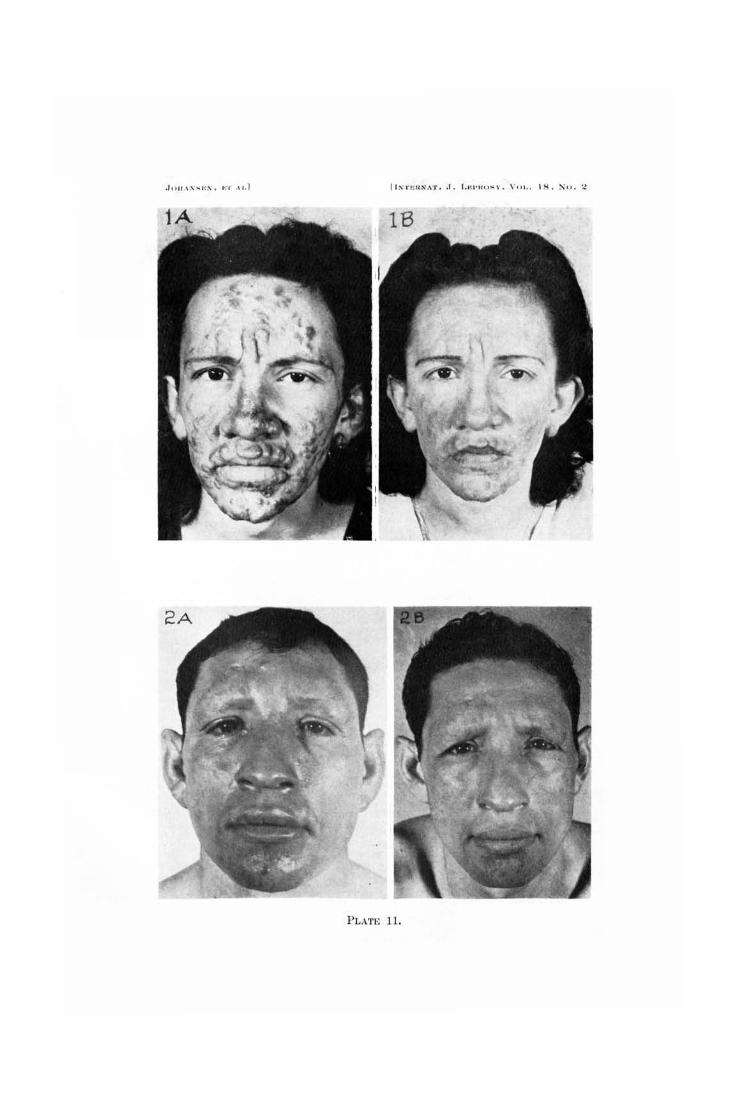
Clinical photographs of representative cases serve further to illustrate the effect of promacetin on leprous lesions (Figs. 1 and 2).
DISCUSSION
The final status of these 27 patients at the end of their individual treatment intervals was on the whole improved. Those who had no prior sulfone treatment, in addition to objective improvement, showed the usual subjective improvement manifested by an air of well being, increase in appetite and weight gain. Those who had long periods of treatment with sulfones prior to promacetin seemed to benefit by a change in treatment as shown by the renewed clearing of their skin and by the further reduction of leprosy bacilli in their specific lesions.
The early changes in macules and infiltrations observed following treatment might be looked upon by the experienced leprologist as possibly due to the spontaneous forces of natural regression aided by rest and hospital care. This is not believed to be a tenable explanation for the improvement in all patients. The change in the various lesions was remarkably uniform in time and appearance in all patients. It was sustained as long as promacetin was administered. Withdrawal of the drug was followed by a reappearance of lesions in due time. All of these considerations point to the drug administered as the causative agent for the improvement observed.
232 {nternational J oU1'nal of L eprosy 1950
Early inl~rovement in infected skin ulcerations suggests that promacetin, in addition to having antileprotic properties, is capable of combating secondary infections. Relief from nasal obstruction and dryness of the throat sustained by patients taking ~he drug also is evidence in favor of its ability to 'combat ordinary infections.
It was found in the determination of. promacetin blood levels that the blood level remained fairly constant between 1.5 and 2.0 mg, per 100 ·cc. of blood on oral doses varying from 3.0 to 4.0 'gm. daily irrespective of the length of teatment, Urine levels behaved diffe~ently. In the early weeks and months of treatment the urine level on the stated dosage averaged around 25 to 50 mg. per 100 cc. of urine. After 6 to .9 months of treatment the urine levels tended to increase going as high as 200 to 250 mg. per too cc. of urine . . Eventually, some patients excreted by way of the kidneys almost as much of the drug daily as was administered. An adequate explanatioJl for the increase observed· in the uriiie promacetin level after prolonged treatment while there was very little increase in the blood 'level of the drug cannot be given at present. After prolonged treatment significant amounts of promacetin were excreted in the urine up to 12 days f611ow- ~ ing discontinuation of the drug.
The opinion expressed by some that sulfones are of no value in tuberculoid leprosy is probably influenced by the feeling that this type of leprosy is not in need of treatment. The improvement noted in the patient of this type treated with promacetin was certainly more striking and rapid than th.at usually observed without treatment.
CONCLUSIONS
Objective clinical improvement of skin and mucous membrane lesions of lepromatous leprosy is observed to occur following the oral administration of promacetin. The improvement noted is urtiform, universal and sustained; therefore, it cannot be wholly attributed to spontaneous factors.
Red4ction in the number of leprosy bacilli in the skin and . mucous membrane follows clinical improvement. Skin smears commence to show a noticeable reduction of bacilli at the end of one year's treatment while many patients show an absence of bacilli in the mucous membrane at this time.
Promacetin is well tolerated orally even upon prolonged administration of doses as high as 3.0 to 4,0 gm. daily. Slight
- ~ depression of the erythrocyte count may occur during the first
0-18,2 Johansen, et al: Promacetin Progress R eport 233
few weeks pf treatment. Unless there are other ~omplications of the disease present, such depressed counts usually return to the original level spontaneously. Before it can be definitely stated that promacetin does or does not have the property of producing severe grades of anemia when used in the treatment of uncomplica:ted leprosy, further blood studies on a larger group of patients are necessary.
Renewed clearing after promacetin therapy of apparently stationary residual lesions in patients previously treated with sulfones suggests that a wider application shoulo be made of alternating or combined methods of treatment in leprosy.
Prpmacetin apparently possesses chemotherapeutic properties against human leprosy. Its lack of protection against tuberculosis of guinea pigs and its apparent effectiveness in another acid-fast infection, human leprosy, present interesting implications as to the future choice of experimental drugs for trial in both human leprosy and human ttfirerculosis. :
REFERENCES
1. FAGET, G. H., POGGE, R. C., JOHANSEN, F. A., DINAN, J. F ., PREJEAN, B. M. and ECCLES, C. G. The promin treatment of leprosy (a progress report). Pub. Health Rep. 58 (1943) 1729.
2. FAGET, G. H., POGGE, R. C. and JOHANSEN, F. A. Present status of diasone in the treatment oof leprosy. Pub. Health Rep. 61 (1946) 960-963. - 0
3. WHARTON, L. H. Preliminary report on a new sulphone drug (sulphetrone). Internat. J . Leprosy 15 (1947) 231.
4. JOHANSEN, F. A. and ERICKSON, P. T. Studies on the therapy of leprosy. Proc. 4th Internat. Congo Trop. Med. & Malaria. Washingto~, D. ,C., Department of State, 1948, Vol. 1, p. 365.
5. SHARP, E. A. and PAYNE, E. H. Th~ present status of the sulfones in o therapy. Internat. J. Leprosy 16 (1948) 157.
6. GRUHZIT, O. M. Description of 1. A. 307. Report from Parke, Davis o and Co., Department of Clinical Investigation.

234 International Journal of L eprosy 1950
DESCRIPTION OF PLATE
PLATE 11.
FIG. 1. Photographs showing response to treatment of extensive nodulation of the skin. A, before treatment; B, after 12 months' treatment.
FIG. 2. Photographs showing response to treatment of diffuse infiltration and small discrete nodules. A, before t r eatment; B, after 4 months' treatment.
. J O II . \ S~ I'; S. 1': '1' ,\1 ,1 f l xn:nNAT •• T. J.1~ I · H O!oO Y. VOL. I S. K o . • )
PLATI~ 11.