oral (po) (non- parenteral) sublingual via feeding tube topical transdermal intranasal ...
TRANSCRIPT

Administration Routes in Small Animals
Special Topics

Routes of Administration
Oral (PO) (Non-parenteral)
Sublingual Via feeding tube Topical Transdermal Intranasal
Inhalation
› Nebulized or volatilized
Rectal (PR) Aural Topical ophthalmic

Parenteral Routes: Administered with a needle and syringe
o Intradermal (ID)o Subcutaneous (SC or
SQ)o Intramuscular (IM)o Intravascular/ intravenous (IV)
o Intraosseouso Intraperitoneal (IP)o Intra-arterial (IA)o Epidural/subduralo Intracardiac (IC)o Intramammary
Many factors are considered before the route of administration for a drug is selected. Some factors are based on
the drug itself; other factors are based on the animal being treated

Drug Factors Some drugs cause one effect when given
parenterally and another effect when given non-parenterally (magnesium sulfate causes muscle relaxation when given IV and diarrhea when given orally)
Some drugs are insoluble in water and can be given IM but cannot be injected IV – Always be aware of label!
Some drugs are destroyed by stomach acid and cannot be given orally
Some injectable drugs must be given very slowly; while others must be given in a bolus
Patient Factors Animals that are actively vomiting cannot
absorb drugs given orally Critically ill patients need to get therapeutic
levels of drug into their bodies rapidly The animal’s temperament must be considered Can/will the owner medicate their animal at
home properly with the medication as prescribed?

Other Factors to Consider Is restraint going to be an issue? What is your time frame? Does this medication require that
special precautions be followed during administration (i.e. gloves, mask)?
Potential side effects? What is the most convenient route of
administration for the owner?

Comparison of Common Parenteral Routes of Drug Administration
Intramuscular 90° Subcutaneous
45°
Intravenous25°
Intradermal10°–15°
Muscle
Epidermis
Dermis
Subcutaneous tissue

Parenteral Routes of Drug Administration

Mini A&P Review of Skin Layers
The skin is made up of three layers: the epidermis, dermis, and subcutaneous layers The epidermis is several cell layers thick and does not contain blood vessels. Its thickness varies greatly from region to region in any animal and varies from species to species
The dermis is composed of blood vessels, lymph vessels, nerve fibers, and accessory organs of the skin (glands and hair follicles)
The subcutaneous layer (hypodermis) is composed of connective tissue and contains a large amount of fat

Always Gather Your Supplies First!
Needles Syringes Medication(s) to be injected Cotton balls (or gauze) in alcohol (venipuncture
only)
Vet Wrap, gauze or dry cotton balls Muzzle or muzzles (E-collars can also act as
great restraint devices) Proficient person to restrain the animal Hydrogen Peroxide (optional and if available)

Anatomy of a Syringe
Needle
Barrel
Needle hub
Luer-lock tip
Bevel
Cap
Rubber stopper
Scale
Plunger
Flange
Thumb rest
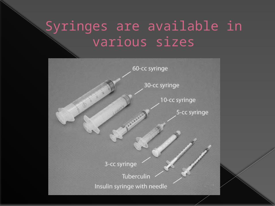
Syringes are available in various sizes

18 gauge x 1 in 25 gauge x 1 in20 gauge x 1 in 22 gauge x 1 in
Various needles commonly utilized with injections
Please memorize the gauges in relation to their colors
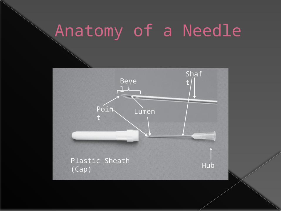
Anatomy of a Needle
Bevel
LumenPoint
Shaft
Plastic Sheath (Cap) Hub

Injectable Drugs Usually supplied as sterilized solutions,
prepackaged syringes with needles for injection, powders that must be reconstituted with sterile solution, or in vials to be drawn up into syringes for injection
May be stored in ampules or (single or multidose) vials


Intradermal Administration

Intradermal Injections
Local anesthetics injected ID to desensitize skin
Allergy skin testing performed with ID injections
Most animals will not tolerate skin testing unless they are sedated
Skin must be shaved with a #40 clipper blade prior to administering injections for allergy testing

ID Allergy Injection Procedure
A fold of skin is lifted and a 25- to 27- gauge needle attached to a 1 ml syringe is inserted with the bevel up into the dermis
A 0.1 ml volume of allergen is injected
The injection site will look like a translucent lump if the injection is performed correctly
The skin is then examined for tissue reaction

Subcutaneous Administration

Subcutaneous Injections
Most easily and most frequently performed Most common route for vaccine
administration Absorption rate may be slow in obese
animals Not recommended in severely dehydrated or
critically ill animals Good route for chronic, at-home fluid
administration Some substances are harmful if injected SC
(>5% dextrose solution can cause skin sloughing)

SC Injection Sites Preferred site for most SC injections is the
dorsolateral region from the neck to the hips
In cats: the intrascapular region should be avoided because of the incidence of vaccine-induced tumors. Feline vaccinations should be administered in as distal a portion of an extremity as possible› Intrascapular region should also be avoided for
insulin injections. Insulin should be injected in alternating sites along dorsolateral or ventrolateral aspect of trunk

SC Injection Procedure
Fold of skin is tented and the needle is inserted at the base of and parallel to the long axis of the fold
Do not insert needle perpendicular to the long axis, it may penetrate both sides of the skin and the medication (or fluids) deposited will be outside of the animal
Retract plunger of syringe slightly and check the needle hub for blood prior to injection
If blood appears in hub, remove needle and reinsert in another location

Skin Tent

SC Injection Procedure

SC Injection Procedure
After injection, briefly massage skin to facilitate drug distribution
If multiple vaccinations or medications are administered, injection sites should be a minimum of several centimeters apart
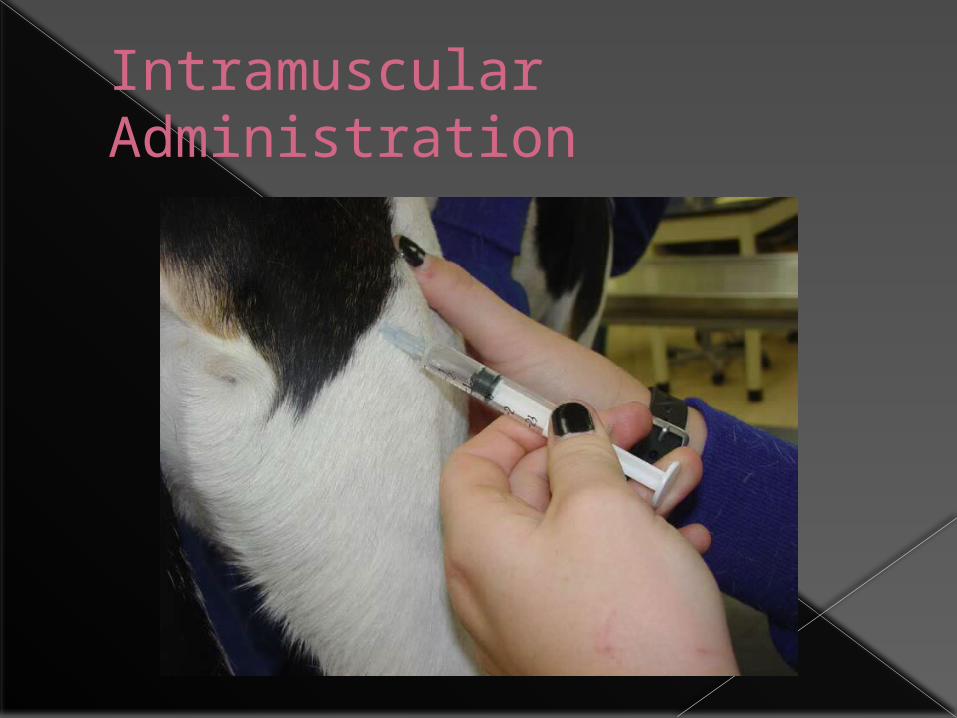
Intramuscular Administration

Intramuscular Injections
Appropriate route for injection of small volumes of medication
Generally, more painful for animals than SC or IV
Never use the muscles of the neck! Avoid the sciatic nerve!

IM Injection Sites Drugs are most often administered in the
lumbosacral musculature lateral to the dorsal spinous processes

IM Injection Sites In the semimembranosus or semitendinosus
muscles of the rear leg Needle should enter the lateral aspect of the
muscle and be directed caudally to prevent penetration of the Sciatic nerve penetration can cause: pain and lameness

IM Injection Sites
Occasionally the triceps muscles on the caudal aspect of the front legs are used as IM injection sites

IM Injection Videos
http://www.youtube.com/watch?v=uevk6K_r3iM
http://www.youtube.com/watch?v=tQaBe-nzkUU&playnext=1&list=PL07102E44BD3D9D14&feature=results_video

IM Injection Procedure Gather all supplies needed. Isolate the muscle between the fingers and
thumb A 22 to 25 gauge needle attached to a syringe
is embedded in the muscle. As with a SQ injection, the needle hub is checked for blood before administration of medication to make certain a vessel is not inadvertently penetrated
Once in the muscle, inject the medication slowly
Massage the site for a few seconds after the injection to help distribute the substance if possible
(Immiticide injections are not typically massaged)
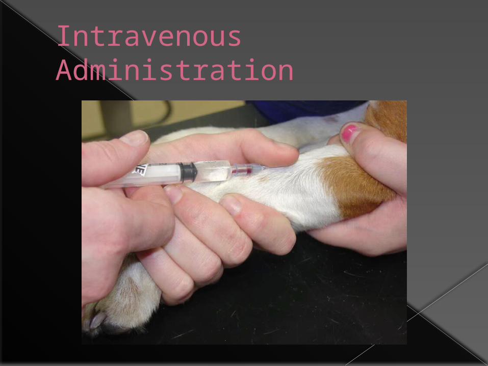
Intravenous Administration

Intravenous Administration Drugs, medications, and/or fluids may be injected
directly into a vein or through an IV catheter IV route:
› Predictable concentration of drug› Produces an immediate response› Rapidly reach high blood levels, achieving a rapid onset of
action Irritating drugs can be given IV (not IM or SC) Increased risk of adverse effects (if drug is given too
rapidly, not sterile, or improperly mixed) In most cases, IV drugs should be given slowly Make sure to remove all air bubbles in substance to
be injected to prevent air emboli which can cause tissue damage or even, potentially death

IV Injection Sites
Cephalic vein Lateral saphenous
vein
Cephalic vein Medial saphenous
vein
DOG CAT
The jugular vein is used to administer injections in both large and small animals IF an
intravenous catheter is in place

Cephalic Vein

Medial Saphenous Vein (cat)

Lateral Saphenous Vein (dog)
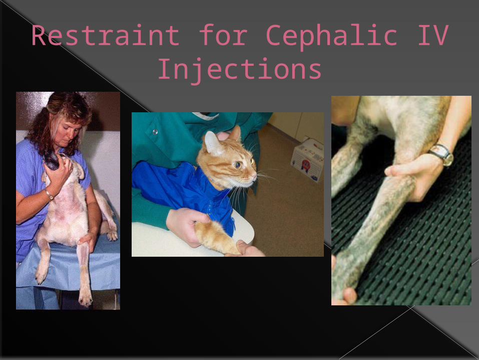
Restraint for Cephalic IV Injections
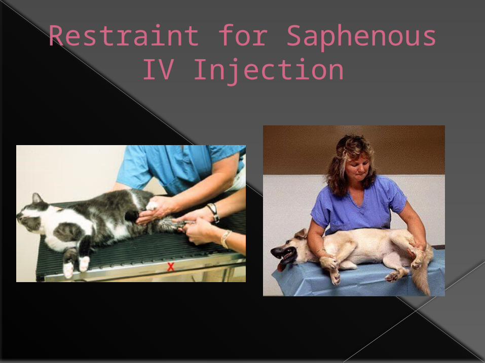
Restraint for Saphenous IV Injection
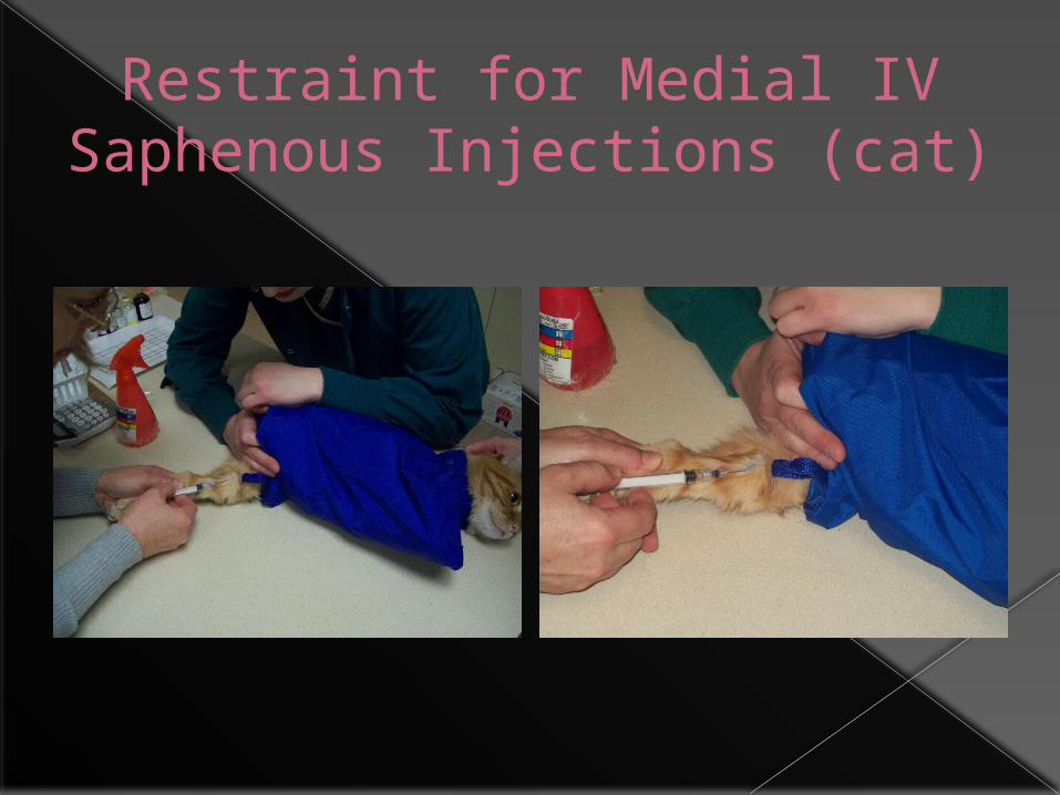
Restraint for Medial IV Saphenous Injections (cat)

IV Injection Instructions Gather your supplies! Expel all air bubbles from the syringe prior
to inserting into the vein Occlude the vessel with digital pressure or
use a tourniquet Grasp the extremity and pull the skin taut in
a distal direction Shave the fur and swab with alcohol or Swab
the skin and hair with an alcohol-soaked cotton ball (go with the fur)
IV Injection Instructions (cont’d) Insert a 22- to 25- gauge needle, bevel up
into the vein Usually blood enters the hub of the needle
at penetration of the vein, BUT, placement is confirmed by aspirating the blood back into the syringe
Have the restrainer release pressure from the vein, and inject the syringe contents into the vein
Let the restrainer know that you are going to withdraw the needle, and apply firm pressure to the injection site until hemostasis/coagulation occurs (~1 minute)

About Tourniquets… Most common: Nye Tourniquet or
Penrose drain Can be very dangerous used
improperly Goal is to visualize and access vein Remove quickly!
Possible Complications with IV Injections
Injecting drug outside of vein “Blowing vein” Hematoma formation Intra-arterial injection of drug Hitting a nerve (pain, lameness,
paralysis) Air-embolus; other embolism Septicemia

IV INJESCTION VIDEO
http://www.youtube.com/watch?v=xOqQ6xqYfjg