β induces mesenchymal stem cells migration and … · il1β induces mesenchymal stem cells...
TRANSCRIPT

IL1β Induces Mesenchymal Stem Cells Migrationand Leucocyte Chemotaxis Through NF-κB
Rubén Carrero & Inmaculada Cerrada & Elisa Lledó &
Joaquín Dopazo & Francisco García-García &
Mari-Paz Rubio & César Trigueros &
Akaitz Dorronsoro & Amparo Ruiz-Sauri &José Anastasio Montero & Pilar Sepúlveda
Published online: 31 March 2012# The Author(s) 2012. This article is published with open access at Springerlink.com
Abstract Mesenchymal stem cells are often transplantedinto inflammatory environments where they are able tosurvive and modulate host immune responses through apoorly understood mechanism. In this paper we analyzed theresponses of MSC to IL-1β: a representative inflammatorymediator. Microarray analysis of MSC treated with IL-1β
revealed that this cytokine activateds a set of genes related tobiological processes such as cell survival, cell migration, celladhesion, chemokine production, induction of angiogenesisand modulation of the immune response. Further moredetailed analysis by real-time PCR and functional assaysrevealed that IL-1β mainly increaseds the production ofchemokines such as CCL5, CCL20, CXCL1, CXCL3,CXCL5, CXCL6, CXCL10, CXCL11 and CX3CL1, interleu-kins IL-6, IL-8, IL23A, IL32, Toll-like receptors TLR2,TLR4, CLDN1, metalloproteins MMP1 and MMP3, growthfactors CSF2 and TNF-α, together with adhesion moleculesICAM1 and ICAM4. Functional analysis of MSC prolifera-tion, migration and adhesion to extracellular matrix compo-nents revealed that IL-1β did not affect proliferation but alsoserved to induce the secretion of trophic factors and adhesionto ECM components such as collagen and laminin. IL-1βtreatment enhanced the ability of MSC to recruit monocytesand granulocytes in vitro. Blockade of NF-κβ transcriptionfactor activation with IκB kinase beta (IKKβ) shRNAimpaired MSC migration, adhesion and leucocyte recruitment,induced by IL-1β demonstrating that NF-κB pathway is animportant downstream regulator of these responses. Thesefindings are relevant to understanding the biological responsesof MSC to inflammatory environments.
Keywords Mesenchymal stem cells . Interleukin1β . Chemotaxis .Migration and adhesion
Introduction
Mesenchymal stem cells have become a therapeutic option forseveral pathologies like myocardial infarction, osteogenesis
Supported by grants from the Instituto de Salud Carlos III for theRegenerative Medicine Program to Centro de Investigación PrincipeFelipe, from the FIS (PI07/784, CP08/80) and from Kutxa.
Electronic supplementary material The online version of this article(doi:10.1007/s12015-012-9364-9) contains supplementary material,which is available to authorized users.
R. Carrero : I. Cerrada : J. A. Montero : P. Sepúlveda (*)Fundación para la Investigación Hospital Universitario La Fe,Avda Campanar 21,Valencia 46009, Spaine-mail: [email protected]
I. Cerrada : E. LledóUniversidad Cardenal Herrera-CEU,Valencia, Spain
J. Dopazo : F. García-GarcíaFunctional Genomics Node, National Institute for Bioinfortmatics,Valencia, Spain
J. Dopazo : F. García-García :M.-P. RubioCentro de Investigación Príncipe Felipe,Valencia, Spain
C. Trigueros :A. DorronsoroFundación Inbiomed,San Sebastián, Spain
A. Ruiz-SauriUniversidad de Valencia,Valencia, Spain
Stem Cell Rev and Rep (2012) 8:905–916DOI 10.1007/s12015-012-9364-9

imperfecta, graft versus host disease and wound healing [1–4].As a part of the cell therapy, MSC are often transplanted inischemic, apoptotic and/or inflammatory environments wherecells survive and promote tissue regeneration by mechanismsthat remain poorly understood. These cells are immunoprivi-leged, and in most of pathologies the induced potential benefitsare related to paracrine activity mediated by their ability tosurvive in ischemic and inflammatory environments [5–7].Despite their therapeutic potential initial, clinical results havebeen disappointing due to reported low benefits. It is believedthat in adequate doses, low engraftment and poor survivalare responsible for these results. We and others reported thatintramyocardial MSC transplantation recruits a number ofinflammatory cells that contribute to the healing of the infarct[8, 9]. Transplanted cells are consistently exposed to tissuesignals, immune cells and mediators that could influencetheir behaviour. Since successful application of stem cellapproaches will depend on the microenvironment of therecipient tissue, we have sought to investigate the response ofMSC to an inflammatory environment. Previous reportsshowed that proinflammatory cytokines were able to in-creased migration of human MSC to many chemotacticfactors [10], to induce MSC to produce chemokines[11] and to stimulate MSC to differentiate into neuralphenotype [12]. Following this rationale we culturedMSC in the presence of inflammatory mediators andanalyzed biological responses implicated in proliferation,survival, adhesion and migration that could aid to predicttheir response in these environments. We focused ourstudies in IL-1β as a prototypical inflammatory mediatorand the results showed that this cytokines promotesspecific biological processes in MSC in part due toactivation of the transcription factor NF-κB (NuclearFactor KAPPA-light-chain-enhancer of activated B cells).
Materials and Methods
Cells and Culture Conditions
Human bone marrow MSC (n04; Inbiomed, San Sebastian,Guipuzcoa, Spain) were cultured following manufacturers´instructions. After centrifugation cells were seeded in tissueculture flasks in low glucose Dulbecco’s modified Eaglemedium (Sigma-Aldrich, St. Louis, MO; htpp://www.sigmaaldrich.com), supplemented with 10% fetal calfserum (Thermo Scientific Hyclone, Rockford, IL; htpp://www.piercenet.com), 4 mM L-Glutamine (Gibco, GrandIsland, NY; htpp://www.invitrogen.com) and 1% antibiotics[50U/mL penicillin, 50 μg/mL streptomycin (Gibco)]. Cellswere maintained in a humidified atmosphere of 95% air and5% CO2 at 37°C. The medium was replaced every 3 days.IL-1β (25 ng/mL), (Millipore, Temecula, CA; htpp://
www.milipore.com), IL-6 (20 ng/mL), (R&D Systems,McKinley Place, MN; htpp://www.RnDSystems.com), IL-8 (20 ng/mL), (Sigma-Aldrich) and TNF-α (50 ng/mL),(Sigma-Aldrich) were added and maintained during differentperiods of time depending on the experiments.
Electrophoresis and Western blotting
Cells were submitted to serum deprivation for 18 h beforeincubation with IL-1β (25 ng/mL) for 30 min. Cells werethen washed with PBS and the monolayer lysed in M-PER®Mammalian Protein Extraction Reagent (Thermo FisherScientific, Rockford, IL; htpp://www.piercenet.com)containing protease inhibitors (Roche, Mannheim, Germany;htpp://www.roche.com)] and phosphatase inhibitors [SodiumOrthovanadate 1 mM (Sigma-Aldrich), Sodium Fluoride1 mM (Sigma-Aldrich)]. Protein concentration was quantifiedusing the Pierce® BCA Protein Assay Kit (Thermo FisherScientific). For western blotting 30 μg of protein was loadedand separated by 10% SDS-PAGE, before transfer to a PVDFmembrane (Thermo Scientific) and blockinged with 5%BLOT-Quick Blocker (Millipore). Primary antibodies wereincubated at 4°C overnight. After, the blots were incubatedwith IgG HRP Conjugated for 1 h at room temperature.Detection was performed with ECL system [ECL PlusTM
Western Blotting Detection Reagents Amersham (GE health-care, Bukinghamshire, UK; htpp://www.gehealthcare.com)].GAPDH was used to determine equal protein loading.Antibodies used were anti-phospho-Akt 1/2/3 (Ser473),anti-phospho-NFκβ p65 (Ser536) (Cell Signaling TecnologyInc., Danvers, MA; htpp://www.cellsignaling.com), anti-GAPHD, anti-ERK 1/2 (MAPK), anti-phospho-ERK 1/2(MAPK), (Chemicon, Temecula, CA; htpp://www.chemicon.-com), anti-Akt 1/2/3, anti-NFκβ(p65), anti-rabbit IgG HRPConjugated (Santa Cruz Biotecnology Inc., Santa cruz,CA; htpp://www.scbt.com) and anti-mouse IgG HRPConjugated (Promega, Madison, WI; htpp://www.promega.com). All antibodies used were assayed at 1:1,000 dilutionexcept anti-Akt 1/2/3 and anti-NFκB (p65) that were usedat 1:500.
Cell Cycle Assay
To analyze the effect of IL-1β in cell cycle, 106 cells wereharvested, fixed with 70% EtOH and kept at −20°C until use.Fixed cells were centrifugated and resuspended in 50 μg/mLpropidium bromide (Sigma-Aldrich) and analyzed by flowcytometry (488 nm excitation, 625 nm emission).
MTT Proliferation Assay
Cell proliferation was determined using a 3-(4, 5-dimethylthiazol-2-thiazolyl)-2,5-diphenyl-2H-tetrazolium
906 Stem Cell Rev and Rep (2012) 8:905–916

bromide (MTT) assay. MSC were plated at a density of104 cells/cm2 in 96-well microplates. 24 h post seeding,cells were treated with recombinant IL-1β (25 ng/mL) orIL-6 (20 ng/mL) for 24 h. Proliferation was assayed byThiazolyl Blue Tetrazolium Bromide (Sigma-Aldrich),acording to manufacturer´s instructions. The absorbance ofthe samples was measured at 570 nm using a microplatereader (Victor3 1420 Multilabel Counter; PerkinElmer Inc.,Waltham, MA; htpp://www.perkinelmer.com). Experimentswere performed in triplicate and results were expressedrelative to the untreated control.
Construction of IKKβ shRNA and Lentiviral Transduction
IKKβ shRNA secuences have been published previously [13]and were purchased from Sigma-Genosys (Sigma-Aldrich): -IKKβi: GGAAGTACCTGAACCAGTTTG. Oligos wereannealed and cloned into pSUPER plasmid carrying H1 pro-moter using BglII-HindIII sites. The H1-shRNA expressioncassette was then excised and cloned into pLVTHM (Addgeneplasmid 12247; Addgene, Cambridge, MA, http://www.addgene.org) using EcoRI-ClaI sites. Viral particleswere produced in human embryonic kidney 293 T cells(ATCC, www.atcc.org). Briefly, 293 T cells were seededin high glucose DMEM containing 10%FBS. psPAX(Addgene plasmid), pVSV-G (Addgene plasmid 12259,www.addgene.org) and the lentiviral vector pLVTHMcontaining GFP reporter gene and shRNA sequenceswere transfected in to the packing cells using calciumphosphate precipitation method. Viral transduction wascarried out using a multiplicity of infection (M.O.I.) of10 in the presence of 8 μg/mL of polybrene (Sigma-Aldrich) in order to achieve 100% infection.
MSC Migration Assay
To study trophism induced inMSC by IL-1β, cells were seededin basal medium (DMEM with 0.5% FBS) at 10,000 cells/cm2
in the top chamber of an 8 μm-pore migration transwell (BDFalcon, Bedford, MA, htpp://www.bd.com). After overnightculture, 25 ng/mL of IL-1β was added to the bottom chamberand cells were fixed with 2% paraformaldehide (PanreacQuímica, Castellar del Vallés, Spain), SDF-1α (20 ng/mL)and 10% FBS were used as positive controls. Briefly, after4 h non migrated cells were removed from the upper sideof the membrane using a cotton bud to remove nonmigrating cells, the membrane was cut and placed in aglass slide with the bottom side upwards and beforestaining with 4´,6 diamidino-2 phenyilindole (DAPI)(Sigma-Aldrich). All assays were performed in duplicatedwells and repeated three times. Migrated cells were countedas fold increase relative to passive MSC cell migration inuntreated wells.
Leucocyte Migration Assay
To determine the nature of human leucocytes that could berecruited in response to paracrine factors secreted by MSC,we established co-culture of MSC and pheripheral bloodleucocytes (PBLs) using a transwell culture system (BDFalcon). MSC (10,000 cells/cm2) seeded in the lower chamberof the transwells were stimulated or not with IL-1β for 2 h.After extensive washes with PBS, wells were filled with freshmedium and human PBLs from buffy coats (100,000 cells)were seeded in the upper chamber. Migrated cells through8 μm-pore size membranes were counted after 5 h of co-culture. Cells were fixed as described above and leucocytepopulations were quantified in hematoxilin stained preparationby morphologic counting. All studies were performed in ablinded fashion.
Adhesion Assay
Cells were seeded in basal medium onto cover slides previouslytreated with 10 μg/cm² of fibronectin (Sigma-Aldrich),2 μg/cm² of laminin (Sigma-Aldrich) and 10 μg/cm²of collagen (Sigma-Aldrich). After 1 h, cells were fixedwith 2% paraformaldehyde, washed with PBS, labelledwith 4´,6-diamidino-2-phenylindole (DAPI) and counted.
Reverse Transcription and Real-Time Quantitative PCR
MSC incubated in different conditions were washed withPBS. RNAwas extracted using TRIzol Reagent (Invitrogen,Carlsbad, CA; htpp://www.invitrogen.com) and purifiedwith RNeasy Plus Mini Kit (Qiagen, Dusseldorf, Germany;htpp://www.qiagen.com). RNA samples were quantified byspectrometry (NanoDrop ND-1000, NanoDrop Technologies,Wilmington, DE; htpp://www.nanodrop.com) and integritywas assessed by agarose gel electrophoresis and the absorbanceratio 260/280 nm. cDNA was obtained by retrotranscriptasereverse transcription using M-MLV Reverse Transcriptase(Invitrogen) from total RNA (1 μg). Primers were designedusing the Primer-Blast online tool (Table 1). The Ct of eachPCR in a reference human MSC cDNA sample (dilution 1/10)is expressed as mean ± standard deviation (SD) of threeindependent PCR experiments. Real-time PCR was performedusing convenient primers and SYBR Green I [1X LightCycler480 SYBR Green I Master (Roche Molecular Biochemical,Mannheim, Germany; htpp://www.roche.com)]. Plates wererun in a real-time thermal cycler (LightCycler 480 Instrument;Roche Diagnostics, Mannhein, Germany, htpp://www.roche.com) following manufacturer´s instructions. Real-timemonitoring of the PCR reaction was performed with theLightCycler 480 Software 1.5, as well as the quantification ofthe products in the exponential phase of the amplification.Relative expression levels were calculated with the Relative
Stem Cell Rev and Rep (2012) 8:905–916 907

Table 1 List of oligonucleotides used for Real time-PCR. Ct and efficiency values are indicated
Gene Forward primer Reverse primer Size (bp) Ct SD Ct Efficencya
ACTB AGAGCCTCGCCTTTGCCGATCC CATGCCGGAGCCGTTGTCGAC 101 16.10 0.025 1.971
CCL2 TCTCAGTGCAGAGGCTCGCGA CCACTTCTGCTTGGGGTCAGCAC 119 17.01 0.006 1.977
CCL3 TTCAGAAGGACACGGGCAGCAGACA GGAATCTGCCGGGAGGTGTAGCT 216 29.64 0.050 1.862
CCL5 TACATTGCCCGCCCACTGCC GGGTTGGCACACACTTGGCG 119 23.82 0.052 2.061
CCL7 CAGCTGCTTTCAGCCCCCAGG GCTTCCCGGGGACAGTGGCTA 148 23.93 0.032 1.923
CCL8 TGGCAGCCACTTTCAGCCCT GCACAGACCTCCTTGCCCCG 190 27.98 0.167 2.040
CCL20 TCTGCGGCGAATCAGAAGCAGC TTCATTGGCCAGCTGCCGTGT 110 21.19 0.068 2.16
CLDN1 CCGGGTTGCCCACCTGCAAA CGTACATGGCCTGGGCGGTC 258 24.49 0.148 1.862
CLDN14 GTCGCTGTGGGCAGGTGGTC AGCCTCCCCTTCCCAGCCTG 192 30.48 0.093 2.084
CSF2 TGCAGCATCTCTGCACCCGC AGGCAGGTCGGCTCCTGGAG 176 24.47 0.188 1.708
CSF3 AGACCCATGGCTGGACCTGCC GTGGCACACTCACTCACCAGCTTC 218 31.56 0.043 2.026
CX3CL1 CTGGCTGGACAGCACCACGG GCTCCTTCGGGTCGGCACAG 175 25.96 0.087 1.880
CXCL1 AGCCTGCAACATGCCAGCCA TGTGCACATACATTCCCCTGCCT 94 21.72 0.036 1.848
CXCL10 TGCAAGCCAATTTTGTCCACGTGT GCAGCCTCTGTGTGGTCCATCC 200 24.10 0.051 1.956
CXCL11 TGTCTTTGCATAGGCCCTGGGGT AGCCTTGCTTGCTTCGATTTGGGA 164 29.72 0.178 1.821
CXCL3 AATGTAAGGTCCCCCGGACCCC ACCACCCTGCAGGAAGTGTCAA 199 20.92 0.036 2.023
CXCL5 AATCTCCGCTCCTCCACCCAGT GCTCTCTCAACACAGCAGCGGC 201 28.49 0.050 1.614
CXCL6 GCACGAGGAAACCAAAGTGCTCTG GTGCAACGCAGCTCTGTCAGCA 234 26.37 0.071 1.890
ELF3 ATTTAGAGCCGGGTAGGGGAGCG GTTGCAGCCATGAGGCTACCGGAGT 132 25.68 0.088 1.912
GAPDH CCCCTCTGCTGATGCCCCCA TGACCTTGGCCAGGGGTGCT 122 16.79 0.015 1.998
IBSP GGAGTACGAATACACGGGCGCC GGTAGCCGGATGCAAAGCCAGA 222 27.12 0.049 2.018
ICAM1 CTGGTCCTGCTCGGGGCTCT GGGCTGGTCACAGGAGGTGC 126 22.42 0.094 1.638
ICAM4 AATACACTTTGCGCTGCCACGTG GGCTCCAAGCGAGCATCAGTGT 264 28.16 0.116 2.057
IL11 TGACCCGCTCTCTCCTGGCG GCACGTGCCGCAGGTAGGAC 192 24.23 0.163 1.803
IL12A CCCAAAACCTGCTGAGGGCCG TGGAGGCCAGGCAACTCCCAT 219 29.08 0.047 1.985
IL15 GCTGCTGGAAACCCCTTGCCAT TAGGTGCTTTGGGCCAACTGGG 214 28.78 0.035 1.899
IL1B AGGCACAAGGCACAACAGGCT AACAACTGACGCGGCCTGCC 277 21.12 0.110 1.965
IL23A TGGCTGTGACCCCCAAGGACTC TGCCATGGCTGGCTGGGACT 246 27.01 0.053 1.893
IL32 TTTGTGCCAGGAAGACTGCGTGC GGCTCGACATCACCTGTCCACG 215 24.41 0.078 2.004
IL34 GAACACCACCATGCCCCGGG CAGCCTGGTGACGTTGGCGA 250 24.59 0.078 1.853
IL6 CATTCTGCCCTCGAGCCCACC GGCAGCAGGCAACACCAGGA 139 17.08 0.088 1.955
IL7 GCTGCTCGCAAGTTGAGGCAATTT TTGTTGGTTGGGCTTCACCCAGG 157 26.58 0.213 1.974
IL8 CGTGGCTCTCTTGGCAGCCTTC TTCCTTGGGGTCCAGACAGAGCTC 229 15.88 0.707 2.005
ITGA9 GCAGTGACCGCTGGCCACTT GCGCACAAGGAGGAGCCGAA 182 27.63 0.098 1.969
ITGB3 TGCCGCCCTGCTCATCTGGA TCCTGCAATCGTGGCACAGGC 239 22.43 0.008 2.192
ITGB8 CTAGCGACACTCGGCCCGC CTGGACCCAGCGCAAGGCAC 292 27.76 0.023 1.850
MMP1 GTGTCTCACAGCTTCCCAGCGAC GCACTCCACATCTGGGCTGCTTC 238 25.01 0.084 2.006
NOD2 AGGCCTACCCGCAGATGCCA GTGGGAGAGAGGCTGGCCCA 298 28.04 0.172 2.062
SDC4 CGGAGTCGCCGAGTCGATCC GGCTCCCAGACCCTGCCCTC 248 20.51 0.030 1.925
SELECTIN TTGTTCCTGCCAGCAGCTGCC AGGGCCAGAGACCCGAGGAG 164 17.64 0.085 2.030
SERPINE1 AGGACGAACCGCCAATCGCAAG ACCCTCACCCCGAAGTCTGAGG 167 25.86 0.034 2.120
TCAM1P CGAGCTTGGCTGTGGCCTCC TCTCCGCCATCCCAGCCTCC 225 27.05 0.078 1.856
TLR2 AGGCAGCGAGAAAGCGCAGC CCCCCAAGACCCACACCATCCA 253 31.23 0.352 2.162
TLR4 CCCTGCGTGGAGGTGGTTCCTA CTCCCAGGGCTAAACTCTGGATGGG 280 26.45 0.024 2.007
TNF CCCTCTGGCCCAGGCAGTCA ATGGGTGGAGGGGCAGCCTT 235 21.58 0.019 2.100
VCAM1 AGGTGACGAATGAGGGGACCACA CCAGCCTCCAGAGGGCCACT 181 17.31 0.010 2.039
a PCR efficiency of each PCR was estimated from a serially diluted standard curves obtained from a reference MSC cDNA sample. The cyclenumber of the crossing point CP vs. Log (dilution factor) were plotted to calculate the slope. The efficiency of each PCR was then calculated withthe equation E ¼ 10½�1=slope�
908 Stem Cell Rev and Rep (2012) 8:905–916

Quantification Analysis software. Results were consideredto be significant with a 2-fold induction. For all real-timeexperiments, gene expression levels were normalized totwo human housekeeping genes ACTB and GAPDH andaverage from triplicate samples.
Microarray Assays
Human bone marrow MSC were treated or not with IL-1β(25 ng/mL) for 24 h at 37°C in a humidified incubator with 5%CO2. Cells were collected and total RNAwas extracted usingthe High Pure RNA Isolation kit (Roche) and quantified byspectrometry (NanoDrop ND-1000, NanoDrop Technologies).800 ng of total RNA were used to produce Cyanine 3-CTP-labeled cRNA using the Low RNA Input Linear Ampli-fication Kit PLUS (Agilent, Santa Clara, CA¸ http://www.chem.agilent.com). Based on the protocol for One-ColorMicroarray-Based Gene Expression Analysis Version 5.5(Agilent p/n 5188–5977), 3 μg of labeled cRNA was hybrid-ized with Whole Human Genome Oligo Microarray Kit(Agilent p/n G4112F) containing 41,000+ unique humangenes and transcripts. Arrays were scanned in an AgilentMicroarray Scanner (Agilent G2565BA) according to themanufacturer’s protocol and data extracted using AgilentFeature Extraction Software 9.5.1. Hybridization wasperformed by the microarray core facility from CentrePrincipe Felipe.
Microarray Data Analysis
Signal was standardized across arrays using quantile normali-zation [14]. Differential gene expression was carried outusing the fold change. Gene set analysis was carriedout for the Gene Ontology terms using FatiScan [15]from Babelomics web tool [16]. GO annotation for thegenes in the microarray where taken from Ensembl 55release (http://www.ensembl.org, Ensembl org, Hinxton,UK), allowing the visualization of functional categorieswithin their biological context. Results were consideredto be significant with a 2-fold induction.
The microarrays data of this study have been deposited inthe Gene Expression Omnibus database under accessionnumber GSE33755.
Statistical Analysis
Data are expressed as mean ± SD. Comparisons betweenexperimental groups were done with unpaired and pairedtwo-samples t-test using the SPSS software (SPSS, Chicago,IL http://www.spss.com). Differences were considered sta-tistically significant at P<0.05.
Results
Global Transcriptome Profiling of MSC Culturedwith IL-1β
To test the effect of IL-1β onMSC, cells were cultured with orwithout 25 ng/mL of IL-1β for 24 h. Gene expression changesinduced by the pro-inflammatory cytokine were evaluated bymicroarray analysis. Further bioinformatics analysis usingBabelomics software (http://www.babelomics4.org) wasperformed to classify genes by function using the GeneOntology (GO) scheme, revealing the main families ofgenes affected by the treatment. Growth in IL-1β resulted inactivation of genes associated to very specific GO categories.In particular, we identified alterations in the expression ofgenes implicated in the following biological processes: i)response to wounding, ii) immune and inflammatory re-sponse, iii) defense response, iv) chemotaxis, v) locomotorybehaviour, vi) regulation of cell proliferation, vii) leukocytechemotaxix, viii) I-kappaB kinase/NF-kappaB cascade, ix)negative regulation of apoptosis, x) blood coagulation, andxi) cell adhesion (Table 2). Fold changes of up-regulatedgenes (negative values) from enriched biological processesin MSC treated with IL-1β (MSC-IL1β) are indicated(Supplemental Table 1).
IL-1β Increases Expression of Multiple Chemokinesand Growth Factors in MSC
After bioinformatic analysis, highly up-regulated genes relatedwith these biological processes were further assayed by real-
Table 2 Enriched biological processes for up-regulated genes inMSC-IL1β versus MSC
GO biological process Process gene set P value
GO:0009611 Response to wounding 2.00E-21
GO:0006955 Inmune response 1.74E-19
GO:0006954 Inflammatory response 2.43E-18
GO:0006952 Defense response 2.43E-17
GO:0006935 Chemotaxis 5.44E-10
GO:0007626 Locomotory behavior 6.15E-9
GO:0042127 Negative regulation ofcell proliferation
1.27E-5
GO:0030595 Leukocyte chemotaxis 3.04E-3
GO:0007249 I-kappaB kinase/NF-kappaBcascade
3.27E-3
GO:0043066 Negative regulationof apoptosis
3.64E-3
GO:0007596 Blood coagulation 1.36E-2
GO:0007155 Cell adhesion 3.26E-2
Stem Cell Rev and Rep (2012) 8:905–916 909
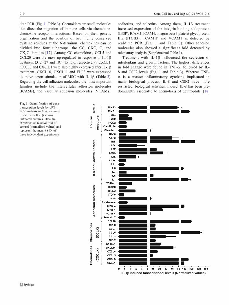
time PCR (Fig. 1, Table 3). Chemokines are small moleculesthat direct the migration of immune cells via chemokine-chemokine receptor interactions. Based on their geneticorganization and the position of two highly conservedcysteine residues at the N-terminus, chemokines can bedivided into four subgroups, the CC, CXC, C, andCX3C families [17]. Among CC chemokines, CCL5 andCCL20 were the most up-regulated in response to IL-1βtreatment (312±27 and 187±15 fold, respectively). CXCL1,CXCL3 and CX3CL1 were also highly expressed after IL-1βtreatment. CXCL10, CXCL11 and ELF3 were expressedde novo upon stimulation of MSC with IL-1β (Table 3).Regarding the cell adhesion molecules, the most importantfamilies include the intercellular adhesion molecules(ICAMs), the vascular adhesion molecules (VCAMs),
cadherins, and selectins. Among them, IL-1β treatmentincreased expression of the integrin binding sialoprotein(IBSP), ICAM1, ICAM4, integrin beta 3 platelet glycoproteinIIIa (ITGB3), TCAM1P and VCAM1 as detected byreal-time PCR (Fig. 1 and Table 3). Other adhesionmolecules also showed a significant fold detected bymicroarray analysis (Supplemental Table 1).
Treatment with IL-1β influenced the secretion ofinterleukins and growth factors. The highest differencesin fold change were found in TNF-α, followed by IL-8 and CSF2 levels (Fig. 1 and Table 3). Whereas TNF-α is a master inflammatory cytokine implicated inmany biological process, IL-8 and CSF2 have morerestricted biological activities. Indeed, IL-8 has been pre-dominantly associated to chemotaxis of neutrophils [18]
Ch
emo
kin
es
(CX
CL
X)
Ch
emo
kin
es
(CC
LX
)A
dh
esio
n m
ole
cule
sIL
s an
d G
row
th F
acto
rsT
oll-
like
rece
pto
rsM
MP
s
IL-1β induced tanscriptional levels (Normalized values)
Fig. 1 Quantification of genetranscription levels by qRT-PCR analysis in MSC culturestreated with IL-1β versusuntreated cultures. Data areexpressed as relative fold ofcontrol (normalized values) andrepresent the mean±S.D. ofthree independent experiments
910 Stem Cell Rev and Rep (2012) 8:905–916

Table 3 Transcriptional levelsof up-regulated genes in MSCafter treatment with IL-1β.Normalized values of MSC-IL1β vs non-treated MSCwere calculated and expressedas mean±standard deviation(SD) of three independentPCR experiments
Chemokines Genebank number Mean Ct SD Ct Normalized values Normalized Error
CXCL1 NM_001511 21.72 0.036 89.1 5.9
CXCL3 NM_002090 20.92 0.036 154 11
CXCL5 NM_002994 28.49 0.050 8.65 0.59
CXCL6 NM_002993 26.37 0.071 16.5 1.27
CXCL10 NM_001565 24.10 0.051 2028 127
CXCL11 NM_005409 29.72 0.178 41.3 3.07
CX3CL1 NM_002996 25.96 0.087 21.1 0.76
CCL2 NM_002982 17.01 0.006 13.1 0.46
CCL3 NM_002963 29.64 0.050 29.2 15.8
CCL5 NM_002985 23.82 0.052 312 27
CCL7 NM_006273 23.93 0.032 13.1 0.90
CCL8 NM_005623 27.98 0.167 12.2 1.00
CCL20 NM_004591 21.19 0.068 187 15
Adhesion molecules
Selectin E NM_000450 17.64 0.085 0.94 0.08
VCAM-1 NM_001078.3 17.31 0.010 5.33 0.19
ICAM-1 NM_000201 22.42 0.094 24.5 0.84
ICAM-4 NM_001544.3 28.16 0.116 87.4 9.1
Syndecan 4 NM_002999.3 20.51 0.030 1.45 0.05
IBSP NM_004967.3 27.12 0.049 6.21 1.64
ITGA9 NM_002207.2 27.63 0.098 0.81 0.05
ITGB3 NM_000212.2 22.43 0.008 2.75 0.11
ITGB8 NM_002214.2 27.76 0.023 1.24 0.07
TCAM-1 NR_002947.1 27.05 0.078 2.17 0.12
Interleukines
IL1 NM_000576.2 21.12 0.110 145 41
IL6 NM_000600.3 17.08 0.088 9.99 1.12
IL7 NM_000880.3 26.58 0.213 1.29 0.27
IL8 NM_000584.3 15.88 0.707 76 14.4
IL11 NM_000641.2 24.23 0.163 0.79 0.18
IL12A NM_000882.3 29.08 0.047 1.16 0.05
IL15 NM_172175.2 28.78 0.035 2.94 0.48
IL23A NM_016584.2 27.01 0.053 25.3 7.8
IL32 NM_001012631.1 24.41 0.078 14.8 0.81
IL34 NM_152456.2 24.59 0.078 3.12 1.44
Growth factors
TNF NM_000594 21.58 0.019 243 21
CSF2 NM_000758 24.47 0.188 39.3 13.2
CSF3 NM_000759 31.56 0.043 1.16 0.03
ELF3 NM_004433 25.68 0.088 656 45
Toll-like receptors
Claudin 1 NM_021101.4 24.49 0.148 5.49 0.23
Claudin 14 NM_144492.2 30.48 0.093 1.30 0.13
NOD2 NM_022162.1 28.04 0.17 1.23 0.11
TLR2 NM_003264.3 31.23 0.352 2.29 0.12
TLR4 NM_138554.3 26.45 0.024 1.50 0.16
Metalloproteins
MMP1 NM_002421.3 25.01 0.084 162 12
MMP3 NM_002426.4 16.56 0.650 47.5 7.02
Stem Cell Rev and Rep (2012) 8:905–916 911

whereas CSF2 is implicated in monocytic differentiation[19].
Other biological processes activated in response to IL-1β were related to host defence and immune response.Microarray analysis and real time PCR experimentsshowed up-regulation of several Toll-like receptors (TLRs),claudins and NOD proteins. These molecules are implicatedin the innate immune response to microbial infection.However, recent reports have revealed that these moleculealso modulates biological processes in MSC such as differen-tiation, migration and immunomodulatory responses [20, 21].
IL-1β Activates the NF-κB Pathway and do not InduceMSC Proliferation
We next analyzed the effect of IL-1β on BM-MSC signaltransduction and cell proliferation. IL-1β promoted phos-phorylation of NF-κB, but not PI3K/AKT and ERK1/2pathways (Fig. 2a), as reported for other cell types [22].However, in correlation with the result of the microarrayanalysis (Table 2), IL-1β did not induce significant cellproliferation as assessed by MTT assay (Fig. 2b). Theseresults were further confirmed by cell cycle analysis usingflow cytometry (Fig. 2c). The percentage of cells in G0-G1was 91.16±2.71 in MSC versus 90.19±2.72 in MSC- IL-1β. The percentage of cells in S phase was 2.28±1.21 inMSC versus 1.60±0.46 in MSC- IL-1β, and finally thepercentage of cells in G2-M phase was 5.43±1.16 versus6.40±1.69, respectively.
IL-1β Induced Migration and Adhesion of MSC is MainlyActivated Through NF-κB Signaling
We and others have previously described that MSC areable to migrate in vivo to ischemic and pro-inflammatoryenvironments [8, 23, 24] and it is believed that thisbehaviour may underlie the ability of these cells toaccelerate wound healing. Migration of MSC towardscytokines, chemokines and growth factors has also beenexplored in vitro [10]. To test if IL-1β was able toincrease migratory ability in MSC, we cultivated MSCin the upper chamber of a transwell and stimulatedmigration by adding SDF-1α, IL-1β or 10% FBS inthe lower chamber (Fig. 3a). A negative control of formigration was achieved using the same proportion offetal bovine serum (0.5% FBS) in the upper and lowerchamber. SDF-1α was used since it is a well-knowntrophic factor for MSC implicated in homing to ischemicareas [24], and 10% FBS was used as positive controlsince it is a rich source in cytokines and growth factors.Surprisingly, the migratory response of MSCs to IL-1βwas in fact more pronounced than it was to to SDF-1α(1.68±0.21 fold increase versus 1.35±0.16), indicating astrong promigratory role for IL-1β Maximum migrationwas achieved towards FBS gradient (1.87±0.12 foldincrease). Similar levels of cell migration were observedwhen TNF-α or IL-8 were used as trophic factors (notshown), indicating that multiple inflammatory mediatorscan exert trophic effects on MSC as reported [24]. We
AKT (P)
AKT
ERK 1/2 (P)
ERK 1/2
GAPDHNFκβ (P)
NFκβ
+IL
1 β
+IL
1β
Co
ntr
ol
Co
ntr
ol
A
BIL-1βControl
DN
A c
on
ten
t
DN
A c
on
ten
t
CMTT Assay
0
0,5
1
1,5
Control IL1β IL6
AU
*
+IL
1β
Co
ntr
ol
Fig. 2 a, Western blot analysisof kinase phosphorylationin MSC treated with IL-1β.Total cell lysates (30 μg)were separated by SDS-PAGE.Anti-GAPDH was used asloading control. b, MTT assayof MSC cultures treated withIL-1β (25 ng/mL) and IL-6(20 ng/mL) for 24 h. Valuesare expressed in fold increaserelative to control. c, Cell cycleanalysis by flow cytometryof MSC cultures treated withIL-1β (25 ng/mL) for 24 h.(*P<0.05)
912 Stem Cell Rev and Rep (2012) 8:905–916

next wanted to investigate whether the signaling pathwaysinduced by IL-1β could be directly linked to MSC migrationtowards trophic factors. NF-κB transcription factors play animportant role in the balance between cell survival andapoptosis and are involved in the regulation of cellproliferation and differentiation of various cell types[25]. IKKβ phosphorylates IκB molecules, the inhibitorsof NF-κB, leading to ubiquitination and proteasomedegradation of the inhibitors, and hence release andactivation of NF-κB [26]. NF-κB has previously beendescribed as the main transcription factor activated inmany pro-inflammatory responses [27]. In these context,regulation of NF-κB cascade members was observedamong the biological processes most positively affectedby IL-1β treatment (Table 2) and phosphorylation ofNF-κB was induced on MSC after IL-1β treatment(Fig. 2). Thus, we sought to evaluate the role of NF-κBsignaling in the biological responses of MSC in response toIL-1β. For this purporse, we constructed a vector containingshRNA targeting IKKβ that was lentiviraly transduced inMSC. We then evaluated the migratory response to IL-1β,SDF-1α and FBS. As shown in Fig. 3a, treatment withIKKβ shRNA reduced trophic response ofMSC towards each
of the 3 trophic factors assayed. An increase in the basalresponse of IKKβ transduced cells of 1.05±0.11 fold wasobserved, and in response to trophic factors this was increasedby 1.21±0.11 towards SDF-1α, 1.45±0.06 towards IL-1β,and 1.58±0.07 towards 10% FBS, strongly suggestingthat NF-κB signaling pathway plays a major role in MSCtrophism.
Migration and invasiveness of adherent cells is in partmediated by changes in the affinity of cells to particularECM components (ECM). To test whether IL-1β had an effecton MSC cell adhesion, we measured the adhesion of MSC tothe main components of ECM. The results showed that IL-1βtreatment increased the adhesion to collagen (3.03±0.29 fold),fibronectin (1.75±0.11 fold) and laminin (2.79±0.15 fold)(Fig. 4b). In similar way to migration experiments, adhesioninduced by IL1β treatment to collagen (1.75±0.15 fold),fibronectin (1.20±0.05 fold) and laminin (1.32±0.07 fold)was impaired in IKKβ-MSC. The fact that IKKβ expressiononly affected the adhesion induced by IL-1β but not thebasal levels of adhesion to extracellular matrix componentsindicates that IKKβ blocks specifically the mechanismsinduced by this cytokine, confirming the importance of NF-κB signaling pathway in the IL-1β mediated biologicalprocesses.
Il-1β Treatment of MSC Increases Recruitmentof Leucocytes In Vitro
MSC have been shown to recruit inflammatory cells suchas neutrophils, eosinophils, macrophages and to suppressproliferation of cytotoxic and helper T cells through therelease of soluble factors such as HGF and TGF-β [11,28–30]. Moreover, infusion of MSC into myocardium and
B
0.0
0.5
1.0
1.5
2.0
2.5
3.0
3.5
Collagen Fibronectin Laminin
Ad
her
entc
ells
(rel
ativ
eto
con
tro
l) MSC
MSC IL 1β
IKK β
IKK β IL 1β
**
******
0
0.5
1
1.5
2
2.5
IKKβMCS
Mig
rati
on
fold
incr
ease
(rel
ativ
eto
con
tro
l)
Untreated
SDF
IL 1β
FBS
**A***
**
*
*
Fig. 3 a, migration of MSC or MSC-IKKβ towards trophic factorsSDF-1α (20 ng/mL), IL-1β (25 ng/mL) and 10% FBS. b, Adhesion ofuntreated MSC (black bars) and MSC-IKKβ (dashed bars) or treatedwith IL-1β (white and grey bars, respectively) to collagen, fibronectinand laminin. Data are represented as fold increase relative to MSCcontrol. (*P<0.05, **P<0.01, ***P<0.001 in both panels)
0
2
4
6
8
10
12
14
16
18
20
Neutrophils Eosinophils Lymphocytes Monocytes
Mea
n o
f m
igra
ted
Leu
kocy
tes/
fie
ld
MSCMSC-IL-1βIKKβIKKβ-IL-1β
*
** ***
Fig. 4 Leucocyte migration assay of human PMNs using a transwellinsert culture system towards different stimuli. Upper chamber werefilled with PMNs and lower chambers were seeded with MSC (blackbars) or MSC-IKKβ (white bars) treated (dashed and grey bars,respectively) or not with IL-1β. Migrated neutrophils, eosinophils,lymphocytes and monocytes are presented as a mean of threeexperiments±S.D. (number of migrated cells/field). *P<0.05, **P<0.01,***P<0.001
Stem Cell Rev and Rep (2012) 8:905–916 913

other tissues is accompanied by marked, paracrine mediatedleucocytic infiltration [4, 8]. In order to test whether IL-1βtreatment had a similar impact in MSC leucocyte recruitment,we cultured control or IL-1β treated MSCs, in a transwellsystem and measured the number and the type of leucocytesthat migrate through a the 8 μm pores of the membrane. After5 h of culture, the mean number of migrated leucocyes/fieldtowards the MSC lower chambers were; 12.06±2.91 neutro-phils, 6.52±2.14 eosinophils, 4.40±2.41 lymphocytes and3.15±1.34 monocytes. However, migration towards IL-1βtreated MSCs increased the number of migrated neutrophils(16.11±2.75, P<0.05), eosinophils (10.44±2.61, P<0.01),lymphocytes (6.31±2.68, n.s.) and monocytes (9.85±2.94,P<0.001) (Fig. 4). The observed increase in chemotacticleucocyte migration induced by IL-1β treated MSC was againimpaired when MSC were transduced with IKKβ (9.78±2.29neutrophils, 5.52±1.74 eosinophils, 3.42±1.98 lymphocytesand 2.68±1.45 monocytes). Thus, IL-1β induced recruitmentof neutrophils, eosinophils and monocytes and NFκB plays amayor role in trophism exerted by MSC on these cellpopulations.
Discussion
MSC have been used to treat a wide variety of diseases.Whilst the contribution of differentiation/transdifferentiationto tissue repair, are often minimal, other positive angiogenicand immunomodulatory effects are exerted by MSC inischemic, apoptotic and pro-inflammatory environments[6].
IL-1β is produced in different tissues, not only as aresponse to pathogens, but also as a danger signals inpathologies such as acute myocardial infarction [31], type2 diabetes [32], neural disorders [33]. In this study wewanted to investigate the response ofMSC to proinflammatorystimuli in terms of survival, proliferation and induced paracrinefactors. Thus, we treatedMSCwith IL-1β and usedmicroarrayto infer the biological response, firstly by gene function andlater by direct gene set with known functional outcomes. Arange of biological responses were activated in response to IL-1β, but perhaps, themost prominent was the potent stimulationof secretion of chemokines and growth factors that in turnwere able to increase migration and adhesion of MSCand to regulate recruitment of monocytes and granulocytes. Itis known that members of the CC family target primarilymonocytes and T cells, whereas CXC chemokines affectmainly neutrophils. It has been previously reported that theexistence of different monocyte subsets expressing differentchemokine receptors display distinct migratory and functionalproperties. Interestingly, the profile by MSCs in responseto chemokines secreted IL-1β, was enriched in CCL5,CCL20 and CX3CL1, that could specifically attract not
only neutrophils and monocytes, but also monocytesexpressing CCR5, CCR6 and high levels of CX3CR1 [34].Although leukocyte chemotaxis and lymphocyte developmentare the main functional properties of chemokines, they possesother biological activities like regulation of angiogenesis,control of cell proliferation and alteration of the expressionof adhesion molecules. Indeed, the structural ERL domainpresent in several members of the CXC chemokine familydetermines their angiogenic potential [35] and the inducedchemokquines CXCL1, CXCL3, and CXCL8 (IL-8) containthis motif. In the same context, CXCL10 is considered a “stopsignal” that limits expansion of the fibrotic reaction triggeredby TGFβ, FGF, and VEGF during myocardial healing [31].The high levels of activation of this chemokine in MSC(Table 3) could account for the potent ability of these cells tocontrol adverse remodeling during myocardial healing[8, 36, 37].
Claudins are transmembrane proteins found in tightjuntions that participate not only in regulating tissuebarrier function and permeability but also in cell motility,adhesion and migration [38]. Claudins (CLDN1 andCLDN14) were up-regulated in MSC after IL-1β treatment.A similar response has been reported in airway smoothmusclecells in response to IL-1β and TNFα [39], indicating similaractivation pathways. It has been described that TLR signallingis linked to NF-κB and MAPK signalling pathways, andthat this induction mediates the secretion chemokines andregulates immunosuppressive activity and recruitment ofinnate immune cells [21, 40, 41]. TLR2 and TLR4 were up-regulated in response to IL-1β. Similar effect had beenpreviously described after stimulation with LPS ofMSC from human parotid glands [42].
We also found differences between the activation patternof MSC in response to different inflammatory mediators.Whereas TNFα increased preferentially CCL2 (MCP-1),CCL5 (RANTES), CXCL1, CXCL5, CXCL8, CXCL10and CCL11 [10], we demonstrate here that IL-1β increasespreferentially CCL3, CCL5, CCL20, CXCL1,CXCL3,CXCL10 and CXCL11. Thus, modulation of MSC biologicalresponses is closely associated with culture conditions and thepresence of immune mediators influence MSC proliferationand multipotency. In this context, culture protocols withmilieu capable of MSC expansion while preserving chromo-some stability have been developed [43]
In summary, our findings show that IL-1β increasesmigration and adhesion of MSC and promotes leucocytechemotaxis through MSC secretion of soluble factors. Asdescribed in other cell types [44], IL-1β activates NF-κBresultings in transcriptional activation of a wide variety ofgenes such inflammatory mediators, adhesion molecules,growth factor or immune response mediator. Since some ofthese molecules are chemotactic for inflammatory leukocytes,like monocytes and neutrophils, these paracrine factors
914 Stem Cell Rev and Rep (2012) 8:905–916

could facilitate infiltration of immune cells for tissuerepair when MSC are transplanted into injured tissues.
Taken together, these findings shed light onMSC behaviourin inflammatory environments and suggest that inflammatorymediators like IL-1β induce a response in MSC that couldtrigger paracrine actions in vivo.
Acknowledgments Supported by grants from the Instituto de SaludCarlos III for the RegenerativeMedicine Program of Valencian Communityto Centro de Investigación Principe Felipe, from the FIS (PI07/784, andCP08/80) and from Kutxa Foundation. This work is also partly supportedby grants BIO2008-04212 from the Spanish Ministry of Science andInnovation (MICINN) and PROMETEO/2010/001, Conselleria deEducación y Ciencia de Valencia, Spain. PS acknowledges supportfrom Miguel Servet and RETICS programs (Instituto de SaludCarlos III). We are grateful to all patients and clinical colleagues P. Solvesand Luis Larrea who donated or collected clinical samples.
Disclosure of Potential Conflicts of Interest The authors indicateno potential conflict of interest.
Open Access This article is distributed under the terms of the Crea-tive Commons Attribution License which permits any use, distribution,and reproduction in any medium, provided the original author(s) andthe source are credited.
References
1. Pereira, R. F., O'Hara, M. D., Laptev, A. V., Halford, K. W.,Pollard, M. D., Class, R., Simon, D., Livezey, K., & Prockop, D.J. (1998). Marrow stromal cells as a source of progenitor cells fornonhematopoietic tissues in transgenic mice with a phenotype ofosteogenesis imperfecta. Proceedings of the National Academy ofSciences of the United States of America, 95, 1142–7.
2. Dezawa, M., Ishikawa, H., Itokazu, Y., Yoshihara, T., Hoshino, M.,Takeda, S., Ide, C., & Nabeshima, Y. (2005). Bone marrow stromalcells generate muscle cells and repair muscle degeneration.Science, 309, 314–7.
3. McFarlin, K., Gao, X., Liu, Y. B., Dulchavsky, D. S., Kwon, D.,Arbab, A. S., Bansal, M., Li, Y., Chopp, M., Dulchavsky, S. A., &Gautam, S. C. (2006). Bone marrow-derived mesenchymal stromalcells accelerate wound healing in the rat. Wound Repair andRegeneration, 14, 471–8.
4. Chen, L., Tredget, E. E., Wu, P. Y., & Wu, Y. (2008). Paracrinefactors of mesenchymal stem cells recruit macrophages andendothelial lineage cells and enhance wound healing. PLoSOne, 3, e1886.
5. Gnecchi, M., He, H., Noiseux, N., Liang, O. D., Zhang, L.,Morello, F., Mu, H., Melo, L. G., Pratt, R. E., Ingwall, J. S., &Dzau, V. J. (2006). Evidence supporting paracrine hypothesis forAkt-modified mesenchymal stem cell-mediated cardiac protectionand functional improvement. The FASEB Journal, 20, 661–9.
6. Kinnaird, T., Stabile, E., Burnett, M. S., Lee, C. W., Barr, S.,Fuchs, S., & Epstein, S. E. (2004). Marrow-derived stromal cellsexpress genes encoding a broad spectrum of arteriogenic cytokinesand promote in vitro and in vivo arteriogenesis through paracrinemechanisms. Circulation Research, 94, 678–85.
7. Uemura, R., Xu, M., Ahmad, N., & Ashraf, M. (2006). Bonemarrow stem cells prevent left ventricular remodeling of ischemicheart through paracrine signaling. Circulation Research, 98, 1414–21.
8. Arminan, A., Gandia, C., Garcia-Verdugo, J. M., Lledo, E.,Trigueros, C., Ruiz-Sauri, A., Minana, M. D., Solves, P., Paya,R., Montero, J. A., & Sepulveda, P. (2010). Mesenchymal stemcells provide better results than hematopoietic precursors forthe treatment of myocardial infarction. Journal of the AmericanCollege of Cardiology, 55, 2244–53.
9. Kawada, H., Fujita, J., Kinjo, K., Matsuzaki, Y., Tsuma, M.,Miyatake, H., Muguruma, Y., Tsuboi, K., Itabashi, Y., Ikeda, Y.,Ogawa, S., Okano, H., Hotta, T., Ando, K., & Fukuda, K. (2004).Nonhematopoietic mesenchymal stem cells can be mobilized anddifferentiate into cardiomyocytes after myocardial infarction.Blood, 104, 3581–7.
10. Ponte, A. L., Marais, E., Gallay, N., Langonne, A., Delorme, B.,Herault, O., Charbord, P., & Domenech, J. (2007). The in vitromigration capacity of human bone marrow mesenchymal stemcells: comparison of chemokine and growth factor chemotacticactivities. Stem Cells, 25, 1737–45.
11. Ren, G., Zhang, L., Zhao, X., Xu, G., Zhang, Y., Roberts, A. I.,Zhao, R. C., & Shi, Y. (2008). Mesenchymal stem cell-mediatedimmunosuppression occurs via concerted action of chemokinesand nitric oxide. Cell Stem Cell, 2, 141–50.
12. Greco, S. J., & Rameshwar, P. (2007). Enhancing effect of IL-1alpha on neurogenesis from adult human mesenchymal stemcells: implication for inflammatory mediators in regenerativemedicine. Journal of Immunology, 179, 3342–50.
13. Bednarski, B. K., Ding, X., Coombe, K., Baldwin, A. S., & Kim,H. J. (2008). Active roles for inhibitory kappaB kinases alphaand beta in nuclear factor-kappaB-mediated chemoresistanceto doxorubicin. Molecular Cancer Therapeutics, 7, 1827–35.
14. Bolstad, B. M., Irizarry, R. A., Astrand, M., & Speed, T. P.(2003). A comparison of normalization methods for highdensity oligonucleotide array data based on variance and bias.Bioinformatics, 19, 185–93.
15. Al-Shahrour, F., Minguez, P., Tarraga, J., Medina, I., Alloza, E.,Montaner, D., & Dopazo, J. (2007). FatiGO +: a functional profilingtool for genomic data. Integration of functional annotation, regulatorymotifs and interaction data with microarray experiments. NucleicAcids Research, 35, W91–6.
16. Medina, I., Carbonell, J., Pulido, L., Madeira, S. C., Goetz, S.,Conesa, A., Tarraga, J., Pascual-Montano, A., Nogales-Cadenas,R., Santoyo, J., Garcia, F., Marba, M., Montaner, D., & Dopazo, J.(2010). Babelomics: an integrative platform for the analysis of tran-scriptomics, proteomics and genomic data with advanced functionalprofiling. Nucleic Acids Res 38:W210-3. doi:10.1093/nar/gkq388.
17. Moser, B., & Loetscher, P. (2001). Lymphocyte traffic control bychemokines. Nature Immunology, 2, 123–8.
18. Spanaus, K. S., Nadal, D., Pfister, H. W., Seebach, J., Widmer,U., Frei, K., Gloor, S., & Fontana, A. (1997). C-X-C and C-Cchemokines are expressed in the cerebrospinal fluid in bacterialmeningitis and mediate chemotactic activity on peripheralblood-derived polymorphonuclear and mononuclear cells invitro. Journal of Immunology, 158, 1956–64.
19. Gordon, S., & Martinez, F. O. (2010). Alternative activation ofmacrophages: mechanism and functions. Immunity, 32, 593–604.
20. Kim, H. S., Shin, T. H., Yang, S. R., Seo, M. S., Kim, D. J., Kang,S. K., Park, J. H., & Kang, K. S. Implication of NOD1 and NOD2for the differentiation of multipotent mesenchymal stem cellsderived from human umbilical cord blood. PLoS One 5:e15369.
21. Tomchuck, S. L., Zwezdaryk, K. J., Coffelt, S. B., Waterman,R. S., Danka, E. S., & Scandurro, A. B. (2008). Toll-likereceptors on human mesenchymal stem cells drive their migrationand immunomodulating responses. Stem Cells, 26, 99–107.
22. Marui, N., Offermann, M. K., Swerlick, R., Kunsch, C., Rosen, C.A., Ahmad, M., Alexander, R. W., & Medford, R. M. (1993).Vascular cell adhesion molecule-1 (VCAM-1) gene transcriptionand expression are regulated through an antioxidant-sensitive
Stem Cell Rev and Rep (2012) 8:905–916 915

mechanism in human vascular endothelial cells. The Journal ofClinical Investigation, 92, 1866–74.
23. Kim, Y. S., Park, H. J., Hong, M. H., Kang, P. M., Morgan, J. P.,Jeong, M. H., Cho, J. G., Park, J. C., & Ahn, Y. (2009). TNF-alphaenhances engraftment of mesenchymal stem cells into infarctedmyocardium. Frontiers in Bioscience, 14, 2845–56.
24. Zhang, D., Fan, G. C., Zhou, X., Zhao, T., Pasha, Z., Xu, M., Zhu,Y., Ashraf, M., & Wang, Y. (2008). Over-expression of CXCR4 onmesenchymal stem cells augments myoangiogenesis in the infarctedmyocardium. Journal of Molecular and Cellular Cardiology, 44,281–92.
25. Hayden, M. S., & Ghosh, S. (2004). Signaling to NF-kappaB.Genes & Development, 18, 2195–224.
26. Mercurio, F., Zhu, H., Murray, B. W., Shevchenko, A., Bennett, B.L., Li, J., Young, D. B., Barbosa, M., Mann, M., Manning, A., &Rao, A. (1997). IKK-1 and IKK-2: cytokine-activated IkappaBkinases essential for NF-kappaB activation. Science, 278, 860–6.
27. Baker, R. G., Hayden, M. S., & Ghosh, S. (2011). NF-kappaB,inflammation, and metabolic disease. Cell Metab, 13, 11–22.
28. Aggarwal, S., & Pittenger, M. F. (2005). Human mesenchymalstem cells modulate allogeneic immune cell responses. Blood, 105,1815–22.
29. Di Nicola, M., Carlo-Stella, C., Magni, M.,Milanesi, M., Longoni, P.D., Matteucci, P., Grisanti, S., & Gianni, A. M. (2002). Human bonemarrow stromal cells suppress T-lymphocyte proliferation inducedby cellular or nonspecific mitogenic stimuli. Blood, 99, 3838–43.
30. Raffaghello, L., Bianchi, G., Bertolotto, M., Montecucco, F.,Busca, A., Dallegri, F., Ottonello, L., & Pistoia, V. (2008). Humanmesenchymal stem cells inhibit neutrophil apoptosis: a model forneutrophil preservation in the bone marrow niche. Stem Cells, 26,151–62.
31. Bujak, M., & Frangogiannis, N. G. (2009). The role of IL-1 in thepathogenesis of heart disease. Archivum Immunologiae et TherapiaeExperimentalis, 57, 165–76.
32. Donath, M. Y., & Shoelson, S. E. (2011). Type 2 diabetes asan inflammatory disease. Nat Rev Immunol, 11, 98–107.
33. Shaftel, S. S., Griffin, W. S., & O'Banion, M. K. (2008). The roleof interleukin-1 in neuroinflammation and Alzheimer disease: anevolving perspective. Journal of Neuroinflammation, 5, 7.
34. Tacke, F., Alvarez, D., Kaplan, T. J., Jakubzick, C., Spanbroek, R.,Llodra, J., Garin, A., Liu, J., Mack, M., van Rooijen, N., Lira, S.A., Habenicht, A. J., & Randolph, G. J. (2007). Monocyte subsetsdifferentially employ CCR2, CCR5, and CX3CR1 to accumulatewithin atherosclerotic plaques. The Journal of Clinical Investigation,117, 185–94.
35. Le, Y., Zhou, Y., Iribarren, P., & Wang, J. (2004). Chemokines andchemokine receptors: their manifold roles in homeostasis anddisease. Cellular and molecular immunology, 1, 95–104.
36. Amado, L. C., Saliaris, A. P., Schuleri, K. H., St John, M., Xie, J.S., Cattaneo, S., Durand, D. J., Fitton, T., Kuang, J. Q., Stewart,
G., Lehrke, S., Baumgartner, W. W., Martin, B. J., Heldman, A.W., & Hare, J. M. (2005). Cardiac repair with intramyocardialinjection of allogeneic mesenchymal stem cells after myocardialinfarction. Proceedings of the National Academy of Sciences of theUnited States of America, 102, 11474–9.
37. Davani, S., Marandin, A., Mersin, N., Royer, B., Kantelip, B.,Herve, P., Etievent, J. P., & Kantelip, J. P. (2003). Mesenchymalprogenitor cells differentiate into an endothelial phenotype,enhance vascular density, and improve heart function in arat cellular cardiomyoplasty model. Circulation, 108(Suppl1), II253–8.
38. Leotlela, P. D., Wade, M. S., Duray, P. H., Rhode, M. J.,Brown, H. F., Rosenthal, D. T., Dissanayake, S. K., Earley,R., Indig, F. E., Nickoloff, B. J., Taub, D. D., Kallioniemi, O.P., Meltzer, P., Morin, P. J., & Weeraratna, A. T. (2007).Claudin-1 overexpression in melanoma is regulated by PKCand contributes to melanoma cell motility. Oncogene, 26,3846–56.
39. Fujita, H., Chalubinski, M., Rhyner, C., Indermitte, P., Meyer, N.,Ferstl, R., Treis, A., Gomez, E., Akkaya, A., O'Mahony, L., Akdis,M., & Akdis, C. A. (2011). Claudin-1 expression in airway smoothmuscle exacerbates airway remodeling in asthmatic subjects.J Allergy Clin Immunol, 127, 1612–21 e8.
40. Liotta, F., Angeli, R., Cosmi, L., Fili, L., Manuelli, C., Frosali, F.,Mazzinghi, B., Maggi, L., Pasini, A., Lisi, V., Santarlasci, V.,Consoloni, L., Angelotti, M. L., Romagnani, P., Parronchi, P.,Krampera, M., Maggi, E., Romagnani, S., & Annunziato, F.(2008). Toll-like receptors 3 and 4 are expressed by human bonemarrow-derived mesenchymal stem cells and can inhibit their T-cell modulatory activity by impairing Notch signaling. Stem Cells,26, 279–89.
41. Romieu-Mourez, R., Francois, M., Boivin, M. N., Bouchentouf,M., Spaner, D. E., & Galipeau, J. (2009). Cytokine modulation ofTLR expression and activation in mesenchymal stromal cells leadsto a proinflammatory phenotype. Journal of Immunology, 182,7963–73.
42. Brandau, S., Jakob, M., Hemeda, H., Bruderek, K., Janeschik, S.,Bootz, F., & Lang, S. (2010). Tissue-resident mesenchymal stemcells attract peripheral blood neutrophils and enhance their inflam-matory activity in response to microbial challenge. J Leukoc Biol, 88,1005–15.
43. Crespo-Diaz, R., Behfar, A., Butler, G. W., Padley, D. J., Sarr,M. G., Bartunek, J., Dietz, A. B., & Terzic, A. (2011). Plateletlysate consisting of a natural repair proteome supports humanmesenchymal stem cell proliferation and chromosomal stability.Cell Transplantation, 20, 797–811.
44. Collins, T., Read, M. A., Neish, A. S., Whitley, M. Z., Thanos, D.,& Maniatis, T. (1995). Transcriptional regulation of endothelialcell adhesion molecules: NF-kappa B and cytokine-inducibleenhancers. The FASEB Journal, 9, 899–909.
916 Stem Cell Rev and Rep (2012) 8:905–916