(마더리스크라운드) human embryology
TRANSCRIPT

Human Embryology
제일병원 병리과 전이경

Critical periods of development for various organ systems and the resultant malformations

First week of development: ovulation to implantation
blastocyst
morule

4.5 days 6 days
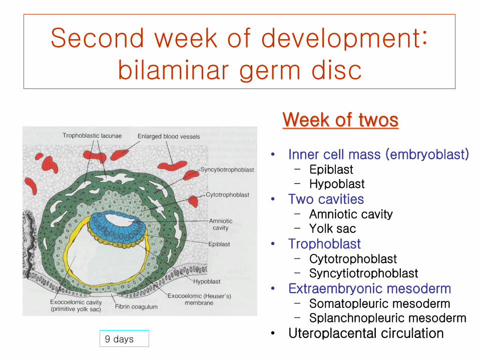
Second week of development:bilaminar germ disc
Week of twos
• Inner cell mass (embryoblast)– Epiblast– Hypoblast
• Two cavities– Amniotic cavity– Yolk sac
• Trophoblast– Cytotrophoblast– Syncytiotrophoblast
• Extraembryonic mesoderm– Somatopleuric mesoderm– Splanchnopleuric mesoderm
• Uteroplacental circulation9 days

Amniotic
cavity
Day 12

Day 13
(primary yolk sac)

Third week of development: Trilaminar germ disc
Gastrulation
• Bilaminar germ disc
-> trilaminar germ disc– Ectoderm
– Mesoderm
– Endoderm
• Primitive streak– Thickened linear band of
epiblast
– 15- to 16-day embryo: clearly visible

Epiblast ->
Source of all of
the germ layers
1. ectoderm
2. mesoderm
3. endoderm
18-day embryo
1.25 mm in length

Derivatives of the ectodermal germ layer
1. Central nervous system
2. Peripheral nervous system
3. Skin, including hair and nails
4. Sensory epithelium of ear, nose, and eye
5. Pituitary, mammary, and sweat glands and enamel of the teeth

Derivatives of the mesodermal germ layer
• Connective tissue, cartilage, bone and striated and smooth muscles
• heart, blood and lymph vessels and cells
• kidney, ovary and testis, genital ducts, serous membranes lining, spleen, and adrenal cortex

Derivatives of the endodermal germ layer
1. Epithelial lining of the gastrointestinal tract, respiratory tract, and urinary bladder
2. Parenchyma of the thyroid, parathyroid, liver and pancreas
3. Epithelial lining of the tympanic cavity and auditory tube

Fate of the primitive streak
• 4주말 이후 급격히 줄어들다사라짐
• Sacrococcygeal teratoma– sacrococcygeal region에남은
primitive streak에서기원
– 신생아에서 가장 흔한 종양

Teratogenesis associated with gastrulation
• The beginning of the third week of development, when gastrulation is initiated, is a highly sensitive stage for teratogenic insult.
• At this time, fate maps can be made for various organ systems, such as the eyes and brain anlage, and these cell populations may be damaged by teratogens.
• For example, high doses of alcohol at this stage kill cells in the anterior midline of the germ disc, producing a deficiency of the midline in craniofacial structures and resulting in holoprosencephaly.
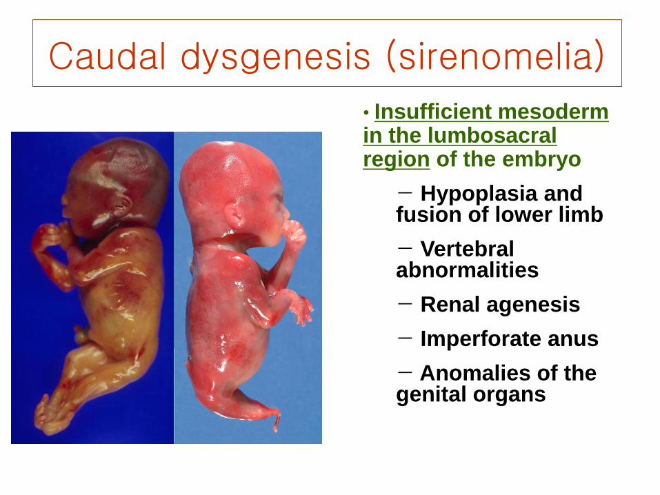
Caudal dysgenesis (sirenomelia)
• Insufficient mesoderm in the lumbosacral region of the embryo
− Hypoplasia and fusion of lower limb
− Vertebral abnormalities
− Renal agenesis
− Imperforate anus
− Anomalies of the genital organs

Formation of the notochord
B C
Notochord:1. Primitive axis of the embryo
2. Induction - neural plate (future nervous system)
Fate of the Notochord:
– Regress in the vertebral bodies
– Intervertebral disc: persist as the nucleus pulposus

20 days
Neurulation includes
the formation of the neural
plate (day 18-19), neural
folds (day 20-21), and the
neural tube (day 22-26); the
latter will develop into the
future brain and spinal
cord

Final closure
Anterior neuropore: 25th day
Posterior neuropore: 27th day

Neural tube defects
• Most common CNS malformation, 1/1000 births
• Primary non-close of the neural tube
• Spectrum– Anencephaly
– Meningocele
– Meningoencephalocele
– Meningomyelocele

Anencephaly• Absence of scalp, calvarium,
and normal brain -> “frog’s face”
• Area cerebrovasculosa
• Hypoplastic adrenal glands with absent fetal zone
• Recurrence rate: 3-5%
• DDx: amniotic disruption sequence
Amnion
disruption
consequence
Anencephaly

Holoprosencephaly
4th week, 4mm, 3 vesicle stage 5th week, 8 mm, 5 vesicle stage
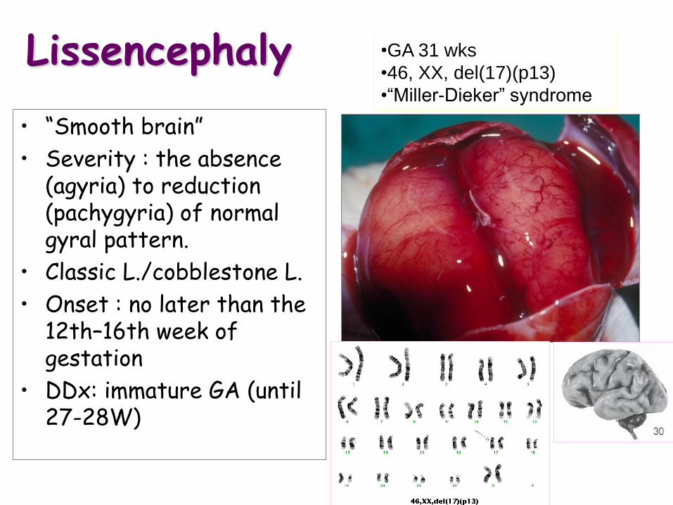
Lissencephaly
• “Smooth brain”
• Severity : the absence (agyria) to reduction (pachygyria) of normal gyral pattern.
• Classic L./cobblestone L.
• Onset : no later than the 12th–16th week of gestation
• DDx: immature GA (until 27-28W)
•GA 31 wks
•46, XX, del(17)(p13)
•“Miller-Dieker” syndrome

Formation and migration of neural crest cells in the spinal cord
22 days

Neural crest cells• Vulnerable cell population
• Easily killed by compounds such as alcohol and retinoic acid.
• Deficient in superoxide dismutase and catalase enzymes that are responsible for scavenging free radicals.
• Neural crest derivatives Connective tissue and bones of the face and skull Dermis in face and neck
☞ Severe craniofacial malformations☞ Treacher-Collins' Syndrome, DiGeorge anomaly...
Conotruncal septum in the heart ☞ cardiac anomalies including persistent truncus arteriosus, TOF and TGA
Cranial nerve ganglia, spinal ganglia, sympathetic chain and preaortic ganglia, parasympathetic ganglia of the gastrointestinal tract, glial cells, schwann cells, adrenal medulla, C cells of the thyroid gland, arachnoid and piamater, melanocytes, odontoblasts

MIGRATION PATHWAYS OF
NEURAL CREST CELLS
from forebrain, midbrain,
and hindbrain regions into
their final locations (shaded
areas) in the pharyngeal
arches and face
Skeletal structures of the
head and face. Mesenchyme
for these structures is
derived from neural crest
(blue), lateral plate
mesoderm (yellow), and
paraxial mesoderm (red).

Treacher-Collins' Syndrome /
mandibulofacial dysostosis
• Characterized by malar hypoplasia due to underdevelopment of cheek bones, mandibular hypoplasia, down-slanting eyes and malformed external ears
• Normal development and intelligence
• AD with variable penetrance
• 60% as new mutation
www.treachercollins.org/main.ht
ml

Velocardiofacial Syndrome/DiGeorge anomaly
• 22q11.2 deletion
• “CATCH 22”
– Cardiac defects
– Abnormal face
– Thymic hypoplasia
– Cleft palate
– Hypocalcemia
• Abnormal development of neural crest cells
• Specific facial features – low-set ears, wide-set eyes,
a small jaw, and a short groove in the upper lip
• Etiology– Genetic causes, exposure to
retinoic acids, alcohol, and maternal DM
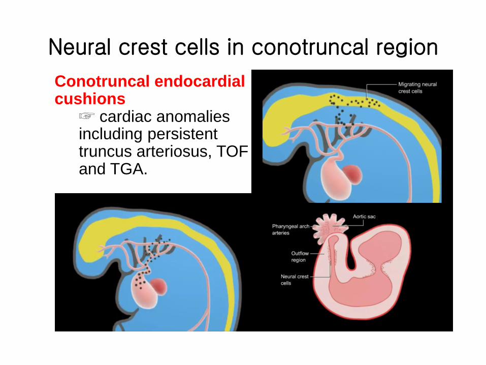
Neural crest cells in conotruncal region
Conotruncal endocardialcushions
☞ cardiac anomalies including persistent truncus arteriosus, TOF and TGA.

Limb growth and development
A. 5-week embryo B. 6-week embryo C. 8-week embryo
The hindlimb buds are less well developed than those of the forelimbs.
The most sensitive period for teratogen-induced limb malformations is the fourth and fifth weeks of development

Nasal pit
Lateral nasal prominence
Medial nasal prominence
5주
10주7주
6주
Medial nasal prominence
Lateral nasal prominence

A.B. 6.5-week embryo
The palatine shelves
are in the vertical
position on each side
of the tongue.
C.D. 7.5-week embryo
The tongue has
moved downward,
and the palatine
shelves have reached
a horizontal position.
E.F. 10-week embryo
The two palatine
shelves have fused
with each other and
with the nasal septum.
C D
FE


3주말
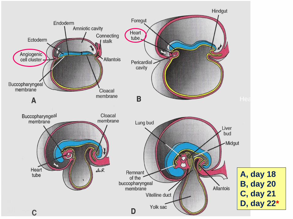
A, day 18
B, day 20
C, day 21
D, day 22*
Heart tube
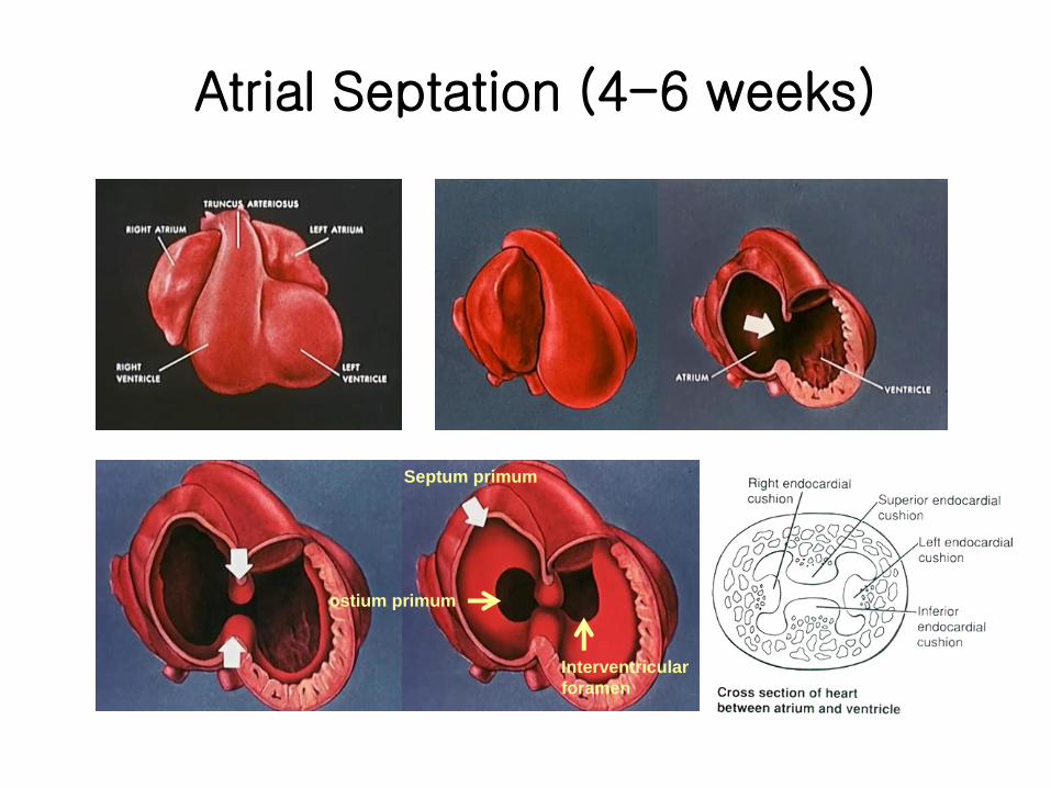
Atrial Septation (4-6 weeks)
Septum primum
ostium primum
Interventricular
foramen

ostium
secundum
Septum
secundum
35 days

Development of conotruncal ridges and closure of the interventricular foramen
6 weeks Beginning of 7 weeks

End of 7 weeks

Third to eighth week:The embryonic period
• Period of organogenesis
• Each of three germ layers gives rise to its own tissues and organs.
• Major features of the external body form recognizable by the end of the second month
