tspace.library.utoronto.ca · expression and characterization of the extracellular amino-terminal...
TRANSCRIPT

Expression and Characterization of the Extracellular Amino-Terminal Domain of the mGluR4 Subtype
Metabotropic Glutamate Receptor
Guangming Han
A thesis submitted in conformity with the requirements for the degree of Master of Science
Graduate Department of Pharmaceutical Science University of Toronto
Q Copyright by Guangming Han 2000
National Library I*I of Canada Bibliothèque nationale du Canada
Acquisitions and Acquisitions et Bibliographie Services services bibliographiques 395 Wellington Street 395, rue Wellington ûttawaON KtAON4 ûîtawaON KlAON4 Canada Canada
The author has granted a non- exclusive licence ailowing the National Library of Canada to reproduce, loan, distribute or seIl copies of this thesis in microform, paper or electronic formats.
L'auteur a accordé une licence non exclusive permettant à la Bibliothèque nationale du Canada de reproduire, prêter, distribuer ou vendre des copies de cette thèse sous la forme de microfiche/fïIm, de reproduction sur papier ou sur format électronique.
The author retains ownership of the L'auteur conserve la propriété du copyright in this thesis. Neither the droit d'auteur qui protège cette thèse. thesis nor substantial extracts £iom it Ni la thèse ni des extraits substantiels may be printed or othenvise de celle-ci ne doivent être imprimés reproduced without the author's ou autrement reproduits sans son permission. autorisation.

Expression and Characterization of the Extracellular Amino-Terminal Domain of the mGluR4 Subtype Metabotropic Glutamate Receptor
Guangming Han, Master of Science Graduate Department of Pharmaceutical Science
University of Toronto, Toronto
Abstract
The mGluR4 subtype metabotropic glutamate receptor is a presynaptic receptor that modulates
neurotransmitter release. We hypothesized that expressing the extracellular m i n o terminal
domain (ATD) of mGluR4 would produce a soluble protein that retains the pharmacological
characteristics of mGluR4.
In this study, I expressed and charactenzed 4 truncated versions of rnGluR4. A receptor
terminated 39 arnino acids upstream of the first putative transmembrane domain (TMD) was
secreted into the culture media of transfected human embryonic kidney cells. The rank order of
potency of metabotropic ligands at this receptor was similar to that of the full-Iength receptor.
Receptors tmcated at the carboxyl-terminus of the ATD and 98 or 174 amino acids upstream
from the first TMD al1 failed to be secreted from the cells and to bind the radioligand. The
truncated mGluR4 and the equivalent mGluR8 were expressed in Escherichia coii and insect
cells. Both systems produced immuno-reactive recombinant receptors that failed to display
ligand binding activity.
Together, these results demonstrate that all of the LIVBP homology region and part of the
cysteine-rich region are required for optimal secretion in a soluble form that retains binding
activity. The successfil expression of a soluble truncated m G l a 4 receptor produced a useful
substrate for the crystallization of the mGIuR4 ligand binding domain, and may provide
information for the design of subtype specific ligands for mGluRs.

Acknowledgements
1 would like to extend my deepest thanks to my supervisor, Dr. David R. Hampson, for his
expertise and guidance that made this project possible. 1 would particularly like to thank him for
providing me such great opportunities to expend my knowledge in the field of metabotropic
glutamate recepton and in medical research in general. 1 would also like to acknowledge my
advisory cornmittee members Drs. James Wells and David MacLeman for their expertise and
advice.
1 must thank the current and previous members of Our laboratory for their support throughout
my study. I would especially like to extend my deep gratitude to Ms. Xi-Ping Huang for al1 her
technical support and generous help. Many thanks also go to Dr. Vanya Peltekova for her heip
and wonderfil discussions on and off science. I also need to thank my fellow students Geoff
Homby, Nima Soleymanlou, Clinton Wong, Erica Rosemond, Mark Naples and many others at
the Faculty of Pharmacy for their continues support.
My sincere thanks must go to my grandparents, who introduced me to the world of research
and encouraged me to develop interests in medical sciences at a young age. 1 would also like to
thank my parents for their love, support and helpfùl insights as researchers and former graduate
students. Last but not the least, special thanks go to my fiancé Feng, for sharing my laughter and
tears, and for his tremendous suppoa and understanding throughout the years.
This project was supported by rny scholarship frorn the Medical Research Council of Canada,
and the Pharrnaceutical Manufacturers Association of Canada Health Research Foundation,
iii

Table of Contents
Abstract
Acknowledgrnents
Table of Contents
List of Abbreviations
List of Figures
List of Tables
1.0 Introduction
1.1 Classification of Metabotropic Glutamate Receptors
1.2 Pharmacology of Group LU. mGluRs
1.3 Distributions of Group III mGluRs
1.4 Possible Physiological Function of Group III mGluRs
1.5 Potential Clinical Use of the Group DI mGluR Agonists
1.6 The Three Structural Domains of mGluRs
1.7 Oligomenzation of mGluRs
1.8 The ATD and the Ligand Binding Domain of mG1uR.s
1.9 Objectives and Rational
2.0 Materials and Methods
2.1 Chemicals and Reagents
2.2 Standard Molecular Biology Procedures
2.2.1 Bactenal Transformation
Page
. . LI
... 111
iv
. . . vi11
xi
xii

2.2.2 Plasmid Preparation
2.2.3 Restriction Enzyme Digestions and Agarose Gel Electrop horesis
2.2.4 Blunt-end Reactions, DNA Dephosphorylation and DNA Ligation
2.3 Plasmids for Expressing Truncated mGluR4 Receptors in Mamrnalian System
2.3.1 c-myc Tagged Full-Length mGluR4a in pcDNA3
2.3.2 Truncated mGluR4 Receptors m4Tr-P586 and m4Tr-548
2.3.3 Plasmids Expressing m4Tr-Y489 and m4Tr-V4 13
2.4 Construction of Plasmids for E. coli Expression
2.4.1 pBAD/Myc-His System for Intracellular Expression in E. coli
2.4.2 PET-22b System for Periplasmic Expression in E. coli
2.5 Baculovirus Transfer Vectors for Protein Expression in hsect Cells
2.5.1 Truncated mGluR4 and mGluR8 with Original Mammalian Signai Peptides
3.5.2 Truncated mGluR4 and mGluRS with an Baculoviral Signal Peptide
2.6 Recombinant Protein Expression in Mammalian Cells
2.6.1 Transfection
2.6.2 Sodium Dodecyl Sulfate Polyacrylarnide Gel Electrophoresis and lmmunoblotting
2.6.3 Irnrnunocytochernistry
2.7 [ 3 ~ ] ~ - ~ ~ 4 Radioligand Binduig Assay
2.7.1 Cnide Membrane Preparation fiom Transfected HEK Cells

2.7.2 Soluble Sarnple Preparation
2.7.3 Membrane Binding Assay
2.7.4 Soluble Bhding Assay
2.8 Deglycosylation and Lectin Binding Test
2.9 Recombinant Protein Expressions in E. coli
2.9.1 L-Arabinose lnduced Expression of m4Tr-pBAD/hfyc-His and rn8Tr-pB ADIMyc-His
2.9.2 IPTG Induced Expression of m4Tr-pET22b and m8Tr-pET22b
2.10 Recombinant Protein Expressions in [nsect Cells
2.10.1 Insect Ce11 Cultures
2.10.2 Generation of Recombinant Baculovinis
2.10.2.1 Co-transfection for pBlueBac4.5 Constructs
2.10.2.2 Transposition and Transfection using Bac-To-Bac BacuIovirus Systern
2.10.2.3 PuriQing Recombinant Baculovinis by Plaque Assay and Titering of Vin1 Stocks
2.10.3 Expression Analysis of Truncated mGluR4 and mGluR8 in Insect CeIls
3.0 Results
3.1 Expression of Truncated Variants of mGluRJ in HEK Cells
3.2 Analysis of the Oligomeric Structure of m4Tr-Y548
3.3 ?rnmunocytochemical Analysis of Transfected HEK Cells
3.4 Ligand Binding Properties of Tnuicated Variants of mGIuR4
3.5 Deglycosylation of m4Tr-Y548
3.6 Expression of Tnincated mGluR4 and rnGluR8 in E. coli

3.7 Expression of Tmcated mGluR4 and mGluR8 in Insect Cells using the Baculovinis Expression System
4.0 Discussiou
4.1 Pharmacology of the Soluble Truncated mGluR4
4.2 Minimal Sequence in mGluR4 Ligand Binding
4.3 Biochemical Characterization of Soluble Truncated mGluR4
4.4 Expression of Truncated mGluR3 and mGluR8 in E. coli
4.5 Expression of Tnincated mGluR4 and mGluR8 using the Baculovirus Expression System
5.0 Conclusions and Future Considerations
6.0 References
vii

List of Abbreviations
IS, 3R-ACPD
lS, 3s-ACPD
AMPA
ATD
ATP
bp
&Il&\
BSA
C A M P
CaR
cDNA
CHO
CNS
CPCCOEt
CPPG
Cyclobutylene-AP5
DTT
EDTA
EGTA
GABAsR
GPCR
( 1 S, 3 R)-aminocyclopentanedicarboxylate
(1 S, 3s)-aminocyclopentanedicarboxylate
a-amino-3 hydroxy-5-methyl-isoxazoIe-4-propionate
arnino terminal domain
adenosine triphosphate
base pair
maximum number of binding sites
bovine serum albumin
c yclic adenosine monophosp hate
calcium-sensing receptor
complementary DNA
Chinese hamster ovary
central nervous system
7-hydroxyiminocyclopropan[P]chromen- 1 a-caroxylic acid ethyl ester
(R,S) a-cycloprop y l-4-p hosphonop henylglycine
(R,S) 1 -amino-3-@hosphonomethylene)cyclobutane
dithiothreitol
ethylenediaminetetra acetic acid disodium salt
ethyleneglyco-bis-N,N,N',N'-tetraacetic acid
type B y-aminobutyric acid receptor
G-protein coupled receptor

HEK
HEPES
IC50
iGluR
PTG
KD
Ki
kDa
KO
L-AP4
LB
L-CCG- t
L N B P
L-SOP
MAP4
MCPG
Mr
mG1uR
MPPG
NMDA
PBP
PBS
PCR
human ernbryonic kidney
N-[2-hydroxyethyl]piperazine-Nt-[Zethanc acid]
half-maximal inhibitory concentration
ionotropic glutamate receptor
isopropy 1-P-D-thiogalactoside
dissociation constant
inhibition constant
kilodalton
knock-out
L-amino-4-phosphonobutyric acid
Luria-Bertani
(X, 2' S, 1 ' S)-2-(carboxycyclopropy1)glycine
leucine-isoleucine-valine binding protein
L-serine-O- phosphate
(S)-2-amino-4-phosphonobutync acid
(R,S) cc-methyl-4-carboxy-phenylglycine
relative molecular weight
metabotropic glutamate receptor
(R,S) a-methyl4phosphonop henylglycine
N-methyl-D-aspartate
periplasmic binding protein
phosphate buffered saline
polymerase chab reaction

PMSF
S.E.M
SDS-PAGE
TMD
UTR
X-g a1
phenylmethylsulfonyl fluoride
standard error of the mean
sodium dodecyl sulfate polyacrylarnide gel electrophoresis
transmembrane domain
untranslated region
5-bromo-4-chloro-3-indoIyl-~-D-galac toside

List of Figures
Figure Number Page
Figure 1. Schematic representation of the proposed structure of an mGluR - 7
Figure 2. Dendrograrn and classification of the members of the mGluR fmily 3
Figure 3. Chemical structures of group UI rnGluR agonists and antagonists used in this study 8
Figure 4. Schematic diagram oCmGluR4 marnmalian expression constnicts 54
Figure 5. Irnmunoblotting analysis of full-length and truncated mGluR4 receptors expressed in HEK cells 57
Figure 6. The effect of the reducing agent DTT on the soluble m4Tr-YS 48 60
Figure 7. Relative level of ['HIL-AP~ binding to the full-length and truncated mGluR4s 63
Figure 8. Inhibition of ['H]L-AP~ binding to full-length mGluR4a and m4Tr-Y548 by ionotropic and metabotropic glutamate receptor ligands 65
Figure 9. Cornpetition curves for the inhibition of [ 'HIL-M~ binding to m4Tr-Y548 by group III mGluR agonists and antqonists 68
Figure 1 O. Enzymatic deglycosylation of rn4Tr-Y548 72
Figure 1 1. immunoblotting analysis of truncated mGluR4 and mGluR8 expressed in E. coii 76
Figure 12. hrnunoblotting analysis of truncated m G l W and mGluR8 expressed in Sf-9 cells 79

List of Tables
Table
1. Binding constants for agonists and antagonists at m4Tr-Y548 and full-length mGIuR4a recepton
2. Faatures of die expression construçts b r truncated rnGiuR4 m d mGluR8 in E. coli and insect cells
Page
70
8 I

1 .O Introduction
1.1 Classification of Metabotropic Glutamate Receptors
Metabotropic glutamate receptors (rnGluRs) are a group of guanine nucleotide binding
protein (G protein)-coupled neurotransrnitter recepton (GPCRs) that are activated by the
neurotransmitter L-glutamate. L-glutamate is the major excitatory neurotransmitter in
mamrnalian central nervous system (CNS) and plays important roles in a wide variety of CNS
functions. L-glutamate activates two different groups of receptors in the CNS: the ionotropic
glutamate receptors (iGluRs), and the mGluRs. The farnily of iGluRs is comprised of the N-
methyl-D-aspartate (NMDA) receptors, the a-arnino-3-hydroxy-5-methyl-isoxazole-4-propioate
(AMPA) recepton, and the kainate receptors. They act as ligand-gated cation channels to
mediate fast excitatory synaptic transmission. The rnGluRs, on the other hand, are involved in
modulating different transduction pathways, including the activation of phospholipase C and the
inhibition of adenylyl cyclase activity.
Sequence cornparisons have revealed the existence of at least three major families of GPCRs.
Receptors homologous to rhodopsin receptor constitute farnily 1. Farnily 2 receptors include
receptors for vasoactive intestinal peptide and glucagon. The mGluRs, the type B y-
aminobutyric acid receptors (GABABRs), the parathyroid calcium sensing receptor (CaR), a
class of pheromone receptors and a new class of taste recepton (Brown et al. 1993; Kaupmann et
al., 1997; Matsunami and Buck, 1997; Hoon et al. 1999) comprise the family 3 GPCR (Bockaert
and Pin, 1999). This receptor family is characterized by a large (- 600 residues in the mGluRs)
extracellular amino-terminal domain (ATD) (Figure 1). Similar to other GPCRs, the family 3
receptors contain an extracellular ATD, seven putative transmembrane domains (TMDs) and an

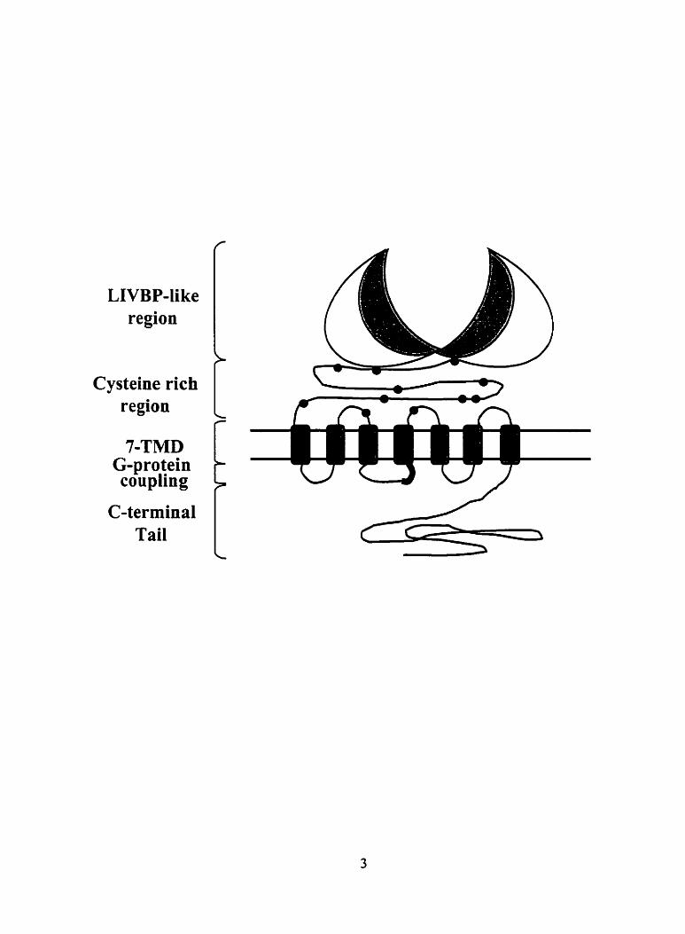
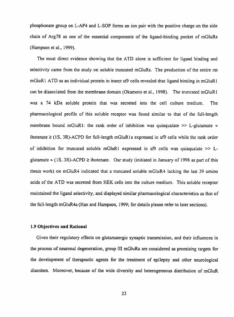
Figure 1 Schematic representation of the proposed structure of an mGluR protein. The
region rich in cysteine residues is indicated with black circles. The segment in the second
intracellular loop that is important for G-protein coupling specificity is indicated in black.
Cysteine rich
7-TMD G-protein coupling

intracellular carboxyl-terminal domain (Figure 1). However, they do not share sequence
homology with receptors fiom the other families. The overall sequence identity between
mGluRs, CaRs and GABAeRs is about 20% (Figure 2). The rnGluRs and the CaRs also share a
highly conserved cysteine-rich region in the ATD, whereas the GABAsRs lack this region.
The cDNA of the first member of mGluR family, the mGluR1, was cloned independently by
hvo proups in 1 % ) 1 (Houarned et al., 199 1; iMasu et al., 1991). To date, eight rnembers of the
mG1uR fmily have been cloned. They have been classified into three subgroups according to
their sequence homology, pharmacological profiles, and signal transduction rnechanisms (COM
and Pin, 1997). The group I mGluRs include mGluRl and mGluR5, the group II mGluRs
include mGluR2 and mGluR3, and the group III mGIuRs include mGluR4, mGluR6, mGluR7
and mGluR8. These rnGluRs share roughly 45% sequence homology arnong the subgroups,
whereas the degree of sequence identity increases to about 70% when comparing mGluRs within
the same subgroup (Figure 2).
The group I mGluRs are coupled to the activation of phospholipase C, resulting in an increase
in phosphoinositide turnover and the release of ~ a " h m intemal stores (Conn et al., 1995). A
recent study suggested that in addition to acting through transduction cascades involving G-
proteins, mGluRl is also able to mediate G-protein independent signal pathway through a Src-
farnily protein tyrosine kinase (Heuss et al., 1999). The splice variants of group 1 mGluRs
include rnGluRl a, L b, lc, Id, 1 C, and mGluRSa and Sb. Recently, a novel splice variant of
mGluRl (mGluRlE55) was cloned from the mouse CNS. The predicted protein product of
mGluRlE5S contains only the extracellular domain of the receptor and may be secreted (Zhu et
al., 1999). The function of this receptor remains unknown as it contains no TiMD or carboxyl-
terminal domain, and cannot couple to G-protein.

Figure 2 Dendrograrn and classification of the memben of the mGluR family, together with
the parathyroid ~a"-sensing receptor (PCaRl) and type B GABA receptor (GABAeRL a).

Group
I
il
1i1

The group iI mGluRs are negatively coupled to adenylyl cyclase and the production of cyclic
adenosine monophosphate (CAMP) through a pertussis toxin-sensitive G-protein (Gi/Go) when
heterologously expressed in ce11 lines (Thomsen et al., 1997; Flor et al., 1997; Corti, et al. 1998).
There is no evidence for the existence of splice variants of mGluR2 and mGluW to date. The
group III rnGluRs and their splice variants (mGluR4a, Jb, mGluR6, mGluR7a, 7b, mGluRSa and
8b) are also negatively coupled to adenylyl cyclase and the formation ofcAMP through a Gi/Go
protein. One exception is the newly cloned human mGluRSc (Malherbe et al.. 1999). The
rnGluR8c cDNA contains a 74-bp out-of-frame insertion which results in a unique 49 amino acid
carboxyl terminus inserted at amino acid 452 in the ATD. Similar to mouse mGluR1 E S , human
mGluR8c appears to be a truncated receptor that lacks the seven putative TMDs and the
carboxyl-terminal domain, and may exist as a soluble form of the receptor.
1.2 PharmacoIogy of Group III rnGluRs
The three groups of mGluRs differ considerably in their ligand selectivity. Group 1 receptors
are selectively activated by agonists quisqualate and 3,5-dihydroxyphenylglycine. Group II
receptors show high affinity for the agonists (2S, 1 'R, 2'R, 3'R)-2-(2, 3-dicarboxycyclopropyl)
glycine, (2S, 2 'S , 1 3)-2-(carboxycyclopropyi)glycine (L-CCG- 1), and 1 S, 3s
aminocyclopentanedicarboxylate (1 S, 3s-ACPD) (Pin et al., 1999).
The group III rnGluRs are characterized pharmacologically by their high affinity towards the
agonists L-amino-4-phosphonobutyric acid (L-AP4) and L-senne-O-phosphate (L-SOP) (Tanabe
et al., 1993; Brabet et al., 1998). Both L-AP4 and L-SOP are structurally similar to L-glutamate,
with the y-carboxyl group of glutamate being substituted for a phosphonate group (Figure 3).
Both L-AP4 and L-SOP are very specific for group DI mGluRs, as they are inactive at other

Figure 3 Chernical structures of group III mGIuR agonists and antagonists used in this
study.

Agonists for Group III mGluRs
L-Glutamic Acid L-AP4 L-SOP
Antagonists for Group III mGluRs
MAP4 CPPG MPPG

mG1uR.s. A number of other group III mGluR ligands appear to be active at group I and/or II
mG1uR.s as well. For example, although a-rnethyl-L-amino-4-phosphonobutyrate (MAP4) is a
more selective mGluR4 antagonist (Figure 3, Table 1), it is also antagonist with low potency on
mGluR2 (Johansen and Robinson, 1995; Gomeza et al., 1996). Most mG1uRl and mGluR.2
antagonists are inactive on group III mGluRs, except (R, S) a-rnethyl-4-
phosphonophenylglycine (MPPG), which appears to be slightly more potent to mGluR4 than to
mGluR2 (Gomeza et al., 1996A).
The pharmacological profiles of the mGluRs have been established using functional assays
that measure the accumulation or inhibition of second messenger molecules. Inhibition o f CAMP
formation is the most commonly used assay for the characterization of the group II and III
mGluRs. An alternative method involves CO-trans fection of receptors with chimeric G proteins
and the rneasurement of phosphoinositides or intracelluiar calcium levels (Gorneza et al., 1 W6A;
Hampson et al., 1999). Studies using the functional assays have established a rank order of
potency for agonists at the group 111 mGluRs of L-AP3 > L-SOP > L-glutamate > L-CCG-I >
(1 S, 3s) -ACPD » ( i S, 3R)-ACPD. Interestingly, functional assays measuring the inhibition of
CAMP revealed that the agonist potencies of mGluR7 were almost 1,000 times lower than the
other group III mGluRs (Okamoto et al., 1994; Saugstad et al., 1994), despite the 70% sequence
homology shared by the group III recepton. This suggests that mGluR7 may either bind ligand
with a much lower affinity or transduce its activation signal with greatly reduced efficacy as
compared with the other goup III mGluRs. Sequence comparison between mGluR7 and the
other group III mGluRs may therefore reveal key amino acid residues involved in the ligand
binding or signal transduction process of the group III mGluRs.

Although L-AP4 and L-SOP are specific for group III recepton, high potency and subtype-
specific ligands for individual group UI recepton have yet to be developed. An analogue of
AMPA, homo-AMPA has been s h o w to be specific for mGluR6. However, the potency of this
agonist is 4-fold lower than that of L-glutamate (Brauner-Osborne et al., 1996). Therefore, the
search for subtype-selective, high affinity ligand for group III mGluRs is still an ongoing
endeavor. A number of new g o u p ILI mGluR agonists and antagonists are now available for
testing, including the selective and potent agonist (R, S)-4-phosphonophenylglycine (PPG)
(Gasparini et al., 1999) and the antagonist (R, S)-cc-cyclopropyl-4-phosphonophenylglycine
(CPPG).
1.3 Distributions of Group III rnGluRs
The group III mGluRs are widely distributed throughout the CNS, with the exception of
rnGluR6, which is expressed exclusively in the retina. Immunocytochemical studies have shown
that the rnGluR4a protein is expressed in discrete neuronal populations in the rat and rnouse
CNS, with the highest level found in the cerebellurn (Kinoshita et al., 1996; Mateos et al., 1998).
A study on mGluR4 knock-out (KO) mice using ['H]L-AP~ autoradiography also confirmed the
presence of high levels of mGluR4 protein expression in the cerebellum (Thomsen and
Harnpson, 1999). Mice lacking mGluR4 displayed a significant decrease in [ 3 ~ ] ~ - ~ ~ 4 binding
compared with the wild-type mice in the molecular layer of the cerebellum. mGluR4 is also
expressed in the hippocampus, cerebral cortex, olfactory bulb, strîtum, pirifonn cortex/amygdala,
and thalamus (Risso Bradley et al., 1996; Phillips et al., 1997; Risso Bradley et
Within the hippocampus, moderate mGluR4a expression is present in the molecular
dentate gynis, while in the cerebrai cortex mGluR4a expression is distributed evenly
al., 1999).
ayer of the
throughout

the cortex at the interface of cortical layers IV and V (Shigernoto et al., 1997; Phillips et al.,
1997). No study on mGluR4b protein expression has been reported to date.
The expression of mGluR6 protein is restricted to a post-synaptic site on rod bipolar cells of
the inner nuclear layer of the retina. It is suggested that this specific localization of mGluR6 is
mediated by a 9.5 kilobase sequence located upstream From the translational initiation site on the
mGluR6 gene (Ueda et al., 1997). This is based on experiments using transgenic mice with a P-
galactosidase reporter jene fused to this 9.5 kilobase fragment. In these mice, expression of the
enzyme was found restricted to the retinal bipolar cells.
The mGluR7a is the most abundant group III mGluR in the brain. Lt is widely distributed
throughout the CNS, with the highest levels in the olfactory bulb, hippocampus, and cerebral
cortex of the adult rat. The mGluR7a is the principal member of the group III mGluRs expressed
in the hippocampus and is labeled with high intensity in the C M , CA3 and dentate gyrus regions
(Shigemoto et al., 1997; Kinoshita et al., 1998). Moderate levels o f mGluR7a are found in the
amygdala, basal ganglia, supenor colliculus an spinal cord and lower levels in the thalamus and
hypothalamus (Ohishi et al., 1995; Kinoshita et al., 1998). The mGluR7b protein is expressed in
a limited fasashion and is almost always CO-localized with the mGluR7a (Kinoshita et al., 1998).
The fùnctional significance of the CO-localization is unclear, and no evidence for the direct
interaction between mGluR7a and mGluR7b has been reported to date.
mGluR8 protein is expressed most prominently in the olfactory bulb, pontine grey, and the
lateral reticular nucleus of the thalamus (Duvoisin et al., 1995; Saugstad et al., 1997). Lower
levels of mGluR8 expression were found in the rat cerebral cortex, cerebellum, hippocarnpus,
and mammillary body (Saugstad et al., 1997). in the hippocampus, mGluRSa shows prominent
expression in the molecular layer of the dentate gyms and the teminal zone of the lateral

perforant path in the CA3 region (Shigemoto et al., 1997). Detailed imrnunohiçtochemistry
studies on rnGluR8b have yet to be conducted.
Except for mGluR6, which is expressed post-synaptically in the retina, immunohistochemical
studies have shown that mGluR4, mGluR7, and mGluR8 are pnmarily located presynaptically at
asymmetric glutarnatergic synapses (Shigemoto et al., 1997). In addition, the presynaptic
localization of mGluR4, mGluR7 and mGluR8 is also observed at the inhibitory GABAergic
synapses in various regions in the brain (Salt et al., 1996; Risso Bradley et al., 1996; Kinoshiia et
al., 1998).
1.4 Possible Physiological Function of Group III rnGluRs
Activation of group III mGluRs has been s h o w to inhibit adenylyl cyclase activity and
CAMP formation in neuronal cultures (Prézeau et al., 1994; Bruno et al., 1995). The negative
coupling to CAMP formation, together with their presynaptic localization at glutamatergic
synapses, suggests a role for mGluR4, 7 and 8 as autoreceptors in regulating glutamate release
from nerve teminals. Furthemore, agonists at group III mGluRs have been shown to inhibit
voltage-gated ca2+ charnels in cultured olfactory bulb neurons, cultured hippocampal cells, and
in isolated pyramidal neurons (Sahara and Westbrook, 1993; Trombley and Westbrook, 1992;
Stefani et al., 1998). Evidence indicates that the inhibition of ~ a " cunents is likely due to
interactions between the G-proteins and the ca2' channels, and is not dependent on protein
kinase A (Herrero et al., 1996). Since voltage-gated ca2' currents are known to play an
important regdatory role in the modulation of central synaptic transmission, these observations
fit well with the hypothesis that group III mGluRs function as presynaptic autoreceptors. Indeed,
group III mGluR agonists have been shown to cause a reduction in synaptic transmission at
glutamatergic synapses (Attwell et al., 1995; Dietrich et al., 1997).
Studies on KO mice lacking mGluRl have provided more direct insights on the biological
roles played by the receptor. These mice showed normal g ros motor performance, but were
deficient on the rotating rod motor-leaming test, suggesting that rnGluR4 KO rnice may have an
impaired ability to lem compiex rnotor rasics (Pekhietski et ai., 1996j. h analysis of
presynaptic short-term synaptic plasticity at the parallel fiber-Purkinje ce11 synapse revealed that
paired-pulse facilitation and post-tetanic potentiation were impaired in the mutant mice, whereas
long-term depression was not impaired (Pekhletski et al., 1996). Taken together, these results
suggested an important function o f mGluR4 in maintaining synaptic efficacy during repetitive
activation. A piausible mechanism is that mGluR.4 may act io conserve the synaptic stores of
glutamate for release upon repetitive stimulation; in mice lacking the receptor, the stored
glutamate would be npidly depleted during repetitive firing (Pekhletski et al., 1996). The
mGluR7 KO mice have been shown to have reduced f e u response and a deficit in conditioned
taste aversion (Masugi et al., 1999). Since the amygdala is the most likely region being involved
in both of the phenotypes, it has been suggested that mGluR7 is cntical in amygdala-dependent
aversion leaming (Masugi et al., 1999).
1.5 Potentiai Clinical Use of the Group III mGluR Agonists
One of the potentid beneficial therapeutic effects of group III mGIuR agonists is reduction of
neuronal damage that occurs after stroke, traumatic brain injury, or in certain neurodegenerative
disorders. Marked increases in the extracellular levels of glutamate are observed following brain
or spinal cord trauma and are correlated to injury severïty (Faden et al., 1989; Panter et al.,

1990). It is well accepted that the excessive release of glutamate causes neurotoxicity.
Activation of iGluRs including the NMDA and AMPA receptors induces large increases in the
concentration of neuronal cytosolic free ~ a " , due to influx through NMDA receptors
andor secondary activation of voltage-gated caZi channels. This will eventually result in
neuronal death, via a pathophysiological process cornmonly known as excitotoxicity (Choi,
i988; Faden et ai., i 989; LVrathaii et ai., i99ij.
It has been s h o w that mGluR4, but not group I or group II mG1u.R has an increased
expression in vulnerable b r i n areas following global ischernia in rats (Iversen et al., 1994). In
addition, reduced level of mGluR4 expression was observed in cerebellar granule neurons
undergone apoptotic neuronal death (Borodezt et al., 1998). Both observations suggest the
possible involvement of mGluR4 in regulating the downstream signaling events that are elicited
following neuronal darnage. Consequently, agonist activation of group III mGluRs may inhibit
the release of glutamate, and impart neuroprotective properties to these receptors. A number of
studies support this hypothesis. Maiese et al. have s h o w that administration of L-AP4 before
nitric oxide exposure significantly increased survival of rat hippocampal neurons in culture
(Maiese et al., 1996). Selective group III agonist also have been shown to protect cortical cells
or cerebellar granule cells in culture against P-arnyloid peptide induced apoptosis (Copani et al.,
1995), or cortical neurons exposed to a toxic pulse of NMDA (Bruno et al., 1995). Moreover,
treatment of L-AP4 or L-SOP to cortical neuronaVglial cultures subjected to mechanical injury
resulted in dose-dependent neuroprotection (Faden et al., 1997).
In addition to their neuroprotective properties in neuronal injury, agonists of the goup III
mGluRs may aiso have ad-convulsant Function. in support of a regulatory role for the group III
mGluRs in epilepsy, an increase in mGluR4 rnRNA levels has been reported in rats exhibiting

status epilipticus (Aronica et al., 1997). A recent study indicated that the expression of mGluR4
in hippocampd neurons Erom patients with chronic mesial temporal lobe epilepsy was up-
regulated at both mRNA and protein levels (Lie et al., 2000). The upregulation of mGluR4
would likely counteract excessive calcium influx and glutamate release, and subsequently reduce
synaptic transmission. Moreover, the sensitivity of the group III mGluRs towards L-AP4 was
found to be enhanced in rat with arnygdala-kindled seizures (Neugebauer et al., 1997). L-AP4
produced depressions of laterai arnygdala evoked monosynaptic excitatory postsynaptic currents
(EPSC) in kindled neurons with a ECjo value of 10.8 nkf, which was 30 times lower than that of
control neurons (297 nM). This observation suggests that during a seinire, the group III rnGluRs
become more sensitive to stimulation with a given level of glutamate, either due to the changes
in receptor affinities or the alterations in second messenger systems. Together, these findings
suggest that the group III rnGluRs c m be useful drug targets in the treatment of epilepsies. In
support for this notion, administration of group III mGluR agonists has been shown to protect
against both chemically and electrically induced seizures in several rodent rnodels of epilepsy
(Abdul-Ghai et al., 1997; Thomsen and Daldy, 1998; Gasparini et al., 1999).
1.6 The Three Structural Domains of mGluRs
The mGluRs are large molecules ranging in size from 95 kDa for mGluR6 to 133 kDa for
rnGluRl a. The mG1uR.s have a large extracellula. ATD, followed by a hydrophobie region
containing seven putative TMDs and an intracellular carboxyl-terminal domain with variable
lengths. The AïDs of the mGluRs are highly conserved. There are 17 conserved cysteine
residues within the predicted ATD of the mGluRs, some of which likely to form disulfide bonds
and play a role in the protein folding and/or signal transduction (Conn and Pin, 1997). One

significant difference between mGluRs and the family 1 and fmi ly 2 GPCRs is the location of
their ligand binding sites. While the ligand binding regions of most GPCRs reside within their
TMDs, ligand binding sites of the mGluRs and the other members of the fmily 3 GPCRs are
contained within the ATD region.
In most conventional GPCRs (Family 1), the third intracellular loop plays a cntical role in the
G-protein coupling specificity. Interestingly, in mGluRs, the second intracellular loop likely
plays a role equivalent to that of the third intracellular loop of the other GPCRs. A study on
chimeric receptors between the phospholipase C-coupled mGluR1 and the adenylyl cyclase-
coupled rnGluR.3 has shown that the second intncellular loop of mGluRs plays a critical role in
G-protein coupling specificity (Gomeza et al., 1996B) (Figurez). Studies also indicated that all
intracellular segments are likely involved in the coupling and activation of the G-protein
(Gomeza et al., 1996B, Pin et al., 1994). The amino acid sequence of the first and third
intracellular loops is highly conserved among al1 the mGluRs and PCaRl, suggesting that these
domains play important roles in G-protein activation. Indeed, a mutation in the third intracellular
loop of the human CaRl that prevents the receptor h m activating phospholipase C has been
found in familial hypocalciuric hypercalcemia patients (Pollak et al., 1993).
In a recent study, 7-hydroxyiminocyclopropan[P]c hromen- 1 a-carboxylic acid ethyl ester
(CPCCOEt) has been shown to act as a noncompetitive mGluRl antagonist which inhibits
receptor signaling without affecting glutamate binding (Litschig et al., 1999). This compound
selectively inhibited glutamate-induced increase in intracellular calcium at human mGluRl b with
an ICjo of 6.5 FM while having no agonist or antagonist activity at human mGluR2, 4, 5 , 7 and
8. It has been suggested that CPCCOEt acts in a noncompetitive manner by decreasing the
efficacy of glutamate-stimulated phospoionsitide hydrolysis without affecting the ECso value of
glutamate. Chimenc and mutagenesis studies have s h o w that Thr8 15 and Ala8 18 of human
rnGluRlb, located at the extracellular surface of the seventh TMD, are critical in the binding of
CPCCOEt. It has been therefore proposed that the interaction of CPCCOEt with Thr815 and
Ala8 1 8 of rnGluR 1 disrupts receptor activation by inhibiting an intnmolecular interaction
behveen the agonist-bound extracellular ATD and the TMDs and very likely the subsequent
intramolecular transduction (Litschig et al., 1999).
The carboxyl-terminal dornain ofgroup III mGluRs has been shown to interact with G protein
py subunits (Gabellini et al., 1993; Prézeau et al.. 1996). Moreover, it has been s h o w that a
subdomain in the carboxyl-terminal tail of group III mGluRs binds calmodulin and G protein py
subunits in a mutually exclusive manner (O'Connor et al., 1999). This result suggests a novel
mechanism of presynaptic modulation in which cal'-calmodulin is required to release G protein
py subunits from the carboxyl-terminal domain of g o u p III mGluRs in order to rnediate
glutarnatergic autoinhibition. However, the carboxyl-terminal domain is likely to have other
functions. For exarnple, chimeric and deletion studies revealed that axon exclusion of rnGluR2
versus axon targeting of mGluR7 is mediated by their carboxyl-terminal domains (Stowell and
Craig, 1999). In addition, this domain may also be the target for a number of protein kinases.
1.7 Oligomerurtion of mGluRs
A unique structural feature shared by al1 eight mGluRs is the presence of 21 conserved
cysteine residues. Seventeen of these are in the ATD and two are in the extracellular loops.
Among the seventeen cysteines in the ATD, nine are at the C-terminal portion of the ATD
(Figure 2). Although the function of these cysteines is currently unclear, the strict conservation
of position irnpiies that their function is shared and important for this family of receptors.

One possible function for these cysteines is mediating dimerization of mGluRs. Under non-
reducing condition, mGluR5 expressed in human embryonic kidney (HEK) cells was shown io
migrate on polyacrylamide gel as a homodimer but not as a mGluRl/rnGIuRS heterodimer
(Romano et al., 1996). The existence of dimer under non-reduced condition was also observed
in our study on tnincated mGluR4 (Han and Harnpson, 1999). Co-irnrnunoprecipitation
expenments have shown that a mutant mGluR5 receptor tnincated a fe r the first TMD was able
to retain the ability to f o m dimer with either itself or the Ml-length mGluR5, suggesting that the
locus for disulfide-mediated dimerization is within the extracellular dornain of the receptor.
Furthemore, using proteolytic removal of al1 or part of the extracellular domain by trypsin
revealed that the cysteine(s) responsible for disulfide bond formation are in the mino-terminal
17 kDa of rnGluR5. In a more recent study, the dimerization of mGluRl a was reported in
membranes isolated from both rat brain and transfected BKK cells (Robbins et al., 1999). The
dimerization was suggested to form in the rndoplasrnic reticulum.
Interestingly, these 17 cysteines are also present in the ATD of CaR at equivalent positions.
A series of studies have shown that intermolecular interactions between dimeric calcium-sensing
receptor monomers are important for its normal function (Bai et al., 1998; Bai et al., 1999;).
Moreover, mutagenesis studies indicated that several cysteine residues within the ATD of CaR
were critical in the dimerization of the receptor (Pace et al., 1999; Ray et al., 1999). The more
distantly related GABABRs lack the cysteine nch region altogether, and form heterodimer by
interactions behveen their carboxyl-terminal tails (Jones et al., 1998; Kaupmann et al., 1998).
1.8 The ATD and the Ligand Binding Domain of mGluRs

In 1993, O'Hara and colleagues reported low homology between the large ATD of the
rnGluRs and a group of bacterial proteins called the periplasmic binding proteins (PBPs); the
most homologous of the PBPs was the leucine-isolucine-valine binding protein (LIVBP)
(O' Hara et al., 1 993). A three-dimensional compatibility searc h that identifies sequences fo lding
into a known structure (Bowie et al., 1991) showed that mGluRl scored in the top 0.1% of al1
database sequences queried with a LIVBP profile. A cornparison of mGluR 1-5 with LIVBP and
a nurnber of other PBPs in a multiple alignment indicated that the observed sirnilarity might be
biologically meaningful (O'Hara, et al., 1993).
The PBPs are a family of proteins that act as transporters to trafic amino acids and sugars
fiom the periplasmic space across the bactenal imer membrane and into the cells. These
proteins have an affinity of approximately 1 pM for their ligands, and fùnction to concentrate
important growth substrates inside the ce11 (Kelleman and Szmelcman, 1971; h e s 1988). The
crystal structure of the LIVBP has been resolved. The LM3P is composed of nvo globular lobes
comected by three short stretches of peptide (the hinge region, Sack et al., 1989). This bi-lobed
structure exists in an open and a closed fom, and acts through the so-called Venus flytrap
mechanism. Upon binding of substrate in the open configuration, the PBP adopts a closed
conformation and interacts with a unique set of membrane-bound proteins that catalyzes the
actual membrane translocation of the substrate (Adams and Oxender, 1989).
Based on the homology, a three-dimensional structure of rnGluRl ATD sequences that
aligned with the available PBPs was modeled. The mode1 has the characteristics of the bactenal
PBPs: two large, globular lobes consisting of P sheets flanked by a helices, linked by a hinge
region consisting of three interdomain crossover segments. The ligand is thought to interact with
a binding site on one of the lobes (O'Hara et al., 1993). The hinge region has been implicated in

the transduction of binding signals. Mutations in close proximity to the hinge region of the
parathyroid calcium sensing receptor results in patients exhibiting genetic disease that affected
calcium homeostasis, which may due to the loss of the fùnction of the receptor (Pollak et al.,
1993, Pollak et al., 1993).
In the PBPs, substrate binding occurs in a clef? and is stabilized by hydrogen bonds. Two
ligand-bound t o m s of PBPs have been observed: an open f o m with ligand initially bound to
one domain, and a closed form in which ligand is bound to both domains and enclosed within the
cleft (Sack et al., 1989). In the open form of LIVBP, Ser79 and ThrlOî are involved in hydrogen
bonding to LIVBP ligands. k g 1 16, Phe276 and Asp323 are involved in the LIVBP-binding
pocket structure (Sack et al., 1989), part of which is a salt bridge between Argl16 and Asp333.
Interestingly, these residues align with identical residues in mGluRs.
A number of experiments have been carried out over the 1st few years providing informative
dues about the localization of the L- lu ta mate binding domain in mGluRs. Takahashi et al. have
produced a series of chimenc receptors by exchanging vanous parts of the ATD of nt mGluRl
to the corresponding segments of mGluR2 (Takahashi et al., 1993). The subsequent
pharmacological characterization of these chimeric receptoe allowed them to conclude that
substitution of up to half of the mGluR2 ATD in the homologous region of mGluR1 ATD is
sufficient to convert the pharmacological profile of rnGluRl into that of rnGluR2. Data obtained
from experiments on chimeric constructs of the ATD of human mGluR4 with the TMDs and
carboxyl-terminal regions of rnGluRlb dso indicated that pharmacological selectivity is
conferred by residues located in the ATDs of mGl& (Tones et al., 1995). in another approach,
polyclonai antibodies against two different portions of the ATD of rat mGluRl was shown to

inhibit the action of L-glutamate on the stimulation of phosphatidylinositol hydrolysis when
tested on CHO cells stably expressing mGluRl (Shigemoto et al., 1994).
In the study conducted by O'Hara et al. (1993), site-directed mutagenesis was used to probe
the mGluRl binding pocket for residues that are essential for ligand binding. As mentioned
previously, the arnino acid residue Ser79 and Th102 side chah atoms in the LIVBP are known
to t o m a hydrogen bond with the a-ammonium and a-carboxylate atoms of the glutamate ligand
(Sack et al. 1989). Alignrnent of the LIVBP with mGluRl shows that Serl65 and T M 8 8
residues of mGluRl correspond to Ser79 and ThrlO2 in the LIVBP. Single point mutations
changing either Ser165 or Thr188 in rnGluRl to alanine resulted in significant decrease in
afinity of mGluRl for its ligand. No functional response was observed with the TI S8A mutant,
and the affinity of mGluRl for both glutamate and quisqualate was reduced by over 10,000-fold
for the S165NT188A double mutant (O'Hara et al., 1993).
In a more recent study, Harnpson et al. (1999) used a molecular model of the tertiary structure
of rat mGluR4 ATD as guiding tool to probe the ligand-binding pocket of mGluR4. In mGluR4,
Ser159 aligns with Ser79 in the LIVBP and Ser165 in rnGluR1. m l 8 2 aligns with ThrlO2 in
the LiVBP and Thr188 in mGluR1. Mutation of Ser159 to alanine in rnGluR4 resulted in a 95 %
reduction in [ 3 ~ ] ~ - ~ 4 binding compared with the wild-type mGluR4, whereas the mutation of
Thr182 to alanine produced a 96% decrease in [ 3 ~ ] L-AP4 binding, despite that the expression
levels of the mutant receptors were comparable to that of the wild-type mGluR4. These
observations are in agreement with the loss of activity seen in the analogous mutation in
mGluR1. Arg78, which is conserved in a11 mGluRs, was identified as the third residue cntical in
ligand-binding to mGluR4. Mutation of R78A in mGluR4 resulted in a nearly complete loss of
[ 3 ~ ~ - A P 4 binding. It has been suggested that the y-carboxy group on L-glutamate or the y-
phosphonate group on L-AP4 and L-SOP foms an ion pair with the positive charge on the side
chain of k g 7 8 as one of the essential components of the ligand-binding pocket of rnGluRs
(Hampson et al., 1999).
The most direct evidence showing that the ATD alone is sufficient for ligand binding and
selectivity carne from the study on soluble tnincated mGluRs. The production of the entire rat
mGiuRl A l D as an individuai protein in insecr SB ceiis reveaied that iigand binding in mUiuRI
can be dissociated From the membrane dornain (Okamoto et al., 1998). The truncated mGluRl
was a 74 kDa soluble protein that was secreted into the ce11 culture medium. The
pharmacological profile of this soluble receptor was found similar to that of the full-length
membrane bound mGluR1: the rank order of inhibition was quisqualate » L-glutamate s
ibotenate 2 ( lS, 3R)-ACPD for full-length mGluRla expressed in sf9 cells while the rank order
of inhibition for truncated soluble mGluRl expressed in SB cells was quisqualate » L-
glutamate = (1 S, 3R)-ACPD 2 ibotenate. Our study (initiated in January of 1998 as part of this
thesis work) on rnGluR4 indicated that a truncated soluble mGluR4 lacking the last 39 arnino
acids of the ATD was secreted from HEK cells into the culture medium. This soluble receptor
maintained the ligand selectivity, and dispiayed similar pharmacological characteristics as that of
the full-length mGluR4a (Han and Hampson, 1999; for details please refer to later sections).
1.9 Objectives and Ritional
Given their regulatory effects on glutarnatergic synaptic transmission, and their influences in
the process of neuronal degeneration, goup iII mGluRs are considered as promising targets for
the development of therapeutic agents for the treatment of epilepsy and other neurologica1
disorden. Moreover, because of the wide diversity and heterogeneous distribution of mGLuR

subtypes, the opportunity exists for developing highly selective drugs that affect a limited
nurnber of CNS functions. More detailed analyses on the ligand binding mechanism of these
receptors are therefore required in order to design subtype specific dnigs.
Since it is usually dificult to conduct structural analysis such as X-ray crystallography on
membrane-bound proteins, a soluble segment of the ATD of mGluR4 that retains similar
pharmacologml profile as the wild-type receptor would be desirable for detailed structural
studies. O'Hara et al.3 mode1 of rnGluR ATD suggested the possibility of isolating such a
segment. A simiiar attempt in the study of' the structure of iGluR ligand-binding core was
successfùl. The iGluRs appear to have an LIWP-like domain, followed by a distinct glutamate-
binding protein-like region, which is split into two by the first three of the four putative
transrnembrane regions (the S1 and S2 regions). By directly comecting the two DNA stretches
that encode the SI and S2 segments of iGluR4, Keinhen and his coIleagues elegantly created a
soluble mini-receptor that lacked the putative transrnembrane domains. This soluble protein
displayed a ligand-binding profile typical of the AMPA-subtype receptors (Kuusinen et al.,
1995). In a sirnilar study on the NMDA receptor NRI, it has been shown that the S M 2
Eragment is responsible for the binding of Glycine (Ivanovic et al., 1998). Using this S M 2
Fragment, the crystal structure of GluR2 ligand-binding region in a cornplex with kainate was
finally solved (Armstrong et al., 1998).
Based on the homology between the extracellular ATD of the mGluR4 receptor and the
L N B P protein, and the evidence From previous studies, we hypothesized that a tmcated
receptor protein containing d l or part of the mGluR4 ATD would be soluble, yet retain the
ligand binding activity of the full-length rnGluR4. Such a receptor protein could be a substrate
for protein crystallization. One objective of my study was to produce a tnincated mGluR4

receptor with only the ATD or part of the ATD. Pharmacological and biochemical
characterization of this tmcated receptor would provide valuable answers to a series of
questions regarding the ligand binding to rnGluR4, including the impact of glycosylation, the
possibility of receptor dimenzation, and most importantly, the role of ATD in ligand selectivity.
A radioligand binding assay using the agonist ['HIL-AP~ provides a means to assess the
pharmacologml profile of the truncated mGluR4, which c m be compared to the wild-type
receptor. A second objective was to define the boundary for amino acid residues required in
ligand binding to mGluR4. The goal was to identify the shortest segment of the XTD of
mGluR4 that c m be expressed as a soluble protein and retain pharmacological activity. This
study would produce a soluble receptor protein containing only regions necessary and sufficient
for ligand binding, and would eliminate regions of the mGluR4 ATD not involved in ligand
binding but could potentially complicate the expression of the protein in other host systems such
as bacteria. This study would also explore the possible existence of other ATD regions outside
the LIVBP homology region that may be important in rnaintaining the pharmacological
characteristics of the receptor.
The third goal of this study was to express the muicated foms of mGluR4 and the closely
related mGluR8 in different expression hosts such as E. d i and insect cells For high-level
protein expression. The purification and production of crystals of tnincated mGluRs are
prerequisites for conducting structural shidies such as X-ray crystallography, which will in tum
provide direct information on the structure of the receptor binding pocket and the interaction
between ligands and the binding site. mGluR4 and rnGluR8 share high sequence homotogy
(over 70% identity). They display simitar a f i t i e s for most of the ligands including L-AP4 L-
SOP and L-glutamate (Pin et al., 1999), but different affinities for ligands such as PPG, which

appears to have higher potency at rnGluR8 than at rnGluR4 (Gasparini et al., L999). Cornparison
bebveen the ligand binding domains of these two recepton would provide usefbl insight for the
design of subtype-specific ligands.
2.0 Materials and Methods
2.1 Chernicals and Reagents
Al1 chemicals and reagents were purchased from Sigma Canada and BDH Inc., unless
otherwise indicated.
2.2 Standard Molecular Biology Procedures
2.2.1 Bacterial Transformation
Al1 solutions and plates used for Escherichia coli (E. coli) competent ce11 preparation and
transformation were prepared using Millipore water and were autoclaved. Bacterial cultures
were grown in Luna-Bertani (LB) medium ((wh): 1.0% bacto-tryptone, 0.5% yeast extract,
1.0% NaCI) and transformed bacteria were piated on LB-agar plates prepared by supplementing
LB medium with 1.5% bacto-agar. Appropriate antibiotics were added to the plates for selection
of transformed bactena. Al1 incubation steps were carried out at 37OC unless othenvise
specified, and ail shaking steps were carried out at 225 rpm on a New Brunswick orbital shaker.
For the preparation of competent E. coli, a single colony of DH5a (Gibco BRL) E. coli
was inoculated in 2 ml of LB medium and incubated at 37°C with shaking for 16 houn. An
aliquot of 15 pl of this overnight culture was added to 25 ml of fiesh LB and incubated at 3 7 T
with shaking and aeration until reaching an of 0.3. Competent cells were then prepared
using the calcium chloride (CaC12) method outlined in Sambrook, Fritsch and Maniatis (1989).
Afier the preparation of the competent DH5a cells, 10 ng of supercoiled plasmid DNA or 50 ng
of ligated relaxed plasmid DNA was added to 200 pl of competent cells and incubated on ice for
40 minutes. The cells were then heat-shocked for 45 to 60 seconds at 42OC and immediately
chilled on ice for 2 minutes. The cells were then incubated in 0.8 ml of fresh LB with shaking at
37OC for 1 hour. The transformed bacteria were then spread on LB agar plates containing 100
pg/ml ampicillin (Boehringer Mannheim). The plates were incubated at 37°C for 16 hours to
allow for selection of ampicillin-resistant colonies.
2.2.2 Plasmid Preparation
Ampicillin-resistant colonies were inoculated in 3 ml of LB medium containing 100
pg/ml ampicillin, and incubated for L6 hours at 37°C with aeration. Ovemight cultures were
harvested by centrifugation in 1.5 ml microfuge tubes at 3000 x g for 1 minutes. Plasmid DNA
was then isolated from the cells using the QIAspin iMiniprep Kit (Qiagen) according to the
manufacturer's protocol. Plasmid DNA concentration and purity were verified by
spectrophotometric analysis at 280 MI and 260 nm. A small arnount of the plasmid DNA
product (approximately 0.2 pg) was subjected to restriction digest and gel electrophoresis to
confirm the presence of the correct plasrnid and orientation of the inserted DNA.
2.2.3 Restriction Enzyme Digestions and Agarose Gel Electrophoresis
Restriction enzyme digestions were used for subcloning purposes and to screen
transformants for the appropriate size and orientation of DNA inserts in plasmids constructed.
Digestions were perfonned in reaction volumes vary from 10 to LOO pl. The digestion reactions
typically contained 1-2 pg DNA, deionized sterile water, specific IOX reaction buffer supplied
by the manufacturer, and 5-10 u n i t s of specific restriction enzyme (New England Biolabs, Gibco
BRL). Al1 reactions, unless specified were incubated at 37' C for 2 hours to ensure cornplete
digestion of the DNA.

Agarose gel electrophoresis was used to visually analyze restriction digests and quantify
purified plasrnid DNA or DNA fragments. Unless specified, electrophoresis was performed
using 0.8% agarose gels dissolved in 0.5 x Tris-acetate/EDTA buffer (20 m M Tris-acetate, 0.5
mM EDTA) supplemented with 0.5 pg/ml ethidium bromide. DNA-gel loading bu ffer (0.25 %
bromophenol blue, 0.25% xylene cyan01 FF, 30% (w/v) glycerol in water) was added to each
sarnple at 116 of the total volume prior to loading on the gel. Samples were run on a Mupid-21
Mini-Gel apparatus (Helixx Technologies) at a setting of 100 volts until adequate separation O f
digested bands had been accomplished. Fluorescent bands were visualized using an ultraviolet
transilluminator (UVP Incorporated). Gels were also loaded with a 1-kilobase or 100 bp ladder
(Gibco B U ) for determination of DNA fragment lengths, and DNA was quantified by
cornparison with a known arnount of Hind III digested J. DNA (Gibco BRL).
2.2.4 Blunt-end Reactions, DNA Dephosphorylation and DNA Ligation
Treatment with T4 polymense was used to create blunt ends in DNA fragments For
subcloning purposes. In a typical reaction, approximate I pg of DNA in a volume of 25 pl was
mixed with 5 pl of 10X reaction buffer, 2.5 pl of bovine serurn albumin (BSA, 1 mg/ml), 4 pl of
dNTPs (1.25 mM), 2 pl of T4 polymerase (3000 u/ml, Pharmacia) and 11.5 pl H20. The
reaction was camed out at 12OC for 20 minutes.
To prevent self-ligation in vectors with blunt ends, these vector were dephosphorylated by
calf intestinal alkaline phosphatase prior to ligation reactions. The typical reaction mixture
contained 1 pg of DNA in 35 pl W20, 5 pl of 10X reaction buffer and 10 pl diluted enzyme,
which was made fiom 0.5 pl calf intestinal alkaline phosphatase (30 units/ml, Gibco BRL) and
28 pl dilution buffer. The reaction was carried out at 37°C for 1 hour. The dephosphorylated
product was precipitated as descnbed above.
In DNA ligation reaction, DNA kagrnents with cornpetitive ends were joined by T4 ligase.
Typically, a 20 pl reaction mixture contained approxirnate 30 ng of insert (50 ng for blunt-ended
ligation) and 10 ng of vector in Tris-HCl (pH8.0), 1 pl of T4 DNA ligase (400,000 unitdml, New
England Biolabs), 2 pl 10X reaction buffer, 2 pl of 10 m M ATP and H 2 0 to make the final
volume of 20 pl. The reaction was done at 16°C for 16 hours.
2.3 Plasmids for Expressing Truncated mGluR4 Receptors in Marnmalian System
2.3.1 C-myc tagged full-length mGluR4a in pcDNA3
Plasmid expressing wild type mGluR4a (mGluR4a-pcDNA3) was constructed by inserting
Bgl II-EcoR 1 hgment of rat mGIuR4a cDNA in the pBluescnpt SK- phagemid (provided by Dr.
S. Nakanishi) into the pcDNA3 rnammalian expression vector (Invitrogen Corp.) at the BumH 1
site. For the construction of c-myc-tagged mGluR4a, the mGluR4a-pcDNA3 plasmid was cut
with B o 1, and the Iarger Fragment containing the pcDNA3 backbone was ligated to itself (the
5'-mGluR4a-pcDNA3 plasmid). The oligonucleotides encoding the c-nzyc epitope 5 ' -
GTCACGAACAAAAGCTTATTTCTGAAGAAGACTTGGATCC-' (f-BstE II-mye) and
5'-GTGACCTGGATCCAAGTCTTCTTCAGAAATAAGCTTTTGTTC-3 (rev-BstE [I-WC)
were phosporylated, annealed to each other, and inserted into the 5'-mGluR4a-pcDNA3 piasmid
at the BstE II site to produce 5'-mGluR4a-pcDNA3. The 93 1-bp Nde 1-Xho 1 Fragment from
S'rnGluR4a-myc-pcDNA3 and a 33 35-bp . n o 1-Not 1 fragment of mGluR4a-pcDNA3 were
subcloned into pcDNA3 at the Nde 1-Nor I sites using a three-piece ligation to produce myc-
mGla4-pcDNA3. Plasmids mGluR4a-pcDNA3 and mGluR4a-myc-pcDNA3 were constnicted
by Dr. Roman Pekhletski.
2.3.2 Truncated mGluR4 receptors m4Tr-P586 and m4Tr-Y548
The DNA riagrnent encoding the ATD of mGluR4 fiom amino acid 1 to 586 was generated
by polyrnerase cham reaction (PCR). The tonvard pnmer
SYTTTCCGAAATGTCCGGGAAGG3' (m4-8 olig) included the start codon, and annealed
upstream to the signal peptide sequence in rnGluR4a-n1y~-pcDNA3. The reverse primer
SyCTTACGGCGAGTCCCACTCCA3' (mR4-tmnc-Rev) created a stop codon after Pro586, the
second Iast amino acid before the beginning of the first TMD. In order to produce PCR product
with phosphorylated ends, the two primers were treated with T4 kinase prior to the PCR reaction.
300 pmol of each primer together with 1 pl of 10X T4 kinase buffer (New England Biolabs), 1 pl
of T4 kinase (10,00Ounits/ml, New England Biolabs), and 1pl of 1mM ATP were incubated in a
1 0 ~ 1 reaction mixture at 37'C for 18 minutes. The PCR reaction consisted of a 100p1 mixture
containing: 2.5 units of Taq polymerase (Pharmacia), 10 pl of IOX Taq reaction buffer
(Pharmacia), 2 pl of the T4 kinase reaction mixture containing 60 pmoi of each primer (added
irnmediately after the 18 minutes phosphorylation reaction descnbed above), 500 ng of
mGluR4a-myc-pcDNA3 as the template, and 125 mole of each of the four dNTPs (Pharmacia).
The PCR products were purified with QIAquick PCR purification kit (Qiagen). Procedures were
carried out according to manufacturer's instruction. The purified PCR fragment was subjected to
blunt-end treatment, and isolated using QIAquick gel extraction kit (Qiagen) according to the
manufacturer's protocol.

The pcDNA3 vector was digested with EcoR V (Pharmacia), and extracted with equal volume
of phenol-chlorofonn-isoamyl alcohol (2524: 1 vlv, Gibco BRL). To precipitate the DNA From
the digestion mix, 1/10 volume of 3M sodium acetate (pK 5.2) and 3 volumes of anhydrous
ethanoi were added. The mixture was left at -20°C for I hour, and centrifuged at 14,000 X g for
10 minutes. The pellet containing the vector DNA was washed with 70% ethanol (v/v), and air-
dned. The DNA was then dephosphorylated.
To prepare the insert for constnict m4Tr-Y548-pcDNA3, a 1686 bp fragment was isolated
from the mGluR4a-~c-pcDNA3 by digesting with Kpri 1 (Gibco BRL). This fragment encoded
mGluR4 signal peptide, myc epitope, and the ATD tmncated afier Tyr548, which was 39 arnino
acids fiom the beginning of the fint TMD.
The ligation reaction for the PCR Fragment and the EcoR V digested pcDNA3 vector had an
approximate 5: 1 insert to vector rnolar ratio. A ratio of 3: 1 was used for the ligation reaction for
the Kpn 1 fragment from rnGluR4a-myc-pcDNA3 and the Kprz I digested pcDNA3 vector.
Ligation products were transfowed into E. coli XL-1 DH5a. Plasrnid was isolated t o m the
ampicillin resistant colonies, and the orientation of the cDNA insetts was checked by restriction
enzyme digestion.
2.3.3 Plasmids Expressing m4Tr-Y489 and m4Tr-V4 1 3
cDNA fragments encoding m4Tr-Y489 and m4Tr-VI13 were generated by PCR with Vent
DNA polyrnerase (New England Biolabs). The PCR primers were synthesized by Gibcol BRL.
Both reactions used the same forward primer, which had the sequence of 5'-
CCGAGGTTCATGGGTCTCTAGATCTT-3' (GM-FI). Restriction sites for EcoR 1 and Bgl II
were incorporated for convenient subcloning. The reverse primers for m4Tr-Y489
(GCACGAGCCGGGTACCTAGTACTC, GM-R-Y489) and m4Tr-V4 13 ( 5 ' -
GCGCGGTACCCATGGCCTACACAG-3', GM-R-V413) had stop codons after the Y489 and
V4 13, respectively, followed by a restriction site for Kpn I in both primers.
The PCR products encoding m4Tr-Y489 and m4Tr-V4 13 were digested with EcoR 1 and Kpn
1 and inserted into the pcDNA3 vector digested with the same enzymes. In the second
subcioning step to construct m4Tr-YJS9-pcDirii\jA ÿnd m4Tr-V4i 5-pçDNA3, ECuX 1 I ragnrnls
containing cDNA sequences were isolated and blunt-ended. The blunt-ended fragments were
inserted into pcDNA3 at the EcoR V site. The orientations of the inserts were examined by
restriction enzyme digest.
2.4 Construction of Plasmids for E. coli Expression
2.4.1 pBADIMyc-His System for Intracellular Expression in E. coli
The DNA Fragment encoding amino acids 1 to 549 of mGluR4 was subcloned into
pBADIMyc-His (version B, Invitrogen), and coding sequence was in-fnme with the vector at
both 5' and 3' junctions. First the mammalian expression plasmid m4Tr-Q549-pcDNA3 was
constmcted. A PCR reaction was carried out using the c-niyc-mGluR4-pcDNA3 as the tempiate
and the GM-FL primer as the Forward primer. Ln the reverse primer For m4Tr-Q549 (5'-
CAGGTCGAATTCTTACTGGTACCC-3', GM-R-Q549), a stop codon was incorporated after
the codon for 4549, and an EcoR 1 site was downstream Erom the stop codon. The PCR product
for m4Tr-Q549 was subjected to restriction digestion by EcoR 1 (Gibco BRL), and ligated with
EcoR 1 digested pcDNA3 vector. The insert for consûucting bacreria mGluR4 expression
plasmid was prepared by fvst digest m4T~Q549-pcDNA3 plasmid with EcoR 1. The digestion
product was blunt-ended and gel-purified. The linear, blunt-end DNA was digested with Kpn 1

and the 17 14 bp fiagment was isolated by gel-extraction. The vector was digested with Bgl 11
and blunt-ended. The product was pwified by gel-extraction and subjected to Kpn 1 digestion.
The final double-digested vector was purified by gel-extraction. To express the m i n o acids 1 to
543 of rat mGluR8 with the pBADIMyc-His vector, the insert was isolated as an EcoR i fragment
From the plasmid m8Tr-pBIueHis2, and inserted into vector pBADliMvc-His version B at the
EcoR 1 site.
The ligation products were transformed into E. coli strain TOP 10 (Invitrogen) following the
transformation procedure described previously. Transformed bacteria was selected by ampicillin
supplemented LB agar plate and the orientation of the inserts was checked by restriction
digestion.
2.4.2 PET-22b System for Periplasmic Expression in E. coli
PET-22b vector was purchased from Novagen. It allows the fusion of the protein in interest
with an E. d i signal peptide, therefore, the production of a recombinant protein secreted into the
periplasmic space. It also provides 6X His tag to the carboxyl-terminus of the protein.
To insert the DNA fiagrnent encoding the amino acids 1 to 549 of rat mGluR4 into PET-22b,
a Hind III-Kpir 1 fngment containing the cDNA sequence was isolated from m4Tr-pBIueBacJ.5.
Both ends of this Fragment were blunted using T4 DNA polymerase. This fragment was then
ligated with pET22b(+) vector, which was digested with EcoR 1 and Nor 1, blunt-ended, and
dephosphorylated. The orientation of the insertion was verified by restriction enzyme digestion.
The reading Erames at both 5' and 3' ends of the insertion were confirmed by DNA sequencing.
To insert the DNA hgment encoding the amino acids 1 to 543 of rat rnGluR8 into pET22b,
an EcoR 1-Ba I fragment was isolated from T40-pcDNA3.lIMyc-His (a mammalian expression

plasmid producing a soluble tmcated rat rnGluR8, constructed by Dr. Vanya Peltekova),
followed by blunt-end reaction. The vector was prepared in the s m e way as for mJTr-pET22b.
2.5 Baculovirus Transfer Vectors for Protein Expression in Insect Cells
2.5.1 Truncated mGluR4 and mGluR8 with Original Marnmalian Signal Peptides
To construct rn4Tr-pBlueBac4.5 plasmid. a 1707 bp Bgl II-EcoR ! Fragment was isolated
From mJTr-QS49-pcDNA3. The baculovirus transfer vector pBlueBacJ.5 (Invitrogen) was also
digested with Bgl II and EcoR 1. In constmcting the transfer vector expressing truncated
mGluR8, a plasmid m8Tr-pBlueHis2 was fint made. To prepare the insert for mSTr-pBlueHis2,
T40-pcDNA3.1Myc-His was digested with ,Du I (Gibco BRL) and blunt-cnded. The DNA was
then digested by Kpn I, and the 1739 bp Fragment was isolated. The pBlueHis2 vector (version
B, Invitrogen) was digested with EcoR I and blunt-ended. The DNA was precipitated as
described in previous section, and subjected to Kpn I digestion and dephosphorylation. The
ligation, transformation and plasmid isolation procedures were carried out as described
previously. An EcoR I fragment containing the cDNA sequence was isolated from m8Tr-
pBlueHis, and was inserted into EcoR I digested and dephosphorylated pB IueBac 4.5. The
plasmids, rn4Tr-pBlueBac4.5 and m8Tr-pBlueBac4.5, were transforrned into DHSu E. coli
strain. The correct orientations of the inserts were verified by restriction enzyme digest.
2.5.2 Truncated mGluR4 and mGluR8 with a Baculoviral Signal Peptide
To replace the rat signal peptides of the truncated rat mGluR4a and mGluR8a with a
baculoviral signal peptide that is specific for secretion in insect cells, cDNA Bagrnents of
mGluR4 and mGluR8 were generated via PCR and inserted h to pFastBac vector derivatives

(pK509-8 and pK503-19) containing a vira1 signal sequence (provided by Dr. K. Keininen). A
PCR product was generated using rnGluR4a-myc-pcDNA3 as the template. The primer
sequences were: 5XCGAGGTTCATGGGTCTCTAGATCTT-3' (TmJ-pKSO9-F) for the
forward pnmer and 5' -TAGCGGTCCACTCTAGACTGGTAC-3' (Trm4-pK509-R) for the
reverse primer. The revene primer contained an B a 1 site, which allowed the cDNA fragment
to be in-hune with the His-tag sequence in the vector. PCR reaction using Vent polymerase
(New England Biolab) was carried out as described previously. The PCR product was digested
with B a 1 and ligated with the vector, which was prepared by digesting pK509-8 with Not 1,
followed by blunt-end reaction and finally digestion with Xba 1. After the construct was
obtained and verified, it was subject to restriction digestion with BamH I. A 1578 bp fragment
was isolated and blunt-ended. This Fragment was inserted into pK503-19 that was digested with
EcoR 1-BamH I and blunt-ended. DNA sequencing confirmed that the PCR fragment was error-
fiee, and the insertion in m4Tr-pK503-19 was in-hame with both 5' viral signal peptide and
FLAG epitope tag and the 3 ' His-tag.
A 287 bp PCR product was generated with the forward primer 5'-
GATCCTCACGCGGCCGCAAAGAACTC-3' (Tm8-pK509-F) and revene pnmer 5'-
CTGGAACACGTGTCAAGGA-3', using the plasrnid T40-pcDNA3.I -M-vclHis as the template.
This fragment incorporated a Nol I restriction site at its 5' end. It was digested with Acc III
(Stretagen) at 60UC for 3 hours and used as the insert in the first subcloning step. To prepare the
vector, T40-pcDNA3.1-Myc/His was transformed into and then isolated Crom E. coli stnin
SCSllO (Stretagen) so that the DNA was not methylated. The isolated plasmid was digested
with EcoR 1, followed by blunt-end reaction and then digestion with Acc III. Ligation and
transformation procedures were descnbed above. A 1558 bp fiagrnent was then isolated from

this construct by digesting with Not 1-Xba 1, and inserted into pK5094 treated with the same
enzymes. The product of the second subcloning step was digested with Not 1 and blunt-ended,
followed by a subsequent ligation of the plasmid itself. The final construct was confirmed by
DNA sequencing to be PCR error-free and i n - h e with the signal peptide, FLAG epitope and
His tag.
2.6 Recombinant Protein Expression in Mammalian Cells
2.6.1 Transfection
Transient transfection of HEK 293 TSA-201 cells was perfomed using the calcium-
phosphate precipitation method descnbed by Goman et al (1990). For plasmid DNA to be used
for transfection, a 2801260 MI absorbence ratio > 1.8 was required. HEK cells were cultured in
100 mm sterile dishes (Nunclon) to a confluency of 80%. These ce11 cultures were maintained in
minimai essential medium (Gibco BRL) supplemented with 6% fetal bovine serurn (Gibco,
BRL) and an antibiotic solution containing penicillin and streptomycin (Gibco, BRL). Cells
were grown at 3 7 T and 5% CO2.
Three hours prior to transfection, 10 ml of fresh supplemented medium were added to
each plate. Transfections were performed using 20 pg plasmid DNA per 100 mm plate. The
plasmid DNA was combined with 450 pl 111 O TE (1 m M Tris-Cl, O. 1 mM EDTA, pH 8.0) and
50 pl of 2.5 M CaC12. This solution was thoroughly mixed and added to 500 pl 2X HEPES
Buffered Saline (HBS: 0.28 M NaCl, 50 rnM HEPES, 3 mM NatHP04) to fonn an opaque
precipitate. This final solution was mixed and added dropwise to each plate. The cells were
incubated at 37OC for 4 houn before exposure to a glycerol shock. The medium was aspirated
fiom each dish and 6 ml of 15% glycerol in phosphate-buffered saline (PBS: 10 m M Na2HP04, 3

rnM KH2P04, 0.12 M NaCl, pH 7.2) was added. Cells were exposed to the glycerol solution for
30 seconds. The glycerol solution was aspirated and the cells were washed with 6 ml of PBS.
The PBS was aspinted, 10 ml of fresh supplemented medium was added to the cells and they
were placed at 37OC incubator. 24 hours post-transfection, each transfected plate was subcultured
into 2 separate dishes and placed at 37OC.
7.6.2 Sodium Dodecyl Sulfate Polyacrylamide Gel Electrophoresis and Irnrnunoblotting
Sodium dodecyl sulfate polyacrylamide gel clectrophoresis (SDS-PAGE) was used in
conjunction with immunoblotting to confirm expression of al1 receptor constructs. Equivalent
amounts of protein were loaded for each sample and the proteins were separated by
electrophoresis on 8% or 10% gels using an SE 250 SDS-PAGE apparatus (Hoeffer Scientific
Instruments). Electrophoresis was carried out followed by placing the gel in a transblotting
cassette for transfer of the proteins to nitrocellulose membrane (0.45 pm pore size, Schleicher
and Schuell). Proteins were transferred for 2 houn at 4°C. under a constant current of 225 mA
and were then placed in blocking solution (10 rnM Tris-CI, 150 mM NaCI, 0.2%
polyoxyethylene-sorbitan monolaurate, 5% powdered mi lk, pH 7.6) ovemight. The membranes
were washed 3 x 15 minutes with wash buffer (10 mM Tris-HCI, 150 mM NaCI, 0.2%
polyoxyethylene-sorbitan monolaurate, pH 7.6), and then incubated for 2 hours with gentle
rocking with the appropriate pnmary antibody diluted in wash buffer. Following incubation with
the primary antibody, blots were washed 3 x 15 minutes with wash buffer. Blots were then
incubated for 2 hours with a honeradish peroxidase-conjugated anti-rabbit or anti-rnouse IgG
secondary antibody (Amenham) diluted at a ratio of 1: 10,000 in wash buf'fer. Mots were
washed for a final 3 x 15 minutes and were then exposed to enhanced cherniluminescence

(Amenham) reagents for hvo minutes. lhmunoreactive bands on the nitrocellulose blots were
visualized on enhanced cherniluminescence hyperfilm.
2.6.3 ~unocy tochern i s t ry
HEK cells transiently transfected with the hvo constructs were examined by irnmunostaining.
Twenty-four hours after the transfection, transfected HEK cells were subcultured and grown in
35rnm plates with a g l a s cover slip attached at the center. The cells were first placed with 200
pl of OptiMEM medium on the central glass cover slip that was pre-incubated with 0.01 % poly-
L-ornithine at 37'C for 14 hours. ARer growing the cells at 37°C for 4 hours, another 800 pl of
OptiMEM medium was added to each plate. The following day, cells were washed for hvo
tirnes, 2 minutes each time with PBS. After the removal of PBS, rnethanol was added onto the
g l a s cover slip of each plate, and the plates were placed at -20°C for 20 minutes. Methanol was
then removed and the plates were air-dried for 10 minutes. The fixed cells were washed with
PBS for two 5 minutes washing periods, and blocked in 10% BSA for 1 hour. The cells were
then incubated with mouse monoclonal anti-c-nzyc IgG (200 pg/rnl, Santa Cruz Biotechnology,
1 : 1400 dilution in 1 % BSA) for 1.5 hours. AAer three 5 minutes washes in PBS, anti-rnouse Ig
biotin conjugate (Sigma, 1:1500 dilution in 1% BSA) was used to incubate the cells for 1 hour.
The cells were washed with PBS for three 5 minute penods, and then incubated in ExtrAvidin
FITC conjugate (Sigma, 1 :600 dilution in 1% BSA) for 1 hour. M e r three 10 minutes washes in
PBS, cells were mounted with 50% glycerol in PBS (v/v). The depositions of the proteins were
visualized as FITC fluorescent under a Zeiss Axiovert 135TV fluorescent microscope.
2.7 [ 3 ~ ] L-AP4 Radioligand Binding Assay
2.7.1 Cnide Membrane Preparation korn Transfected HEK cells
The membrane preparation of transfected HEK cells was carried out according to the
methods used by Eriksen and Thomsen, 1995. Transfected cells were harvested 48 hours post-
transfection by gently pipetting them off of the plates. The cells were then centrifuged at 1,380
x g (3,000 rprn for JA-14 rotor, Beckman) for 10 minutes and then resuspended in 20 ml lysis
buifer (30 mM HEPEJ, 1 mM EDTA, 5 mM MgC12, pH 7.4) and polytroned (Polytron. Lnc) for
5 seconds at level 5 . The cells were then centnhged at 48,400 x g (20,000 rpm for JA-20 rotor,
Beckman) for 20 minutes. The pellet was resuspended in 15 ml lysis buffer supplemented with
0.08% Triton X-100 and incubated at 37°C for 10 minutes. An additional 12 ml of non-
supplemented lysis buffer was added, and the ce11 Iysate was centrifuged again at 48,400 x g.
The pellet was resuspended in 1 5 ml binding assay buffer (30 mM HEPES, 0. t M NaCl, 1.2 m M
MgC12-6Hz0, 5 mM KCI, 2.5 m i l CaClz, pH 8.0) and centnfuged as before. For al1 membrane
preparations. both assay buffer and lysis bu ffer were supplemented with the protease inhibitor
phenymethylsulfonyl fluoride (PMSF) to a final concentration of 0.1 mk1. Finally, the pellet was
resuspended in 1-5 ml assay buffer (according to desired concentration of final membrane
fraction) and homogenized with a 5 ml glass homogenizer. The homogenized membranes were
aliquoted into 1 ml fractions and frozen immediately at -70°C. A small aliquot of membrane was
also removed for protein concentration measurement. Detemination of the protein concentration
was done according to the method of Bradford (1976) using BSA as a protein standard.
2.7.2 Soluble Sample Preparation
At 24 hours post-trmsfection, medium was changed to OptiMEM serurn reduced medium
(Gibco BRL). Each plate of transfected cells was subcultured into two plates with 7 ml of

OptiMEM medium in each plate. At 48 hours post-transfection, media and cells were collected
separately. The medium was centrifbged at 48,400 x g for 30 minutes at 4OC, and the
supernatant was subjected to dialysis to remove the glutamate in the medium. Dialysis was
canied out in 2,400 ml of dialysis buffer (sarne as assay buffer above) supplemented with 0.1
mM PMSF and 1 mM EGTA. Samples were dialyzed at J°C with gentle stir. Dialysis buffer
was changed 3 times over a 24-hour penod. After the dialysis, the sarnples were pooled,
transferred to microfùge tubes as 1 ml aliquots, and stored at -70°C.
2.7.3 Membrane Binding Assay
[ ' H I - L - ~ 4 and other mGluR ligands used in the radioligand binding assays were purchased
h m Tocns Cookson. The E ~ H ] - L - M ~ binding assay was performed on ice using a constant
concentration of ['HI-L-AP~ (30 nM) and a range of concentrations for the cold ligands (10 nM
- 1 mM). The concentration of 30 nM of [ 3 ~ ] - ~ - ~ ~ 4 was employed, as this represented the
lowest concentration of ligand that could speci fically label the group DI mGluRs. The assay for
membrane-bound receptors was camed out as descnbed by Enksen and Thomsen (1993) and
Thomsen et al., (1997) except that 300 pM L-SOP was used for blanks. Each assay point was
performed in triplicate with each tube in the assay containing 25 pl assay bufferkompeting drug,
200 pl diluted membranes with a total protein concentration of 0.625 rnglml, and 25 pl ['HI-L-
AP4 (300 nM), for a final volume of 250 pl. AH drugs used in the binding assays were
dissolved in assay buffer (+PMSF) and were prepared as 10X stocks. L-SOP (300 pM) was used
as the 'blank' in the assays to determine the amount of non-specific binding, as at such a high
concentration, it effectively saturates al1 receptor binding sites. Reagents for the assay were
added in the following order: 25 y1 of assay bufferk-SOPlcompeting drug, followed by 200 pl

of diluted membranes, and at last 25 pl of ['HI-L-AP~. After the addition of all reagents, the
tubes were vortexed and incubated on ice, with shaking (154 rpm) for 30 minutes. After the
incubation period, the mixtures were centrifuged at 13,000 x g for 2 minutes. The supernatant
was aspirated and the pellets washed once with 1 ml of binding assay buffer without PMSF.
Pellets were dissolved in 500 pl 1M NaOH for 16 hours at 2-C. Sarnples were then transferred
to Pony liquid scintillation vials (Packard) and supplemented with 5 ml of Ultima Gold
scintillation cocktail (Packard). Sarnples were left for 3 hours to allow for equilibration of the
samples with the scintillation fluid, and radioactivity was then measured by liquid scintillation
spectrometry. Triplicate sets of'binding counts were summed and the mean values calculated.
The mean of the non-specific or 'blank' binding was subtracted from the mean binding counts
for each of the assay points to calculate each point's specific binding. This number was divided
by the total specific counts (from sarnples without cornpeting drug) to yidd a numerical value
representing each point's perceniage of total binding. Al1 radioligand binding data analyses were
perfomed using the GraphPad P r i m software prognm. Percent total binding values were fitted
into a one-site competition mode1 for ligand binding to determine an ICjo value for each curve.
Ki values were also calculated using GraphPad Prism progam. The calculations were based on
the ICJo value of each competition assay and a KD value of470 nM for L-AP4 at the full-length
mGluR4a receptor. Cornpetition curves for each drug were perfonned in tnplicate and mean
ICso and Ki values were deterrnined.
2.7.4 Soluble Binding Assay
Each sarnple of the soluble [ 3 ~ ~ - ~ ~ 4 binding assay was in a total volume of 250 pl. The
addition of each reagent was in following order: for each tube, first add 25 pl 10X competing

dmg, 25 pl of 3 mM L-SOP (for blank) or 25 p1 of assay buffer (for total binding), then LOO pl
assay buffer, followed by addition of 100 pl dialyzed sample, and finally 25 pl of 300 n M ['HIL-
AP4 to give a 30 nM final concentration of [ 3 ~ ] ~ - ~ ~ 4 (except for autocompetition expenments
with L-AP4 in which 10 nM final concentration was used). The solutions were mixed, then
incubated on ice (shake at 154 rpm) for 40 minutes. AAer incubation, 20 pl of 17 mg/ml y-
globulin (425 pg) was added to each tube, followed by the addition of 200 pl 30%
polyethyleneglycol (w/v). The mixture was vortexed, and incubated on ice For 2-4 minutes. It
was then centrifuged at 14,000 x g for 2 minutes. The supernatant was aspirated. The pellet was
washed with cold 15% polyethyleneglycol, then dissolved in 500pl 1M NaOH. The rest
Followed the same procedure for binding assay with membrane sample, as described above. h
IC50 value for L-AP4 at the soluble receptor was generated from autocompetition curve obtained
with GraphPad Pnsm software, and the KD value for L-AP4 at the soluble receptor was
calculated using the equation ICjo = Ko (1 + p]/K,) where [Pl is the concentration of the labeled
probe ([3~]~-AP4, 10 nM), and Kp is the Ko of the labeled probe. Unpaired t test was conducted
using Prism s o b a r e for statistic analysis.
2.8 DeglycosyIation and Lectin Binding Test
For each 23 pl dialyzed sample, 3 pl 1OX G7 Buffer (New England BioLabs) and 3 pl 10%
NP40 were added. 1 pl PNGase F (500,00OU/ml, New England BioLabs) was added to each
mixture, and the reactions were incubated at OUC, room temperature, and 37°C for 1 hour,
respectively. After incubation, 4X SDS sample buffer was added to the reaction mixtures, and
the mixtures were subjected to immunoblotting analysis with anti-c-myc antibody.
Culture medium samples containing m4Tr-Y548 protein were dialyzed in buffer A (50 mM
Tris-HC1, 2 mM CaC12 and 100 mM NaCl, pH 7.5) for 16 houn in cold room. Wheat germ
agglutinin-sepharose was washed with 1 ml of buffer A, and centrifuged at 6,000 x g for 4
minutes at 4OC. After the addition of 1 ml of the dialyzed sample, the sarnple-lectin mixture was
incubated for 2 hours at 4°C with shaking. The mixture was then centrifuged at 10,000 x g for 1
minute. An aliquot of the supernatant was saved for immunoblotting analysis. The pellet was
washed with 1 ml of buffer A. The elution was carried out by incubating the pellet with 1 ml of
buffer A containing 300 mM N-acetyl-D-galactosamine for 2 hours at 4°C with shaking. The
mixture was centnhged at 10,000 x g for 1 minute and the supernatant was saved for
immunoblotting analysis.
2.9 Recombinant Protein Expressions in E. coii
2.9.1 L-Arabinose Induced Expression of m4Tr- pB ADlMvc-His and m8Tr-p BAD/~b[vc-His
In the pilot expression test, 2 ml of LB containing 50 @ml of ampicillin was inoculated with
a single colony from each transformant. The cultures grew at 37°C with shaking for 16 hours.
For each transformant, 5 tubes each containing 2 ml of Fresh LB supplemented with 50 pgml of
ampicillin were inoculated with 15 pl of the ovemight culture. The cells were incubated at 37'C
with shaking to an ODbao = 0.5. An aliquot of 1 ml ce11 culture of each transformant was
removed and centrifuged at 14,000 x g for 30 seconds in a 1.5 ml microcentrifuge tube. The
supernatant was aspinted and the ce11 pellet was fiozen at -20°C to serve as the zero tirne point
samples in later analysis. Five L-arabinose solutions were made, at concentrations of 20%, 1%,
0.2%, 0.02% and 0.002% (w/v). For each of the 5 tubes Ekom the same transformant, 20 pl of
each L-arabinose stock solution was added to. The cells were g rom for 4 hours at 37OC with

shaking. Cells were collected as described above at the end of the incubation and kept at -20°C
for m e r analysis.
After al1 the sarnples had been collected, the pellets were resuspended in 100 pl of bactena
lysis buffer (3 mM KH2P04, 17 mM K2HP04, 400 mM NaCI, 100 mM KCl, 10% glyceroal
(v/v), 0.5% Triton X-100 (v/v), 10 mM imidazole, pH7.8). The sarnples were placed on ice and
sonicated for 2 x 10 seconds. To separate soluble proteins fiom the insoluble ones, the sonicated
samples were centrifbged at 100,000 x g For 45 minutes in an ~ i r f & e ~ " air-driven
ultracentrifige (Beckrnan). The supematants and pellets were subjected to SDS-PAGE and
immunoblotting experiments separately.
For radioligand binding analysis on the soluble and insoluble samples from the transfomed
E. coli, the expression experiment was scaled up accordingly. The soluble fractions were
dialyzed against binding assay buffer supplernented with PMSF and EGTA. The [ 3 ~ ] ~ - A P 4
binding assay was camed out as the same procedure for mammalian soluble samples.
2.9.2 IPTG Induced Expression of m4Tr-pET22b and m8Tr-pET22b
Plasmids were transformed into E. coli strain BL2 1 (DE3) (Novagen) following the
transformatiofi procedure descnbed previously. For pilot test, single colonies were inoculated in
3 ml of fresh LB supplemented with 100 pg/ml ampicillin. The growth of the bactena was
monitored every hour until the density of the culture reached ODbo0 = 0.4. Each tube of bacterial
culture was divided into two tubes with equal volume. IPTG was added to one of the tube at a
Final concentration of 0.4 mM, whereas the other tube served as the negative control. The
incubation was then continued for another 3.5 hours, and samples were collected at the end of the
incubation tirne.

Each 1.5 ml of bacterial culture was transferred to a microcentrifuge tube and subjected to
centrifùgation at 14,000 x g for 2 minutes. The ce11 pellet was resuspended in 1 ml of PBS by
gentle vortex. The centrifugation step was repeated and the pellet was resqended in 150 p1 of
cold 20% (w/v) sucrose in lOmM Tris-HC1 (pH7.5). AAer vortex, 25 pl of O.lM EDTA (pH8)
was added to the ce11 suspension. A 50 pl aliquot was saved as the whole ce11 fraction for SDS-
PAGE analysis. The remainder was incubated on ice of 10 minutes. The samples were
centrifuged for 5 minutes at 14,000 x g, and the ce11 pellet was resuspended in 100 p1 of 2.5 miM
MgCl?. The sarnples were again incubated on ice for 10 minutes, and the centrifugation step was
repeated. The supernatant fractions were collected as the periplasmic extracts for SDS-PAGE
analysis. The pellets were resuspended in LOO pl of cold HzO and collected as the
cytoplasmic/membrane kactions. Saniples for SDS-PAGE were prepared by mixing with equal
volume of 2X SDS sarnple buffer. To prepare periplasmic extracts for radiolabeled ['HIL-AP4
binding assay, the bacterial culture was 30 ml in volume and the entire procedure was scaled up
accordingly. The periplasmic extracts were dialyzed against binding assay buffer supplemented
with PMSF and EGTA, as described previously. The ['HIL-AP4 binding assay was carried out
as the same procedure for mammalian soluble samples.
2.10 Recombinant Protein Expressions in Insect Cells
2.10.1 Insect Ce11 Cultures
EX-CELL 400TM s e m free insect medium (EX-400) was purchased from IRH Biosciences.
Fetal bovine senim and ce11 culture media supplements were purchased fiom Gibco BRL. Ce11
culture flasks, dishes and pipettes were fiom Coming, Falcon Becton-Dickenson Labware, Nunc
and Sarstedt.
Insect Sf-9 cells (Spodoptera jkgiperda) were obtained from Invitrogen Corporation. The
cells were cultured and maintained in EX-400 media supplemented with 2% fetal bovine serum,
1 % fungizone and 0.1 % gentamicin at 27OC. To establish a culture, a via1 (1 .O ml) of frozen cells
(2 X 10' cells) in bozen media consisting of 10% dimethylsulfoxide, 10% fetal bovine serum and
80% culture media (vh) was taken out of liquid nitrogen storage and thawed rapidly with gentle
agitation in a 37°C water bath. The cells were then transferred to 6 ml of culture media in a 25
cm' tissue culture flask and incubated at 27°C for 1-5 days with media changes evet-y 3 days.
Once the cells reached 80-90% confluence, they were suspended in culture media by agitation.
The ce11 viability and density was determined using a hemocytomerer and staining with 0.045
(v/v) trypan blue. The cells were replated either at 1 x 10%ells per 100 mm culture plate as a
monolayer culture, or in a 100 ml spinner flask with 85-90 ml of completed media. The spimer
flask was kept at 27'C with constant stirring at 80 rpm.
2.10.2 Generation of Recombinant BacuIoviruses
2.10.2.1 Co-transfection for pBlueBac4.5 constmcts
SB cells were seeded at 2 x 10⁶ cells per 60 mm tissue culture plates with completed EX-400
media. Afer the plates were rocked gently to evenly distribute the cells, the cells were lefi
undisturbed for 15 minute to attach to the bottom of the plates. Meanwhile, in a 1.5 ml
microcentrifuge tube, 10 pl (0.5 pg) of the Bac-N-BlueTM DNA (Invitrogen), 4 pl of
recombinant transfer plasmid (1 pgpl), 1 ml of FBS-free EX-400 media and 20 ml of Insectin-
plusM Liposomes (hvitrogen) were added and mixed by vortexing vigorously for 10 seconds.
The transfection mixture was incubated at 2Z°C for 15 minutes. While the mixture was
incubating, the medium was removed fiom the cells without disrupting the monolayer and the

cells were washed by 2 ml of fiesh FBS-fiee EX400 media. Afer the media was again
rernoved, the entire transfection mix was added into the dish dropwise. The dish was then
incubated at 22OC for 4 hours on a side-to-side, rocking platform. Following the Chour
incubation period, 1 ml of complete EX400 medium was added to each 60 mm dish. The plates
were place in a sealed plastic bag with paper tower wetted with 5 mM EDTA inside and were
incubated at 27°C for 72 hours. EDTA was used to prevent the growth of mildew and bacteria
on the paper towels.
2.10.2.2 Transposition and Transfection using Bac-To-Bac Baculovirus S ystem
The vectors used for the construction of mJTr-pK503-19 and m8Tr-pK509-8 are derived
From pFastBac donor plasmid, which is a component of the Bac-To-Bac baculovims expression
system (Gibco B K ) . The generation of viral DNA using this system involves transposition of
insert £kom donor plasmid to the bacmid within E. coli cells. For transposition, I ng of each
recombinant donor plasmid was transformed into MAX EFFICIENCY DH~OBAC competent cells
(Gibco BK). E. coli containing recombinant bacmid DNA was selected on LB plate
containging 50 pg/ml kanamycin, 7 pg/ml gentamicine, 10 pgirnl tetracycline, 100 pg/ml 5-
bromo-4-chloro-3-indolyl-P-D-galactoside (X-gal) and 40 pglml PTG. M e r 40 houn
incubation at 37OC, white colonies were selected fkom the plates and replated on another flesh
plate for reconfirmation. The recombinant bacmid DNA was isolated From the white colonies
using Plasmid Mini Kit (Qiagen), following manufacturer's instruction for large size DNA
isolation.
Transfection of SB cells with recombinant bacmid DNA was camied according to
manufacturer's instruction. CELLFECTIN was purchased fkom Gibco B K . Cells were seeded at

9 x 10' cells per 35-mm well in 2 ml of serum free medium with 50 unitslml of penicillin and 50
pg/ml of streptomycin. The cells were washed with medium without antibiotics, and the wash
medium was aspirated. For each transfection, 0.5 pg of DNA was diluted into 100 pl of serum
free medium without antibiotics, whereas 6 pl of CELLFECTM was also diluted into 100 pl of
serum fiee medium without antibiotics. The two solutions were combined and incubated for 30
minutes at 20°C. Each 200 pl of the mixture was fùrther diluted with 800 pl of medium, and the
diluted lipid-DNA complexes were overlaid onto the cells. The cells were incubated at 27°C for
5 hours, and the transfection mixtures were removed, and replaced by 2 ml of serum free
medium with antibiotics, The cells were incubated for another 72 hours at 27°C. The
transfection procedure was done by Ms. Xi-Ping Huang.
2.10.2.3 Purifjmg Recombinant Baculovinis by Plaque Assay and Titering of Viral Stocks
At 72 houn post-tnnsfection, the media From the transfection dishes, or transfection viral
stock, was collected by centrifugation. The transfection viral stock was vigorously vortexed
before used to prepare 10-fold serial dilutions in complete EX-400 media. For each dilution, the
final volume was 1 ml. The dilutions made were 10'~, 10" and IO-' dilutions. Cells were
carehilly seeded at a density of 5 x Io6 cells/100 mm plate in 5 ml complete EX-400 medium to
give a 50% confluence. Three plates were prepared for each viral dilution. The plates were rock
at a speed of 8 side-to-side motions per minutes for 10 minute ai 22OC to form an evenly
distributed ce11 monolayer. The plates were left standing at a level position for 15 minutes the
rocker, and undenvent another 30 minutes incubation at 22°C to ailow the cells to attach the
bottom of the plates.

Once the cells had h l y attached to the plates, 3 ml of medium was removed fkom each
plate. One ml of viral dilution was added dropwise to each plate. The infected plates were then
incubated at 22°C on a slowly rocking platform for 1 hour. The medium was then aspirated from
each plate, and 5 ml of agarosemedium mix was withdrawn using a sterile 10-ml plastic pipette.
The mix was prepared during the incubation time by combining 50 ml of 2.5% baculovims
aguosr: with 50 mi of çompieie EX-Jûû medium suppiementeci with i5G py'rni o i %Gai, anci
kept in 47OC water both. The agarose was slowly strearned against the side edge of the plate so
that it flew down the side and across the plate with minimum disturbance of the ce11 monolayer.
After the agarose was set, the plates were sealed in a plastic bag with paper towels slightly
darnpened with 5 m M EDTA to prevent the plates From drying out. The sealed plates were
incubated at 27°C for 5-6 days, or until plaques were well formed.
To isolate viral DNA From the recombinant blue plaques, a stenle pipette tip was used to
penetrate the agarose and remove the cell monolayer containing the plaque. The agarose plug
was transferred to one of the wells on a 12-well microtiter plate in which 5 x 10' SB cells were
seeded with 2 ml of complete EX-400 medium. The microtiter plate was then placed in a sealed
plastic bag and incubated at 27°C for 72 hours. The entire procedure was repeated using the viral
stock purified kom the fint plaque assay to further improve the purity of the stock.
High titer viral stocks were generated by infecting 100 ml (1x10~ cells/ml) of Sf-9 cell culture
in a 100-ml spimer flask and incubating for 7 days at 27°C with constant stimng at 80 rpm. At 7
days post-infection, the culture medium was collected by centrifugation at 1,500 x g for IO
minutes at 22OC. The supernatant, denoted the P3 stock, was transferred to new tubes and stored
at 4'C, while an aliquot for the purpose of long-term storage was taken and kept at -70UC.

Once the P3 stock was generated, the titer of the virus was determined by an end-point-
dilution assay. The procedures for plaque assay and titration of baculovirus stocks were carried
out by Ms. Xi-Ping Huang.
2.10.3 Expression Analysis of Truncated rnGluR4 and mGluR8 in insect Cells
Both the iniected Si-3 ceils and the cuinire media were coilected 48 hour post-infection, and
subjected to SDS-PAGE and immunoblotting analysis. To examine the solubility of the
recombinant proteins, P3 stocks were used to infect Sf-9, MG-1 and High 5 cells. For each type
of cells, four 100-mm plates of cells were infected with mJTr-549-pBlueBac4.5 P3 stock. Cells
were harvested at 48 hours post-infection. One ml of the harvested cell-medium mix was
removed fiom each culture, and centrifuged at 14,000 x g in a 1.5 ml microcentrifuge tube. The
ce11 pellets were resuspended in 100 pI of PBS and subjected to sonication for hvo 10-second
penods at level 14 (Heat System, Ultrasonics hc.). The samples were then centrifuged at
100,000 x g for 45 minutes in an ~ i r f b ~ e ~ ~ air-dnven ultracentrifuge (Beckman). The pellets
and the supernatants were analyzed by immunoblotting sepantely.
The rest of the cell cultures were subjected to centrifugation at 1,380 x g, and crude
membrane preparations were obtained as described in previous section. To test the ligand-
binding ability of the truncated mGluR4 expressed in insect cells, these membrane sarnples were
subjected to single point [ 3 ~ ] ~ - ~ ~ 4 binding assay using 30nM [ 3 ~ ] L-AP4. Same procedures
were repeated for SB cells infected with m8Tr-pBlueBac4.5 P3 stock.
S B cells infected with m4Tr-pK50349 and m8Tr-pK509-8 were subjected to
immunoblotting expenment to examine the protein expression. Samples were collected 72 hours
post-transfection by washing the cells off the plate with culture media. Whole ce11 kactions were
separated from the culture media by centrifuging at 1,380 x g for 5 minute at 72°C. The
supernatant was subject to centrifugation at 100,000 x g for 45 minutes before undergoing
dialysis. For sample preparation for ['HIL-M~ binding assay, soluble sarnples were dialyzed
against the binding assay buffer supplemented with 0.1 mM PMSF and I mM EGTA. The
dialyzed samples were subjected to soluble binding assay as described previously. The binding
test was done in bvo repeats.
3.0 Results
3.1 Expression of Truncated Variants of mGluR4 in HEK Cells
A series of plasmids were constnicted to produce segments of varying length at the ATD of
the mGluR4 receptor. The purpose was to find suitable constructs that were soluble and retained
the pharmacological characteristics of the full-length receptor. Al1 the constructs were made
using the pcDNA3 mammalian expression vector. This vecior contains the transcription
enhancer and promoter elements from the irnmediate-early gene of human cytomegalovirus for
high-level, constitutive expression. By using PCR and restriction endonuclease digestion, five
constructs were made From the original full-length rat mGluRla cDNA constnict tagged with a
myc epitope immediately downstream from the receptor signal sequence. A schematic diagram
of the full-length and truncated mGluR4 receptors analyzed in this study is shown in Figure 4.
m4Tr-P586 encodes a receptor that is truncated one amino acid upstream of the junction of
ATD and the first TMD. In contains the entire LIVBP homology region and the cysteine-nch
region. m4Tr-Y548 terminates 39 upstream of the first TMD. This region includes the entire
LIVBP hornology region and part of the cysteine-rich region. Both m4Tr-P586 and m4Tr-YS48
contain 115 bp of the 5' untranslated region (UTR) from the original rat cDNA. m4Tr-Y489,
which has only 24 bp of the 5' UTR, terminates at the 3' end of LM3P homology region. The
shortest truncated constnict, m4Tr-V413, lacks the last 98 amino acids of the ATD, and
terminates 76 amino acids upstream of the 3' end of LIVBP hornology region.
Since al1 the truncated constructs retained the original signal peptide fkom the rat mGluR4a
receptor, and none of the constnicts contained any putative TMDs, we initially predicted that al1
the proteins produced from the constructs would be secreted fiom HEK cells into the ce11 culture

Figure 4 Schematic diagram of mGluR4 mammaiian expression constructs
The truncated mGluR4 recepton are named according to the last amino acid they encode
(numbering includes amino acids in the putative signal peptide regions). The dashed lines
represent 5' UTR. Open boxes represent the rat signal peptide. Gray boxes represent the seven
putative TMDs. Black bars indicate the position of the c-myc epitope.

c-myc
B
LIVBP I

medium. Furthemore, since m4Tr-P586, m4Tr-YS48 and m4Tr-Y489 al1 include the entire
LIVBP homology region, they were likely to retain ligand-binding properties. Accordingly, we
also predicted that rn4Tr-V413, in which the LIVBP homology region was tnincated, would not
have intact binding activity.
The expression plasmids were transiently expressed in HEK cells using calcium phosphate
method. Two days atter the transtection, both the whole cells and culture media portions of the
transfected cultures were collected and examined by immunoblotting. The results of
imrnunoblotting experiments are shown in Figure 5. Immuno-reactive bands were detected in
the cellular samples from cells transfected with the expression plasmids, but not the sample from
cells subjected to rnock transfection. The irnmuno-reactive proteins appeared to be either very
close to or slightly larger than their predicted molecular masses (Figure 5). Glycosylation most
likely accounts for the differences between the calculated rnolecular masses of these proteins and
the apparent molecular masses in immunoblot. However, in contrast to the prediction, while
immuno-reactive proteins were detected in cellular portions from al1 the transfected cultures,
m4Tr-YS43 was the only truncated receptor that was secreted into the culture medium.
Immunoreactive receptor protein was detected in the medium sample from cells expressing
m4Tr-Y548, but not in the medium samples from cells expressing full-length mGluR4a, m4Tr-
P586, rn4Tr-Y489 or m4Tr-V413.
3.2 Analysis of the Oligomeric Structure of m4Tr-Y548
The effects of the reducing agent DTT on the tnincated receptor m4Tr-Y548 were examined
using SDS-PAGE and immunoblotting. In the presence of high concentrations of DTT (1 - 100
mM), the receptor migrated as a monomer with a M, = 71,000 (Figure 6). Lowering the

Figure 5 Immunoblotting anlilysis of fulblength and truncated mGluR4 receptors
erpressed in HEK cells. Total cellular proteins (panel A) and media-secreted proteins (panel B)
were isolated fiom transiently transfected HEK cells. Sarnples containing 12 pg of total cellular
protein or 2 pg of total media protein were subjected to SDS-PAGE, trançferred to nitrocellulose
membranes, and probed with an anti-c-myc mouse monoclonal antibody. The standard
molecular weights are indicated in kilodaltons. For both panel A and B: lane 1: mock transfected
HEK cells; lane 2: full-length c-myc-mGluR4a; lane 3: m4Tr-P586; lane 4: mJTr-Y548; lane 5:
m4Tr-Y489; lane 6: m4Tr-V4 13.


concentration of DTT fiom 1 mM to 0.1 mM produced an abrupt shift in the pattern of the
immunoreactive bands; the band corresponding to the monomer disappeared, and bvo higher
immunoreactive bands appeared. The lower molecular weight band migrated at M, = 145,000,
whereas the upper band mignted with an estimated M, = 202,000; these likely correspond to
dimers and trimers of the truncated receptor. The pattern of bands observed in the presence of
o.ol mM D'TT was identicai to that seen with ü.1 mM DTT. These resuits inciicate that
oligomers of the truncated receptors are formed by intermolecular disulfide bonds.
3.3 Immunocytochernical Analysis on Transfected HEK cells
Immunocytochemistry was carried out using anti-c-myc antibody to further examine the
localization of the expressed truncated receptors in HEK cells. Transfected cells were fixed with
methanol, and Labeled with an anti-c-mye antibody. In cells expressing the full-length mGluR4a.
intense fluorescent labeling was seen around the penphery of the cell, indicating ce11 surface
expression. In cells expressing the mJTr-P586, the immunolabeling was detected in the
intracellular cornpartment. The intracellular labeiing was also present in cells expressing m4Tr-
4549, m4Tr-Y548 and m4Tr-Y489 (data not show).
3.4 Ligand Binding Properties of Truncated Variants of rnGluR4
High a fh i t y binding of L-AP4 and L-SOP are unique pharmacological features of group III
mGluRs. The binding of goup III mGluR specific agonist L-AP4 to the truncated recepton was
accessed in a ['Hl L-AP4 binding assay. Binding was examined in both the soluble fractions and
the membrane fractions. in the soluble fractions, no specific binding of [ 3 ~ ] L-AP4 was detected
in sarnples nom hill-length rnGluRk, m4Tr-P586, m4Tr-Y489 and m4Tr-V4 13. However, in

Figure 6 The effect of the reducing agent DTT on the soluble truncated m4Tr-Y548. The
electrophoresis sarnples were treated with various concentrations of DTT as indicated, separated
on a 10% polyacrylarnide gel, and transferred to nitrocellulose. The concentration of DTT in
each sample is indicated in the figure. The immunoblot was probed with an anti-c-myc mouse
monoclonal antibody.
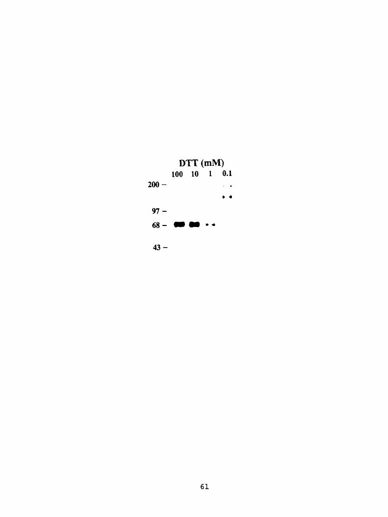
DTT (mM) 100 10 1 0.1
200 - . .

the soluble fraction of the cells expressing m4Tr-Y548, a hi& level of specific binding was
consistently observed (Figure 7, panel A). In the membrane £kactions, the amount of specific
binding for ail the truncated rnGluR4 receptors was very low cornpared with the Ml-length
membrane bound mGluR4a (Figure 7, panel B).
To examine the ligand selectivity of m4Tr-Y548 as a group III mGluR, the binding properties
of the soluble tnincated mGluR4 were compared with the membrane-bound full-length receptor
using glutamate receptor ligands including the nonselective mGluR agonist ( 1 S, 3R)- l-arnino
cyclopentane-1.3-dicarboxylic acid (lS, 3R ACPD), the group I mGluR antagonist (R, S) a-
methyl-4-carboxyphenylglycine (MCPG), and the ionotropic glutamate receptor agonists kainic
acid and NMDA (Figure 8). Each drug was tested at a concentration of 100 PM. At the
truncated receptor m4Tr-Y548, kainic acid, NMDA and MCPG produced less than 10%
inhibition of [)HI L-AP4 binding; a similar low level of inhibition was observed with the full-
length mGluR4a. The very low potency of MCPG for mGluR4 has also been reported for
expenments on human mGluR4 expressed in CHO cells (Flor et al, 1995). (1S, 3R) ACPD
showed a greater degree of inhibition at m4Tr-Y548 (27% of control binding) compared with the
full-length mGluR4a (61% of control binding), indicating that this agonist may be more potent at
the tnincated receptor. This difference in degrees of inhibition was statistically very significant,
with a p value = 0.005. The relatively low potency of (lS, 3R) ACPD compared with group III
mGluR ligands such as L-AP4 and L-SOP is consistent with previous binding data kom rnGluR4
expressed in Sf9 insect cells (Thomsen et al., 1997).
The inhibitory potency of the agonists L-AP4, L-SOP, L-glutamate, and cyclobutylene A P 5
and the group III mGluR antagonists M M 4 CPPG, and MPPG for the soluble m4Tr-Y548 were
compared with the full-length mGluR4a. The rank order of potency of the agonists at m4Tr-

Figure 7 Relative level of [ 3 ~ ] L-AP4 binding to the full-length mGluR4a and its truncated
variants. Al1 binding assays were carried out using 30 nM [ 3 ~ ] L-AP4. Panel A: the soluble,
media-secreted fiactions containing 7 pg protein from the HEK ce11 culture transfected with
trmcated mGluR4. The data are expressed as percentages relative to the soluble m4Tr-Y548.
No specific binding was detected in the soluble fnction of the m4Tr-Y489 and mlTr-V413.
Panel B: membrane fractions were prepared tiom HEK cells trms fected with full-length or
tmncated variants of mGluR4a, containing 125 pg protein in each sample. The relative arnounts
of binding of the tmncated variants are presented as percentage relative to the full-length c-niyc-
mG1uRJa. Each colurnn depicts the mean * SEM of 3 determinations conducted in triplicate.

Culture Medium Fractions
Cell Membrane Fractions

Figure 8 Inhibition of [ 3 ~ ] ~ - ~ 4 binding to the full-length mGluR4a and m4Tr-Y548 by
ionotropic and metabotropic glutamate receptor ligands. The specific binding of [-'H]L-AP~
to the full-length c-myc-mGluR4a is represented by the open bar, and the specific binding of [ 3 ~ ]
L-AP4 to m4Tr-Y548 is represented by the solid bar. Each drug was tested at a concentration of
100 FM. Each column is the mean i SEM of three experiments.
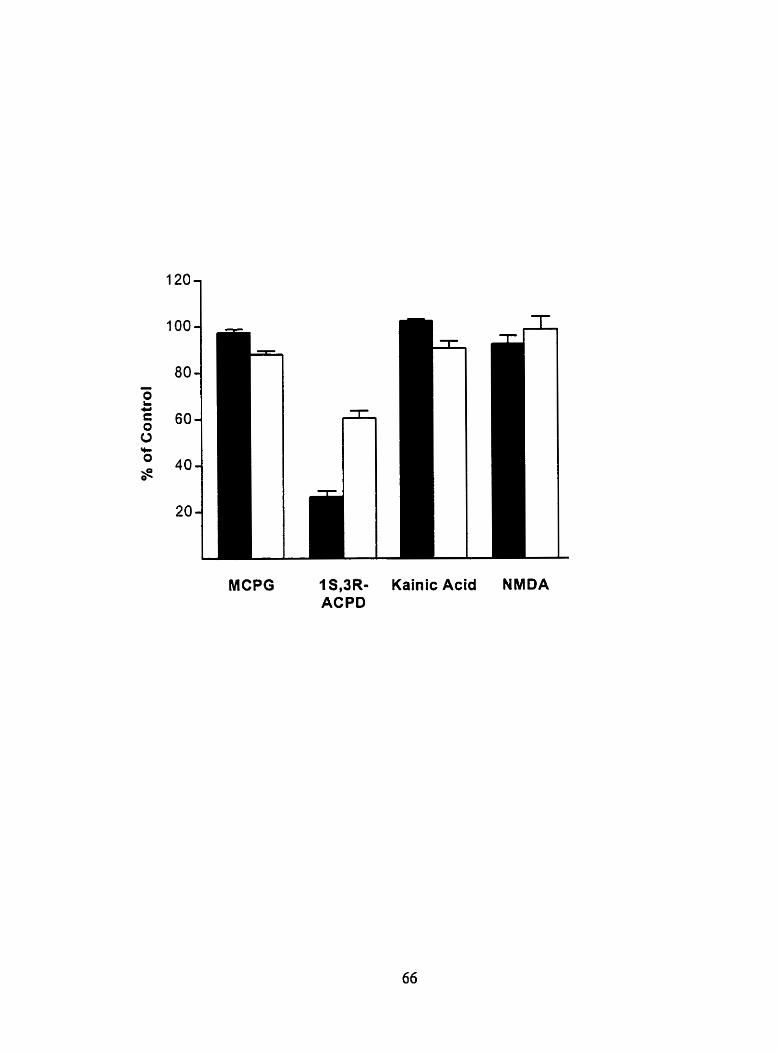
MCPG 1 S,3R- ACPD
Kainic Acid NMDA

Y548 was identical to that of the full-length receptor. The affinhies of all four agonists were
higher for the tnincated receptor compared with the full-length receptor. This was also
consistent with observation that (lS, 3R) ACPD had higher inhibition of [-'H]L-M~ binding at
the truncated receptor. The afinity for the endogenous ligand L-glutamate at mJTr-Y548 was
about 7-fold higher than that at the membrane-bound Ml-length mGluRJa (Figure 9; Table 1).
Except for CPPG, whch had a 7-fold decrease in affinity for m4Tr-Y548, the antagonists
displayed similar a f ~ t i e s for m4Tr-Y548 compared with the membrane-bound &Il-length
mGluR4a. The rank order of the antagonists at m4Tr-Y548 (MAPJ > MPPG > CPPG) was
similar to that at the full-length mGluR4a (CPPG = MAPJ > MPPG) except for CPPG, which
was the most potent of the three compounds for the full-length receptor but was the lease potent
at the soluble receptor (Table 1; Figure 9). The unpaired t test indicated that the dirferences in
affinities of agonists at the truncated and full-Iength receptors were statistically significant or
very significant (the p values are 0.003 for L-AP4 0.0 12 for L-SOP. 0.0 16 for L-glutamate and
0.016 for cyclobutylene-AP5). M i l e the difference in affinity of CPPG at the tnincated and
full-lenyth receptors was statistically very significant ( p = 0.007), the differences for the other
hvo antagonists were not statistically significant @ = 0.139 for MAP4 and p = 0.670 for MPPG).
3.5 Deglycosylation of m4Tr-Y548
The soluble tnincated receptor migrated on SDS-PAGE with a M, = 71,000; this is
approximately M, = 8,000 larger than the predicted molecular weight based on the amino acid
sequence, suggesting that the tnincated receptor was glycosylated. Treatment of the soluble
fraction containing m4Tr-Y548 with Endo H, which cleaves the chitobiose core of high mannose
and some hybnd otigosaccharides fiom asparagine-linked glycoproteins, produced a small shift

Figure 9 Cornpetition curves for the inhibition of t 3 ~ ] L-AP4 binding to m4Tr-Y548 by the
agonists L-giutamate, L-AP4, L-SOP and cyciobutyiene-AP5 (A) and the intagonists
MAP4, MPPG, and CPPG (B). The soluble binding assay was conducted in triplicate using 30
nM ['HI L-AP4 (except for L-AP4, for which 10 nM [ j ~ ] L-AP4 was used). Each point
represents the mean * SEM of three determinations on samples obtained from hvo to three
di fferent preparations.

0 L-AP4 r L-SOP
Glutamate Cyclobutylene-AP5
I I 1 1 I I I 1 -9 -8 -7 -6 -5 -4 -3 -2
Log [Agonist] (M)
0 MPPG MAP-4
A CPPG
I 1 I I 1 I I t -8 -7 -6 -5 4 -3 -2 -1
Log [Antagonist] (M)

Table 1. Binding Constants for Agonists and Antagonists at the Full-Length mGluR4a and m4Tr-548
L-SOP 0.23I0.03
Antaeonists
CPPG
MAP4
MPPG
Al1 values represent the mean f SEM of 3 independent measurernents. The KD and B,, values for the truncated receptor were 120 & 10 nM and 33 t 4 pmoVmg protein, while the Ko and B,, values for the full-length receptor were 470 + 30 n M and 9 + 0.4 pmoUmg protein.

in the immuno-reactive band fiom M, = 71,000 to Mr = 69,000 (data not shown). However,
treatment with PNGase F, which cleaves al1 asparagine-linked carbohydrates, produced a larger
shift to Mr = 61,500 (Figure 10, panel A). This molecular weight estimate was slightly smaller
than the predicted molecular mass of the nonglycosylated protein (62,700 daltons), but close to
that of the nonglycosylated protein f i e r the cleavage of signal peptide (59,300 daltons).
The binding of ['HI L-AP4 to the deglycosylated receptor was also assessed. L'HI L-AP4
binding to samples treated with PNGase F was 99% of control samples (average of two
independent detenninations) that were not treated with the enzyme (Figure 10. panel B),
indicating that agonist binding to Inincated mGluR4 is not dependent upon asparagine-linked
carbohydrates. Approximately 0.15 pg of m4Tr-Y548 was incubated with 100 pl of wheat germ
agglutinin-sepharose in 50 mM Tris-HCI and 10 mM NaCl (pH7.0) at JUC for 16 hours, and
followed by elution with N-acetyl-D-glucosamine, which often used to replace the bound
glycoprotein on lectins. Immunoblotting expenment indicated that small arnount of the
tnincated receptor was bound to the lectin, but most of the protein remained unbound.
Moreover, N-acetyl-D-glucosamine failed to elute the bound receptors off the lectin-coupled
sepharose (data not shown).
3.6 Expression of Truncated mGluR4 and mGluR8 in E. coli
The amount of the soluble receptor m4Tr-Y548 secreted From HEK cells was estimated to be
0.15 pdml of culture medium. Since the amount of pwified protein required for crystallization
experiment is typically in the milligram range, an alternative expression system producing higher
amounts of the soluble protein would be desirable. To obtain high level of expression for solubie
tnincated mGluRs on a large scale, two bacterial expression systems were tested.

Figure 10 Enzymatic deglycosylation of m4Tr-Y548. Panel A: lane 1, ce11 culture medium
t o m HEK celis transfected with m4Tr-Y548 and incubated at 37°C for I hour; lane 2. ce11
culture medium fkom E K cells transfected with m4T~Y548 and treated with PNGase F at 37OC
for 1 hour. Panel B: histogram summarizing the results of [k] L-AP4 binding (30 nM) to m4Tr-
Y548 with and without PNGase F. Each column is the average of 2 independent experiments.


The DNA fra,grients encoding the mGluR4 ATD up to Gln549 and the equivalent constmct
containing the mGluR8 ATD up to Gly543 (Peltekova et al., 2000) were used as the coding
sequences for expressions in E. d i . The DNA Bagments were inserted into the pBAD/hfyc-His
vector. The pBAD plasmids are pUC-derived expression vectors designed for inducible, dose-
dependent recombinant protein expression and purification in E. coli. The Ievel of expression
can be controlled by the L-anbinose concentration in the culture media. Recombinant proteins
expressed in this vector were tagged at the carboxyl-terminus with a hljc epitope tag and a 6X
His tag (Table 2). The insertions were designed so that the tnincated receptors would be
translated in-fiame with the vector for both the 5' start codon and the 3' sequences of the M'yc
and His tags.
The expression plasmids were transformed into the TOP10 E. coli strain. This strain is
capable of L-arabinose uptake, but not metabolizing it. This is important for expression studies
as the level of L-arabinose will be constant inside the ce11 and not decrease over time. To find
the optimal expression condition, L-arabinose was added at final concentrations of 0.2%, 0.02%.
0.002%, 0.0002% and 0.00002%. Immunoblotting showed that the level of expression increased
initially as th? concentration of L-arabinose increased. However, very Iittle difference in the
expression levds was found between the samples induced with 0.02% L-arabinose and that with
0.2% L-arabinose. Therefore, the subsequent expression test were done using 0.02% as the final
concentration of L-arabinose.
To test the solubility of the truncated recepton, bactenal cells were sonicated in 100 y1 of
bacteria lysis buffer and cenaifuged at 100,000 x g for 45 minutes to separate the soluble and
insoluble proteins. No immuno-reactive band was found in negative control samples (E. coli
transformed with pBADIMyc-His vector). Immunoblotting analysis revealed that imrnuno-

reactive bands were present only in the insoluble portions of the samples fiom the m4Tr-
pBADIMyc-His and m8Tr-pBADlMyc-His transfonnants (Figure 11, panel A). The immuno-
reactive proteins fi-om m4Tr-pBAD/Myc-His and m8Tr-pBAD/Myc-His both miprated at an
estimated M, = 63,000, which was very close to the predicted molecular masses for the tnuicated
mGIuR4 and mGIuR8 (62,700 daltons for truncated mGluR4 and 63,000 daltons for truncated
rnGluR8). Results from the radioligand binding assay using ['HI L-AP4 failed to detect specific
binding in either the soluble or the insoluble samples.
The cytoplasmic space of E. d i is known to be in a reduced state that prevents the proper
formation of disulfide bond. The rnincated mGluR4 and rnGluR8, each contains 14 conserved
cysteines, may therefore be misfolded when expressed intracellularly in E. d i . The two
constructs using pET22b vectors, mJTr-pET22b and m8Tr-pET22b. produced truncated mGluRJ
(arnino acids 1 to 549) and rnGluR8 (amino acids 1 to 543) with an E. coli signal peptide
attached to their amino-termini. The signal peptide rnay localize the truncated recepton to the
periplasmic space, which is a more oxidative environment than the cytoplasmic space. Both
tnincated recepton had a 6X His tag attached to their carboxyl-termini for affinity purification
(Table 2). Afier the addition of IPTG to the growth media, the expressions of these proteins
were observed in the E. coli strain BL2 1 (DE3) transfomed with the plasmids. Immunoblotting
analysis revealed that m4Tr-pET22b and m8Tr-pET22b proteins were expressed in both
cytoplasrnic/membrane fractions and penplasmic extracts (Figure 11, panel B). The truncated
receptors in the cytopIasmic/mernbrane fractions migrated at a M, of 62,400, which is similar to
the predicted molecular masses for truncated receptors. The recepton in the penplasmic extracts
migrated on SDS-PAGE at Mr = 61,000, which is slightly smaller then the predicted molecular
masses. In both the cytoplasmic/mernbrane fractions and penplasmic extracts, the expression

Figure 11 Immunoblotting analysis of truncated mGluR4 and mGluR8 expressed in E.
coli. Panel A: Intracellular expression of truncated mGluR4 and mGluR8 in E. coli (TOPIO)
using pBADIMyc-His vector. Lanes 1, 2 and 3 contain insoluble intracellular fractions from the
transformed E. c d i cells. Lane 1: pBAD1Myc-His transformant (negative control); lane 2: m4Tr-
pBADlhlyc-His transformant; lane 3: m8Tr-pBADlibfyc-His transformant. Lanes 4, 5 and 6
contain the soluble intracellular fiactions fiom the transformed E. coli cells. Lane 4:
pBADIMyc-His transformant (negative control); lane 5 : m4Tr-pBAD/h&c-His transformant; lane
6: m8Tr-pBADIMyc-His transformant. Samples contained 8 pg of total insoluble proteins or I
pg of total soluble proteins were subjected to SDS-PAGE, transferred to nitrocellulose
membrane, and labeled with an anti-c-nlyc mouse monoclonal antibody. Resemble
immunoblotting results were obtained from 2 independent experiments. Panel B: Periplasmic
expression using the pET22b vector. Lmes 1, 2 and 3 contain cytoplasmic and membrane
fractions from transformed E. coli cells. Lane 1: pET22b transformant (negative control); Lane
2: m4Tr-pET22b transformant; Lane 3: m8Tr-pETZ2b transformant. Lanes 1, 5 and 6 contain
periplasmic extractions from the transformed E. coli cells. Lane 4: pET22b transformant
(negative control); Lane 5: m4Tr-pET22b transformant; Lane 6: m8Tr-pET22b transformant.
Samples contained 8 pg of total cytoplasrnic/membrane proteins or 0.8 pg of total periplasmic
proteins from the m4Tr-pET22b transformant and 0.1 pg of total periplasmic proteins fiom the
rn8Tr-pET22b or pET22b transformant were subjected to SDS-PAGE, transferred to
nitroce1:ulose membrane, and labeled with an anti-mGluR48 rabbit polyclonal antibody. The
b Io ts shown are representative of 3 independent expenments. Arrow indicates the periplasmic
expressions of m4Tr-pET22b and m8Tr-pET22b.
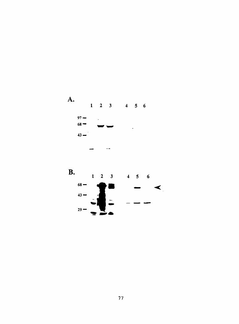

level of m4Tr-pET22b was much higher than that of m8Tr-pET2îb. In the
cytoplasmic/membrane fraction of the bacteria expressing m4Tr-pET22b, multiple
immunoreactive bands migrating at smaller MG than the predicted receptor molecular mass were
observed (Figure 11, panel B). These smaller bands may represent the fragments produced by
protease degradation of the receptor protein. The periplasmic extracts were subjected to [-'H]L-
AP4 binding analysis. No significant specific binding of the radiolabeled agonist was seen in
either of the truncated receptors (data not show).
3.7 Expression of Truncated mGluR4 and mGluR8 in Insect Cells using the Baculovirus
Expression System
DNA Eragrnents encoding tmncated mGluR4 containing amino acids 1 to 549 and mGluR8
containing amino acids 1 to 543 were inserted into baculovirus transfer vector pBlueBacJ.5
(Table 2). Afier obtaining the recombinant viruses, they were used to infect three different insect
ce11 lines: SF-9, MG-1, and High 5. At 72 hours post-infection, whole cells and culture media
were collected separately and subjected to immunoblotting analysis. Immunoreactive proteins
were found in cells infected with the recombinant vinises, but not in the uninfected cells.
immunoreactive bands were present at high level in the cellular portions of al1 ce11 types, but not
in the culture media (Figure 12, panel A). The truncated mGluR4 migrated at a M, = 64,900,
which is slightly larger than the predicted molecular mass for the nonglycosylated mincated
mGluR4 (62,700 daltons). The M, of the truncated mGluR8 appeared to be approximately
68,000, which is also larger then the predicted molecular mass for the nonglycosylated truncated
mGluR8 (63,000 daltons). Multiple immunoreactive bands with lower molecular weights were

Figure 12 Irnmunoblotting analysis of truncrted mGluR4 and mGluR8 expressed in sf-9
insect cells. Panel A: expression analysis on samples from SB cells infected with m4Tr-
pB lueBac4.5 and m8Tr-pBLueBac4.5. Lane 1 : whole ce11 fraction of uninfected sf9 cells. Lane
2: whole ce11 fraction of cells infected with rn4Tr-pBlueBacJ.5. Lane 3: whole ce11 fraction of
the cells infected with m8Tr-pBlueBac4.5. Lane 4: medium fiaction of uninfected si9 cells.
Lane 5: medium tiaction of cells infected with m4Tr-pBlueBac4.5. Lane 6: medium tiaction of
cells infected with m8Tr-pBlueBac4.5. Panel B: expression analysis on sarnples from sf9 cells
infected with m4Tr-pK503- 19 and rn8Tr-pK509-8. Lane 1 : whole ceIl fraction of uninfecied SB
cells. Lane 2: whole ce11 Fnction of cells infected with m4Tr-pK503-19. Lane 3: whole ce11
fiaction of the cells infected with m8Tr-pK509-8. Lane 4: medium fraction of uninfected sf9
cells. Lane 5: medium fraction of cells infected with m4Tr-pK503-19. Lane 6: medium hct ion
of cells infected with mSTr-pKSO9-S. Samples contained 8 pg of total cellular proteins or 1 pg
of total medium proteins were subjected to SDS-PAGE. transferred to nitrocellulose membrane,
and labeled with an anti-mGluR4/8 rabbit polyclonal antibody. The blots show are
representative of 3 independent experiments.


Table 2. Features of the Expression Constructs for Truncated mGluR4 and mGluR8 in E. coli and Insect Cells
Receptor Expression Host Vector Signal Ex pec ted Tag * Subtype Host Strain System Sequence localization
TOP 10 pBAD/Myc- Rai Intracellular M: C-terminal His H: C-terminal
E. coli BL2 1 pET22b Bactena Periplasmic M: After Signal Peptide (DE3) Space H: C-terminal
Truncated mGluR4 S B pBlueBac4.5 Rat Extracellular M: AAer Signal Peptide
Insect Cells SB pFastBac 1 Viral Extracellular F: ARer Signal Peptide
Derivative H: C-terminal
TOP 10 pBAD1Myc- Rat Intracellular M: C-terminal His H: C-terminal
E. coli BL2 1 pET22b Bacteria Periplasmic H: C-terminal
Truncated (DE31 Space mGluR8
SIP pBlueBac4.5 Rat Extracellular No Tag Attached lnsect Cells
SB pFastBac 1 Viral Extracellular F: AAer Signal Peptide Derivat ive H: C-terminal
* M = Myc epitope tag; H = His tag; F = FLAG epitope tag.

detected in the cellular fractions of cells expressing truncated mGluR8; these bands most likely
resulted from protease degradation. uifected cells were collected and sonicated in PBS. After
high-speed centrifugation in an ultracentrifuge at 100,000 x g for 45 minutes to separate the
soluble pro teins frorn the insoluble proteins, al1 samples were analyzed by imrnunoblotting
experiments. Only low amounts of the receptor proteins were detected in the supernatants.
whereas higher levels were found in the insoluble samples (data not show). Cell membranes,
soluble cytoplasmic fractions and ce11 culture media from infected Sf-9, MG-1 and High5 cells
were prepared and subjected to radioligand binding assay. However, no specific binding of [ j ~ ]
L-AP4 was detected in any of the samples.
The original cDNAs of rat mGluR4a and mGluRIa encode signal peptides of the receptors.
which were 32 and 33 arnino acids in length respectively. To test whether replacing the rat
signal sequence with a signal peptide originating fiom baculoviral protein AcNPV ecdysone
glucotransferase would facilitate the secretion of the truncated receptors from insect cells, DNA
lngments encoding truncated rnGluRJ arnino acids 33 to 548 and mGluRS amino acids 34 to
543 were generated by PCR and subsequent subclonings. These DNA fragments, lacking the rat
receptor signai sequences, were inserted into pK50349 (tnincated mGluR4) and pK509-8
(truncated rnGluR8), both denvatives of pFastBac vector. These vectors contained the sequence
encoding the baculoviral signal peptide upstream of their multiple cloning regions (Table 2). As
the result, the rat receptor signal peptides were replaced by the baculoviral signal peptide when
the truncated receptors were expressed in sf9 cells. The recombinant proteins were tagged with
FLAG epitope at the amino-termini and 6X His tag at the carboxyl-termini.
immunobloning analysis detected the expression of the tnuicated receptors in samples fiom
sfP cells transfected with m4Tr-pK503-19 and with m8Tr-pK509-8. The tnincated receptors

were detected in the both culture media and the whole ceil fractions (Figure 12, panel B). In the
whole ce11 fractions, both the tnincated rnGluR4 and the tnincated mGluR8 migrated at a M, =
64,900. The approxirnately 2,000 dalton differences between the relative molecular weights and
the predicted molecular masses for the truncated receptors are likely due to the glycosylation of
the receptors. The tnincated mGluR4 and mGluR8 receptors were detected in the ce11 media.
They migrated at M, = 63,800, which is approximately M, = 4,000 larger than the predicted sizes
for the nonglycosylated truncated recepton with the signal peptides cleaved (59,700 daltons).
Samples of the ce11 culture media were dialyzed against binding buffer and subjected to [ 3 ~ ] ~ -
AP4 binding analysis. However, in hvo experiments, no specific binding was detected (data not
show).

4.0 Discussion
4.1 Pharmacology of the Soluble Truncated mGluR4
The cDNA constnict encoding m4Tr-Y548 produced a protein that extended from the amino
terminus to Tyr548, which is located 39 amino acids upstream fiom the junction of the ATD and
the first putative TMD of mGluR4. This region of ATD includes the entire LIVBP homology
region and part of the cysteine-rich region. The protein was secreted into the ce11 culture
medium, indicating that the protein did not possess regions of sufficient hydrophobicity to cause
retention in the endoplasmic reticulum or plasma membrane of the cell and that it was soluble in
an aqueous environrnent.
A pharmacological analysis of the m4Tr-Y548 was conducted by assessing the ability of
various mGluR4 ligands to compete with the binding of the radiolabeled agonist [ 3 ~ ] ~ - ~ 4 .
The results indicate that the binding properties displayed by the soluble rn4Tr-Y548 reflect the
unique pharmacological profile of the group III mGluRs. Ligands at group I mGluRs or
ionotropic glutamate recepton showed little or no affinity for either the full-length mGluR4a or
m4Tr-Y548. Except for the antagonist CPPG, the rank order of potency for a senes of group III
mGluR ligands at m4Tr-Y548 was the sarne as at the full-length membrane-bound mGluR4a.
These observations suggest that the primary determinants of ['HIL-AP~ binding are conferred by
residues present in the ATD of mGluR4. However, the inhibition constants for L-glutamate, L-
AP4, L-SOP, and cyclobutylene AP5 were lower than those of the full-length receptor, indicating
that the tmcated soluble receptor displayed higher affinities for agonists compared with the full-
length receptor. The differences in affinity of the ligands were likely not caused by diEerences
in assay conditions for the soluble and membrane-bound receptors, since the amnities of the

soluble receptor for antagonists were either similar (to MPPG and MAP4) or lower ((0 CPPG)
compared with the full-length receptor.
Stnkingly similar results were reported in the study of a truncated mGluR8 receptor (m8Tr-
G543), which terminates 40 amino acids upstream of the first putative TMD of
mGl~EU(Pe1tekova et al., 2000). This receptor displayed a similar pharmacological profile to the
full-length mGluR8. However, as observed with the soluble mGluR4 receptor, the affinities of
the truncated mGluR8 for agonists were higher than that of the Full-length mGluR8 receptor.
The antagonist CPPG had 10-fold decrease in afinity for the soluble receptor, whereas another
antagonist, MAP4, displayed simila. affinities for the soluble and full-length mGluR8 receptors.
These observations suggest that the increase in affinities for agonists and the decrease in affinity
for CPPG are shared features of the soluble tnincated mGluR4 and mGluR8.
Interestingly, in a recent study of a soluble truncated GABAeRl receptor, the agonist-binding
affinities of a soluble GABAeRl receptor were also increased compared with the full-length
GABAeRl (Malitschek et al., 1999). This increase was also observed with a truncated
GABAeRl receptor that contained a single TMD and was therefore membrane-anchored.
Moreover, despite the increased dfinity to agonists, the rank order of affinities was unchanged
by the üuncations. These observations are rernarkably similar to our findings with mGluRs.
However, unlike our results on mGluR4, the affinities of antagonists at the truncated GABABRl
receptors did not differ from those at the full-length receptor.
The change in the ligand af'finities &er the truncation suggests that regions of the full-length
receptor excluded fiom m4Tr-Y548 and m8Tr-G543 might influence the affinities. These
regions include the TMDs, the extracellular loops between the TMDs, and the 39 amino acids
between Tyr548 and the first TMD. In the fuil-length receptor, the ATD may possibly interacts

with one or more of these regions after ligand binding, which in turn induces changes in the
affinity for the ligand. in a mode1 proposed by Pin and Bockaert (1995), it has been suggested
that agonist binding to the mGluRs may induce a confornational change that allows the ATD to
interact with a region within the TbiDs. Such an interaction rnay result in a decrease in agonist
f in i ty . According to this model, the TMDs do not influence the affinity of antagonists because
antagonists are not expected to promore the interaction between the ATD and the TMDs. Pin
and Bockaert's model exp Iains the higher agonist affinities observed w ith the m4Tr-Y 5 48,
m8Tr-G543 and the truncated GABAeRls, since the TMDs do not exist in these receptors. The
observation that antagonists MAP4 and MPPG displayed similar affinities for the m4Tr-Y548
and the full-length mGluR4 is also supported by the model. However, the model fails !O address
the 7-fold decrease in the affinity of CPPG for the soluble receptor compared to the full-length
mGluR4. h alternative explmation is that the high-affinity agonist site observed in the
truncated receptors represents an altemate conformational state of the lower-affinity site present
in the full-length receptor. A possible reason for the increase in agonist affinity is that the
binding domain of the soluble mGluR4 is locked in a slightly changed state due to the structural
constraints imposed by the truncation. The new conformation may also have reduced affinity for
larger molecules such as CPPG.
Whether or not ligand binding in other mGluRs is affected by regions outside the ATDs
remains to be determined. Aithough the pharmacological results reported by Okamoto et al.
(1998) on the binding of [3~]quisqualic acid to the soluble mGluRl receptor are difficult to
compare with ours because they did not report ICso values for L-glutamate or other compounds
that were assessed in this study, it appears that the a E t y for [3~quisqualic acid for the soluble
receptor was very similar to the affinity measured for the full-length rnGluRl receptor. One

difference between the truncated mGluR1 receptor and the m4Tr-Y548 is that the former
contained the entire ATD of mGluR1, whereas the latter did not include the 39 amino acids
immediately upstrearn from the tint TMD. Our results demonstrated that this region is not
required for high affinity binding of ['HIL-AP4 to mGluR4. This observation is consistent with
mutational and chimeric studies conducted on mGluRl and mGluR4, which showed that several
residues located in the more amino-terminal regions of the ATDs of the receptors are required
for ligand binding (O'Hara et al., 1003; Takahashi et al., 1993; Harnpson et al., 1999). However,
we cannot nile out the possibility that these 39 amino acids of mGluR4 may affect the affinity of
ligands for the binding site.
Of interest in this regard are studies on the receptor for the glycoprotein hormones luteinizing
hormone and choriogonadotropin. The luteinizing hormone/choriogonadotropin receptor is a
member of the G-protein-coupled receptor fmily and, like mGluRs, it has a large ATD that can
be separated From the seven TMDs and still retain ligand binding activity. Radiologand binding
studies using ~"~-labeled human choriogonadotropin have shown that the exodornain (equivalent
to the ATD) alone has a higher affinity for the glycoprotein hormone than the full-length
receptor (Xie et al., 1990; Ryu et al., 1998). A structural analysis of this receptor using site-
directed mutagenesis has s h o w that several amino acids in the second extracellular loop within
this TMD region affect the affinity of the ligand for the receptor. It was suggested that residues
within this loop constrain the affinity of the homone for the receptor (Ryu et al., 1998).
Although the amino acid sequences of the mGluRs are not homologous with the glycoprotein
hormone receptors, it is possible that in mGluR4 the extracellular loops or the regions
immediateiy upstrearn of the TMDs are involved in a similar mechanism to constrain the
affinities of the agonists for the binding site located in the first 548 arnino acids of the receptor.

Many GPCRs display biphasic binding c w e s indicative of the presence of high and low
affinity binding sites. The commonly accepted explmation for high and low affinity binding
sites is that they reflect receptors with bound and unbound G-protein, respectively. However,
some GPRCs, such as rnuscarinic acetylcholine receptors, show multiple states of' affinity in the
absence of G-proteins (Pertenon et al., 1984; Wreggett and Wells, 1995). Convenely, other
GCRPs such as the cloned mGluR4 receptors display only a single class of sites in radioligand
binding assays (Eriksen and Thomsen, 1995; Thomsen et al., 1997); it is possible that lower
affinity sites may exist but cannot be detected with binding assay. Altematively, it is possible
that the unitary nature of the binding state of the cloned membrane-bound mGuR4 receptor
reflects only a low affinity state due to the absence of the appropriate G-proteins in the host ceil
lines. However, it has been observed that CO-expression of full-length mGluR4 with various
marnmalian G-protein subunits including Gii, Gi2, Gi3, and Go in inseci SB cells does not
significantly increase the affinity or the capacity (Bmx) of ['HIL-AP4 for mGluR4 (L. Blythe and
D. R. Hampson, unpublished observation). Moreover, [ 3 ~ ] ~ - ~ ~ 4 binding experiments
conducted in nt (Hudtloff and Thomsen, 1998) and mouse (Thomsen and Hampson, 1999) brain
have detected only a single class of sites with a KD value very similar to that seen with the cloned
receptor. Thus, the binding site in the full-length receptor with an affinity for ['HIL-AP~ in the
400 - 500 nM range rnay represent the high afhi ty state of mGluR4. If this is tnie, the
comparable affinity for ['HIL-AP4 at m4Tr-Y548, which lacks G-protein coupling region,
suggests that the G-protein coupling may not be required for the high affinity state of the
receptor.
Although the radioligand binding assays conducted in this study and a number of previous
studies revealed only one binding site in mGluR4, the possible existence of a second binding sire

is suggested by a recent study on a rnGluR4 variant that functions as a taste receptor in lingual
tissue (Chaudhari et al., 2000). Compared to the brain form mGluR4a, this receptor starts at
Met308 and lacks about 50% of the ATD (amino acids 1 to 588 in brain mGluR4a).
Interestingly, none of the amino acid residues that are believed to be critical in ligand binding to
the brain form mGluR4, including Arg78, Ser159 and Thr182, are present in this taste form
mGluR4. A fmctional assay rneasunng the inhibition of CAMP formation revealed that the taste
receptor has a -100-fold lower sensitivity to L-AP4 than the full-length brain form. The ECjo
value of L-AP4 for taste form mGluR4 was about 100 PM, whereas that for the brain form
mGluR4 was between 0.5 to 1.0 FM. The low binding affinity displayed by the taste form
receptor may represent the binding of agonists to a second binding site in mGluR4. This low
affinity binding site could be in the carboxyl-terminal half of the ATD that remained in the taste
form mGluR4. Altematively, it could reside in the TMDs or extracellular Ioops. In the
radioligand binding assay, a binding site with such a low affinity would not be detected.
4.2 Minimal Sequence in mGluR4 Ligand Binding
A pnmary objective of this study was to probe the limits of truncation of the mGluR4 ATD in
tems of producing tnincated proteins that retained the ability to be secreted and bind ligands.
Since the shortest constnict analyzed in this study (m4Tr-V413) contains a truncated LIVBP
homology domain, it was not surprising that this protein did not possess binding activity. The
second shortest construct (m4Tr-YJ89) also failed to display any binding activity in the soluble
or membrane fractions, despite the fact that this protein does include the entire L N B P homology
region. This observation is in agreement with the results obtained with a mGluRl receptor,
which is truncated at a site equivalent to 8 amino acids downstream of the carbo~yl~te~minus of

m4Tr-Y489. This truncated mGluRl (mGluR103), like m4Tr-Y489, was incapable of binding
agonists (Okarnoto et al., 1998). The low expression level of mGluR103 was suggested as the
possible reason for the protein to lack binding ability. It is possible that the LIVBP homology
region by itself is sufficient for imparting ligand-binding capabilities to mGluRs. However, the
segment of amino acid sequence between the site of tnincation at 39 arnino acids and 98 amino
acids upstream tiom the first TMD, contains in~ormation that greatly facilitates the secretion of
the tnincated receptors. This region contains 6 of the 17 cysteine residues that are conserved in
the ATDs of al1 8 rnGluRs and the homologous calcium-sensing receptor; it is Iikely that one or
more of these 6 cysteines are required for maintaining the 3-dimensional structure of the receptor
in its native conformation.
Further cornparisons between our results and those obtained with chimeric and truncated
GABAeRl receptors are informative. Of particular interest are experiments conducted using
chimeric recepton possessing portions of the ATD of the GABAsRla subunit fused to
downstrearn sequences of the rnGluRlb receptor. A chimeric receptor containing the ATD of
the GABAeRla subunit encompassing ail of the ATD except the last 10 amino acids
immediately upsneam of the fint TMD (termed aN550) bound a GABAe receptor photoaffinity
ligand, whereas a constnict fbsed 60 amino acids upstream of the first TMD (termed aN530) did
not bind the photoaffïnity label (Malitschek et al., 1999). These observations are strikingly
sirnilar to our results with tnincated mGluR4 receptors.
We also examined the constnict that encoded the entire ATD of mGluR4 (m4Tr-P586). This
protein was ~ c a t e d at the junction of the ATD and the fim putative TiMD. Surprisingly,
aithough it contained no TMD, the protein was not secreted fiom transfected cells and did not
bind ['KJL-AP~. This observation suggested that one or more residues in the intervening

sequence between the first TMD and the truncation site 39 amino acids upstream, inhibited
protein secretion. The lack of binding ability rnay be directly due to the presence of the 39
arnino acid segment, or altematively, as a consequence of the intracellular retention of the
protein. One study on a mGluR8 receptor teminated at the junction of the ATD and the first
TMD reported results resemble our observations on m4Tr-P586 (Peltekova et al., 2000). This 39
amino acid segment of polypeptide contains 3 conserved cysteines. The 3 cysteines are also
present in the truncated mGluR8. It is possible that one or more of these 3 cysteines might
inhibit secretion and binding by fonning aberrant intramolecular disulfide bonds with other
proteins in the endoplasrnic reticulum and therefore inhibit the normal folding of the receptor.
In support of this speculation, a cysteine-to-serine mutation in the calcium-sensing r e q t o r
was required in a conserved cysteine residue located immediately upstream of the first TMD for
secretion of the ATD (Goldsmith et al., 1993). These results, however, differ from those
obtained tiom the analogous construct of mGluR1. A truncated mGluRl containing the entire
ATD was secreted and bind agonists when expressed in insect cells with a baculovirus
expression system (Okamoto et al., 1998). Although the reason for this difference in secretion is
not obvious, one possibility may be the presence of N-linked carbohydrate side chahs near the
carboxyl-terminal end of the mincated proteins. mGluR4 and mGluR8 both have a consensus
sequence for N-glycosylation 19 residues upstream from the first TMD; this site is missing in
mGluRl and the GABAeRl receptor subunit in which the entire ATD cm also be expressed as a
secreted protein (Malitschek et al., 1999). Although this carbohydrate side chain, if present in
the wild-type receptor, might confer some usefbl function, the presence of the carbohydrate close
to the carboxyl terminus of the tnuicated receptors might impair secretion nom the cell.

Together these results suggest that the cntical boundaries for secretion and activity in group III
mGluRs and in GABABRs are similar. Our results suggest that there are strict limitations on the
proper folding of tnincated versions of the ATDs of mGluR4 and mGluR8. Importantly, the
LIVBP homology region in conjunction with part of the cysteine-rich region is required for
optimal secretion in a soluble form that retains ligand binding activity.
4.3 Biochemical Chnracterization of the Soluble Truncated mGluR4
The soluble tnincated rnGluR4, m4Tr-Y548, contains four consensus sequences for
asparagine-linked glycoslyation. The decrease in the molecular weight of the truncated pmtein
by about M, = 8,000 f i e r complete deglycosylation indicates that one or more of these sites are
glycosyiated. An analysis of the effects of deglycosylation on the binding of [ 3 ~ ] ~ - A P 4 to the
soluble receptor demonstrated that the bound carbohydrate is not required for agonist binding.
Although our data show that asparagine-linked oligosaccharides are not required for ligand
binding to mGluR4, it is possible that the bound carbohydrates may be important in the
subcellular targeting of this receptor in the nervous system. Despite the presence of
oligosaccharides in the muicated receptor protein, wheat germ agglutinin did not effectively
remove the receptor protein fiom ce11 culture medium, indicating that wheat germ agglutinin is
not the most suitable lectin for the purification of m4T~Y548. The type of lectin suitable for
protein purification may depend on the type of oligosaccharides on the protein. For example,
RCA-1 lectin was determined to be the most efficient lectin to qualitatively purify a soluble,
truncated human CaR (Goldsmith et al., 1999). Pilot test such as screening with a lectin affinity
chromatography screening kit may identiQ the most efficient lectin io isolate m4Tr-Y548 from
the ce11 culture medium.

The soluble m4Tr-Y548 exists as a monomer in the presence of high concentrations of a
reducing reagent and as a dimer and trimer in the presence of low concentrations of the reducing
agent. Dirneric foms of al1 the mGluR subtypes have been observed on immunoblots of brain
tissue and transfected cells (Pickering et al., 1993; Harnpson et al., 1994; Petralia et al., 1996;
Pekhletski et al., 1996; Romano et al., 1996; Nomura et al., 1994; Shigemoto et al., 1997).
Romano et al. (1996) have s h o w that rnGluR5 migrated on SDS-PAGE as a dimer under
nonreducing conditions and as a monomer under reducing conditions, indicating that the dimers
are formed by intermolecular disulfide bonds. Analysis of the electrophoretic mobility of a
truncated construct of mGluR5 indicated that the intermolecular disulfide bonds are fonned
between cysteine residues located in the ATD of mGluR5 (Romano et al., 1996). Modulation of
monomer and dimeric forms by reducing agents was also seen with the soluble tnincated form of
the mGluRl receptor (Okamoto et al., 1998; Robbins et al., 1999). The dimenzation of soluble
tnincated mGluR1 ATD occurred in the endoplasmic reticulum, and is not dependent on protein
glycosylation as it was not prevented by treatment of the cells with htnicamycin.
Despite the studies outlined above providing evidence for dimers of mGluRs, no data exist
demonstrating a direct link between the ability to bind ligands or activate signal transduction
pathways and a particular oligomenc configuration of an mGluR. However, in this study the
[ 3 ~ ] ~ - A P 4 radioligand binding assay was carried out in the absence of reducing agents, whereas
the addition of reducing agent DTT decreased binding activity in a concentration-dependent
fashion. Despite the observation that at a concentration of 1mM or higher DTT caused the
soluble truncated mGluR4 to migrate on SDS-PAGE as a monoer, the bindings of ['HIL-AP4 to
samples containing DTT at concentrations of OmM, O.OlrnM, O. lmM, ImM, 1OmM and 1OOrn.M
decreased sequentially. The effect of DTT in reducing [ 3 ~ ] ~ - ~ ~ 4 binding is likely due to the

disruption of both the intrarnolecular and the intermolecular disulfide bonds. It is likely that in
the typical oxidatiodreduction environment of the extracellular space in nerve tissue, mGluRs
exist as oligomenc complexes. The oligomeric configurations of mGluRs as they exist in nerve
cells in vivo are not known. However, our observations on the ATD of rnGluR4 together with
the results on mGluRl and mGluRS noted above al1 suggest that intemolecular disulfide bonds
are a general structural feature of the mGluR farnily of receptors and that the active form of the
receptors is likely to be a multimeric cornplex.
4.4 Expression of Truncated mGluR4 and rnGluR8 in E. coli
To investigate the possibility of higher level expression, m4Tr-Y548 and rn8Tr-G543 were
expressed in bacteria using two different expression systems. Compared to mammalian ce11
culture, bac terial expression systems have a number of advantages, including the rapid ce11
growth, low cost of growth medium, and high expression level. In some cases yields of purified
recombinant proteins in E. coli from expression vecton containing T7 promoter have typically
been in the range of 10 - 100 mg per liter of culture (Chang et al., 1999). However, expression
of mGluRs in a prokaryotic system has not been reported to date. Ln this study we found that the
üuncated versions of mGluR4 and mGluRS maintained ligand binding ability when expressed in
mamrnalian cells, but not when expressed in E. coli.
One cornmon problern in bacterial expression systems is that proteins are often expressed in
an inactive form and require refolding. The production of inactive protein may be due not only
to the properties inherent in the protein expressed, but also to the rate of accumulation of the
protein. The extraordinary protein yields ofT7-based systems such as PET system may ofien be
achieved at the expense of solubility (Sun et al., 1997). in many cases, such as the

crystallization of the GluR.2 subtype of iGluR, the inactive protein c m be denatured by
chaotrophic agents such as urea and guanidine-Hl1 and then refolded into active f o m with
proper conditions (Armstrong et al., 1998). Sometirnes the denatured recombinant protein could
not be refolded into the active form using conventional methods, but was reactivated by direct
solubilization of the inclusion bodies with Triton X-100 (Sunitha et al., 2000). However, in
other cases, proteins rnay resist al1 attempts to renature them.
The fint bacterial expression system I examined was the araB expression system in E. coli.
In this system, recombinant protein expression is tightly controlled and readily induced with L-
arabinose. The expression level of the recombinant protein with araB system is typically lower
than that of T7-based system. However, the truncated mGluR4 and mGluRS were found
insoluble and inactive. Therefore, I expressed the tmncated receptors with a bacterial signal
peptide for potential penplasmic localization of the recombinant proteins. Since the ATDs of
mGluRs are homologues of PBPs that naturally expressed in the bacterial penplasmic space,
periplasmic space might provide a bener environment for the correct folding of the truncated
ATDs. The processes mediating translocation across the ce11 membrane of E. coli are
incompletely undentood. The signal peptide is known to be necessary, but not sufficient for
export proteins into the periplasm. Periplasmic localization may also depend on determinants
within the mature protein (Wickner et al., 1991; Lory, 1992). In the expression of GluR2
subtype iGluR subunit in E. coli with a bactenal signal peptide, the recombinant protein was
successfully translocated into the penplasmic space and demonstrated ligand binding activity
(Arvola and Keiniinen, 1996). However, the level of the receptor was low when expressed in
penplasmic space, and cytoplasmic expression was ultimately used for generating crystal
(Armstrong et al., 1998). In our study, the tmcated receptor proteins found in periplasmic

extracts were at very low levels, whereas most of the proteins remained in the
c ytoplasmic/membrane fractions. Furthemore, the small amount of proteins present in the
periplasmic extracts were incapable of binding to ['HIL-AP~. One possible reason that the
mGluR ATDs lack binding activity when expressed in the bacterial periplasmic space is that the
ATDs of mGluRs contain cysteine rich regions and the disulfide bonds formed behveen these
cysteines are necessary for the correct folding of the ATDs. The periplasmic space, although
more oxidative than the cytoplasmic cornpartment, may still be too reduced for the proper
formation of the disulfide bonds in mGiuRs.
4.5 Expression of Truncated mGluR4 and rnGluR8 using the Baculovirus Expression
S ystem
The third expression host 1 tested in this study was insect cells. Compared with mammalian
ce11 lines, insect cells typicaily express recombinant proteins at higher levels. Protein
insolubility is generally not a concem when expressing proteins in insect cells, and only simple
N-linked glycosylation will be present in the protein.
I have used two different vector systems to express the tnincated mGluR4 and mGluR8 in SB
cells. in the first system, the rat receptor signal peptide was used, whereas in the second system,
the rat signal peptide was replaced by a baculoviral signal peptide. Irnmunoblotting analysis
indicated a better secretion of the expressed proteins with the baculoviral signal peptide than that
with the rat signal peptide. These observations are different Erom what Okarnoto et al. (1998)
reported for mGluRl ATD, which was secreted into the culture medium fiorn the insect cells by
utilizing its original (rat) signal peptide. Our results, however, do bear some similarity to the
results obtained in the study of soluble tnincated GABABRl (Malitschek et al., 1999). In that

study, the ATD of GABAeRla with the rat signal peptide was secreted from the insect cells and
into the culture medium. However, the soluble secreted GABAeRIa was detected on the
irnmunoblot at a iower level than the soluble secreted GABAeRlb (the other alternatively
spliced form of GABAeR1), which had a baculoviral signal peptide. It was not reported whether
GABAeRlb wiîh the rat signal peptide was secreted into the culture medium. Interestingly, the
two truncated receptors that were secreted into the culture media with their original signal
peptides have shorter signal sequence (18 arnino acids for mGluRL and 17 amino acids for
GABAeRLa) than that of GABAeRlb (29 arnino acids). The lengths of the signal peptides for
mGluRJa (32 amino acids) and rnGluR8a (33 amino acids) are close to that of GABAoRlb.
Therefore, one could speculate that the lengths of the original signai peptides for these receptors
might affect their secretion. With some proteins, a short mammalian signal peptide might be
sufficient for the secretion in insect cells. whereas a longer one may interfere with the proper
secretion.
in my study, replacing the rat signal peptides of tmncated mGIuR4 and mGluR8 with the
baculoviral signal peptide did facilitate the secretion of the receptors into ce11 media. Our
observation on the tmcated mGluR4 and rnGluR8 with the baculoviral signal sequence,
however, differs nom that of the tmncated GABAeRl b in ternis of binding activity. With a
baculoviral signal sequence, GABABRlb was secreted from the insect cells and
pharmacologically active (Malitschek et al.. 1999). The rn4Tr-pK503- 19 and m8Tr-pK509-8, on
the other hand, failed to display binding to ['H]L-AP~. One possible reason for these truncated
receptors to lack binding activity is that, glycosylation on the ATDs of mGluR4 and mGluRS
may affect the proper folding of the proteins. Compared with mammalian cells, insect cells are
not capable of producing protein with compiex polysaccharides. While our results in this study

indicated that, after m4Tr-Y548 was properly folded, N-linked carbohydrates were not required
for binding to [ 3 ~ ] ~ - ~ ~ 4 , it is still possible that these polysaccharides are involved in protein
folding. Another possible explanation for the lack of binding activity seen in the insect ce11
medium sarnples containing tnincated mGluR4 and mGluR8 is the low levels of proteins present
in the sarnples. As in the study of the tnincated rnGluR103, the soluble protein was detected in
the cuinire medium wirh immunobiorring bur was not derected by binding assay using
[ ' ~ ]~u i s~ua la t e (Okamoto et al., 1998)

5.0 Conclusions and Future Considerations
Previous studies using site-directed mutagenesis and chimeric recepton have suggested that
the ligand binding pockets of mGluRs are located in the ATDs. My results on tmncated mGluR4
receptors (Han and Harnpson, 1999, Peltekova et al., 2000) provide a direct evidence that the
first 548 arnino acids of mGluR4 ATD contain al1 of the structural information that is necessary
and suficient for ligand selectivity and *nity for the receptor.
A tmncated mGluR4 receptor containing only the first 548 amino acids of the full-length
mGluR4a was successfully produced in HEK cells as a soluble, secreted protein. The results
frorn [ ~ H I L - A P ~ binding assay indicate thnt this soluble truncated mGluR4 receptor displays a
pharmacological profile very similar to that of the full-length wild-type mGluR4a. The affinities
for agonists to the soluble receptor were slightly higher compared to that of the full-length
mGluR4a. We therefore conclude that the first 548 amino acids OC mGluR3, which contain the
entire L N B P homology region and part of the cysteine-rich region, are sufficient to maintain the
ligand binding activity and ligand selectivity. Other regions of the receptor may also regulate the
ligand binding to mGluR4 through their interactions with the ATD and the subsequent
conformational changes in the ligand binding pocket.
The second conclusion fiom this study is that, there are strict limitations on the proper folding
of tmncated versions of mGluR4 ATD. Shorter recepton lacking the cysteine-rich region and a
longer truncated receptor containing the entire ATD failed to be secreied fiom the cells and
display binding to the agonist ['HIL-AP~. Therefore, when expressed in mammalian cells, the
L I W P homology region and part of the cysteine-rich region are both required for optimal
secretion in a soluble form of mGluR4 that retins ligand binding activity.

In the hird part of my study, 1 attempted to express the soluble truncated mGluR4 and its
mGluR8 analog in E. d i and insect cells. The expressed proteins did not bind [ 3 ~ ] ~ - ~ ~ 4 .
These results suggest that these expression systems may lack the cellular mechanisms for protein
modifications, such as disulfide bonding formation and/or glycosylation, which could be crucial
for the correct folding of the ATDs of mGluRs.
Future studies could attempt to express the truncated receptor with protein disulfide isomerase
in a mutant strain of E. coii (FA1 13) that has a more oxidative cytoplasmic environment. In E.
coli, disulfide isomerases such as DsbA and DsbC could enhance the rate of disulfide bond
isomenzation and greatly assist the folding of proteins containing multiple disulfide bonds. This
system has been reported to facilitate folding of intracellularly expressed proteins with multiple
disulfide bonds (Bessette et al., 1999). If the lack of stable disulfide bonds accounts for the
receptor proteins being inactive in E. coli, this mutant system could provide the suitable
machinery to produce functional tmcated receptors. The second approach is to establish a
stable mammalian ceIl line expressing the soluble tmncated recepton. Since we have
demonstrated that mJTr-Y548 is soluble and active when expressed in HEK cells, this approach
may be the most promising one for the production of a functional soluble receptor. However, as
in the study of the soluble tnincated human CaR (Goldsmith et al, 1999), a large bioreactor may
be required to produce sufficient amount of protein for purification.
In surnmary, work conducted in this study has (1) demonstrated a role of ATD in ligand
binding to mGluR4, and (2) produced a soluble receptor that pharmacologically closely
resembles the full-length mGluR4. This soluble receptor provides a starting point for conducting
m e r detailed structural analysis, such as X-ray crystallization, on the binding site of mGluR4.
6.0 References
Abdul-Ghani, A.-S., Attwell, P.J.E., Sin& Kent, N., Bradford, H.F., Croucher, M.J., Jane, D.E. (1997). Anti-epileptogenic and anticonvulsant activity of L-2-amino-4-phosphonobutyrate, a presynaptic glutamate receptor agonist. Brain Research 755,202-212.
Adams, M.D., Oxender D.L. (1989). Bacterial penplasmic binding protein tertiary structures. The Journal of Biological Chemistv 264, 15739- 15742.
Ames, G.F. (1 988). S tnicture and mechanism of bacterial penplasrnic transport systems. Journal of Bioenergetics arzd Biomembranse 20, 1 - 18.
Armstrong, N., Sun, Y., Chen, G.-Q., Gouaux, E. (1998). Structure of a glutamate-receptor Ligand-binding core in complex with kainate. Nature 395, 91 3-91 7.
Aronica, E.M., Gorter, I.A., Paupard, M X . , Grooms, S.Y., Bennett, M.V.L., Zukin, R.S. (1997). Status epilepticus-induced alterations in metabotropic glutamate receptor expression in young and adult rats. n e Journal of Netlroscience 17(21), 8588-8595.
Arvola, M. and Keiniinen, K. (1996). Charactenzation of the ligand-binding domains of glutamate receptor (G1uR)-B and GluR-D subunits expressed in Escherichia Coli as periplasmic proteins. The Journal of Biological Chemisv 271(26), 15527- 15532.
Attwell, P.J.E., Kaura, S., Sigala, G., Bradford, H.F., Croucher, M.J., Jane, D.E., Watkins. I.C. (1995). Blockade of both epileptogenesis and glutamate release by (1s. 3s)-ACPD, a presynaptic glutamate receptor agonist. Brain Research 698, 155- 162.
Bai, M., Trivedi, S., Brown, E.M. (1998). Dimerkation of the extracellular calcium-sensing receptor (Ca) on the ce11 surface of CaR-transfected KEK793 cells. The Journal of Biologieal Chernistry 273(36), 23605-236 10.
Bai, M., Trivedi, S., Kifor, O., qui^, S.J., Brown E.M. (1999). Intemolecular interactions between dirnenc calcium-sensing receptor monomers are important for its normal function. Proc. Natl. Acad. Sei. USA 96,2834-2839.
Bessette, P.H., Aslund, F e , Beckwith, J., Georgiou, G. (1999). Efficient folding of proteins with multiple disulfide bonds in the Escherichia coli cytoplasm. Proc. Natl. Acad. Sci. USA 96, 13703-1 3708.
Bockaert, J., Ph, J.-P. (1999). Molecular tinkering of G protein-coupled receptors: an evolutionary success. EMBO Journal 18, L 723-1 729.
Borodezt, K. and DYMello, S.R. (1998). Decreased expression of the metabotropic glutamate receptor-4 gene is associated with neuronal apoptosis. Journal of Nezrroscience Research 53, 53 2-541,

Bowie, J.U., Luthy, R., Eisenberg, D. (1991). A method to identiS, protein sequences that fold into a b o w n three-dimensional structure. Science 253, 164- 170.
Brabet, L, Parmentier, M.-L., De Colle, C., Bockaert, J., Acher, F., Pin, I.-P. (1998). Comparative effect of L-CCG-1, DCG-IV and y-carboxy-L-glutamate on a11 cloned metabotropic glutamate receptor sub types. Neuropliarmacology 37, 1 043- 105 1.
Bradford, M.M. (1976). A rapid and sensitive method for the quantitation of microgram qumtities of protein utilizing the principle of protein-dye binding. Analytical Biochernisny 72, 248-253.
Brauner-Osborne, H., Slsk, F.A., Skjaerbaek, N., Ebert, B., Naohiro, S., Nakanishi, S., Krogsgaard-Larsen, P. (1996). A new highly selective metabotropic excitatory amino acid agonist: 2-Amino-4-(3-hydroxy-5-methylisoxazol-4-yl)butyric acid. Jotirnal of Medicinal Chenzistv 39,3 188-3 194.
Brown, E.M., Gamba, G., Riccardi, D., Lombardi, M., Butters, R., Kifor, O., Sun, A., Hediger, M.A., Lytton, J., Hebert, S.C. (1993). Cloning and chancterization of an extracellular ci?'- sensing receptor fiom bovine parathyroid. Nature 366, 575-580.
Bruno, V., Copani, A., Knopfel, T., Kuhn, R., Casabona, G. (1995). Activation of metabotropic glutamate receptors coupled to inositol phospholipid hydrolysis amplifies NMDA-induced neuronal degeneration in cultured cortical cells. Netiropharmacoiogy 34, 108% 1098.
Chaudhari N., Landin, A.M., Roper S.D. (2000). A metabotropic glutamate receptor variant functions as a taste receptor. Natitre Neziroscience 3(2), 1 13- 1 19.
Chang, M., Bolton J.L., Blond, S.Y. (1999). Expression and purification of hexahistidine- tagged human glutathione S-tram ferase P 1 - l in Escherichia coli. Protein Ekpression and Ptirz$cafion 17,443 -448.
Choi, D.W. (1988). Glutamate neurotoxicity and diseases of the nervous system. Nertron 8, 623-634.
Choi, D.W. and Hartley, D.M. (1993). Calcium and glutarnate-induced cortical neuronal death. Research Publicarions. Association for Research in Nervoirs and Mental Disease 71, 23-34.
Conn, P.J. and Pin, J.-P. (1997). Pharmacology and functions of metabotropic glutmate recepton. Anntial Review of Pharmacology and Toxicology 37,205-237.
Conn P.J., Chung D., Winder DG., Gereau, R.W., Boss, V. (1995). Biochemical transduction systems operated by excitatory amino acids. In CNS Nairotrunsmitters and Neiiromoduhtors, ed. TW Stoned. pp. 18 1-200. New York: CRC Press

Copa . , A., Bruno, V., Battaglia, G., Leanza, G., Pellitten, R., Russo, A., Stanzani, S., Nicoletti, F. (1 995). Activation of metabotropic glutamate receptors protects cultured neurons against apoptosis induced by beta-amyloid peptide. Molecular Pharmacology 47(5), 890-897.
Corti, C., Restituito, S., Rimland, LM., Brabet, I., Corsi, M., Pin, .i.-P., Ferraguti, F. (1998). Cloning and characterization of alternative mRNA forms for the rat metabotro pic glutamate receptors mGluR7 and mGluR8. European Journal of Neuroscience 10,3629-3641.
Dietrich, D., Beck, H., Kral, T., Clusmann, H., Elger, C.E., Schramm, 1. (1997). Metabotropic glutamate receptors modulate synaptic transmission in the perforant path: phmacology and localization of two distinct receptors. Brain Research 767, 220-227.
Duvoisin, R.M., Zhang, C., RamoneII, K. (1995). A novel metabotropic glutamate receptor expressed in the retina and olfactory bulb. The Joitrnal of Neitroscierice 15(4), 3075-3083.
Eriksen, L. and Thomsen, C. (1995). ['~]-~-2-amino-~-~hos~honobutyrate labels a metabotropic glutarnate receptor, mGluR4a. British Journal of Pliarmacologv 116, 3279-3287.
Faden, AL, Demeiuk, P., Panter, S.S., Vink, R. (1989). The role of excitatory amino acids and NMDA recepton in traumatic brain injury. Science 244, 798-800.
Faden, AL, Ivanova, S.A., Yakovlev, A.G., Mukhin, A.G. (1997). Neuroprotective effects of group III mGluR in traumatic neuronal injury. Joiirnal of Neiirotratrnln 14(12), 885-895.
Flor, P.J., Lukic, S., Rüegg, D., Leonhardt, T., Knopfel, T., Kuhn. R. ( 1995). Molecular cloning, Functional expression and phaxmacological characterization of the human metabotropic glutamate receptor type 4. Neziropharnracology 34(2), 149- 155.
Flor, P.J., van der Putten, K., Rüegg, D., Lukic, S., Leonhardt, T., Bence, M., Sansig, G., Knopfel, T., Kuhn, R. (1997). A novel splice variant of a metabotropic glutamate receptor, human mGluR7b. Neuropharmacolog~ 36(2), 153- 159.
Gabellini, N., Manev, R.M., Candeo, P., Favaron, M., Manev., H. (1993). Carboxyl domain of glutamate receptor directs its coupling to metabolic pathways. NezrroReport J,53 1-534.
Gasparini, F., Bruno, V., Battaglia, G., Lukic, S., Leonnhardt, T., Inderbitzin, W., Laurie, D., Sommer, B., Vmey, M.A., Hess, S.D., Johnson, E.C., Kuhn, R., Urwyler, S., Sauer, D., Portet, C., Schmutz, M., Nicoletti, F., Flor, P.J. (1 999). (R,S)-4-Phosphonophenylglycine, a po tent and selective group III metabotropic glutamate receptor agonist, is anticonvulsive and neuroprotective in vivo. The Jozirnal of Pharmacology and Erperimental Therupeutics 290, 1678-1 687.
Goldsmith, P.K., Fan, G.-F., Ray, K., Shiloach, J., McPhie, P., Rogers, K.V., Spiegel, A.M. (1999). Expression, purification, and biochemical characterization of the arnino-terminal extracellular domain of the human calcium receptor. The Jorirnal of Biological Chemistty 274(16), 11303-1 1309.
Gomeza, J., Joly, C., Kuhn, R., Knopfel, T., Bockaert, J., Pin, J.-P. (1996 B). The second intracellular loop of metabotropic glutamate receptor 1 cooperates with the other intracellular domains to control coupling to G-proteins. nie Journal of Biologicol Chemistry 271, 2199- 2205.
Gomeza, J., Mary, S., Brabet, I., Parmentier, M.-L., Restituito, S., Bockaert, J.. Pin, J.-P. (1996 A). Coupling of metabotropic gluamate receptors 2 and 4 to Ga15, G a 16 and chimeric Guq/i proteins: characterization of new antagonist. Molenrlar Pharmacology 50,923-93 0.
Hampson, D.R., Theriault, E., Huang, X.-P., Kristensen, P., Pickering, D.S., Franck, J.E., Mulvihill, E. (1994). Characterization of two alternatively spliced forms of a rnetabotropic glutamate receptor in the central nervous system of the rat. Neuroscience 60, 325-336.
Hampson, D.R., Pekhletski, R., Peltekova, V., Homby, G., Huang, X.P., Thomsen, C., Tbgersen. (1999). Probing the ligand binding domain of the mGluR4 subtype of metabotropic glutamate receptor. The Jotrrnal of Biological Cheniistv 274(47), 33488-33495.
Han, G. and Hampson, D.R. (1999). Ligand binding to the amino-terminal domain of the mGluR4 subtype of metabotropic glutamate receptor. Journal of Biological Cliemistry 274(15), 10008-10013.
Herrero, L, Vizquez, E., Miras-Portugal, M.T., Siinchez-Prieto, 1. (1996). A decrease in [~a"] , but not in CAMP mediates L-AP4 inhibition of glutamate release: PKC-mediated suppression of this inhibitory pathway. Ezrropean Jo~rmal of Neuroscience 8,700-709.
Heuss, C., Scanziani, M., Gahwiler, B.H., Gerber, U. (1999). G-protein-independent signaling mediated by metabotropic glutamate receptors. iVatrrre Nelrroscie>tce 2(12), 1070- 1077.
Hoon, M.A., Alder, E., Lindemeier, J., Batley, J.F., Ryba, N.J., Zuker, C S . (1 999). Putative marnrnalian taste receptors: a class of taste-specific GPCRs with distinct topographie selectivity. Cell96(4), 541 -55 1.
Houamed, K.M., Kuijper, J.L., Gilbert, T.L., Haldeman, B.A., O'Hara, P.J., Mulvihill, E.R., Almers, W., Hagen, F.S. (1991). Cloning, expression, and gene structure of a G protein-coupled glutamate receptor fiom rat brain. Science 252,13 18-2 1 .
Hudtloff. C., Thomsen, C. (1998). Autoradiographic visualization of group UI metabotropic glutamate recepton using ['~]-~-2-amino-4-~hos~honobut~ate. British Jotmal of Pharmacology 124(5), 97 1-977.
Ivanovic, A., Reiliinder, H., Laube, B., Kuhse, J. (1998). Expression and initial characterization of a soluble glycine binding domain of the N-methyl-D-aspartate receptor NR1 subunit. The Journal of Biologicul Chernisw 273(32), 19933-19937.

Iversen, L., Mulvihill, E., Haldeman, B., Diemer, N.H., Kaiser, F. (1994). Changes in metabotropic glutamate receptor mRNA levels following global ischemia: increase of a putative presynaptic subtype (mGluR4) in highiy vulnerable rat brain areas. Journal of Nezirochemis»y 63,623-633.
Johansen, P.A. and Robinson, M.B. (1995). Identification of 2-amino-2-methyl-l- phosponobutanoic acid as an antagonist at the mGluR4a receptor. Eziropenn Journal of Pharmacology Molecltlar Pharmacology 290, R1 -M.
Jones, K.A., Borowsky, B., Tamm, J.A., Craig, D.A., Durkin, M.M., Dai, M., Yao, W.-J., Johnson M., Gunwaidsen, C., Huang, L X . , Tang, C., Shen, O., Salon, J.A., Morse, K., Laz T., Smith, K.E., Naganthnam, D., Noble, S.A., Branchek, T.A., Gerald, C (1998). GABAe receptors fùnction as a heteromeric assembly of the subunits GABAeRl and GABAeR2. iVatzire 396,683-687.
Kaupmann, K., Huggel, K., Heid, J., Flor, P.J., Bischoff, S., Mickel, S.J., McMaster, G., Angst, C., Bittiger, H., Froestl, W., Bettler, B. (1997). Expression cloning of GABAe receptors uncovers similarity to metabotropic glutamate recepton. Nature 396, 674-679.
Kauprnann, K., Malitschek, B., Schuler, V., Heid, J., froestl, W., Beck, P., Mosbacher, J., Bischoff, S., Kulik, A., Shigemoto, R., Karschin, A., Bettler, B. (1998). GABAB receptor subtypes assemble into functionai heteromeric complexes. Nature 296,239-246.
Kellermann, 0. and Smelcman, S. ( 1974). Active transport of maltose in Escherichia coli K12. Involvement of a "penplasmic" maltose binding protein. Ewopecin Jozirnal of Biochenlistry 47(1), 139-149.
Kinoshita, A., Ohishi, H., Nomura, S., Shigemoto, R., Nakanishi, S., Mimno, N. (1996). Presynaptic localization of a metabo tropic glutamate receptor, mGluR4a, in the cerebellar cortex: a light and electron microscope study in the rat. Neziroscience Letters 207, 199-202.
Kinoshita, A., Shigemoto, R., Ohishi, H., van der Putten, H., Minino, N. (1998). Immunohistochemical localization of metabotropic glutamate receptors, mGluR7a and mGluR7b, in the central nervous system of the adult rat and rnouse: a light and electron microscopie study. The Jozirnal of Comparative Neurology 393,332-352.
Kuusinen, A., Arvola, M., Keiniinen, K. (1995). Molecular dissection of the agonist binding site of an AMPA receptor. EMBO Journal 14,6327-6332.
Lie A.A., Becker, A., Behle, K., Malitschek, B., Corn, P.J., Kuhn, R., Nitsch, R., Plashke, M., Sc-, J., Elger C.E., Wiestler, O.D., Blumcke, 1. (2000). Up-regulation of the metabotropic glutamate receptor mGluR4 in hippocampd neurons with reduced seinire vulnerability. Annols of Newrology 47(1), 26-35.
Litschig, S., Gasparini, f., Rueegg, D., Stoehr, N., Flor, P.J., Vranesic, L, Prezeau, L., Pin, S.-P., Thomsen, C., Kuhn R. (1999). CPCCOEt, a noncornpetitive metabotropic glutamate receptor 1

antagonist, inhibits receptor signaling without affecting glutamate binding. Ilfoleailar Pharmacology 55,453-46 1.
Lory, S. (1992). Determinants of extracellular protein secretion in gram-negative bactena. Journal of Bacreriology 1 74,3423-3428.
Maiese, K., Swiriduk, M., TenBroeke, M. (1996). Cellular rnechanisms of protection by metabotropic glutamate receptors during anoxia and nitric oxide toxicity. Joiwnal of Neurochemistry 66(6), 24 19-2428.
Makoff, A., Lelchuk, R., Orer, M., Hmington, K., Emson, P. (1996). bloircular çiiaraçtzri~ation and localization of human rnetabotropic glutamate receptor type 4. kfolecular Brain Research 37(1-2), 239-248.
Malherbe, P., Kratzeisen, D., Lundstrom, K., Richards, J.G., Faull, R.L.M., Mutel, V. (1999). Cloning and fùnctional expression of alternative spliced variants of the human metabotropic glutamate receptor 8. hfolenilar Braiti Research 67,20 1-2 10.
Malitschek, B., Schweizer, C., Keir, M., Heid, J., Froestl, W., Mosbacher, J., Kuhn, R.. Henley, I., Joly, C., Pin, LP., Kaupmann, K., Bettler, B. (1999). The N-terminal domain of y- aminobutyric acide receptors is sufficient to speciQ agonist and antagonist binding. Molecirlar Pharmacology 5 6 , 4 4 8 4 4 .
Masu, M., Tanabe, Y., Shigemoto, R., Nakanishi, S. (1991). Sequence and expression of a metabotropic glutamate receptor. Nature 349, 760-765.
Masugi, M., Yokoi, M., Shigemoto, R., Muguruma, K., Watanabe, Y., Sansig, G., van der Putten, H., Nakanishi, S. (1999). Metabotropic glutamate receptor subtype 7 ablation causes deficit in fear response and conditioned taste aversion. The Journal of Netiroscience 19(3), 953- 963.
Mateos, LM., N t u e , J., Sania, R., Kuhn, R., Grandes, P., Knopfel, T. (1998). Localization of the mGlu4a me!*:jotropic glutamate receptor in rat cerebellar cortex. Histochemical Cell Siology 109, 135-139.
Matsunami, H. and Buck, L.B. (1 997). A multigene family encoding a diverse array of putative pheromone receptors in mammals. Ce11 90(4), 775-784.
Neugebauer, V., Keele, N.B ., Shinnick-Gallagher, P. ( 1997). Ep ileptogenesis in vivo enhances the sensitivity of inhibitory presynaptic metabotropic glutamate recepiors in basolateral amygdala neurons in vitro. The Joiirnal of Net<roscience 17(3), 983-995.
Nomura, A., Shigemoto, R., Nakamura, Y., Okamoto, N., M i m o , N., Nakanishi, S. (1994). Developmentally regulated postsynaptic localization of a metabotropic glutamate receptor in rat rod bipolar cells. Cell77(3), 361-369.

O'Conner, V., El Far, O., Bofill-Cardona, E., Nanoff, C., Freissmuth, M., Karschin, A., Airas, J.M., Betz, H., Boehm, S. (1999). Calmodulin dependence of presynaptic metabotropic glutamate receptor signaling. Science 286, 1 180- 1 184.
O'Hara, P.J., Sheppard, P.O., Thragersen, Venezia, D., Haldeman, B.A., McGrane, V., Houamed, K.M., Thornsen, C., Gilbert, T.L., Mulvihill, E.R. (1993). The ligand-binding domain in metabotropic glutamate receptors is related to bacterial penplasmic binding proteins. Neiiron 11, 41-52.
Ohishi, H., Nomura, S., Ding, Y.-Q., Shigemoto, R., Wada, E., Kinoshita, A., Li, J.-L., Neki, A., Naitanishi, S., Mizuno, N. (1~95). Presynaptic localization of a metabotropic glutamate receptor, mGluR7, in the pt-imary afferent neurons: an irnmunohistochemical study in the rat. Nerrroscience Letters 202, 85-88.
Okamoto, N., Hon, S, Akazawa, C., Hayashi, Y., Shigemoto R., Nizuno, N., Nakanish S. (1994). Molecular charactenzation of a new metabotropic glutamate receptor mGluR7 coupled to inhibitory cyclic AMP signal transduction. The Journal of Biological Chemistry 269, 123 1 - 1236.
Okamoto, T., Sekiyama, N., Otsu, M., Shirnada, Y., Sato, A., Nakanishi, S., Jingami, H. (1998). Expression and purification of the extracellular ligand binding region of metabotropic glutamate receptor subtype 1. The Jotrrnal of Biological Chemistry 273(2 l), 1 3089- 1 3096.
Pace, A.L., Gama, L., Breihvieser, G.E. (1999). Dimerization of the calcium-sensing receptor occurs within the extracellular domain and is eliminated by Cys - Ser mutations at c ~ s " ' and C ~ S ' ~ ~ . n e Jolrrnai of Biofogical Chemistv 274(17), 1 1629-1 1634.
Panter, S.S. and Faden, A.I. (1994). Biochemical changes and secondary injury from stroke and trauma, in: Principfes and Practice of Restorative Neirrology. Butterworth: New York, pp. 32- 52.
Pekhletski, R., Gerlai, R., Overstreet, L.S., Huang, X.-P., Agopyan, N., Slater, NT., Abramow- Newerly, W., Roder, J.C., Hampson, D.R. (1996). Impaired cerebellar synaptic plasticity and motor performance in mice lacking the mGluR4 subtype of metabotropic glutamate receptor. The Journal of Neuroscience 16(20), 6364-6373.
Peltekova, V., Han, G., Soleymanlou N., Hampson, D.R. (2000). Constraints on proper folding of the amino terminal domains of group III metabotropic glutamate receptors. ltlolenilar Brain Research 76(1), 1 80-1 90.
Peterson, G.L., Herron, G.S., Yamaki, M., Fullerton, D.S., Schimerlik, M.I. (1 984). Purification of the muscarinic acetylcholine receptor from porcine atria. Proc. Acad. Sci. USA 81,4993-4997
Petralia, R.S., Wang, Y.X., Niedzielski, A.S., Wenthold, R.J. (1996). The metabotropic glutamate receptors, mGluR2 and mGluR3, show unique postsynaptic, presynaptic and glial localizations. Neuroscience 71(4), 949-976.

Phillips, T., Makoff, A., Brown, S., Rees, S., Emson, P. (1997). Localization of mGluR4 protein in the rat cerebral cortex and hippocampus. NeuroReport 8,3349-3354.
Pickering, D.S., Thornsen, C., Suzdak, P.D., Fletcher, E.J., Robitaille, R., Salter, M.W., MacDonald, J.F., Huang, X.P., Hampson, D.R. (1993). A comparison of two alternatively spliced foms of a rnetabotropic glutamate receptor coupled to phosphoinositide turnover. Journal of Nmrochemist?y 6 1 (1), 85-92
Pin, J.-P. and Bockaert, J. (1995). Get receptive to metabotropic glutamate receptors. Crrrrerir Opinion in Nettrobiology 4, 342-349
Pin, J.-P., De Colle, C., Bessis, A.-S., Acher, F. (1 999). New prespectives for the development of selective metabotropic glutamate receptor ligands. European Journal of Pharmacology 375, 277-294.
Pin, J.-P., Joly, C., Heinemann, S.F., Bockaert, 1. (1994). Domains involved in the specificity of G protein activation in phospholipase C-coupled metabotropic glutamate receptors. EM3O Journal 13,342-48
Pollak, M.R., Brown, E.M., Chou, Y.-H.W., Hebert, C.C., M m , S. (1993). Mutations in the human caB-sensing receptor gene cause familial hypocalciuric hypercalcemia and neonatai severe hyperparathyroidism. Ce
Risso Bradley, S., Standaert, D.G., Rhodes, K.J., Rees, H.D., Testa, C.M., Levey, A.I., Corn, P.J. (1999). Immunohistochernical localization of subtype 4a metabotropic glutamate receptors in the rat and mouse basal gangiia. The Journal of Comparative Netirology 407'33-46.
Robbins, M.J., Ciniela, F., Rhodes, A., McIlhinney R.A.J. (1999). Characterization of the dimerization of metabotropic glutamate receptors using an N-terminal tnincation of mGluRla. Jozirnal of Neurochemistry 72(6), 2539-2547.
Romano, C., Yang, W.-L., O'Malley, K.L. (1996). Metabotropic glutamate receptor 5 is a disulfide-linked dimer. The Jounial of Biological Chemirtry 271 (45). 286 12-286 16.
Ryu, K.-S., Lee, H.-Y., Kim, S.-P., Beauchamp J., Tung, C.-S., Isaacs, N.W., Ji, I., Ji, T.H. (1998). Modulation of high affinity hormone binding. The Journal of Biological Chernisoy 273(1 l), 6285-6291-
Sack, J.S., Saper, M.A., Quiocho, F.A. (1989). Periplasmic binding protein structure and function. Refined X-ray structures of the leucine/isoleucine/valine-binding protein and its complex with leucine. Jorrrnal of Molearlar Biology 206, 171 - 19 1.
Sahara, Y. and Westbrook, G.L. (1993). Modulation of calcium currents by a metabotropic glutamate receptor involves fast and slow kinetic componets in cultured hippocampal neurons. The Journal of Netiroscience 13,3041 -3050.
Salt, T.E., Eaton, S.A., Turner, J.P. (1996). Characterization of the metabotropic glutamate receptors (rnGluRs) which modulate GABA-mediated inhibition in the ventrobasal thalamus. Nerirochemistry International 29(3), 3 17-322.
Sarnbrook, J., Fritsch, E.F., Maniatis, T. (1989). Molecular Cloning A Laboratorv Manual Second Edition, Cold Spring Harbor Laboratory Press.
Saugstad, J.A., Kinzie, J.M., Mulvihill, E.R., Segenon T.P., Westbrook, G.L. (1994). Cloning and expression of a new member of the L-2-amino-4phosphonobutyric acid-sensitive class of metabotropic glutamate receptors. Molecular Pharmacology 45,367-72.
Saugstad, J.A., Kinzie, J.M., Shinohara, M.M., Segerson, T.P., Westbrook, G.L. (1 997). Cloning and expression of rat metabotropic glutamate receptor 8 reveals a distinct pharmacological profile. Moleatlar Pharmacology 51, 1 19-125.
Shigemoto, R., Abe, T., Nomura, S., Nakanishi, S., Hirano, T. (1 994). Antibodies inactivating mGluRl metabotropic glutamate receptor block long-term depression in cultured Purkinje cells. Netrron 12(6), 1245-55
Shigemoto, R., Kinoshita, A., Wada, E., Nomura, S., Ohishi, H., Takada, M., Flor, P.J., Neki, A., Abe, T., Nakanishi, S., Mizuno, N. (1997). Differential presynaptic localization of metabotropic glutamate receptor subtypes in the rat hippocarnpus. The Journal of Neuroscience 17(19), 7503- 7522.

Stefani, A., Spadoni, F., Bemardi, G. (1998). Group III metabotropic glutamate receptor agonists modulate high voltage-activated ca2+ currents in pyramidal neurons of the adult rat. Experimental Brain Research l l9 ,23 7-244.
Stowell, J.N. and Craig, A.M. (1999). Axoddendnte targeting of metabotropic glutamate receptors by their cytoplasmic carboxy-terminal domains. Netiron 22, 525-536.
Sun, Y., Jiang, Y., Zhaag, P., Zhang, S.J., Zhou, Y., Li, B.Q., Toomey, N.L., Lee, M.Y. (1997). Expression and charactenzation of the small subunit of human DNA polymerase delta. The Journal of Biological Chemistry 272, 13013-1301 8.
Sunitha, K., Chung, B.H., Jang, K.-H., Song, K.-B., Kim, C.H., Rhee, S.-K. (2000). Refolding and purification of Zymomonas rnobilis levansucrase produced as inclusion bodies in fed-batch culture of recombinant Escherichia coii. Protein Ekpression and Piir~~cution 18, 3 88-393.
Takahashi, K., Tsuchida, K., Tanabe, Y., Masu, M., Nakanishi, S. (1993). Role of the large extracellular domain of metabotropic glutamate recepton in agonist selectivity determination. The Journal of Biological Chemistry 268(26), 1934 1 - 19345.
Tanabe, Y., Nomura, A., Masu, M., Shigemoto, R., Mizuno, N., Nakanishi, S. (1993). Signal transduction, pharmacological properties, and expression patterns of two n t metabotropic glutamate receptors, mGluR3 and mGluR4. Jotrrnal of ~Veiiroscience 13(1), 1372- 1375.
Thomsen, C. (1997). The L-AP4 receptor. General Pharmacology 2, 15 1 - 158.
Thomsen, C. and Dalby, N.O. (1998). Roles of metabotropic glutamate receptor subtypes in modulation of pentylenetetrazole-induced seinire activity in mice. ~Veziropharniacology 37, 1465-1473.
Thomsen, C. and Hampson, D.R. (1999). Contribution of metabotropic glutamate receptor mGluR4 to ~-2-[~~]amino-4-~hos~honobut~rate binding in mouse brain. hiirncil of Nairocheniistry 72(2), 835 -840.
Thomsen, C., Pekhletski, R., Haideman, B., Gilbert, T.A., O'Hara, P.J., Harnpson D.R. (1997). Cloning and charactenzation of a metabotropic glutamate receptor, mGluR4b. Neuropharmacology 36,2 1-30.
Tones, M.A., Bendali, N., Flor, P.J., Knopfel, T., Kuhn, R. (1 995). The agonist selectivity of a class III metabotropic glutamate receptor, human mGluR4a, is determined by the N-terminal extracellular domain. NairoReport 7, 1 1 7- 120.
Trombley, P.Q. and Westbrook, G.L. (1992). L-AP4 inhibits calcium currents and synaptic transmission via a G-protein-coupled glutamate receptor. The Journal of Neuroscience 12,2043- 2050.

Ueda, Y., Iwakabe, H., Masu, M., Suniki, M., Nakanishi, S. (1997). The mGluR6 5' upstrearn transgene sequence directs a cell-specific and developmentally regulated expression in retinal rod and ON-type cone bipolar cells. Journal of Neuroscience 17(9), 3014-3023.
Wickner, W., Driessen, A.J., Hartl, F.U. (1991). The enzymology of protein translocation across the Escherichia coli plasma membrane. Annual Review of Biochemistry 60, 10 1 - 124
Wrathall, J.R., Teng, Y.D., Choiniere, C., Mundt, D.J. (1992). Evidence that local non-NMDA receptors contribute to bct ional deficits in contusive spinal cord injury. Brnin Research 586, i40-iJ3.
Wreggett, K.A. and Wells, J.W. (1995). Cooperativity manifest in the binding properties of punfied cardiac muscarinic receptors. The Journal of Biolological Chemistry 270(38), 22488- 32499.
Xie, Y.-B., Wang, H., Segaloff', D.L. (1 990). Extracellular domain of lutropin/choriogonadotropin receptor expressed in transfected cells binds choriogonadotropin with high afinity. The Journal of Bioiogical Chernisrry 265(35), 2141 1-214 14.
Zhu, H., Ryan, K., Chen, S. (1999). Cloning of novel splice variants of mouse mGluR1. Moiearlar Brain Research 73, 93- 103.