monographs.iarc.fr · created date: 7/3/2012 10:27:25 am
TRANSCRIPT
41
1. Exposure Data
1.1 Identification of the agents
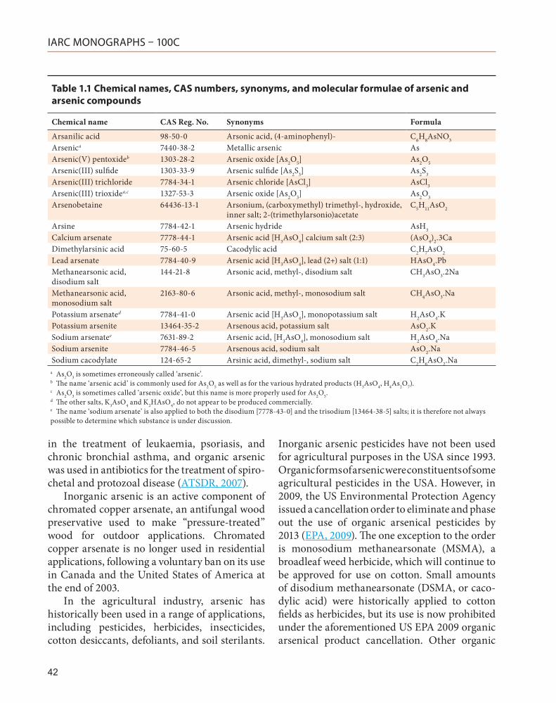
Information on the physical and chemical properties of arsenic and arsenic compounds can be found in Table 1.1, for further details please refer to IARC (1980). The list is not exhaus-tive, nor does it comprise necessarily the most commercially important arsenic-containing substances; rather, it indicates the range of arsenic compounds available.
1.2 Chemical and physical properties of the agents
Arsenic (atomic number, 33; relative atomic mass, 74.92) has chemical and physical proper-ties intermediate between a metal and a non-metal, and is often referred to as a metalloid or semi-metal. It belongs to Group VA of the Periodic Table, and can exist in four oxidation states: –3, 0, +3, and +5. Arsenite, AsIII, and arse-nate, AsV, are the predominant oxidation states under, respectively, reducing and oxygenated conditions (WHO, 2001; IARC, 2004).
From a biological and toxicological perspec-tive, there are three major groups of arsenic compounds:
-inorganic arsenic compounds, -organic arsenic compounds, and -arsine gas. Of the inorganic arsenic compounds, arsenic
trioxide, sodium arsenite and arsenic trichloride are the most common trivalent compounds, and arsenic pentoxide, arsenic acid and arse-nates (e.g. lead arsenate and calcium arsenate) are the most common pentavalent compounds. Common organic arsenic compounds include arsanilic acid, methylarsonic acid, dimethyl-arsinic acid (cacodylic acid), and arsenobetaine (WHO, 2000).
1.3 Use of the agents
Arsenic and arsenic compounds have been produced and used commercially for centuries. Current and historical uses of arsenic include pharmaceuticals, wood preservatives, agricul-tural chemicals, and applications in the mining, metallurgical, glass-making, and semiconductor industries.
Arsenic was used in some medicinal applica-tions until the 1970s. Inorganic arsenic was used
ARSENIC AND ARSENIC COMPOUNDSArsenic and arsenic compounds were considered by previous IARC Working Groups in 1979, 1987, and 2002 (IARC, 1980, 1987, 2004). Since that time, new data have become avail-able, these have been incorporated in the Monograph, and taken into consideration in the present evaluation.
IARC MONOGRAPHS – 100C
in the treatment of leukaemia, psoriasis, and chronic bronchial asthma, and organic arsenic was used in antibiotics for the treatment of spiro-chetal and protozoal disease (ATSDR, 2007).
Inorganic arsenic is an active component of chromated copper arsenate, an antifungal wood preservative used to make “pressure-treated” wood for outdoor applications. Chromated copper arsenate is no longer used in residential applications, following a voluntary ban on its use in Canada and the United States of America at the end of 2003.
In the agricultural industry, arsenic has historically been used in a range of applications, including pesticides, herbicides, insecticides, cotton desiccants, defoliants, and soil sterilants.
Inorganic arsenic pesticides have not been used for agricultural purposes in the USA since 1993. Organic forms of arsenic were constituents of some agricultural pesticides in the USA. However, in 2009, the US Environmental Protection Agency issued a cancellation order to eliminate and phase out the use of organic arsenical pesticides by 2013 (EPA, 2009). The one exception to the order is monosodium methanearsonate (MSMA), a broadleaf weed herbicide, which will continue to be approved for use on cotton. Small amounts of disodium methanearsonate (DSMA, or caco-dylic acid) were historically applied to cotton fields as herbicides, but its use is now prohibited under the aforementioned US EPA 2009 organic arsenical product cancellation. Other organic
42
Table 1.1 Chemical names, CAS numbers, synonyms, and molecular formulae of arsenic and arsenic compounds
Chemical name CAS Reg. No. Synonyms Formula
Arsanilic acid 98-50-0 Arsonic acid, (4-aminophenyl)- C6H8AsNO3
Arsenica 7440-38-2 Metallic arsenic AsArsenic(V) pentoxideb 1303-28-2 Arsenic oxide [As2O5] As2O5
Arsenic(III) sulfide 1303-33-9 Arsenic sulfide [As2S3] As2S3
Arsenic(III) trichloride 7784-34-1 Arsenic chloride [AsCl3] AsCl3
Arsenic(III) trioxidea,c 1327-53-3 Arsenic oxide [As2O3] As2O3
Arsenobetaine 64436-13-1 Arsonium, (carboxymethyl) trimethyl-, hydroxide, inner salt; 2-(trimethylarsonio)acetate
C5H11AsO2
Arsine 7784-42-1 Arsenic hydride AsH3
Calcium arsenate 7778-44-1 Arsenic acid [H3AsO4] calcium salt (2:3) (AsO4)2.3CaDimethylarsinic acid 75-60-5 Cacodylic acid C2H7AsO2
Lead arsenate 7784-40-9 Arsenic acid [H3AsO4], lead (2+) salt (1:1) HAsO4.PbMethanearsonic acid, disodium salt
144-21-8 Arsonic acid, methyl-, disodium salt CH3AsO3.2Na
Methanearsonic acid, monosodium salt
2163-80-6 Arsonic acid, methyl-, monosodium salt CH4AsO3.Na
Potassium arsenated 7784-41-0 Arsenic acid [H3AsO4], monopotassium salt H2AsO4.KPotassium arsenite 13464-35-2 Arsenous acid, potassium salt AsO2.KSodium arsenatee 7631-89-2 Arsenic acid, [H3AsO4], monosodium salt H2AsO4.NaSodium arsenite 7784-46-5 Arsenous acid, sodium salt AsO2.NaSodium cacodylate 124-65-2 Arsinic acid, dimethyl-, sodium salt C2H6AsO2.Na
a As2O3 is sometimes erroneously called ‘arsenic’.b The name ‘arsenic acid’ is commonly used for As2O5 as well as for the various hydrated products (H3AsO4, H4As2O7).c As2O3 is sometimes called ‘arsenic oxide’, but this name is more properly used for As2O5.d The other salts, K3AsO4 and K2HAsO4, do not appear to be produced commercially.e The name ‘sodium arsenate’ is also applied to both the disodium [7778-43-0] and the trisodium [13464-38-5] salts; it is therefore not always possible to determine which substance is under discussion.

Arsenic and arsenic compounds
arsenicals (e.g. roxarsone, arsanilic acid and its derivatives) are used as feed additives for poultry and swine to increase the rate of weight gain, to improve feed efficiencies, pigmentation, and disease treatment and prevention (EPA, 2000, 2006; FDA, 2008a, b).
Arsenic and arsenic compounds are used for a variety of other industrial purposes. Elemental arsenic is used in the manufacture of alloys, particularly with lead (e.g. in lead acid batteries) and copper. Gallium arsenide and arsine are widely used in the semiconductor and electronics industries. Because of its high electron mobility, as well as light-emitting, electromagnetic and photovoltaic properties, gallium arsenide is used in high-speed semiconductor devices, high-power microwave and millimetre-wave devices, and opto-electronic devices, including fibre-optic sources and detectors (IARC, 2006). Arsine is used as a doping agent to manufacture crystals for computer chips and fibre optics.
Arsenic and arsenic compounds are used in the manufacture of pigments, sheep-dips, leather preservatives, and poisonous baits. They are also used in catalysts, pyrotechnics, antifouling agents in paints, pharmaceutical substances, dyes and soaps, ceramics, alloys (automotive solder and radiators), and electrophotography.
Historically, the USA has been the world’s largest consumer of arsenic. Prior to 2004, about 90% of the arsenic consumed, as arsenic trioxide, was in the manufacture of wood preservatives. Since the voluntary ban on chromated copper arsenate in residential applications came into effect at the end of 2003, the consumption of arsenic for wood preservation has declined, drop-ping to 50% in 2007 (USGS, 2008). During 1990–2002, approximately 4% of arsenic produced was used in the manufacture of glass, and 1–4% was used in the production of non-ferrous alloys (NTP, 2005).
1.4 Environmental occurrence
Arsenic is the 20th most common element in the earth’s crust, and is emitted to the environ-ment as a result of volcanic activity and indus-trial activities. Mining, smelting of non-ferrous metals and burning of fossil fuels are the major anthropogenic sources of arsenic contamination of air, water, and soil (primarily in the form of arsenic trioxide). The historical use of arsenic-containing pesticides has left large tracts of agri-cultural land contaminated. The use of arsenic in the preservation of timber has also led to contamination of the environment (WHO, 2000, 2001).
1.4.1 Natural occurrence
In nature, arsenic occurs primarily in its sulfide form in complex minerals containing silver, lead, copper, nickel, antimony, cobalt, and iron. Arsenic is present in more than 200 mineral species, the most common of which is arsenopy-rite. Terrestrial abundance of arsenic is approxi-mately 5 mg/kg, although higher concentrations are associated with sulfide deposits. Sedimentary iron and manganese ores as well as phosphate-rock deposits occasionally contain levels of arsenic up to 2900 mg/kg (WHO, 2001).
1.4.2 Air
Arsenic is emitted to the atmosphere from both natural and anthropogenic sources. Approximately one-third of the global atmos-pheric flux of arsenic is estimated to be from natural sources (7900 tonnes per year). Volcanic activity is the most important natural contrib-utor, followed by low-temperature volatilization, exudates from vegetation, and windblown dusts. Anthropogenic sources are estimated to account for nearly 24000 tonnes of arsenic emitted to the global atmosphere per year. These emissions arise from the mining and smelting of base metals, fuel combustion (e.g. waste and low-grade brown
43

IARC MONOGRAPHS – 100C
coal), and the use of arsenic-based pesticides (WHO, 2000, 2001).
Arsenic is present in the air of suburban, urban, and industrial areas mainly as inorganic particulate (a variable mixture of AsIII and AsV, with the pentavalent form predominating). Methylated arsenic is assumed to be a minor component of atmospheric arsenic (WHO, 2000). Mean total arsenic concentrations in air range from 0.02–4 ng/m3 in remote and rural areas, and from 3–200 ng/m3 in urban areas. Much higher concentrations (> 1000 ng/m3) have been measured in the vicinity of industrial sources, such as non-ferrous metal smelters, and arsenic-rich coal-burning power plants (WHO, 2001).
1.4.3 Water
Arsenic, from both natural and anthropo-genic sources, is mainly transported in the envi-ronment by water. The form and concentration of arsenic depends on several factors, including whether the water is oxygenated (for example, arsenites predominate under reducing condi-tions such as those found in deep well-waters), the degree of biological activity (which is asso-ciated with the conversion of inorganic arsenic to methylated arsenic acids), the type of water source (for example, open ocean seawater versus surface freshwater versus groundwater), and the proximity of the water source to arsenic-rich geological formations and other anthropogenic sources (WHO, 2000, 2001).
The concentration of arsenic in surface fresh-water sources, like rivers and lakes, is typically less than 10 µg/L, although it can be as high as 5 mg/L near anthropogenic sources. Concentrations of arsenic in open ocean seawater and groundwater average 1–2 µg/L, although groundwater concen-trations can be up to 3 mg/L in areas with volcanic rock and sulfide mineral deposits (WHO, 2001).
Exposure to high levels of arsenic in drinking-water has been recognized for many decades in some regions of the world, notably in the People’s
Republic of China, Taiwan (China), and some countries in Central and South America. More recently, several other regions have reported having drinking-water that is highly contami-nated with arsenic. In most of these regions, the drinking-water source is groundwater, natu-rally contaminated from arsenic-rich geological formations. The primary regions where high concentrations of arsenic have been measured in drinking-water include large areas of Bangladesh, China, West Bengal (India), and smaller areas of Argentina, Australia, Chile, Mexico, Taiwan (China), the USA, and Viet Nam. In some areas of Japan, Mexico, Thailand, Brazil, Australia, and the USA, mining, smelting and other industrial activities have contributed to elevated concen-trations of arsenic in local water sources (IARC, 2004).
Levels of arsenic in affected areas may range from tens to hundreds or even thousands of micrograms per litre, whereas in unaffected areas, levels are typically only a few micrograms per litre. Arsenic occurs in drinking-water primarily as AsV, although in reducing environ-ments significant concentrations of AsIII have also been reported. Trace amounts of methylated arsenic species are typically found in drinking-water, and higher levels are found in biological systems. More complete data on arsenic in water may be found in the previous IARC Monograph (IARC, 2004).
1.4.4 Soil and sediments
Natural and anthropogenic sources contribute to the levels of arsenic found in soil and sedi-ments. Mean background concentrations in soil are often around 5 mg/kg, but can range from as low as 1 mg/kg to as high as 40 mg/kg. This vari-ation in levels of naturally occurring arsenic in soils is associated with the presence of geological formations (e.g. sulfide ores, mineral sediments beneath peat bogs). Soils contaminated with arsenic from anthropogenic sources (e.g. mine/
44

Arsenic and arsenic compounds
smelter wastes, agricultural land treated with arsenical pesticides) can have concentrations of arsenic up to several grams per kilogram. Mean sediment arsenic concentrations range from 5–3000 mg/kg, with the higher levels occurring in areas of anthropogenic contamination (WHO, 2001).
1.5 Human exposure
1.5.1 Exposure of the general population
The primary route of arsenic exposure for the general population is via the ingestion of contaminated food or water. The daily intake of total arsenic from food and beverages is gener-ally in the range of 20–300 µg/day.
Inhalation of arsenic from ambient air is generally a minor exposure route for the general population. Assuming a breathing rate of 20 m3/day, the estimated daily intake may amount to about 20–200 ng in rural areas, 400–600 ng in cities without substantial industrial emission of arsenic, about 1 µg/day in a non-smoker and more in polluted areas, and up to approximately 10 µg/day in a smoker (WHO, 2000, 2001).
1.5.2 Occupational exposure
Inhalation of arsenic-containing particulates is the primary route of occupational exposure, but ingestion and dermal exposure may be significant in particular situations (e.g. during preparation of timber treated with chromated copper arsenate). Historically, the greatest occu-pational exposure to arsenic occurred in the smelting of non-ferrous metal, in which arsenif-erous ores are commonly used. Other industries or industrial activities where workers are or were exposed to arsenic include: coal-fired power plants, battery assembly, preparation of or work with pressure-treated wood, glass-manufac-turing, and the electronics industry. Estimates of the number of workers potentially exposed to
arsenic and arsenic compounds have been devel-oped by the NIOSH in the USA and by CAREX in Europe. Based on the National Occupation Exposure Survey (NOES), conducted during 1981–83, NIOSH estimated that 70000 workers, including approximately 16000 female workers, were potentially exposed to arsenic and arsenic compounds in the workplace (NIOSH, 1990). Based on occupational exposure to known and suspected carcinogens collected during 1990–93, the CAREX (CARcinogen EXposure) database estimated that 147569 workers were exposed to arsenic and arsenic compounds in the European Union, with over 50% of workers employed in the non-ferrous base metal industries (n = 40426), manufacture of wood and wood and cork prod-ucts except furniture (n = 33959), and construc-tion (n = 14740). CAREX Canada estimates that 25000 Canadians are exposed to arsenic in their workplaces (CAREX Canada, 2011). These industries include: sawmills and wood preser-vation, construction, farms, non-ferrous metal (except aluminium) production and processing, iron and steel mills and ferro-alloy manufac-turing, oil and gas extraction, metal ore mining, glass and glass-product manufacturing, semi-conductor manufacturing, and basic chemical manufacturing.
1.5.3 Dietary exposure
Low levels of inorganic and organic arsenic have been measured in most foodstuffs (typical concentrations are less than 0.25 mg/kg). Factors influencing the total concentration of arsenic in food include: food type (e.g. seafood versus meat or dairy), growing conditions (e.g. soil type, water, use of arsenic-containing pesticides), and food-processing techniques. The highest concen-trations of arsenic have been found in seafood (2.4–16.7 mg/kg in marine fish, 3.5 mg/kg in mussels, and more than 100 mg/kg in certain crustaceans), followed by meats, cereals, vegeta-bles, fruit, and dairy products. Inorganic arsenic
45
IARC MONOGRAPHS – 100C
is the predominant form found in meats, poultry, dairy products and cereal, and organic arsenic (e.g. arsenobetaine) predominates in seafood, fruit, and vegetables (WHO, 2000, 2001).
Regional differences are seen in the daily intake of total arsenic through food, and are mainly attributable to variations in the quan-tity of seafood consumed. For example, the daily dietary intake of total arsenic in Japan is higher than that in Europe and the USA (WHO, 2000). Based on the limited data available, it is estimated that approximately 25% of daily dietary arsenic intake is from inorganic sources. Arsenic intake is typically higher in men than it is in women and children, with estimated levels ranging from 1.3 µg/day for infants under 1 year of age, 4.4 µg/day for 2-year olds, 9.9 µg/day for 25–30-year-old men, 10 µg/day for 60–65-year-old women, and 13 µg/day for 60–65-year-old men (WHO, 2001).
1.5.4 Biomarkers of exposure
Arsine generation atomic absorption spec-trometry (AAS) is the method of choice for biolog-ical monitoring of exposure to inorganic arsenic (WHO, 2000). The absorbed dose of arsenic can be identified and quantified in hair, nail, blood or urine samples. Because arsenic accumulates in keratin-rich tissue, total arsenic levels in hair, fingernails or toenails are used as indicators of past exposures. In contrast, because of its rapid clearing and metabolism, blood arsenic, urine arsenic, and urine arsenic metabolites (inorganic arsenic, monomethylarsonic acid [MMAV] and dimethylarsinic acid [DMAV]) are typically used as indicators of recent exposure.
The concentration of metabolites of inorganic arsenic in urine generally ranges from 5–20 µg/L, but may exceed 1000 µg/L (WHO, 2001). Time-weighted average (TWA) occupational exposure to airborne arsenic trioxide is significantly corre-lated with the inorganic arsenic metabolites in urine collected immediately after a shift or just
before the next shift. For example, at an airborne concentration of 50 µg/m3, the mean concentra-tion of arsenic derived from the sum of the three inorganic arsenic metabolites in a post-shift urine sample was 55 µg/g of creatinine. In non-occupationally exposed subjects, the sum of the concentration of the three metabolites in urine is usually less than 10 µg/g of creatinine (WHO, 2000).
2. Cancer in Humans
The epidemiological evidence on arsenic and cancer risk comes from two distinct lines of population studies, characterized by the medium of exposure to arsenic. One set of studies addresses the cancer risk associated with inhalation. These studies involve populations that are largely worker groups who inhaled air contaminated by arsenic and other agents, as a consequence of various industrial processes. The second set of studies was carried out in locations where people ingested arsenic in drinking-water at high concentrations over prolonged periods of time.
2.1 Types of human exposure circumstances studied
2.1.1 Arsenic exposure by inhalation
The cohort studies and nested case–control studies considered in this Monograph that are relevant to airborne arsenic include workers in metal smelters and refineries, and miners of various ores. Case–control studies within the general population addressed occupational exposures more generally. Consequently, the exposure to inhaled arsenic was accompanied by exposures to other potentially toxic and carci-nogenic by-products of combustion, such as sulfur oxides with copper smelting, polycyclic aromatic hydrocarbons, and particulate matter.
46
Arsenic and arsenic compounds
Most studies did not attempt to estimate sepa-rately exposures to the full set of agents in the inhaled mixtures, leaving open the possibility of some confounding or modification of the effect of arsenic by synergistic interactions.
2.1.2 Arsenic exposure by ingestion
For most human carcinogens, the major source of evidence contributing to causal infer-ences arises from case–control and cohort studies. In contrast, for arsenic in drinking-water, ecological studies provide important information on causal inference, because of the large exposure contrasts and the limited popula-tion migration. For arsenic, ecological estimates of relative risk are often so high that potential confounding with known causal factors could not explain the results. Although food may also be a source of some ingested arsenic, in several regions of the world where the concentrations of arsenic in drinking-water is very high, arsenic intake through food consumption contributes a relatively small cancer risk to the local residents (Liu et al., 2006a).
The strongest evidence for the association of human cancer with arsenic in drinking-water comes from studies in five areas of the world with especially elevated levels of naturally occurring arsenic: south-western and north-eastern Taiwan (China), northern Chile, Cordoba Province in Argentina, Bengladesh, West Bengal (India), and other regions in the Ganga plain. Although data contributing to our understanding also come from many other places, the current review is largely restricted to the major studies from these regions. Some of the oral exposure may occur via seafood. However, no epidemio-logical studies were available with regard to the cancer risk associated with arsenic exposure via seafood, in which arsenic may occur as partic-ular organic compounds such as arsenobetaine and arsenocholine.
(a) Taiwan (China)
Exposure to arsenic was endemic in two areas of Taiwan (China): The south-western coastal area (Chen et al., 1985), and the north-eastern Lanyang Basin (Chiou et al., 2001). Residents in the south-western areas drank artesian well-water with high concentrations of arsenic from the early 1910s to the late 1970s, with levels mostly above 100 μg/L (Kuo, 1968; Tseng et al., 1968). In the Lanyang Basin, residents used arsenic-contaminated water from household tube wells starting in the late 1940s. Arsenic in the water of 3901 wells, tested in 1991–94 ranged from undetectable (< 0.15 μg/L) to 3.59 mg/L (median = 27.3 μg/L) (Chiou et al., 2001).
(b) Northern Chile
The population-weighted average concentra-tion of arsenic in drinking-water in Region II, an arid region of northern Chile, was about 570 μg/L over 15 years (1955–69) (Smith et al., 1998). With the introduction of a water-treatment plant in 1970, levels decreased. By the late 1980s, arsenic levels in drinking-water had decreased to less than 100 μg/L in most places. With minor excep-tions, water sources elsewhere in Chile have had low concentrations of arsenic (less than 10 μg/L) (Marshall et al., 2007).
(c) Cordoba Province, Argentina
Of the 24 counties in Cordoba Province, two have been characterized as having elevated expo-sure to arsenic in drinking-water (average level, 178 μg/L), six as having medium exposure, and the remaining 16 rural counties as having low exposure (Hopenhayn-Rich et al., 1996, 1998).
(d) Bangladesh, West Bengal (India), and other locations in the Ganga plain
Millions of tube wells were installed in West Bengal (India), Bangladesh, and other regions in the Ganga plain of India and Nepal starting in the late 1970s to prevent morbidity and mortality
47

IARC MONOGRAPHS – 100C
from gastrointestinal disease (Smith et al., 2000). Elevated arsenic in wells in Bangladesh was confirmed in 1993 (Khan et al., 1997). In a Bangladesh survey by the British Geological Survey of 2022 water samples in 41 districts, 35% were found to have arsenic levels above 50 μg/L, and 8.4% were above 300 μg/L, with an estimate of about 21 million persons exposed to arsenic concentrations above 50 μg/L (Smith et al., 2000).
2.2 Cancer of the lung
2.2.1 Exposure via inhalation
Several ecological studies were conducted on populations exposed to arsenic through industrial emissions. The worker studies primarily provide information on lung cancer. The case–control studies are also mostly directed at lung cancer, with one on non-melanoma skin cancer (see Table 2.1 available at http://monographs.iarc.fr/ENG/Monographs/vol100C/100C-01-Table2.1.pdf).
The cohort studies reviewed previously and here consistently show elevated lung cancer risk in the various arsenic-exposed cohorts compared with the general population or other comparison groups, with most values in the range of 2–3 (see Table 2.2 available at http://monographs.iarc.fr/ENG/Monographs/vol100C/100C-01-Table2.2.pdf and Table 2.3 available at http://monog r aphs . ia rc . f r/ ENG/Monog r aphs/vol100C/100C-01-Table2.3.pdf).
The studies incorporate diverse qualitative and quantitative indices of exposure that include measures of average airborne concentration of exposure, cumulative exposure across the work experience, and duration of exposure. There is consistent evidence for a positive exposure–response relationship between the indicators of exposure and lung cancer risk. Case–control studies nested within occupational cohorts provided similar evidence with regard to expo-sure–response relationships.
Several analyses further explored the relation-ship between arsenic exposure and lung cancer risk using models based on either empirical, i.e. descriptive, or biological data (see Table 2.4 available at http://monographs.iarc.fr/ENG/Monographs/vol100C/100C-01-Table2.4.pdf).
Using data from the Tacoma, Washington smelter workers, Enterline et al. (1987) modelled the relationship between lung cancer risk and airborne arsenic exposure using power func-tions, and found that the exposure–response relationship was steeper at lower concentrations than shown in conventional analyses, and was concave downwards at higher concentrations. By contrast, the relationship of risk with urine arsenic concentration was linear. Lubin et al. (2000, 2008) analysed the exposure–response relationship of lung cancer risk with arsenic expo-sure in the cohort of Montana smelter workers, now followed for over 50 years. Overall, a linear relationship of risk with cumulative exposure was found; however, the slope of the relation-ship increased with the average concentration at which exposure had taken place, that is, the effect of a particular cumulative exposure was greater if received at a faster rate.
For a comparison of the different studies, see Table 2.5 available at http://monog r aphs . ia rc . f r/ ENG/Monog r aphs/vol100C/100C-01-Table2.5.pdf.
2.2.2 Exposure via ingestion
A summary of the findings of epidemio-logical studies on arsenic in drinking-water and risk for lung cancer are shown in Table 2.6 (water exposures) available at http://monographs.iarc.fr/ENG/Monographs/vol100C/100C-01-Table2.6.pdf, and online Tables 2.1 to 2.4 (air exposures).
48

Arsenic and arsenic compounds
(a) Ecological studies
Ecological studies, based on mortality records, were conducted in the arseniasis endemic area of south-western Taiwan (China) (Chen et al., 1985, 1988a; Wu et al., 1989; Chen & Wang, 1990; Tsai et al., 1999). All studies found elevated risks for lung cancer mortality associ-ated with levels of arsenic in drinking-water, or surrogate measurements.
In Chile, Rivara et al. (1997) found an elevated relative risk (RR) for mortality from lung cancer in 1976–92 in Region II compared with Region VIII, a low-exposure area. Smith et al. (1998) found an elevated standardized mortality ratio (SMR) of approximately 3 for lung cancer for both sexes in Region II, using the national rate as standard. In Cordoba Province, Argentina, significant increases in lung cancer mortality were associated with increasing exposure to arsenic (Hopenhayn-Rich et al., 1998). Smith et al. (2006) found an elevated lung cancer mortality (RR, 7.0; 95%CI: 5.4–8.9) among the 30–49-year-old residents of Antofagasta and Mejillones born in the period 1950–57, just before the period of exposure to high arsenic levels (1958–70). They were exposed in early childhood to high levels of arsenic through the drinking-water. The temporal pattern of lung cancer mortality rate ratios in Region II compared with that in Region V (a low-exposure area) from 1950 to 2000, showed an increase about 10 years after the onset of high arsenic exposure, and peaked in 1986–87, with relative risks of 3.61 (95%CI: 3.13–4.16) and 3.26 (95%CI: 2.50–4.23) for men and women, respectively (Marshall et al., 2007).
(b) Case–control and cohort studies
In northern Chile, a case–control study of 151 cases and 419 controls reported significantly increasing risks with increasing levels of arsenic during the 1958–70 high-exposure period, with an odds ratio increasing to 7.1 (95%CI: 3.4–14.8) (Ferreccio et al., 2000).
In a cohort from south-western Taiwan (China), Chen et al. (1986) observed a dose–response relationship between the duration of consumption of artesian well-water containing high levels of arsenic and lung cancer mortality risk, showing the highest age- and gender-adjusted odds ratio among those who consumed artesian well-water for more than 40 years compared with those who never consumed artesian well-water. Another cohort study from south-western Taiwan (China) endemic for arsenic found a smoking-adjusted increased risk for lung cancer in relation to increasing average concentrations of arsenic and increasing cumulative exposure to arsenic (Chiou et al., 1995).
A further study of combined cohorts in south-western (n = 2503) and north-eastern (n = 8088) Taiwan (China) found a synergistic interaction between arsenic in drinking-water and cigarette smoking (Chen et al., 2004).
A case–control study from Bangladesh, conducted in 2003–06, found an elevated risk (odds ratio [OR], 1.65; 95%CI: 1.25–2.18) for male smokers consuming tube well-water with arsenic levels of 101–400 μg/L (Mostafa et al., 2008). In non-smokers, the study did not report an increased risk with increasing arsenic expo-sure. [The Working Group noted the ecological nature of the exposure estimates, the possibility of greater sensitivity to arsenic exposure among smokers, and the relatively short latent period, with almost two-thirds of the wells put in place in 1990 or later.]
2.3 Cancer of the urinary bladder and of the kidney
The results of the epidemiological studies on arsenic in drinking-water and the risk for cancers of the urinary bladder and of the kidney are summarized in Table 2.7 available at http://monographs.iarc.fr/ENG/Monographs/vol100C/100C-01-Table2.7.pdf.
49

IARC MONOGRAPHS – 100C
2.3.1 Ecological studies
In south-western and north-eastern Taiwan (China), the relation between cancer of the urinary bladder and of the kidney and drinking-water containing arsenic was evaluated in many of the studies cited above (Chen et al., 1985, 1988a; Wu et al., 1989; Chen & Wang, 1990; Tsai et al., 1999). Each reported an elevation in mortality from these cancers during various time periods in 1971–94 associated with levels of arsenic in well-water from rural artesian wells, with many reporting a dose–response relationship among both men and women. An additional study, based on incidence records, found comparable risks for bladder cancer (Chiang et al., 1993).
In Region II of Chile, two studies found markedly high SMRs for cancer of the urinary bladder and of the kidney in 1950–92 (Rivara et al., 1997) and in 1989–93 (Smith et al., 1998). In the latter study, mortality from chronic obstruc-tive pulmonary disease was at the expected level, suggesting that smoking was not involved. The temporal pattern of bladder cancer mortality in Region II from 1950–2000 was compared with that in Region V (Marshall et al., 2007). Increased relative risks were reported about 10 years after the start of exposure to high arsenic levels, with peak relative risks of 6.10 (95%CI: 3.97–9.39) for men, and 13.8 (95%CI: 7.74–24.5) for women in the period 1986–94. In Cordoba Province, Argentina, positive trends in SMRs were reported for bladder and kidney cancers associated with estimates of exposure to arsenic in drinking-water (Hopenhayn-Rich et al., 1996, 1998), again with no findings for chronic obstruc-tive pulmonary disease.
[The Working Group noted that kidney cancers consist of both renal cell carcinoma and transitional cell carcinoma of the renal pelvis, the latter often being of the same etiology as bladder cancer. As arsenic causes transitional cell carci-noma of the bladder, merging of the two types of
kidney cancer may result in a dilution of the risk estimate for total kidney cancer.]
2.3.2 Case–control and cohort studies
In a case–control study using death certifi-cates (1980–82) from the area in Taiwan (China), endemic for Blackfoot disease, Chen et al. (1986) reported increasing trends in odds ratios with increasing duration of consumption of artesian well-water containing arsenic. The highest risks were seen for over 40 years of exposure, with an odds ratio of 4.1 (P < 0.01) for bladder cancer in a multivariate analysis, after adjusting for smoking and other factors from next-of-kin interviews.
In case–control studies of incident bladder cancer that included analysis of arsenic species in urine samples, a higher risk associated with arsenic was found among persons with higher MMAV:DMAV ratios or, alternatively, with a higher percentage of MMAV (Chen et al., 2003, 2005a; Steinmaus et al., 2006; Pu et al., 2007a; Huang et al., 2008).
Cohort studies from south-western and north-eastern Taiwan (China) (Chen et al., 1988b; Chiou et al., 1995, 2001; Chen & Chiou, 2001) Japan (Tsuda et al., 1995), and the United Kingdom (Cuzick et al., 1992) each observed elevated bladder cancer risk following long-term exposure to ingested arsenic, with dose–response relationships found where the numbers of cases permitted such an analysis. The study from Taiwan (China), also found an elevated risk of kidney cancer (OR, 2.8; 95%CI: 1.3–5.4, based on nine cases) (Chiou et al., 2001).
2.4 Cancer of the skin
The recognition of arsenic as a carcinogen first came from case series describing skin cancers following the ingestion of medicines containing arsenicals (Hutchinson, 1888; Neubauer, 1947), and exposure to arsenical pesticide residues, and arsenic-contaminated wine (Roth, 1957; Grobe,
50
Arsenic and arsenic compounds
1977) or drinking-water, originating from many countries. The characteristic arsenic-associated skin tumours include squamous cell carcinomas arising in keratoses (including Bowen disease), and multiple basal cell carcinomas.
Findings of epidemiological studies on arsenic in drinking-water and risk for skin cancer are summarized in Table 2.8 available at http://monographs.iarc.fr/ENG/Monographs/vol100C/100C-01-Table2.8.pdf.
2.4.1 Ecological studies of prevalence
In south-western Taiwan (China), Tseng et al. (1968) found an 8-fold difference in the preva-lence of skin cancer lesions from the highest (> 600 µg/L) to the lowest category (< 300 µg/L) of arsenic concentration in artesian wells, after an extensive examination survey of 40421 inhab-itants in 37 villages.
2.4.2 Ecological studies based on mortality from cancer of the skin
Studies in Taiwan (China) (Chen et al., 1985, 1988a; Wu et al., 1989; Chen & Wang, 1990; Tsai et al., 1999) analysed skin cancer mortality in relation to levels of arsenic in well-water. These investigations found consistent gradients of increasing risk with average level of arsenic in drinking-water, as measured on the township or precinct level.
Rivara et al. (1997) observed an SMR for skin cancer of 3.2 (95%CI: 2.1–4.8), comparing mortality from skin cancer in 1976–92 between Region II and the unexposed control Region VIII of Chile. Later, Smith et al. (1998) found SMRs of 7.7 (95%CI: 4.7–11.9) among men and 3.2 (95%CI: 1.3–6.6) among women for the years 1989–93 in Region II of Chile, using national mortality rates as reference. [The Working Group noted that the histological type of skin cancer was reported in only a few instances. Although skin cancer mortality can be influenced by access to health
care, the SMRs reported here are so large as to not be explained by any possible confounding.]
2.4.3 Cohort studies
A retrospective cohort study of 789 (437 men, 352 women) of Blackfoot disease patients in Taiwan (China) reported an SMR of 28 (95%CI: 11–59) for skin cancer deaths (based on seven observed deaths), using Taiwan (China) regional rates as reference (Chen et al., 1988b).
In a cohort of 654 persons in south-western Taiwan (China), an observed incidence rate of 14.7 cases of skin cancer/1000 person–years was found (Hsueh et al., 1997), with risks significantly related to duration of living in the area endemic for Blackfoot disease, duration of consumption of artesian well-water, average concentration of arsenic, and index for cumulative exposure to arsenic. Similar findings were observed in a nested case–control study conducted within this cohort (Hsueh et al., 1995).
In Region II of Chile, a decrease in incidence rates of cutaneous lesions (leukoderma, melano-derma, hyperkeratosis, and squamous cell carci-noma) was observed during 1968–71 after a lowering of waterborne arsenic levels from a filter plant, which started operation in 1970 (Zaldívar, 1974).
2.5 Cancer of the liver
2.5.1 Ecological studies
The relation between liver cancer risk and drinking-water contaminated with arsenic was evaluated in many of the studies from south-western Taiwan (China), cited above (Chen et al., 1985, 1988a; Wu et al., 1989; Chen & Wang, 1990; Chiang et al., 1993; Tsai et al., 1999; see Table 2.9 available at http://monographs.iarc.fr/ENG/Monographs/vol100C/100C-01-Table2.9.pdf), with positive associations found in all studies.
51

IARC MONOGRAPHS – 100C
In northern Chile, Rivara et al. (1997) observed a relative risk for liver cancer mortality of 1.2 (95%CI: 0.99–1.6) in arsenic-exposed Region II compared with Region VIII. Liver cancer mortality in Region II of northern Chile during the period 1989–93 among persons ≥ 30 years of age was not significantly elevated, using national rates as reference (Smith et al., 1998). SMRs were 1.1 (95%CI: 0.8–1.5) both for men and for women. Liaw et al. (2008) found an elevated relative risk (10.6; 95%CI: 2.9–39.3, P < 0.001) for liver cancer among children in Region II of Chile born in 1950–57 and exposed in utero or shortly thereafter, compared to rates in Region V of Chile.
In Cordoba Province, Argentina, SMRs were not related to arsenic exposure (Hopenhayn-Rich et al., 1998).
[The Working Group noted that the finding of an association with liver cancer in Taiwan (China), but not in South America may reflect a more sensitive population in the former region, due to endemic hepatitis B. The elevated risk of those exposed in utero and as young children may reflect a combination of greater biological vulnerability in early life (Waalkes et al., 2007) plus the fact that young children consume 5–7 times more water per kilogram body weight per day than adults (NRC, 1993).]
2.5.2 Case–control studies
In a case–control study investigating the consumption artesian well-water containing high concentrations of arsenic and mortality from liver cancer in four townships of south-westernern Taiwan (China), Chen et al. (1986) observed an exposure–response relationship between the duration of consumption of the contaminated well-water and risk for liver cancer, adjusted for cigarette smoking, habitual alcohol and tea drinking, and consumption of vegetables and fermented beans.
2.6 Cancer of the prostate
Studies conducted in Taiwan (China) (Chen et al., 1985, 1988a; Wu et al., 1989; Chen & Wang, 1990; Tsai et al., 1999) analysed prostate cancer mortality in relation to levels of arsenic in well-water, with some overlap among the respective study populations. Using several methodolog-ical approaches and comparison populations including direct and indirect standardization of rates, all studies reported significant dose–response relationships between the level of arsenic in drinking-water and the risk for pros-tate cancer mortality (see Table 2.10 available at http://monographs.iarc.fr/ENG/Monographs/vol100C/100C-01-Table2.10.pdf).
In Chile, Rivara et al. (1997) found a relative risk of 0.9 (95%CI: 0.54–1.53) for prostate cancer, comparing the 1990 mortality rate for prostate cancer of Region II with that of Region VIII.
2.7 Synthesis
The Working Group reviewed a large body of evidence that covers ecological studies, case–control studies and cohort studies in a variety of settings and populations exposed either by ingestion (primarily to AsIII and AsV in drinking-water) or inhalation (with exposure to a mixture of inorganic arsenic compounds). The evidence also relates to historical exposure from pesticidal and pharmaceutical uses. The epidemiological evidence from drinking-water exposure permits the evaluation of the carcinogenicity that is related to exposure to AsIII and AsV. The epidemi-ological evidence from inhaled arsenic mixtures permits the evaluation of the carcinogenicity that is related to inorganic arsenic compounds. However, it does not allow a separation of the carcinogenic risk associated with particular arsenic species that occur in these mixtures.
The observed associations between exposure to arsenic in drinking-water and lung cancer, and between exposure to arsenic in air and lung
52

Arsenic and arsenic compounds
cancer, cannot be attributed to chance or bias. The evidence is compelling for both the inhala-tion and ingestion routes of exposure. There is evidence of dose–response relationships within exposed populations with both types of exposure.
The observed association between exposure to arsenic in drinking-water and bladder cancer cannot be attributed to chance or bias. There is evidence of dose–response relationships within exposed populations.
The observed association between exposure to arsenic in drinking-water and skin cancer cannot be attributed to chance or bias. There is evidence of dose–response relationships within exposed populations. The evidence is primarily for squamous cell carcinoma of the skin.
Although the data for kidney cancer are suggestive of a relationship with exposure to arsenic in drinking-water, overall, the small possibility of chance or bias cannot be completely ruled out.
The evidence for an association between liver cancer and long-term exposure to arsenic in drinking-water relies on mortality data. Although the data strongly suggest a causal asso-ciation with some evidence of a dose–response relationship, the Working Group could not rule out possible chance or bias. The evidence comes mainly from Taiwan (China) where hepatitis B is highly prevalent.
The evidence for an association for pros-tate cancer and long-term exposure to arsenic in drinking-water relies on mortality data. In the studies from Taiwan (China), there is some evidence of a dose–response relationship. However, the data from South America are not consistent with this observation. Although the evidence on prostate cancer suggests the possi-bility of a causal association, the Working Group could not rule out the possibility of chance or bias.
3. Cancer in Experimental Animals
Over the years, it has proved difficult to provide evidence for the carcinogenesis of inor-ganic arsenic compounds. More recent work has focused on methylated arsenic metabolites in humans or exposure to inorganic arsenic during early life, and has provided the informa-tion to show potential links between arsenic and carcinogenesis.
Studies published since the previous IARC Monograph (IARC, 2004) are summarized below.
3.1 Oral administration
3.1.1 Mouse
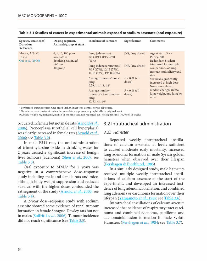
The oral administration of sodium arsenate in drinking-water for 18 months increased lung tumour multiplicity and lung tumour size in male strain A/J mice (Cui et al., 2006; see Table 3.1).
Similarly, drinking-water exposure to the organo-arsenical DMAV for 50 weeks or more increased the incidence and multiplicity of lung adenoma or carcinoma in strain A/J mice (Hayashi et al., 1998), and increased lung tumours in mutant Ogg−/− mice (which cannot repair certain types of oxidative DNA damage) but not in Ogg+/+ mice (Kinoshita et al., 2007; see Table 3.2).
3.1.2 Rat
In male F344 rats, the oral administration of DMAV in drinking-water for up to 2 years produced clear dose–response relationships for the induction of urinary bladder transitional cell carcinoma and combined papilloma or carci-noma (Wei et al., 1999, 2002).
When DMAV was added to the feed of male and female F344 rats for 2 years, a clear dose–response relationship for urinary bladder benign and/or malignant transitional cell tumours
53
IARC MONOGRAPHS – 100C
occurred in female but not male rats (Arnold et al., 2006). Preneoplasia (urothelial cell hyperplasia) was clearly increased in female rats (Arnold et al., 2006; see Table 3.2).
In male F344 rats, the oral administration of trimethylarsine oxide in drinking-water for 2 years caused a significant increase of benign liver tumours (adenoma) (Shen et al., 2007; see Table 3.3).
Oral exposure to MMAV for 2 years was negative in a comprehensive dose–response study including male and female rats and mice, although body weight suppression and reduced survival with the higher doses confounded the rat segment of the study (Arnold et al., 2003; see Table 3.4).
A 2-year dose–response study with sodium arsenite showed some evidence of renal tumour formation in female Sprague-Dawley rats but not in males (Soffritti et al., 2006). Tumour incidence did not reach significance (see Table 3.5).
3.2 Intratracheal administration
3.2.1 Hamster
Repeated weekly intratracheal instilla-tions of calcium arsenate, at levels sufficient to caused moderate early mortality, increased lung adenoma formation in male Syrian golden hamsters when observed over their lifespan (Pershagen & Björklund, 1985).
In a similarly designed study, male hamsters received multiple weekly intratracheal instil-lations of calcium arsenate at the start of the experiment, and developed an increased inci-dence of lung adenoma formation, and combined lung adenoma or carcinoma formation over their lifespan (Yamamoto et al., 1987; see Table 3.6).
Intratracheal instillations of calcium arsenite increased the incidence of respiratory tract carci-noma and combined adenoma, papilloma and adenomatoid lesion formation in male Syrian Hamsters (Pershagen et al., 1984; see Table 3.7).
54
Table 3.1 Studies of cancer in experimental animals exposed to sodium arsenate (oral exposure)
Species, strain (sex) Duration Reference
Dosing regimen, Animals/group at start
Incidence of tumours Significance Comments
Mouse, A/J (M) 18 mo Cui et al. (2006)
0, 1, 10, 100 ppm arsenate in drinking-water, ad libitum 30/group
Lung (adenomas): 0/19, 0/13, 0/15, 4/30 (13%)
[NS, (any dose)]a Age at start, 5 wk Purity, NR Redundant Student t-test used for multiple comparisons of lung tumour multiplicity and size Survival significantly increased at high dose Non-dose-related, modest changes in bw, lung weight, and lung bw ratio
Lung (adenocarcinomas): 9/19 (47%), 10/13 (77%), 11/15 (73%), 19/30 (63%)
[NS, (any dose)]a
Average tumours/mouse lung: 0.59, 1.1, 1.3, 1.4b
P < 0.01 (all doses)
Average number tumours > 4 mm/mouse lung: 17, 32, 44, 60b
P < 0.01 (all doses)
a Performed during review. One-sided Fisher Exact test–control versus all treated.b Numbers are estimates at review because data are presented graphically in original work.bw, body weight; M, male; mo, month or months; NR, not reported; NS, not significant; wk, week or weeks

Arsenic and arsenic com
pounds55
Table 3.2 Studies of cancer in experimental animals exposed to dimethylarsinic acid, DMAV (oral exposure)
Species, strain (sex) Duration Reference
Dosing regimen Animals/group at start
Incidence of tumours Significance Comments
Mouse, A/J (M) 50 wk Hayashi et al. (1998)
0, 50, 200, 400 ppm DMAV in drinking-water, ad libitum 24/group
Number of mice with lung papillary adenomas or adenocarcinomas: 2/14 (14%), 5/14 (36%), 7/14 (50%), 10/13 (77%)
P < 0.01 (high dose) Age at start, 5 wk Purity, NR Survival unremarkable [Only histologically confirmed tumours were considered by the Working Group]
Mouse, Ogg1-/- and Ogg1+/+ (M, F) 72 wk Kinoshita et al. (2007)
0, 200 ppm DMAV in drinking-water, ad libitum; controls received tap water 10/group (Ogg1-/-) 12/group (Ogg1+/+)
Ogg1-/-: Tumour-bearing mice (any site): 0/10, 10/10 (100%)
P < 0.01 Age at start, 14 wk Purity, 99% Bw and food and water consumption unremarkable Left lobe and visible lung nodules used for histopathological tumour analysis Treated Ogg1−/− showed modest decreased survival (~20%) late compared to phenotypic control Small groups
Lung lesions–Hyperplasias: 10/10 (100%), 10/10 (100%)Adenomas: 0/10, 2/10 (20%)
NS
Adenocarcinomas: 0/10, 3/10 (30%)Total lung tumours: 0/10, 5/10 (50%)
P < 0.05
Tumours/mouse: 0, 0.5
P < 0.05
Ogg1+/+: Tumour-bearing mice (any site): 5/10 (50%), 6/10 (60%)Lung lesions–Hyperplasias: 2/10 (20%), 10/10 (100%)
[P < 0.01b]
Adenomas: 1/10 (10%), 0/10
NS
Adenocarcinomas: 0/10, 0/10
NS
Total tumours: 1/10 (10%), 0/10
NS
Tumours/mouse: 0.1, 0
NS
Tumours/mouse: 0.1, 0
NS

IARC M
ON
OG
RAPH
S – 100C
56
Species, strain (sex) Duration Reference
Dosing regimen Animals/group at start
Incidence of tumours Significance Comments
Rat, F344 (M) 104 wk Wei et al. (1999)d, 2002)
0, 12.5, 50, 200 ppm DMAV in drinking-water, ad libitum 36/group
Urinary bladder (hyperplasias): 0/28, 0/33, 12/31 (39%), 14/31 (45%)
P < 0.01 (middle and high dose) Age at start, 10 wk Purity, 99% Survival and food intake unaltered Transient bw suppression early with high and middle dose but then similar to control Water intake increased at highest two doses Incidence rates based on rats at risk (surviving to time of the first bladder tumour at 97 wk) Extensive necropsy
Urinary bladder (papillomas): 0/28, 0/33, 2/31 (2%), 2/31 (2%)
NS
Urinary bladder (carcinomas): 0/28, 0/33, 6/31 (19%), 12/31 (39%)
P < 0.05 (middle dose) P < 0.01 (high dose)
Urinary bladder (papillomas or carcinomas): 0/28, 0/33, 8/31 (26%), 12/31 (39%)
P < 0.01 (middle and high dose)
Rat, F344 (M, F) 104 wk Arnold et al. (2006)
0, 2, 10, 40, 100 ppm DMAV in feed, ad libitum 60/group
Females Urothelial cell (hyperplasias, simple, nodular and papillary): 0/60, 1/59 (2%), 0/60, 29/59 (49%), 48/60 (80%)
P < 0.01 (trend) [P < 0.01 (highest, and second highest dose)]b
Purity > 99%; age, 5 wk Complete necropsies performed No treatment-related differences in mortality or bw
Urinary bladder (papillomas): 0/60, 0/59, 0/60, 0/59, 4/60 (7%)
Sporadic changes in food consumption not treatment-related Water consumption increased with treatment
Urinary bladder (carcinomas): 0/60, 0/59, 0/60, 0/59, 6/60 (10%)
[NS (high dose)]b P < 0.01 (trend)c
Urinary bladder (papillomas and carcinomas combined): 0/60, 0/59, 0/60, 0/59, 10/60 (3%)
[P < 0.05 (high dose)]b P < 0.01 (trend)c [P < 0.05 (high dose)]b
Males Urothelial cell (hyperplasias, simple, nodular and papillary): 0/60, 0/59, 0/60, 6/58 (10%), 40/59 (68%)
P < 0.01 (trend) [P < 0.01 (high dose)]b
Water consumption increased with treatment
Urinary bladder (papillomas): 0/60, 0/59, 1/60 (2%), 1/58 (2%), 0/59
[NS (high dose)]b P < 0.01 (trend)c
Urinary bladder (carcinomas): 0/60, 1/59 (2%), 0/60, 0/58, 2/59 (3%)
[NS (high dose)]b P < 0.01 (trend)c
Urinary bladder (papillomas and carcinomas combined): 0/60, 1/59 (2%), 1/60 (2%), 1/58 (2%), 2/59 (3%)
[NS (high dose)]b
Table 3.2 (continued)

Arsenic and arsenic com
pounds57
Species, strain (sex) Duration Reference
Dosing regimen Animals/group at start
Incidence of tumours Significance Comments
Mouse, B6C3F1 (F) 104 wk Arnold et al. (2006)
0, 8, 40, 200, 500 ppm DMAV in feed, ad libitum 56/group
Females No treatment-related changes in urinary bladder preneoplasia or tumour incidence noted
Age at start, 5 wk Purity 99% Complete necropsies performed Survival, bw and water consumption unchanged Sporadic, small changes in food consumption early Fibrosarcomas not considered related to treatment by authorsBw reduced at 500 ppm throughout study
Any organ (fibrosarcomas): 3/56 (5%), 0/55, 1/56 (2%), 1/56 (2%), 6/56 (11%)
P < 0.01 (high dose)
Males No treatment-related changes in urinary bladder preneoplasia or tumour incidence noted
a Data also included descriptive statistics (i.e. SD).b Performed during review. One-sided Fisher exact test control versus treated.c Trend analysis performed after combination of female and male data for urinary bladder lesions from this same study (Arnold et al., 2006). d Short communication of tumour data only.e On a C57BL/6 background.f As stated by the authors.g The lack of information on group size and the lack of descriptive statistics makes these data impossible to independently re-evaluate for statistical significance.bw, body weight; F, female; M, male; NR, not reported; NS, not significant; wk, week or weeks
Table 3.2 (continued)

IARC MONOGRAPHS – 100C
3.3 Intravenous administration
3.3.1 Mouse
Multiple intravenous injections of sodium arsenate in male and female Swiss mice provided no evidence of elevated tumour formation (Waalkes et al., 2000; see Table 3.8).
3.4 Transplacental and perinatal exposures
3.4.1 Mouse
Pregnant mice were treated subcutaneously with arsenic trioxide on a single specific day during gestation (Days 14, 15, 16 or 17), and the offspring were then treated subcutaneously on postpartum Days 1, 2 and 3 with arsenic trioxide. The offspring initially treated on Day 15 of gestation developed an excess of lung adenoma compared to controls, and the other groups did not (Rudnai & Borzsanyi, 1980, 1981; see Table 3.9).
Pregnant C3H mice were exposed to various doses of sodium arsenite in the drinking-water from Days 8–18 of gestation. They were allowed to give birth and their offspring were put into gender-based groups at weaning. Over the next 90 weeks, arsenic-treated female offspring
developed dose-related benign and/or malig-nant ovarian tumours, and lung adenocarci-noma. During the next 74 weeks, a dose-related increase in the incidences of liver adenoma and/or carcinoma, and adrenal cortical adenoma was observed in the male offspring (Waalkes et al., 2003).
A second study looked at the carcino-genic effects in C3H mice of various doses of sodium arsenite (two levels) in the maternal drinking-water from Days 8 to 18 of gestation, with or without subsequent 12-O-tetradecanoyl phorbol-13-acetate (TPA) applied to the skin of the offspring after weaning from 4–25 weeks of age. Over the next 2 years, with arsenic alone, the female offspring developed an increased inci-dence of ovarian tumours. The male offspring developed arsenic dose-related increases in the incidences of liver adenoma and/or carcinoma and adrenal cortical adenoma (Waalkes et al., 2004).
Pregnant CD1 mice received sodium arsenite (one level) in the drinking-water from gestation Days 8 to 18, were allowed to give birth, and the female (Waalkes et al., 2006a) or male (Waalkes et al., 2006b) offspring were treated with diethyl-stilbestrol or tamoxifen subcutaneously on post-partum Days 1, 2, 3, 4 and 5. In female offspring over the next 90 weeks, arsenic exposure alone
58
Table 3.3 Studies of cancer in experimental animals exposed to trimethylarsine oxide (oral exposure)
Species, strain (sex) Duration Reference
Dosing regimen Animals/group at start
Incidence of tumours Significance Comments
Rat, F344 (M) 2 yr Shen et al. (2003)
0, 50, 200 ppm trimethylarsine oxide in drinking-water, ad libitum 42–45; 42 controls
Liver (adenomas): 6/42 (9%), 10/42 (14%), 16/45 (24%)
P < 0.05 (high dose)
Age at start, 10 wk Purity, 99% Body weights, food intake, water intake, survival rate, and average survival unaltered with treatment Extensive necropsy performed Various other sites negative
bw, body weight; M, male; yr, year or years

Arsenic and arsenic com
pounds59
Table 3.4 Studies of cancer in experimental animals exposed to monomethylarsonic acid, MMAV (oral exposure)
Species, strain (sex) Duration Reference
Dosing regimen Animals/group at start
Incidence of tumours Significance Comments
Mouse, B6C3F1 (M, F) 104 wk Arnold et al. (2003)
0, 10, 50, 200, 400 ppm MMAV in feed, ad libitum 52/group/sex
No treatment-related changes
Age at start, 6 wk Purity, 99% Bw reduced at 400 ppm throughout study Food and water consumption similar or increased at the two higher doses Survival unremarkable Complete necropsy performed
Rat, F344 (M, F) 104 wk Arnold et al. (2003)
0, 50, 400, 1 300a ppm MMAV in feed, ad libitum 60/group/sex
No treatment-related changes
Age at start, 6 wk Purity, 99% Bw reduced at two highest doses in second half of study Food consumption generally similar Water consumption similar or increased at the two higher doses Survival reduced at high dose Complete necropsy performed
a Due to a high mortality in male and female rats fed this level, it was reduced to 1000 ppm during Week 53, and further reduced to 800 ppm during Week 60.bw, body weight; F, female; M, male; wk, week or weeks

IARC MONOGRAPHS – 100C
increased the incidence of tumours of the ovary, uterus, and adrenal cortex. In the male offspring, prenatal arsenic exposure alone increased liver adenoma and/or carcinoma, lung adenocarci-noma, and adrenal cortical adenoma (see Table 3.10).
3.5 Studies in which arsenic modifies the effects of other agents
3.5.1 Mouse
Mice exposed to DMAV in drinking-water after subcutaneous injection of 4-nitroquino-line 1-oxide showed an increase in lung tumour multiplicity compared to mice exposed to the organic carcinogen alone (Yamanaka et al., 1996). In K6/ODC mice first treated topically with 7,12-dimethylbenz[α]anthracene (DMBA) then with DMAV in a cream applied to the same skin area for 18 weeks, the organo-arsenical doubled the skin tumour multiplicity compared to treatment with DMBA alone (Morikawa et al., 2000; see Table 3.11). [The Working Group noted that this study had too few DMAV controls for an appropriate interpretation.]
In the studies of Germolec et al. (1997, 1998), oral sodium arsenite was given to Tg.AC mice with TPA by skin painting, and an approxi-mately 4-fold increase in skin tumour response was reported.
Combined treatment with oral sodium arsenite in drinking-water and multiple expo-sures to excess topical UV irradiation in Crl:SKl-hrBR hairless mice showed that arsenic treatment alone was consistently without carcinogenic effect, but markedly enhanced UV-induced skin tumours including squamous cell carcinoma (Rossman et al., 2001; Burns et al., 2004; Uddin et al., 2005). In another skin study, mice exposed to topical 9,10-dimethyl-1,2-benzanthracene for 2 weeks concurrently with oral sodium arsenate in drinking-water for 25 weeks showed that arsenic treatment alone was without carcino-genic effect, but enhanced skin tumour multi-plicity and tumour size when combined with the organic carcinogen compared to the organic carcinogen alone (Motiwale et al., 2005; see Table 3.12).
When pregnant Tg.AC mice were treated with oral sodium arsenite in drinking-water from Days 8–18 of gestation, and their offspring were topically exposed to TPA from 4–40 weeks
60
Table 3.5 Studies of cancer in experimental animals exposed to sodium arsenite (oral exposure)
Species, strain (sex) Duration Reference
Dosing regimen Animals/group at start
Incidence of tumours Significance Comments
Rat, Sprague-Dawley (M, F) 167 wk (lifespan) Soffritti et al. (2006)
0, 50, 100, 200 mg/L NaAsO2 in drinking-water, ad libitum from onset to 104 wk 50/group
Kidney (tumours): F– 1/50 (2%), 1/50 (2%), 5/50 (10%), 5/50 (10%)c M– 0/50, 2/50 (4%), 2/50 (4%), 0/50
NS for both sexes Age at start, 8 wk Purity 98% Complete necropsy performed Reduced water and food intake especially at two highest doses Dose-related reduced bw
a As stated by the authors.b The lack of information on group size and lack of descriptive statistics makes the data from this work impossible to re-evaluate for statistical significance.c Includes three carcinomas at the high dose and one at the second highest dose in females and a carcinoma in females at the second highest dose.Bw, body weight; F, female; M, male; NS, not significant; wk, week or weeks

Arsenic and arsenic com
pounds61
Table 3.6 Studies of cancer in experimental animals exposed to calcium arsenate (intratracheal instillation)
Species, strain (sex) Duration Reference
Dosing regimen Animals/group at start
Incidence of tumours Significance Comments
Hamster, Syrian golden (M) ~145 wk (lifespan) Pershagen & Björklund (1985)
0, ~3 mg As/kg bw in 0.15 mL saline once/wk for 15 wk 41; 29 controls
Lung (adenomas): 0/26, 4/35 (11%)
P < 0.05
Age at start, 8 wk Purity, ultrapure Mortality during dosing ~15%; mortality increased in arsenate group during second yr Dose approximate
Hamster, Syrian golden (M) Up to 115 wk in treated animals, and 121 wk in controls (lifespan) Yamamoto et al. (1987)
0, 0.25 mg As in 0.1 mL saline once/wk for 15 wk 30; 22 controls
Lung (adenomas): 0/22, 6/25 (24%)
[P < 0.01a]
Age at start, 8 wk Purity, chemical grade Instillations caused 10% mortality and reduced survival ~10% post-instillation Bw not recorded during experiment
Lung (carcinomas): 1/22 (4%), 1/25 (4%)
NS
Lung (adenomas and carcinomas combined): 1/22 (4%), 7/25 (3%)
P-value not reported but stated as significant [P < 0.01a]
a Calculated by the Working Group. One-sided Fisher exact test control versus treated.bw, body weight; M, male; NS, not significant; wk, week or weeks

IARC M
ON
OG
RAPH
S – 100C
62
Table 3.7 Studies of cancer in experimental animals exposed to arsenic trioxide (intratracheal instillation)
Species, strain (sex) Duration Reference
Dosing regimen Animals/group at start
Incidence of tumours Significance Comments
Hamster, Syrian golden (M) Up to ~140 wk (lifespan) Pershagen et al. (1984)a
0 or ~3 mg As/kg bw in 0.15 mL saline once/wk for 15 wk 67; 68 controls
Larynx, trachea, bronchus, or lung (carcinomas): 0/53, 3/47 (6%)
NS Age at start, 7–9 wk Purity, 99.5% Doses approximate Instillation mixture for arsenic contained carbon dust and 2 mM sulfuric acid (not in controls) Significant mortality during dosing (29%) “Adenomatoid lesion” not defined, presumably focal hyperplasia
Larynx, trachea, bronchus, or lung (adenomas, adenomatoid lesions, and papillomas combined): 7/53 (13%), 24/47 (51%)
[P < 0.01]
a Arsenic trioxide was also given with benzo[a]pyrene and the combination appeared to increase combined adenoma, adenocarcinoma and adenosquamous carcinoma in the bronchi and lungs compared to benzo[a]pyrene alone but the data are listed (total tumours/group and not incidence) such that this cannot be independently confirmed.bw, body weight; M, male; NS, not significant; wk, week or weeks

Arsenic and arsenic com
pounds63
Table 3.8 Studies of cancer in experimental animals exposed to sodium arsenate (intravenous exposure)
Species, strain (sex) Duration Reference
Dosing regimen Animals/group at start
Incidence of tumours Significance Comments
Mouse, Swiss CR:NIH(S) (M, F) 96 wk Waalkes et al. (2000)
0, 0.5 mg As/kg bw in 10 mL/kg in saline once/wk for 20 wk staring at onset; controls received salinea 25/group/sex
M Lymphomas: 1/25 (4%), 1/25 (4%)
NS
Age at start, 8 wk Purity, NR Survival and bw not remarkable No leukaemias were observedTesticular interstitial cell
hyperplasias: 8/25 (32%), 16/25 (64%)
P < 0.05
Skin hyperkeratosis: 1/25 (4%), 5/25 (20%)
NS
F Lymphomas: 5/25 (20%), 3/25 (12%)
NS
Uterine cystic hyperplasias: 5/25 (20%), 14/25 (56%)b
P < 0.05
a Based on the treatment regimen of Osswald & Goerttler (1971).b A uterine adenocarcinoma was also observed with arsenate treatment that is noteworthy because of its spontaneous rarity in historical controls of this strain.bw, body weight; F, female; M, male; NR, not reported; NS, not significant; wk, week or weeks

IARC M
ON
OG
RAPH
S – 100C
64
Table 3.9 Studies of cancer in experimental animals exposed to arsenic trioxide (perinatal exposure)
Species, strain (sex) Duration Reference
Dosing regimen Animals/group at start
Incidence of tumours Significance Comments
Mouse, CFLP (NR) 1 yr Rudnai & Borzsanyi (1980), Rudnai & Borzsanyi (1981)a
Single dose of 1.2 mg/kg arsenic trioxide bw s.c. at gestation Day 14, 15, 16, or 17 Test offspring: 5 µg arsenic trioxide/mouse s.c. postpartum Day 1, 2 and 3 Controls untreated Offspring group sizes at start (NR)
Lung (adenomas and adenocarcinomas):b Control–3/17 (17%) Day 14–14/36 (39%) Day 15–12/19 (63%) Day 16–3/20 (15%) Day 17–6/20 (30%)
P < 0.01 (Day 15)b Purity stated as “purum” Pregnancy verified by smear and when positive designated Day 0 Dam number used to derive offspring groups NR Lung and gross lesions histologically examined Survival and bw NR Gender NR and probably mixed Numbers of specific lung tumours NR
a In Hungarian. Tumour incidence data are numerically the same for this and the Rudnai & Borzsanyi (1980) manuscript, but vary in that the treatment day of pregnancy which lead to a significant increase in lung adenoma in the first paper (Day 15) shifted to one day later in the second paper (Day 16). Communication with the primary author revealed that this discrepancy in the re-reporting (Rudnai & Borzsanyi, 1981) is due to a difference in calling the first day on which pregnancy was indicated Day 1 of gestation rather than Day 0 as in the original report (Rudnai & Borzsanyi, 1980). Thus, the treatment regimen and data from the primary paper are herein reported.b The gestational treatment day is given in parentheses before incidence or after indication of significance.bw, body weight; NR, not reported; s.c., subcutaneously; yr, year or years

Arsenic and arsenic com
pounds65
Table 3.10 Studies of cancer in experimental animals exposed to sodium arsenite (transplacental exposure)
Species, strain (sex) Duration Reference
Dosing regimen Animals/group at start
Incidence of tumours Significance Comments
Mouse, C3H/HeNCr (M, F) 90 wk (postpartum) for F 74 wk (postpartum) for M Waalkes et al. (2003)
Maternal exposure: 0, 42.5, 85 ppm As in drinking-water, ad libitum from gestation Day 8–18 Offspring; 25/group/sex
Females Ovary (tumours): Benign–2/25 (8%), 4/23 (17%), 8/24 (33%)
P < 0.05 (high dose plus trend)
Purity,a NR 10 Pregnant mice used to derive each group of offspring Offspring weaned at 4 wk Maternal water consumption and bw unaltered Offspring bw unaltered Survival in offspring unaltered in females Survival reduced at high dose in due to liver carcinoma in males
Malignant–0/25, 2/23 (9%), 1/24 (4%) NSBenign or malignant combined– 2/25 (8%), 6/23 (26%), 9/24 (37%)
P < 0.05 (high dose) P < 0.05 (trend)
Lung (carcinomas): 0/25, 1/23 (4%), 5/24 (20%)
P < 0.05 (high dose) P < 0.05 (trend)
Males Liver (adenomas): 9/24 (37%), 9/21 (43%), 20/23 (87%)
P < 0.01 (high dose)
Liver (hepatocellular carcinomas): 2/24 (8%), 8/21 (38%), 14/23 (61%)
P < 0.05 (high dose) P < 0.01 (trend)
Liver (adenomas or hepatocellular carcinomas): 10/24 (42%), 11/21 (52%), 20/23 (87%)
P < 0.05 (high dose) P < 0.01 (trend)
Liver tumours/mouse: 0.87, 1.81, 4.91
P < 0.05 (high dose) P < 0.01 (trend)
Adrenal cortex (adenomas): 9/24 (37%), 14/21 (67%), 21/23 (91%)
P < 0.05 (high dose) P < 0.01 (trend)
Adrenal adenomas/mouse: 0.71, 1.10, 1.57
P < 0.05 (high dose) P < 0.05 (trend)

IARC M
ON
OG
RAPH
S – 100C
66
Species, strain (sex) Duration Reference
Dosing regimen Animals/group at start
Incidence of tumours Significance Comments
Mouse, C3H/HeNCr (M, F) 104 wk (postpartum) Waalkes et al. (2004)
Maternal exposure: 0, 42.5, 85 ppm As in drinking-water, ad libitum from gestation Day 8–18 Offspring exposure: topical 2 µgb TPA/0.1 mL acetone, twice/wk from 4–25 wk of age applied to shaved back, controls received acetone Offspring groups: 25/group/sex
Females Liver (adenomas or hepatocellular carcinomas):
Purity,a NR 10 Pregnant mice used to derive each group of offspring Litters culled at 4 d postpartum to no more than 8 pups Maternal water consumption and bw unaltered Small bw reductions (~10%) occurred late (> 95 wk) in the high-dose (85 ppm) female offspring TPA did not alter bw Survival unaltered Inclusion of TPA did not have an impact on skin cancers
Without TPA–3/24 (12%), 6/23 (26%), 4/21 (19%)
NS
With TPA–3/24 (12%), 6/22 (27%), 8/21 (38%)
P < 0.05 (high dose and trend)
Liver tumour multiplicity (tumours/mouse):Without TPA–0.13, 0.41, 0.29 NSWith TPA–0.13, 0.32, 0.71 P < 0.05 (high dose
and trend)Ovary (tumours):c
Without TPA–0/24, 5/23 (22%), 4/21 (19%)
P < 0.05 (both doses)
With TPA–0/24, 5/22 (23%), 4/21 (19%) P < 0.05 (both doses)Lung (adenomas):Without TPA–1/24 (4%), 2/23 (9%), 2/21 (9%)
NS
With TPA–1/24 (4%), 2/22 (9%), 6/21 (29%)
P < 0.05 (high dose and trend)
Males Liver (tumours): Adenomas without TPA–10/24 (42%), 12/23 (52%), 19/21 (90%)
Arsenic group not given TPA due to liver carcinoma (males)
P < 0.05 (high dose) P < 0.01 (trend)
Adenomas with TPA–8/23 (35%), 12/23 (52%), 16/21 (76%)
P < 0.05 (high dose) P < 0.01 (trend)
Hepatocellular carcinomas without TPA–3/24 (12%), 8/23 (35%), 10/21 (48%)
P < 0.05 (high dose) P < 0.01 (trend)
Hepatocellular carcinomas with TPA–2/23 (9%), 6/23 (26%), 7/21 (33%)
P < 0.05 (high dose and trend)
Adenomas or hepatocellular carcinomas without TPA–12/24 (50%), 14/23 (52%), 19/21 (90%)
P < 0.05 (high dose) P < 0.01 (trend)
Table 3.10 (continued)

Arsenic and arsenic com
pounds67
Species, strain (sex) Duration Reference
Dosing regimen Animals/group at start
Incidence of tumours Significance Comments
Waalkes et al. (2004) (contd.)
Adenomas or hepatocellular carcinomas with TPA–9/23 (39%), 15/23 (65%), 18/21 (90%)
P < 0.05 (high dose) P < 0.01 (trend)
Multiplicity without TPA: 0.75, 1.87, 2.14
P < 0.05 (both doses) P < 0.01 (trend)
Multiplicity with TPA: 0.61, 1.44, 2.14 P < 0.05 (both doses) P < 0.01 (trend)
Adrenal cortex (adenomas): Without TPA–9/24 (37%), 15/23 (65%), 15/21 (71%) With TPA–7/23 (30%), 15/23 (65%), 12/21 (57%)
P < 0.05 (high dose and trend) P < 0.05 (low dose)
Lung (adenomas): Without TPA–4/24 (17%), 6/23 (26%), 5/21 (24%) With TPA–2/23 (9%), 10/23 (43%), 5/21 (24%)
NS P < 0.05 (low dose)
Mouse, CD1 (M, F) 90 wk (postpartum) Waalkes et al. (2006a, b)k
Maternal exposure: 0, 85 ppm As in drinking-water, ad libitum from gestation Day 8–18 Offspring exposure: Postpartum Day 1, 2, 3, 4, and 5 2 µg DESd/pup/d s.c., or 10 µg TAMe/pup/d s.c., or vehicle (corn oil; control) (control, As, DES, TAM, As + DES, As + TAM) 35/group/sex
Females Ovary (tumours):h 0/33, 7/34 (21%), 2/33 (6%), 1/35 (3%), 9/33 (26%), 5/35 (14%)
P < 0.05 (As, As + DES, As + TAM)
Purity 97.0% NaAsO2 12 Pregnant mice used to derive each group of offspring Litters culled after birth to no more than 8 pups Maternal water consumption unaltered Maternal and offspring bw unaltered
Uterus (adenomas): 0/33, 3/34 (9%), 0/33, 0/35, 0/33, 0/35
NS
Uterus (carcinomas): 0/33, 2/34 (6%), 0/33, 2/35 (6%), 7/33 (21%), 2/35 (6%)
P < 0.05 (As + DES)
Uterus (adenomas or carcinomas): 0/33, 5/34 (15%), 0/33, 2/35 (6%), 7/33 (21%), 2/35 (6%)
P < 0.05 (As, As + DES)
Vagina (carcinomas): 0/33, 0/34, 1/33, 0/35, 5/33g (15%), 0/35
P < 0.05 (As + DES)
Adrenal cortex (adenomas): 1/33 (3%), 9/34 (26%), 3/33 (9%), 2/35 (6%), 8/33 (24%), 7/35 (20%)
P < 0.05 (As, As + DES, As + TAM)
Urinary bladder lesions:
Table 3.10 (continued)

IARC M
ON
OG
RAPH
S – 100C
68
Species, strain (sex) Duration Reference
Dosing regimen Animals/group at start
Incidence of tumours Significance Comments
Waalkes et al. (2006a, b) (contd.)
Hyperplasias– 1/33 (3%), 5/34 (15%), 1/33 (3%), 0/35, 10/33 (30%), 9/35 (26%)
P < 0.05 (As + DES, As + TAM)
Papillomas– 0/33, 0/34, 0/33, 0/35, 0/33, 1/35 (3%)
NS
Carcinomasi– 0/33, 0/34, 0/33, 0/35, 3/33 (9%), 0/35
NS
Total proliferative lesionsj– 1/33 (3%), 5/34 (15%), 1/33 (3%), 0/35, 13/33g (38%), 10/35g (29%)
P < 0.05 (As + DES, As + TAM)
Liver (tumours any type): 0/33, 4/34 (12%), 1/33 (3%), 0/35, 5/33 (15%), 4/35 (11%)
P < 0.05 (As + DES)
Males Liver (tumours):
Purity sodium arsenite 97.0%; DES 99%, TAM 99% Bw transiently reduced (~15%) by DES or TAM early but recovery to control levels by 5–20 wk postpartum Survival unaltered by prenatal arsenic alone. Survival reduced in all other treatment groups (DES, TAM, As + DES, As + TAM) from ~20 wk on compared to controll (males)
Adenomas– 2/35 (6%), 8/35 (23%), 1/33 (3%), 0/30, 12/29 (41%), 9/30 (30%)
P < 0.05 (As, As + DES, As + TAM)
Hepatocellular carcinomas– 0/35, 5/35 (14%), 0/33, 0/30, 4/29 (14%), 5/30 (17%)
P < 0.05 (As, As + DES, As + TAM)
Adenomas or carcinomas– 2/35 (6%), 11/35 (31%), 1/33 (3%), 0/30, 14/29 (48%), 14/30 (47%)
P < 0.05 (As, As + DES,)
Lung (adenocarcinomas): 2/35 (6%), 9/35 (26%), 2/33 (6%), 0/30, 4/29 (14%), 6/30 (20%)
P < 0.05 (As)
Adrenal cortex (adenomas): 0/35, 13/35 (37%), 0/33, 0/30, 9/29 (31%), 11/30 (37%)
P < 0.05 (As, As + DES, As + TAM)
Urinary bladder lesions: Hyperplasias– 0/35, 3/35 (9%), 4/33 (12%), 3/30 (10%), 13/29g (45%), 9/30g (30%)
P < 0.05 (As + DES, As + TAM)
Papillomas– 0/35, 0/35, 0/33, 0/30, 0/29, 3/30 (10%)
NS
Table 3.10 (continued)

Arsenic and arsenic com
pounds69
Species, strain (sex) Duration Reference
Dosing regimen Animals/group at start
Incidence of tumours Significance Comments
Waalkes et al. (2006a, b) (contd.)
Carcinomasi– 0/35, 0/35, 0/33, 0/30, 1/29 (3%), 1/30 (3%)
NS
Papillomas or carcinomas– 0/35, 0/35, 0/33, 0/30, 1/29 (3%), 4/30g (13%)
P < 0.05 (As + TAM)
Total proliferative lesionsj– 0/35, 3/35 (9%), 4/33 (12%), 3/30 (10%), 13/29g (45%), 14/30g (40%)
P < 0.05 (As + DES, As + TAM)
a Purity given in Waalkes et al. (2006a) using same chemical source is 97.0%.b 12-O-tetradecanoyl phorbol-13-acetate.c Exclusively epithelial and primarily adenoma.d Diethylstilbestrole Tamoxifenf Included benign and malignant epithelial and mesenchymal tumours within components of the urogenital system (ovary, oviduct, uterus, cervix, vagina, kidney, and urinary bladder).g Incidence for arsenic plus DES or arsenic plus TAM was significantly (P < 0.05) higher than arsenic alone.h Primarily adenoma.i Exclusively transitional cell carcinoma.j Defined by the authors as the incidence of mice bearing at least one uroepithelial preneoplasia (hyperplasia), papilloma, or carcinoma.k Run concurrently with and derived from the same mothers as the females in Waalkes et al. (2006a) study but reported separately. l Reduced survival in these groups appeared dependent on moderate to extensive kidney damage due to DES and TAM in male mice and appeared unrelated to arsenic exposure.m Two renal tumours also occurred in this group including, an adenoma and a renal cell carcinoma, against none in control, which are noteworthy because of their rare spontaneous occurrence in mice.d, day or days; DES, diethylstilbestrol; F, female; M, male; NR, not reported; NS, not significant, s.c., subcutaneously; TAM, tamoxifen; wk, week or weeks
Table 3.10 (continued)

IARC M
ON
OG
RAPH
S – 100C
70
Table 3.11 Studies where arsenicals given after other agents enhance carcinogenesis while having no effect alone in experimental animals
Species, strain (sex) Duration Reference
Dosing regimen Animals/group at start
Incidence of tumours Significance Comments
Mouse, ddy (M) 25 wk Yamanaka et al. (1996)
Initiation 10 mg 4NQOe/kg bw s.c. then 200 or 400 ppm DMAV in drinking-water for 25 wk Groups: 4NQO alone, 4NQO + 200 ppm DMA, 4NQO + 400 ppm DMA 9–13/group
Macroscopic lung tumours/mouse: 0.22, 3.92, 4.38
P < 0.05 (high dose)
Age at start, 6 wk DMAV purity, NR Bw and survival unremarkable DMAV alone group not included Lung only Microscopic analysis of lung tumours not reported (largely confirmed as tumours) Small group sizes
Mouse, K6/ODC (C57BL/6J background) 20 wk Morikawa et al. (2000)
Single 50 µg dose of DMBAf/mouse topical dorsal skin at Week 1; then 3.6 mg DMAV/mouse in “neutral cream” to dorsal skin twice/wk, Week 2–19 Groups: DMBA, DMBA + DMAV 7; 8 controls (DMBA)
Macroscopic skin tumours/mouse: 9.7, 19.4
P < 0.05
Age at start, 10–14 wk DMAV purity, NR Bw and survival unremarkable DMAV-alone group had only 2 mice; skin tumours not reported Small group sizes Skin only No quantitative microscopic analysis of skin tumours
Rat, Wistar (M) 175 d Shirachi et al. (1983)
Sodium arsenite Partial heptectomy, 18–24 h later 30 mg DENa/kg i.p.; 7 d later 160 ppm As in drinking-water Number at start, NR
Renal tumours: 0/10, 1/7 (14%), 0/9, 7/10 (70%)
P < 0.05
Age at start, NR Purity, NR Arsenic lowered bw and water intake Limited reporting and never reported in full
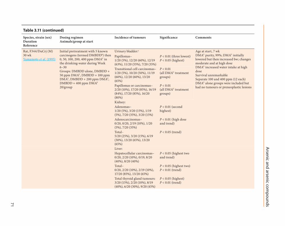
Arsenic and arsenic com
pounds71
Species, strain (sex) Duration Reference
Dosing regimen Animals/group at start
Incidence of tumours Significance Comments
Rat, F344/DuCrj (M) 30 wk Yamamoto et al. (1995)
Initial pretreatment with 5 known carcinogens (termed DMBDDb) then 0, 50, 100, 200, 400 ppm DMAV in the drinking-water during Week 6–30 Groups: DMBDD alone, DMBDD + 50 ppm DMAV, DMBDD + 100 ppm DMAV, DMBDD + 200 ppm DMAV, DMBDD + 400 ppm DMAV 20/group
Urinary bladder:c Age at start, 7 wk DMAV purity, 99%; DMAV initially lowered but then increased bw; changes moderate and at high dose DMAV increased water intake at high dose Survival unremarkable Separate 100 and 400 ppm (12 each) DMAV alone groups were included but had no tumours or preneoplastic lesions
Papillomas– 1/20 (5%), 12/20 (60%), 12/19 (63%), 11/20 (55%), 7/20 (35%)
P < 0.01 (three lowest) P < 0.05 (highest)
Transitional cell carcinomas– 1/20 (5%), 10/20 (50%), 11/19 (60%), 12/20 (60%), 13/20 (65%)
P < 0.01 (all DMAV treatment groups)
Papillomas or carcinomas– 2/20 (10%), 17/20 (85%), 16/19 (84%), 17/20 (85%), 16/20 (80%)
P < 0.01 (all DMAV treatment groups)
Kidney:Adenomas– 1/20 (5%), 3/20 (15%), 1/19 (5%), 7/20 (35%), 3/20 (15%)
P < 0.01 (second highest)
Adenocarcinomas– 0/20, 0/20, 2/19 (10%), 1/20 (5%), 7/20 (35%)
P < 0.01 (high dose and trend)
Total– 5/20 (25%), 3/20 (15%), 6/19 (30%), 13/20 (65%), 13/20 (65%)
P < 0.05 (trend)
Liver:Hepatocellular carcinomas– 0/20, 2/20 (10%), 0/19, 8/20 (40%), 8/20 (40%)
P < 0.05 (highest two and trend)
Total– 0/20, 2/20 (10%), 2/19 (10%), 17/20 (85%), 13/20 (65%)
P < 0.05 (highest two) P < 0.01 (trend)
Total thyroid gland tumours: 3/20 (15%), 2/20 (10%), 8/19 (40%), 6/20 (30%), 9/20 (45%)
P < 0.05 (highest) P < 0.01 (trend)
Table 3.11 (continued)

IARC M
ON
OG
RAPH
S – 100C
72
Table 3.11 (continued)
Species, strain (sex) Duration Reference
Dosing regimen Animals/group at start
Incidence of tumours Significance Comments
Rat, F344 (M) 36 wk Wanibuchi et al. (1996)
Pretreatment with BBNd 0.05% in drinking-water for 4 wk then 0, 2, 10, 25, 50, or 100 ppm DMAV in drinking-water for 32 wk Groups: BBN alone, BBN + 2 ppm DMAV, BBN + 10 ppm DMAV, BBN + 50 ppm DMAV, BBN + 100 ppm DMAV 20/group
Urinary bladder: Papillary/nodular hyperplasias– 14/20 (70%), 13/20 (65%), 14/20 (70%), 18/19 (95%), 20/20 (100%), 20/20 (100%)
P < 0.05 (highest two doses)
Age at start, ~6 wk DMAV purity, 99% Separate 0 and 100 ppm control and DMAV alone groups were included (12 each) but showed no urinary bladder tumours or preneoplastic lesions Bw, water intake and survival unremarkable Urinary bladder only
Papillomas– 3/20 (15%), 2/20 (10%), 7/20 (35%), 11/19 (58%), 13/20 (65%), 17/20 (85%)
P < 0.01 (highest three doses)
Carcinomas– 1/20 (5%), 2/20 (10%), 3/20 (15%), 7/19 (37%), 10/20 (50%), 12/20 (60%)
P < 0.05 (third highest dose) P < 0.01 (highest two doses)
a Diethylnitrosamineb The organic carcinogen treatment consisted of a single dose of diethylnitrosamine (100 mg/kg, i.p.) at the start of the experiment) andN-methyl-N-nitrosourea (20 mg/kg, s.c.) on experimental Days 5, 8, 11 and 14. Thereafter, rats received 1,2-dimethylhydrazine (40 mg/kg, s.c.) on Days 18, 22, 26, and 30). During the same period (experimental Days 0–30) the rats received N-butyl-N-(4-hydroxybutyl)nitrosamine (0.05% in the drinking-water Weeks 1 and 2) and N-bis(2-hydroxypropyl)nitrosamine (0.1% in the drinking-water, Weeks 3 and 4). Altogether this was defined as DMBDD treatment. Rats received no treatment for 2 wk after DMBDD exposure and before DMA exposure.c For brevity, only significant proliferative lesions are noted for each tissued N-butyl-N-(4-hydroxybutyl)nitrosaminee 4-Nitroquinolinef 7,12-dimethylbenz[α]anthraceneg Estimated from graphical presentation.d, day or days; DMA, dimethylarsinic acid; F, female; i.p., intraperitoneal; M, male; NR, not reported; s.c., subcutaneously; wk, week or weeks

Arsenic and arsenic com
pounds73
Table 3.12 Studies where arsenicals given concurrently with other agents enhance carcinogenesis while having no effect alone in experimental animals
Species, strain (sex) Duration Reference
Dosing regimen Animals/group at start
Incidence of tumours Significance Comments
Mouse, Tg.AC homozygous (F) 14 wk Germolec et al. (1997)
0 or 0.02% As in drinking-water, ad libitum throughout experiment 0 or 2.5 µg TPAa/mouse in acetone topical to shaved dorsal skin twice/wk, Week 5 and 6 Groups: control, As alone, TPA, As + TPA 20/group
Macroscopic skin papillomas/mouse: none in control or arsenic alone, intermediate in TPA alone (~0.5/mouse),b “4-fold higher” (~2.1/mouse)b in arsenic + TPA
NR Age at start, NR Purity, NR Survival unremarkable Specific quantitative microscopic analysis of skin tumours not included but confirmed as papillomas at termination Skin lesions only Incomplete reporting makes independent statistical analysis impossible
Mouse, Tg.AC homozygous (F) 24 wk Germolec et al. (1998)
0 or 0.02% As in drinking-water, ad libitum throughout experiment 0, 1.25, 2.5 µg TPA/mouse in acetone topical to shaved dorsal skin twice/wk, Week 5 and 6 Groups: control, As alone, 1.25 TPA, 2.5 TPA, As + 1.25 TPA, As + 2.5 TPA 20/group
Macroscopic skin papillomas/mouse: 0 in control, As alone, and 1.25 TPA alone; As + 1.25 TPA maximal ~5/mouse,b 2.5 TPA ~3/mouse,b in arsenic + 2.5 TPA ~7/mouseb
NR Age at start, 8 wk Purity, NR Survival impacted by high-dose TPA co-treatment but specifics not given Quantitative microscopic analysis of skin tumours not included but confirmed as papillomas at termination Skin lesions only Incomplete reporting makes independent statistical analysis impossible

IARC M
ON
OG
RAPH
S – 100C
74
Species, strain (sex) Duration Reference
Dosing regimen Animals/group at start
Incidence of tumours Significance Comments
Mouse, Crl: SKl-hrBR (hairless) (F) 29 wk Rossman et al. (2001)
0, 10 mg/L sodium arsenite in drinking-water throughout experiment plus topical 1.7 kJ/m2 solar irradiation (85% UVB, < 1% UVC, 4% UVA, remainder visible; termed UVRc) 3x/wk starting 3 wk after As until termination Groups: control, As alone, UVR alone, As + UVR 5–15; 5 controls
Skin (tumours): Macroscopic and microscopic analysis–0/5, 0/5 (control and As alone)
Age at start, 3wk Purity, NR Survival and bw unremarkable Small control groups
Macroscopic analysis– Time to first occurrence: As + UVR earlier than UVR
P < 0.01
Microscopic analysis– Total tumours all mice: 53 (UVR), 127 (As + UVR)Highly invasive squamous cell carcinoma: 14/53 (26%; UVR), 64/127 (50%; As + UVR)
P < 0.01
Tumour volume: UVR smaller than As + UVR
P < 0.01
Mouse, SKl (hairless), (NR) 29 wk Burns et al. (2004)
Experiment 1: 0, 1.25, 2.50, 5.00, 10.0 mg/L sodium arsenite in drinking-water from onset plus topical 0 or 1.0 kJ/m2 solar irradiation (UVRc) 3x/wk, starting 3 wk after As to termination
Experiment 1: Skin tumours/moused: 2.4 (UVR), 5.4 (1.25 As + UVR), 7.21 (2.5 As + UVR), 11.1 (5.0 As + UVR), 6.8 (10.0 As + UVR)
[P < 0.01 all groups vs UVR alonee]
Age, 3 wk Survival and bw unremarkable Specific quantitative microscopic analysis of skin tumours not reported but confirmed as primarily squamous cell carcinomas at termination Experiment 1 shows clear arsenic dose–response in enhancement through 5.0 mg/L by various criteria
Experiment 2: 10.0 mg/L sodium arsenite in drinking-water from onset plus topical 1.7 kJ/m2 UVRc 3x/wk starting 3 wk after As to termination
Experiment 2: Skin tumours/mouse:d 3.5 (UVR), 9.6 (As + UVR) Skin tumour incidence: 0/10, 0/10 (control and As alone both experiments)
[P < 0.01f]
Table 3.12 (continued)

Arsenic and arsenic com
pounds75
Species, strain (sex) Duration Reference
Dosing regimen Animals/group at start
Incidence of tumours Significance Comments
Mouse, Crl: SKl-hrBR (hairless) (F) Duration, NR Uddin et al. (2005)
0, 5 mg/L sodium arsenite in drinking-water from onset; diet unsupplemented or with added vitamin E (62.5 IU/kg diet; basal 49.0 IU/kg) or p-XSCg (10 mg/kg diet) from onset. Topical 1.0 kJ/m2 UVRc 3x/wk starting 3 wk after As to termination. Groups: UVR alone, UVR + As, UVR + As + Vitamin E, UVR + As + p-XSCh 10; 30 controls (UVR)
Macroscopic skin tumours/mouse: 3.60 (UVR alone), 7.00 (UVR + As), 3.27 (UVR + As + Vitamin E), 3.40 (UVR + As + p-XSC)
P < 0.01 (UVR vs UVR + As) P < 0.01 (UVR + As vs UVR + As + either dietary supplement)
Age at start, 3 wk Sodium arsenite, purity (NR), p-XSC Purity > 99% Survival and bw unremarkable Small control groups Vitamin E and p-XSC added as antioxidants Specific quantitative microscopic analysis of skin tumours not reported but random sampling (10 tumours/group) confirmed primarily squamous cell carcinomas at termination No untreated control or arsenic alone groups included
Mouse, Swiss-bald hairless (M) 25 wk Motiwale et al. (2005)
Treatment with 2 mg BAi /mL 25 µL topical once/wk for 2 wk Sodium arsenate 0 or 25 mg/L drinking-water for 25 wk Groups: Control, BA, As, BA + As 10/group
Macroscopic skin tumours/mouse: 0, 2.0, 0, 3.2b % large papillomas (≥ 3 mm) of total papillomas: 0, 16, 0, 65d
P < 0.05 (As + BA vs BA) P < 0.05 (As + BA vs BA)
Age at start, 8 wk Purity, NR Survival unremarkable Small group sizes Quantitative microscopic skin tumour incidence or multiplicity not reported though histologically confirmed
a 12-O-tetradecanoyl-13-acetate.b Estimated from graphical presentation. No descriptive statistics included.c UVR as defined in Rossman et al. (2001) above.d Data included descriptive statistics.e Using Dunnett’s multiple comparison test and not including arsenic alone and untreated control groupsf Using Student’st-test.g 1,4-Phenylbis(methylene)selenocyanate a synthetic organoselenium compound.h Some control groups are not discussed for the sake of brevity (UVR + Vitamin E and UVR +p-XSC).i 9,10-dimethyl-1,2-benzanthracene.F, female; M, male; NR, not reported; wk, week or weeks
Table 3.12 (continued)

IARC MONOGRAPHS – 100C
of age, although arsenic treatment alone had no effect, it markedly increased the multiplicity of squamous cell carcinoma when combined with TPA compared to TPA alone (Waalkes et al., 2008; see Table 3.13).
Prenatal sodium arsenite exposure via maternal drinking-water when combined with postnatal topical TPA exposure increased the liver tumour incidence and multiplicity in an arsenic-dose-related fashion (female offspring), and lung tumours (male offspring) compared to controls; effects not seen with TPA or arsenic alone (Waalkes et al., 2004). Prenatal arsenic exposure followed by postnatal diethylstilbestrol increased uterine carcinoma, vaginal carcinoma, urinary bladder total proliferative lesions, and liver tumours in female offspring compared to controls; effects not seen with diethylstilbestrol or arsenic alone. In female offspring, prenatal arsenic exposure followed by postnatal tamox-ifen administration similarly increased urinary bladder total proliferative lesions (Waalkes et al., 2006a).
In male offspring, prenatal arsenic exposure followed by postnatal diethylstilbestrol increased the liver tumour response and urinary bladder total proliferative lesions effects when compared to controls; effects not seen with diethylstilbestrol or arsenic alone. In male offspring, prenatal arsenic exposure followed by postnatal tamox-ifen increased liver tumour response, urinary bladder total tumours, and urinary bladder total proliferative lesions (Waalkes et al., 2006b).
3.5.2 Rat
Rats that underwent partial hepatectomy followed by diethylnitrosamine injection and one week later by oral administration of sodium arsenite in the drinking-water for approximately 24 weeks showed an increased incidence of renal tumours, but arsenic treament alone had no effect (Shirachi et al., 1983).
In a comprehensive study, rats were given an initial pretreatment with a mixture of organic carcinogens (including diethylnitrosamine, N-methyl-N-nitrosourea, 1,2-dimethylhydra-zine, N-butyl-N-(4-hydroxybutyl)nitrosamine, and N-bis(2-hydroxypropyl)nitrosamine) by various routes, no treatment for 2 weeks and then DMAV (at four levels) in the drinking-water for 24 weeks, rats developed an increased incidence of tumours of urinary bladder with the combined carcinogen treatment and arsenical (Yamamoto et al., 1995).
In another study in rats, N-butyl-N-(4-hydroxybutyl)nitrosamine in the drinking-water was used as an initiator for 4 weeks followed by four levels of DMAV for 32 weeks, and the combined treatment increased urinary bladder hyperplasia, papilloma, and carcinoma, but the arsenical treatment alone had no effect (Wanibuchi et al., 1996).
3.6 Gallium arsenide
A single study (NTP, 2000) was judged to provide evidence for the carcinogenicity of gallium arsenide in rodents. In this report, B6C3F1 mice and F344 rats were exposed via inhalation to various levels of gallium arsenide particulate for up to ~2 years, and the tumour response was assessed in various tissues (see Table 3.14).
3.6.1 Mouse
No treatment-related tumours were observed, but in both males and females, dose-related increases in the incidence in lung epithelial alve-olar hyperplasia were reported.
3.6.2 Rat
In female rats, dose-related responses were reported for the incidence of lung alveolar/bronchiolar tumours and atypical hyperplasia
76
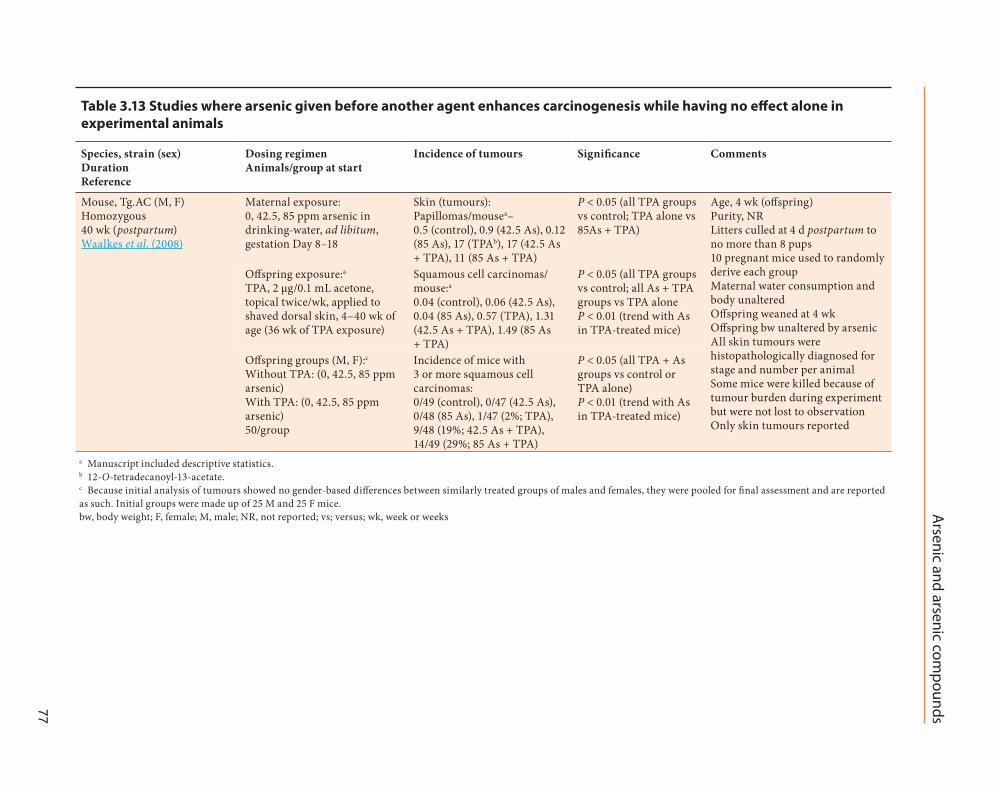
Arsenic and arsenic com
pounds77
Table 3.13 Studies where arsenic given before another agent enhances carcinogenesis while having no effect alone in experimental animals
Species, strain (sex) Duration Reference
Dosing regimen Animals/group at start
Incidence of tumours Significance Comments
Mouse, Tg.AC (M, F) Homozygous 40 wk (postpartum) Waalkes et al. (2008)
Maternal exposure: 0, 42.5, 85 ppm arsenic in drinking-water, ad libitum, gestation Day 8–18
Skin (tumours): Papillomas/mousea– 0.5 (control), 0.9 (42.5 As), 0.12 (85 As), 17 (TPAb), 17 (42.5 As + TPA), 11 (85 As + TPA)
P < 0.05 (all TPA groups vs control; TPA alone vs 85As + TPA)
Age, 4 wk (offspring) Purity, NR Litters culled at 4 d postpartum to no more than 8 pups 10 pregnant mice used to randomly derive each group Maternal water consumption and body unaltered Offspring weaned at 4 wk Offspring bw unaltered by arsenic All skin tumours were histopathologically diagnosed for stage and number per animal Some mice were killed because of tumour burden during experiment but were not lost to observation Only skin tumours reported
Offspring exposure:a TPA, 2 µg/0.1 mL acetone, topical twice/wk, applied to shaved dorsal skin, 4–40 wk of age (36 wk of TPA exposure)
Squamous cell carcinomas/mouse:a 0.04 (control), 0.06 (42.5 As), 0.04 (85 As), 0.57 (TPA), 1.31 (42.5 As + TPA), 1.49 (85 As + TPA)
P < 0.05 (all TPA groups vs control; all As + TPA groups vs TPA alone P < 0.01 (trend with As in TPA-treated mice)
Offspring groups (M, F):c Without TPA: (0, 42.5, 85 ppm arsenic) With TPA: (0, 42.5, 85 ppm arsenic) 50/group
Incidence of mice with 3 or more squamous cell carcinomas: 0/49 (control), 0/47 (42.5 As), 0/48 (85 As), 1/47 (2%; TPA), 9/48 (19%; 42.5 As + TPA), 14/49 (29%; 85 As + TPA)
P < 0.05 (all TPA + As groups vs control or TPA alone) P < 0.01 (trend with As in TPA-treated mice)
a Manuscript included descriptive statistics.b 12-O-tetradecanoyl-13-acetate.c Because initial analysis of tumours showed no gender-based differences between similarly treated groups of males and females, they were pooled for final assessment and are reported as such. Initial groups were made up of 25 M and 25 F mice.bw, body weight; F, female; M, male; NR, not reported; vs; versus; wk, week or weeks

IARC M
ON
OG
RAPH
S – 100C
78
Table 3.14 Studies of cancer in experimental animals exposed to gallium arsenide (inhalation exposure)
Species, strain (sex) Duration Reference
Dosing regimen Animals/group at start
Incidence of tumours Significance Comments
Mouse, B6C3F1 (M, F) 105 wk for M 106 wk for F NTP (2000)
0, 0.1, 0.5, 1.0 mg/m3 6 h/d, 5 d/wk 50/group/sex
Females Lung (epithelial alveolar hyperplasias):2/50 (4%), 5/50 (10%), 27/50 (54%), 43/50 (86%)
P ≤ 0.01 (high dose) P ≤ 0.01 (mid-dose)
Age at start, 6 wk Purity > 98% MMAD, 0.9–1.0 µm GSD, 1.8–1.9 µm Chamber controls used No reduced bw with treatment Survival unaltered No increases in tumour incidence
Lunga (adenomas or carcinomas): 7/50 (14%), 4/50 (8%), 4/50 (8%), 6/50 (12%)
NS
Males Lung (epithelial alveolar hyperplasias):4/50 (8%), 9/50 (18%), 39/50 (78%), 45/50 (90%)
P ≤ 0.01 (high dose) P ≤ 0.01 (mid-dose)
Lunga (adenomas or carcinomas): 15/50 (30%), 14/50 (28%), 16/50 (32%), 13/50 (26%)
NS
Rat, F344 (F) 105 wk NTP (2000)
0, 0.01, 0.1, 1.0 mg/m3 6 h/d, 5 d/wk 50/group/sex
Females Lunga (adenomas): 0/50, 0/50, 2/50 (4%), 7/50 (14%)
P ≤ 0.01 (high dose) P ≤ 0.01 (trend)
Age at start, 6 wk Purity > 98% MMAD, 0.9–1.0 µm GSD, 1.8–1.9 µm Chamber controls used Minimal decrease in body weight at high dose in second yr Survival unaltered No increases in tumour incidence in males
Lung (carcinomas): 0/50, 0/50, 2/50 (4%), 3/50 (6%)
NS
Lung (adenomas or carcinomas): 0/50, 0/50, 4/50 (8%), 9/50 (18%)
P ≤ 0.01 (high dose) P ≤ 0.01 (trend)
Adrenal medulla:b 4/50 (8%), 6/49 (12%), 6/50 (12%), 13/49 (27%)
P ≤ 0.01 (high dose) P ≤ 0.01 (trend)
Mononuclear cell leukaemia: 22/50 (44%), 21/50 (42%), 18/50 (36%), 33/50 (66%)
P ≤ 0.05 (high dose) P ≤ 0.01 (trend)
Males Lung (atypical hyperplasias): 0/50, 2/49 (4%), 5/50 (10%), 18/50 (36%)
P ≤ 0.01 (high dose) P ≤ 0.05 (mid-dose)
Lunga (adenomas): 1/50 (2%), 0/49, 3/50 (6%), 2/50 (4%)
NS
Lung (carcinomas): 2/50 (4%), 0/49, 2/50 (4%), 1/50 (2%)
NS
Lung (adenomas or carcinomas): 3/50 (6%), 0/49, 5/50 (10%), 3/50 (6%)
NS
a All lung tumours were of avelolar/bronchiolar origin.b All tumours were benign pheochromocytoma except one which was malignant in the low-dose group.d, day or days; F, female; h, hour or hours; M, male; NS, not significant; wk, week or weeks; yr, year or years

Arsenic and arsenic compounds
of the alveolar epithelium. In male rats, though treatment-related tumours were not observed, a dose-related increase in the incidence of atyp-ical hyperplasia of the lung alveolar epithelium occurred. Atypical hyperplasia of the lung alve-olar epithelium is considered potentially preneo-plastic. In the female rats, dose-related increases in the incidence of adrenal medulla pheochro-mocytomas and an increase in mononuclear cell leukaemia at the highest dose were also reported (NTP, 2000).
3.6.3 Hamster
Another study using intratracheal instilla-tion of gallium arsenide in hamsters (Ohyama et al., 1988) was judged inadequate due to critical design flaws (short duration, small groups, etc.) with no indication of tumours.
3.7 Synthesis
Oral administration of sodium arsenate and DMAV induced lung tumours in mice. Calcium arsenate induced lung tumours in hamsters by oral and intratracheal administration. Pre- and postnatal exposure in mice to arsenic trioxide, through subcutaneous injections (maternal and postnatal), induced lung tumours in the offspring. Transplacental exposure via maternal oral exposure in mice to sodium arsenite during gestation induced lung, liver, ovary and adrenal tumours in the offspring in several studies, and the uterus in one study. Early life transplacental and perinatal exposure to sodium arsenite appears to be a time of particular sensitivity in terms of carcinogenesis.
Oral exposure to DMAV induced urinary bladder tumours in several studies in rats and among studies in mice, only one showed nega-tive results. Oral trimethylarsine induced liver tumours in rats. Chronic oral exposure to MMAV did not produce tumours in rats and mice. [The Working Group considered that previous
traditional bioassays for arsenicals for adult rodents were frequently negative in their final evaluations.]
Inhalation of gallium arsenide causes lung and adenal tumours in rats but not in mice.
In multiple studies, initiating, promoting or co-carcinogenic activity was demonstrated in the urinary bladder, skin, female reproductive tract, kidney, lung, liver and thyroid after exposure to inorganic arsenicals or DMAV in drinking-water or by transplacental exposure.
4. Other Relevant Data
4.1 Absorption, distribution, metabolism, and excretion
Most inorganic arsenic compounds are readily absorbed after oral exposure (about 80–90% for soluble compounds, and a smaller percentage for less soluble compounds), less well absorbed after inhalation (better for small particulates and soluble arsenicals), and least well absorbed after dermal exposure (NRC, 1999; IARC, 2004). Large airborne arsenic-containing particulates that are deposited in the upper airways may also be absorbed in the intestine if they are later swal-lowed. Hamsters exposed to gallium arsenide by the oral route or by intratracheal instillation showed the presence of AsIII in blood and urine, but the majority of the gallium arsenide was excreted in faeces, indicating that absorption was limited by its insolubility. Absorption was about 30 times higher after intratracheal installation than by the oral route (Carter et al., 2003).
The transport of AsV is thought to take place via phosphate transporters (Csanaky & Gregus, 2001). The sodium-coupled phosphate trans-porter NaPi-IIb may be responsible in part for the intestinal and hepatic uptake of AsV (Villa-Bellosta & Sorribas, 2008). AsIII enters the cell by aquaglyceroporins 9 and 7 (Liu et al., 2004),
79

IARC MONOGRAPHS – 100C
although another major pathway for the uptake of AsIII and MMAIII (see below) is probably via hexose permeases (Rosen & Liu, 2009). Because AsV is rapidly reduced to AsIII once it enters the cell (Carter et al., 2003), the faster rate of cellular uptake of AsIII, compared with AsV, may be part of the explanation for the greater toxicity of AsIII (Bertolero et al., 1987; Dopp et al., 2004). However, the much higher chemical reactivity of AsIII, compared to that of AsV is the major expla-nation. Some data suggests that glyceraldehyde 3-phosphate dehydrogenase (GAPDH) func-tions as a cytosolic AsV reductase in vivo (Németi et al., 2006), although there are other candidate enzymes for this reaction (Aposhian et al., 2004). AsIII can react with cellular glutathione (GSH), either spontaneously or enzymatically, to form the tri-glutathione complex As(SG)3 (Leslie et al., 2004; Rey et al., 2004).
AsIII is metabolized by stepwise methylation, mainly in the liver. Although some details of inorganic arsenic metabolism remain uncertain (Aposhian & Aposhian, 2006), it is clear that the enzyme arsenic (+3 oxidation state) methyl-transferase (AS3MT) is involved (Thomas et al., 2007). Two schemes have been proposed for the methylation.
Reduction: AsV + thiol → AsIII
Oxidative methylation: AsIII + SAM → monomethylarsonate (MMAV)Reduction: MMAV + thiol → MMAIII
Oxidative methylation: MMAIII + SAM → dimethylAsV (DMAV)Reduction: DMAV + thiol → DMAIII
Scheme 1: Inorganic arsenic metabolic pathway in mammals. AsIII methylation is cata-lysed by AS3MT using S-adenosylmethionine (SAM) as a methyl donor and thioredoxin (or, less efficiently, other thiols such as glutaredoxin or lipoic acid) as a reductant. MMAIII: mono-methylarsonous acid; MMAv: monomethyl-arsonic acid; DMAIII: dimethylarsinous acid; DMAv: dimethylarsinic acid
As(SG)3 + SAM → MMAIII (SG)2MMAIII (SG)2 + SAM → DMAIII (SG)
Scheme 2: The use of As(SG)3 (tri-glutathione complex) as a substrate for methylation (Hayakawa et al., 2005). Each of the glutathione (GSH) complexes can also decompose to yield GSH and MMAIII or DMAIII, which can then form MMAV and DMAV, respectively.
Neither reaction scheme necessarily goes to completion in vivo.
Evidence shows that exposure to arsine gas (AsH3) results in the same metabolites as described above, but arsenobetaine found in seafood does not get metabolized in humans (Crecelius, 1977; Luten et al., 1982; Le et al., 1993, 1994; Buchet et al., 1996; Schmeisser et al., 2006). Information is not currently available on the other organo-arsenic compounds in seafood (Lai et al., 2004).
Dimethylthioarsinic acid (DMMTAV) and dimethyldithioarsinic acid (DMDTAV) can be formed from DMAIII in red blood cells, and possibly in other cells (Naranmandura et al., 2007; Suzuki et al., 2007). These compounds have been observed in the urine of arsenic-exposed individuals (Raml et al., 2007). They may have been misidentified as MMAIII and DMAIII in most studies (Hansen et al., 2004).
Most organisms detoxify inorganic arsenic by cellular efflux (Rosen & Liu, 2009). In fibrob-lasts and other non-methylating cells, protection against arsenic takes place by specific mecha-nisms for As(SG)3 efflux catalysed by multidrug-resistance-associated protein-transport ATPases MRP1 and MRP2, and maybe others (Kala et al., 2000; Leslie et al., 2004). These efflux pumps may also remove methylated arsenic–glutathione (As–GSH) complexes.
The rat is not a good model for the human in studying the toxicokinetics of arsenic because rat haemoglobin has a much higher affinity for trivalent arsenic species compared with human haemoglobin (Lu et al., 2004). In mice, chronic
80

Arsenic and arsenic compounds
exposure (12 weeks) to AsV via drinking-water led to total tissue arsenic accumulation in the following ranking: kidney > lung > bladder > > > skin > blood > liver (Kenyon et al., 2008). Monomethylated arsenic species (MMAs) predominated in the kidney, and dimethylated arsenic species (DMAs) predominated in the lung. Urinary bladder and skin had about equal ratios of inorganic arsenic and DMAs. The proportions of different arsenic species in urinary bladder tissue did not match those in urine.
In a study of intratracheal instillation of gallium arsenide, although substantial levels of arsenic were detected in blood and urine, no gallium was detected except for the amount that was left in the lung (Carter et al., 2003).
Human exposure to arsenic is mainly via drinking-water. Trivalent arsenicals are elimi-nated via the bile, and pentavalent arsenicals are mainly eliminated by urinary excretion (Gregus et al., 2000; Kala et al., 2000; Csanaky & Gregus, 2002). Most population groups exposed mainly via drinking-water excrete 60–70% DMAs and 10–20% MMAs, the remainder 10–30% being inorganic compounds (Vahter, 2000). [The Working Group noted that this study did not include thiolated compounds, which had not yet been discovered.] Interindividual differences in methylation patterns may reflect genetic poly-morphisms in AS3MT, and/or variability in the activities of different reductants (Thomas et al., 2007).
4.2 Genetic and related effects
Arsenicals do not react directly with DNA, but cells treated with low concentrations of triva-lent arsenicals show increased oxidative DNA damage (Wang et al., 2002; Schwerdtle et al., 2003; Shi et al., 2004; Ding et al., 2005; Wang et al., 2007a). AsIII and MMAIII are equally potent inducers of oxidative DNA damage in human urothelial cells, where they are equally toxic (Wang et al., 2007a). Cytotoxic concentrations
of trivalent arsenicals also cause DNA strand breaks and/or alkali-labile sites (Kligerman et al., 2003; Klein et al., 2007). In mice, DMAV causes lung-specific DNA damage attributed to the DMA peroxy radical (CH3)2AsOO (Yamanaka & Okada, 1994), which can also induce DNA strand breaks and DNA–protein crosslinks in cultured cells (Tezuka et al., 1993).
Gallium arsenide and other arsenicals are not mutagenic in the Ames test (NTP, 2000; IARC, 2004). There was no increase in frequency of micronucleated erythrocytes in mice exposed to gallium arsenide by inhalation for 14 weeks (NTP, 2000).
Despite the fact that low (non-toxic) concentra-tions of trivalent arsenicals cause oxidative DNA damage such as 8-hydroxy-2′-deoxyguanosine, which is expected to cause G→T transversions, neither AsIII, MMAIII nor DMAIII are significant point mutagens (Rossman, 2003; Klein et al., 2007). This may be due to the efficient removal of oxidative DNA lesions (Fung et al., 2007; Pu et al., 2007b). At toxic concentrations, AsIII increased large-deletion mutations in human/hamster hybrid cells through a mechanism mediated by reactive oxygen species (Hei et al., 1998). MMAIII and DMAIII are weakly mutagenic in mouse lymphoma L5178Y cells, but only at toxic concentrations, and yield mostly deletions (Moore et al., 1997; Kligerman et al., 2003).
Using a transgenic cell line that readily detects deletions as well as point mutations, statistically significant mutagenesis was never observed for DMAIII, and was only seen for AsIII or MMAIII at toxic concentrations. MMAIII yielded a mutant fraction about 4-fold over background at 11% survival, and 79% of these mutants were dele-tions (Klein et al., 2007).
AsIII, MMAIII, and DMAIII can induce chromosomal aberrations in vitro (Oya-Ohta et al., 1996; Kligerman et al., 2003). Statistically significant increases in chromosomal aber-rations occur only at toxic doses (Klein et al., 2007), except as a secondary effect of genomic
81

IARC MONOGRAPHS – 100C
instability in long-term, low-dose treatment protocols (Sciandrello et al., 2004). An analysis of micronuclei induced by AsIII in human fibro-blasts shows that at lower (relatively non-toxic) doses, AsIII acts as an aneugen by interfering with spindle function and causing micronuclei with centromeres, but at high (toxic) doses, it acts as a clastogen, inducing micronuclei without centromeres (Yih & Lee, 1999). Aneuploidy is seen after treatment with AsIII concentrations lower than those that cause chromosomal aberrations (Yih & Lee, 1999; Ochi et al., 2004; Sciandrello et al., 2002, 2004). Aneuploidy associated with disruption of spindle tubulin has been reported in other cells treated with arsenicals (Huang & Lee, 1998; Kligerman & Tennant, 2007; Ramírez et al., 2007). Disrupted mitotic spindles and induced persistent aneuploidy were maintained even 5 days after AsIII removal (Sciandrello et al., 2002). Humans exposed to high concentrations of inorganic arsenic in drinking-water also show increased micronuclei in lymphocytes, exfoli-ated bladder epithelial cells and buccal mucosa cells, and sometimes chromosomal aberra-tions and sister chromatid exchange in whole-blood lymphocyte cultures (Basu et al., 2001). Micronuclei and chromosomal aberrations are also induced in mice after intraperitoneal treat-ment with AsIII (IARC, 2004).
Long-term low-dose treatment of human osteosarcoma cells with AsIII (but not MMAIII) resulted in increased mutagenesis and trans-formation as a secondary effect of genomic instability (Mure et al., 2003). In Chinese hamster V79–13 cells grown in the presence of low concentrations of AsIII, genomic instability (measured by chromosomal aberrations in later generations) followed earlier changes in DNA methylation and aneuploidy (Sciandrello et al., 2002, 2004). Other studies report gene ampli-fication (Lee et al., 1988; Rossman & Wolosin, 1992), and changes in gene expression, e.g. by DNA methylation changes (Liu et al., 2006b; Klein et al., 2007; Reichard et al., 2007; Liu &
Waalkes, 2008). Alterations of DNA methyla-tion, along with histone modification, were seen in cells treated with AsIII and MMAIII (Jensen et al., 2008; Zhou et al., 2008). Global DNA hypomethylation, along with hypermethylation of specific genes, was demonstrated in several AsIII-transformed cells (Benbrahim-Tallaa et al., 2005a; Liu & Waalkes, 2008). Oxidative damage to DNA has been shown to cause changes in DNA methylation (Cerda & Weitzman, 1997), suggesting a mechanism by which AsIII may induce this effect. Changes in DNA methyla-tion patterns could also result from altered SAM pools or downregulation of DNA methyltrans-ferases (Hamadeh et al., 2002; Benbrahim-Tallaa et al., 2005a; Reichard et al., 2007; Liu & Waalkes, 2008). Altered DNA methylation has also been observed in arsenic-exposed humans (Chanda et al., 2006; Marsit et al., 2006).
Although not a mutagen, AsIII can enhance the mutagenicity of other agents (Rossman, 2003; Danaee et al., 2004; Fischer et al., 2005). Co-mutagenesis may occur by interference with both nucleotide-excision repair and base-exci-sion repair (Hartwig et al., 2002; Rossman, 2003; Danaee et al., 2004; Wu et al., 2005; Shen et al., 2008). Nucleotide-excision repair was blocked in human fibroblasts with the following potency: MMAIII > DMAIII > AsIII (Shen et al., 2008). AsIII is not a very effective inhibitor of DNA-repair enzymes (Snow et al., 2005). Rather, it appears to affect DNA-damage signalling events that control DNA repair. One of these is poly(ADP-ribose) polymerase (PARP) (Hartwig et al., 2003; Qin et al., 2008). PARP-1, the major PARP, is involved in base-excision repair by interacting with DNA-repair protein XRCC1, DNA poly-merase β, and DNA ligase III. This might explain the inhibition of the ligation step of base-excision repair by AsIII (Li & Rossman, 1989). MMAIII and DMAIII are more effective PARP inhibitors than is AsIII (Walter et al., 2007). The inhibition of PARP (and other proteins such as XPA) may be
82

Arsenic and arsenic compounds
mediated by the displacement of zinc (Zn) at Zn fingers (Schwerdtle et al., 2003; Qin et al., 2008).
Another important signal pathway affected by AsIII is that mediated by tumour-suppressor gene Tp53. AsIII was shown to prevent the activation of the P53 protein and the downstream expression of p21 after genotoxic insult (Vogt & Rossman, 2001; Tang et al., 2006; Shen et al., 2008). This has the effect of overriding the growth arrest at G1 (normally an opportunity for DNA repair to take place before DNA replication) in cells with DNA damage, and might explain part of the co-mutagenic effect (Vogt & Rossman, 2001; Hartwig et al., 2002; Mudipalli et al., 2005). p53 is also required for proficient global nucleotide-excision repair (Ferguson & Oh, 2005). The inhi-bition of thioredoxin reductase by AsIII, MMAIII and DMAIII (Lin et al., 1999) would cause the accumulation of oxidized thioredoxin, which may be partially responsible for p53 malfunc-tion, as is shown in yeast (Merwin et al., 2002). The upregulation of positive growth genes such as cyclin D by low concentrations of AsIII would also tend to drive cells to cycle inappropriately (Trouba et al., 2000; Vogt & Rossman, 2001; Luster & Simeonova, 2004).
In addition to inhibiting particular proteins, AsIII (at slightly toxic concentrations) can down-regulate expression of some DNA repair genes (Hamadeh et al., 2002; Andrew et al., 2006; Sykora & Snow, 2008). However, very low, non-toxic concentrations, may have the opposite effect of upregulating DNA repair, concomitant with antioxidant defenses (Snow et al., 2005; Sykora & Snow, 2008).
4.3 Co-carcinogenic and in utero carcinogenic effects
There are several non-genotoxic actions of AsIII (sometimes demonstrated also for its triva-lent metabolites) that may contribute to arsenic-induced carcinogenesis. The effects of AsIII on
preventing blockage of the cell cycle after geno-toxic insult by a second agent were discussed above. In addition, low concentrations of AsIII in the absence of a second agent can also stimu-late cell proliferation in vitro (Germolec et al., 1997; Trouba et al., 2000; Vogt & Rossman, 2001; Benbrahim-Tallaa et al., 2005b; Komissarova et al., 2005), and in vivo (Germolec et al., 1998; Burns et al., 2004; Luster & Simeonova, 2004). The concentration-dependent increase in prolif-eration of human keratinocytes after 24 hours of treatment with arsenicals followed the potency trend: DMAIII > MMAIII > AsIII (Mudipalli et al., 2005). AsIII upregulates pro-growth proteins such as cyclin D1, c-myc, and E2F-1 (Trouba et al., 2000; Vogt & Rossman, 2001; Ouyang et al., 2007). The increased proliferation in mouse skin by AsIII alone (in drinking-water) is not sufficient to induce skin cancer (Burns et al., 2004), but may contribute to its co-carcinogen-esis with solar ultraviolet. AsIII was found to block the differentiation of skin cells, resulting in increased numbers of keratinocyte stem cells, the cells that proliferate (Patterson & Rice, 2007; Waalkes et al., 2008). Because tumours may arise from stem cells, this would increase the pool of target cells for cancer of the skin.
Another mechanism for arsenic-related carcinogenesis might be acquired resistance to apoptosis. Long-term growth of human skin cells (HaCaT) in the presence of low concentra-tions of AsIII resulted in cells with a generalized resistance to apoptosis (Pi et al., 2005). This may allow the survival of cells with DNA damage, thus facilitating tumorigenesis. Even short-term exposure to AsIII affected the apoptotic response to solar UV in a mouse keratinocyte cell line (Wu et al., 2005) or to UVB in normal human keratinocytes (Chen et al., 2005b). It is possible that the loss of the P53 function partially medi-ates the reduction in apoptotic response (Chen et al., 2005b).
Numerous studies report increased inflam-mation after AsIII exposure (NRC, 1999; Straub
83

IARC MONOGRAPHS – 100C
et al., 2007). The transcription factor NF-κB is involved in the inflammatory response, and AsIII causes oxidant-dependent activation of NF-κB (Barchowsky et al., 1999). Activation of the NF-κB inflammatory signalling pathway was seen in infants born to AsIII-exposed mothers in Bangladesh (Fry et al., 2007).
AsIII can disrupt the signalling of the estrogen receptor, glucocorticoid receptor, and of other steroids in vivo and in vitro (Benbrahim-Tallaa et al., 2005b, 2007; Liu et al., 2007; Davey et al., 2008). Submicromolar concentrations of AsIII stimulate the transcription of several steroid receptors, but slightly higher concentrations (1–3 µM) are inhibitory (Bodwell et al., 2006). Exposure of mice in utero to AsIII in a protocol leading to hepatocarcinogenesis resulted in altered expression of numerous genes involved in estrogen signalling or steroid metabolism, as well as hypomethylation of estrogen receptor α (Liu & Waalkes, 2008).
Angiogenesis, which provides a blood supply to developing tumours, is stimulated by very low concentrations of AsIII (Mousa et al., 2007; Straub et al., 2007). This activity can be blocked by sele-nium compounds (Mousa et al., 2007), which also blocks AsIII-induced co-carcinogenesis with UV and delays mutagenesis (Uddin et al., 2005).
Many of these effects depend on altered gene expression that can result from genetic and epigenetic effects discussed above. Changes in gene expression by AsIII can also be mediated by the alteration of miRNA patterns (Marsit et al., 2006). Some short-term changes in gene expres-sion (e.g. changes in the expression of DNA-repair proteins or DNA methyltransferases) can result in long-term changes. Genome-wide changes in gene expression and signal transduction induced by arsenicals have been reported in several publi-cations (Su et al., 2006; Kumagai & Sumi, 2007; Ghosh et al., 2008).
4.4 Synthesis
In the human body, inorganic arsenic compounds are converted to AsIII and AsV. AsV is rapidly converted to AsIII. AsIII species are more toxic and bioactive than are AsV species, both because of the greater chemical reactivity of AsIII, and because AsIII enters cells more easily.
For inorganic arsenic and its metabolites, the evidence points to weak or non-existent direct mutagenesis, which is seen only at highly cyto-toxic concentrations. On the other hand, long-term, low-dose exposure to inorganic arsenic – more relevant to human exposure – is likely to cause increased mutagenesis as a secondary effect of genomic instability, perhaps mediated by increased levels of reactive oxygen species, as well as co-mutagenesis with other agents. The major underlying mechanisms observed at low concentrations include the rapid induction of oxidative DNA damage and DNA-repair inhibi-tion, and slower changes in DNA-methylation patterns, aneuploidy, and gene amplification. Gene amplification, altered DNA methylation, and aneuploidy lead to altered gene expression, and genomic instability. Inhibition of DNA repair leads to co-mutagenicity as well. These effects are consistent with the animal carcinogenicity data, in which AsIII is a transgenerational carcinogen – with exposure being present during many cell generations – and in results observed in co-carci-nogenicity studies.
For bladder tumours induced by high doses of DMAV in the rat, the mechanism is likely to involve sustained cytotoxicity followed by stress-related cell proliferation, leading to genomic instability.
Inflammation and cytotoxicity may play a role in lung tumours induced by gallium arse-nide in female rats.
84
Arsenic and arsenic compounds
5. Evaluation
There is sufficient evidence in humans for the carcinogenicity of mixed exposure to inorganic arsenic compounds, including arsenic trioxide, arsenite, and arsenate. Inorganic arsenic compounds, including arsenic trioxide, arsenite, and arsenate, cause cancer of the lung, urinary bladder, and skin. Also, a positive association has been observed between exposure to arsenic and inorganic arsenic compounds and cancer of the kidney, liver, and prostate.
There is sufficient evidence in experimental animals for the carcinogenicity of dimethylars-inic acid, calcium arsenate, and sodium arsenite.
There is limited evidence in experimental animals for the carcinogenicity of sodium arse-nate, gallium arsenide, arsenic trioxide, and trimethylarsine oxide.
There is inadequate evidence in experimental animals for the carcinogenicity of monomethyl-arsonic acid and arsenic trisulfide.
In view of the overall findings in animals, there is sufficient evidence in experimental animals for the carcinogenicity of inorganic arsenic compounds.
Arsenic and inorganic arsenic compounds are carcinogenic to humans (Group 1).
Dimethylarsinic acid and monomethylar-sonic acid are possibly carcinogenic to humans (Group 2B).
Arsenobetaine and other organic arsenic compounds not metabolized in humans, are not classifiable as to their carcinogenicity to humans (Group 3).
The Working Group made the overall evaluation on ‘arsenic and inorganic arsenic compounds’ rather than on some individual arsenic compounds, based on the combined results of epidemiological studies, carcino-genicity studies in experimental animals, and data on the chemical characteristics, metabo-lism, and modes of action of carcinogenicity.
Elemental arsenic and inorganic arsenic species share the same metabolic pathway: arsenate→arsenite→methylarsonate→dimethylarsenite. Thus, independent of the mechanisms of the carcinogenic action, and independent of which of the metabolites is the actual ultimate carcin-ogen, different inorganic arsenic species should be considered as carcinogenic.
References
Andrew AS, Burgess JL, Meza MM et al. (2006). Arsenic exposure is associated with decreased DNA repair in vitro and in individuals exposed to drinking water arsenic. Environ Health Perspect, 114: 1193–1198. doi:10.1289/ehp.9008 PMID:16882524
Aposhian HV & Aposhian MM (2006). Arsenic toxi-cology: five questions. Chem Res Toxicol, 19: 1–15. doi:10.1021/tx050106d PMID:16411650
Aposhian HV, Zakharyan RA, Avram MD et al. (2004). A review of the enzymology of arsenic metabolism and a new potential role of hydrogen peroxide in the detoxi-cation of the trivalent arsenic species. Toxicol Appl Pharmacol, 198: 327–335. doi:10.1016/j.taap.2003.10.027 PMID:15276412
Arnold LL, Eldan M, Nyska A et al. (2006). Dimethylarsinic acid: results of chronic toxicity/oncogenicity studies in F344 rats and in B6C3F1 mice. Toxicology, 223: 82–100. doi:10.1016/j.tox.2006.03.013 PMID:16677751
Arnold LL, Eldan M, van Gemert M et al. (2003). Chronic studies evaluating the carcinogenicity of monomethyl-arsonic acid in rats and mice. Toxicology, 190: 197–219. doi:10.1016/S0300-483X(03)00165-3 PMID:12927375
ATSDR (2007). Toxicological Profile for Arsenic. Atlanta, Georgia.
Barchowsky A, Klei LR, Dudek EJ et al. (1999). Stimulation of reactive oxygen, but not reactive nitrogen species, in vascular endothelial cells exposed to low levels of arsenite. Free Radic Biol Med, 27: 1405–1412. doi:10.1016/S0891-5849(99)00186-0 PMID:10641735
Basu A, Mahata J, Gupta S, Giri AK (2001). Genetic toxi-cology of a paradoxical human carcinogen, arsenic: a review. Mutat Res, 488: 171–194. doi:10.1016/S1383-5742(01)00056-4 PMID:11344043
Benbrahim-Tallaa L, Waterland RA, Styblo M et al. (2005a). Molecular events associated with arsenic-induced malignant transformation of human prostatic epithelial cells: aberrant genomic DNA methylation and K-ras oncogene activation. Toxicol Appl Pharmacol, 206: 288–298. doi:10.1016/j.taap.2004.11.017 PMID:16039940
85
IARC MONOGRAPHS – 100C
Benbrahim-Tallaa L, Webber MM, Waalkes MP (2005b). Acquisition of androgen independence by human pros-tate epithelial cells during arsenic-induced malignant transformation. Environ Health Perspect, 113: 1134–1139. doi:10.1289/ehp.7832 PMID:16140617
Benbrahim-Tallaa L, Webber MM, Waalkes MP (2007). Mechanisms of acquired androgen independence during arsenic-induced malignant transformation of human prostate epithelial cells. Environ Health Perspect, 115: 243–247. doi:10.1289/ehp.9630 PMID:17384772
Bertolero F, Pozzi G, Sabbioni E, Saffiotti U (1987). Cellular uptake and metabolic reduction of pentava-lent to trivalent arsenic as determinants of cytotoxicity and morphological transformation. Carcinogenesis, 8: 803–808. doi:10.1093/carcin/8.6.803 PMID:3608077
Bodwell JE, Gosse JA, Nomikos AP, Hamilton JW (2006). Arsenic disruption of steroid receptor gene activation: Complex dose-response effects are shared by several steroid receptors. Chem Res Toxicol, 19: 1619–1629. doi:10.1021/tx060122q PMID:17173375
Buchet JP, Lison D, Ruggeri M et al. (1996). Assessment of exposure to inorganic arsenic, a human carcinogen, due to the consumption of seafood. Arch Toxicol, 70: 773–778. doi:10.1007/s002040050339 PMID:8896724
Burns FJ, Uddin AN, Wu F et al. (2004). Arsenic-induced enhancement of ultraviolet radiation carcinogenesis in mouse skin: a dose-response study. Environ Health Perspect, 112: 599–603. PMID:15064167
CAREX Canada (2011). Available at: http://w w w . c a r e x c a n a d a . c a / e n / a r s e n i c /occupational_exposure_estimates/phase_2/
Carter DE, Aposhian HV, Gandolfi AJ (2003). The metabolism of inorganic arsenic oxides, gallium arsenide, and arsine: a toxicochemical review. Toxicol Appl Pharmacol, 193: 309–334. doi:10.1016/j.taap.2003.07.009 PMID:14678742
Cerda S & Weitzman SA (1997). Influence of oxygen radical injury on DNA methylation. Mutat Res, 386: 141–152. doi:10.1016/S1383-5742(96)00050-6 PMID:9113115
Chanda S, Dasgupta UB, Guhamazumder D et al. (2006). DNA hypermethylation of promoter of gene p53 and p16 in arsenic-exposed people with and without malig-nancy. Toxicol Sci, 89: 431–437. doi:10.1093/toxsci/kfj030 PMID:16251483
Chen C-J & Chiou H-Y (2001). Chen and Chiou Respond to ‘Arsenic and cancer of the urinary tract’ by Cantor. Am J Epidemiol, 153: 422–423. doi:10.1093/aje/153.5.422 PMID:11226972
Chen C-J, Chuang Y-C, Lin T-M, Wu H-Y (1985). Malignant neoplasms among residents of a blackfoot disease-endemic area in Taiwan: high-arsenic artesian well water and cancers. Cancer Res, 45: 5895–5899. PMID:4053060
Chen C-J, Chuang Y-C, You S-L et al. (1986). A retrospec-tive study on malignant neoplasms of bladder, lung and
liver in blackfoot disease endemic area in Taiwan. Br J Cancer, 53: 399–405. PMID:3964542
Chen C-J, Kuo T-L, Wu M-M (1988a). Arsenic and cancers. Lancet, 331: 414–415. doi:10.1016/S0140-6736(88)91207-X PMID:2893213
Chen C-J & Wang C-J (1990). Ecologicalal correlation between arsenic level in well water and age-adjusted mortality from malignant neoplasms. Cancer Res, 50: 5470–5474. PMID:2386951
Chen C-J, Wu M-M, Lee S-S et al. (1988b). Atherogenicity and carcinogenicity of high-arsenic artesian well water. Multiple risk factors and related malignant neoplasms of blackfoot disease. Arteriosclerosis, 8: 452–460. PMID:3190552
Chen CL, Hsu LI, Chiou HY et al.Blackfoot Disease Study Group. (2004). Ingested arsenic, cigarette smoking, and lung cancer risk: a follow-up study in arseniasis-endemic areas in Taiwan. JAMA, 292: 2984–2990. doi:10.1001/jama.292.24.2984 PMID:15613666
Chen P-H, Lan C-CE, Chiou M-H et al. (2005b). Effects of arsenic and UVB on normal human cultured keratinocytes: impact on apoptosis and implication on photocarcinogenesis. Chem Res Toxicol, 18: 139–144. doi:10.1021/tx049834b PMID:15720117
Chen YC, Su HJ, Guo YL et al. (2003). Arsenic methyla-tion and bladder cancer risk in Taiwan. Cancer Causes Control, 14: 303–310. doi:10.1023/A:1023905900171 PMID:12846360
Chen YC, Su HJ, Guo YL et al. (2005a). Interaction between environmental tobacco smoke and arsenic methylation ability on the risk of bladder cancer. Cancer Causes Control, 16: 75–81. doi:10.1007/s10552-004-2235-1 PMID:15868449
Chiang HS, Guo HR, Hong CL et al. (1993). The incidence of bladder cancer in the black foot disease endemic area in Taiwan. Br J Urol, 71: 274–278. doi:10.1111/j.1464-410X.1993.tb15942.x PMID:8477313
Chiou HY, Chiou ST, Hsu YH et al. (2001). Incidence of transitional cell carcinoma and arsenic in drinking water: a follow-up study of 8,102 residents in an arseniasis-endemic area in northeastern Taiwan. Am J Epidemiol, 153: 411–418. doi:10.1093/aje/153.5.411 PMID:11226969
Chiou HY, Hsueh Y-M, Liaw K-F et al. (1995). Incidence of internal cancers and ingested inorganic arsenic: a seven-year follow-up study in Taiwan. Cancer Res, 55: 1296–1300. PMID:7882325
Crecelius EA (1977). Changes in the chemical specia-tion of arsenic following ingestion by man. Environ Health Perspect, 19: 147–150. doi:10.2307/3428467 PMID:908293
Csanaky I & Gregus Z (2001). Effect of phosphate trans-porter and methylation inhibitor drugs on the dispo-sition of arsenate and arsenite in rats. Toxicol Sci, 63: 29–36. doi:10.1093/toxsci/63.1.29 PMID:11509741
86
Arsenic and arsenic compounds
Csanaky I & Gregus Z (2002). Species variations in the biliary and urinary excretion of arsenate, arsenite and their metabolites Comp Biochem Physiol, 131: Part C355–365. PMID:11912060
Cui X, Wakai T, Shirai Y et al. (2006). Chronic oral expo-sure to inorganic arsenate interferes with methylation status of p16INK4a and RASSF1A and induces lung cancer in A/J mice. Toxicol Sci, 91: 372–381. doi:10.1093/toxsci/kfj159 PMID:16543296
Cuzick J, Sasieni P, Evans S (1992). Ingested arsenic, keratoses, and bladder cancer. Am J Epidemiol, 136: 417–421. PMID:1415161
Danaee H, Nelson HH, Liber H et al. (2004). Low dose exposure to sodium arsenite synergistically inter-acts with UV radiation to induce mutations and alter DNA repair in human cells. Mutagenesis, 19: 143–148. doi:10.1093/mutage/geh010 PMID:14981161
Davey JC, Nomikos AP, Wungjiranirun M et al. (2008). Arsenic as an endocrine disruptor: arsenic disrupts retinoic acid receptor-and thyroid hormone receptor-mediated gene regulation and thyroid hormone-mediated amphibian tail metamorphosis. Environ Health Perspect, 116: 165–172. doi:10.1289/ehp.10131 PMID:18288313
Ding W, Hudson LH, Liu KJ (2005). Inorganic arsenic compounds cause oxidative damage to DNA and proteins by inducing ROS and RNS generation in human keratinocytes. Mol Cell Biol, 279: 104–112.
Dopp E, Hartmann LM, Florea AM et al. (2004). Uptake of inorganic and organic derivatives of arsenic asso-ciated with induced cytotoxic and genotoxic effects in Chinese hamster ovary (CHO) cells. Toxicol Appl Pharmacol, 201: 156–165. doi:10.1016/j.taap.2004.05.017 PMID:15541755
EPA. (2000). National Primary Drinking Water Regulations: Arsenic and Clarifications to Compliance and New Source Contaminants Monitoring; Proposed Rule [40 CFR Parts 141 and 142]Fed Regist, 65: 38888–38983.
EPA (2006). Revised Reregistration Eligibility Decision for MSMA, DSMA, CAMA, and Cacodylic Acid (EPA 738-R-06–021).
EPA (2009). Organic Arsenicals; Product Cancellation Order and Amendments to Terminate Uses (EPA–HQ–OPP–2009–0191; FRL–8437–7).
FDA (2008a). Food and Drug Administration: Arsanilic acid (21CFR558.62), pp. 413–414.
FDA (2008b). Food and Drug Administration: Roxarsone (21CFR558.530), pp. 500–503.
Enterline PE, Henderson VL, Marsh GM (1987). Exposure to arsenic and respiratory cancer. A reanalysis. Am J Epidemiol, 125: 929–938. PMID:3578251
Ferguson BE & Oh DH (2005). Proficient global nucle-otide excision repair in human keratinocytes but not in fibroblasts deficient in p53. Cancer Res, 65: 8723–8729. doi:10.1158/0008-5472.CAN-05-1457 PMID:16204041
Ferreccio C, González C, Milosavjlevic V et al. (2000). Lung cancer and arsenic concentrations in drinking water in Chile. Epidemiology, 11: 673–679. doi:10.1097/00001648-200011000-00010 PMID:11055628
Fischer JM, Robbins SB, Al-Zoughool M et al. (2005). Co-mutagenic activity of arsenic and benzo[a]pyrene in mouse skin. Mutat Res, 588: 35–46. PMID:16242380
Fry RC, Navasumrit P, Valiathan C et al. (2007). Activation of inflammation/NF-kappaB signalling in infants born to arsenic-exposed mothers. [not in text]PLoS Genet, 3: e207 doi:10.1371/journal.pgen.0030207 PMID:18039032
Fung H, Liu P, Demple B (2007). ATF4-Dependent Oxidative Induction of the DNA Repair Enzyme Ape1 Counteracts Arsenite Cytotoxicity and Suppresses Arsenite-Mediated Mutagenesis. Mol Cell Biol, 27: 8834–8847. doi:10.1128/MCB.00974-07 PMID:17938202
Germolec DR, Spalding J, Boorman GA et al. (1997). Arsenic can mediate skin neoplasia by chronic stimu-lation of keratinocyte-derived growth factors. Mutat Res, 386: 209–218. doi:10.1016/S1383-5742(97)00006-9 PMID:9219559
Germolec DR, Spalding J, Yu H-S et al. (1998). Arsenic enhancement of skin neoplasia by chronic stimula-tion of growth factors. Am J Pathol, 153: 1775–1785. PMID:9846968
Ghosh P, Banerjee M, Giri AK, Ray K (2008). Toxicogenomics of arsenic: Classical ideas and recent advances. Mutat Res, 659: 293–301. doi:10.1016/j.mrrev.2008.06.003 PMID:18638567
Gregus Z, Gyurasics A, Csanaky I (2000). Biliary and urinary excretion of inorganic arsenic: monomethy-larsonous acid as a major biliary metabolite in rats. Toxicol Sci, 56: 18–25. doi:10.1093/toxsci/56.1.18 PMID:10869450
Grobe JW (1977). Expert-testimony and therapeutic findings and observations in wine-dressers of the Mosel-region with late sequelae of arsenic intoxication Berufsdermatosen, 25: 124–130. PMID:143935
Hamadeh HK, Trouba KJ, Amin RP et al. (2002). Coordination of altered DNA repair and damage path-ways in arsenite-exposed keratinocytes. Toxicol Sci, 69: 306–316. doi:10.1093/toxsci/69.2.306 PMID:12377979
Hansen HRA, Raab A, Jaspars M et al. (2004). Sulfur-containing arsenical mistaken for dimethylarsinous acid [DMA(III)] and identified as a natural metabolite in urine: major implications for studies on arsenic metabolism and toxicity. Chem Res Toxicol, 17: 1086–1091. doi:10.1021/tx049978q PMID:15310240
Hartwig A, Asmuss M, Ehleben I et al. (2002). Interference by toxic metal ions with DNA repair processes and cell cycle control: molecular mechanisms. Environ Health Perspect, 110: Suppl 5797–799. PMID:12426134
Hartwig A, Pelzer A, Asmuss M, Bürkle A (2003). Very low concentrations of arsenite suppress poly(ADP-ribosyl)
87

IARC MONOGRAPHS – 100C
ation in mammalian cells. Int J Cancer, 104: 1–6. doi:10.1002/ijc.10911 PMID:12532412
Hayakawa T, Kobayashi Y, Cui X, Hirano S (2005). A new metabolic pathway of arsenite: arsenic-glutath-ione complexes are substrates for human arsenic methyltransferase Cyt19. Arch Toxicol, 79: 183–191. doi:10.1007/s00204-004-0620-x PMID:15526190
Hayashi H, Kanisawa M, Yamanaka K et al. (1998). Dimethylarsinic acid, a main metabolite of inor-ganic arsenics, has tumorigenicity and progression effects in the pulmonary tumors of A/J mice. Cancer Lett, 125: 83–88. doi:10.1016/S0304-3835(97)00484-9 PMID:9566700
Hei TK, Liu SX, Waldren C (1998). Mutagenicity of arsenic in mammalian cells: role of reactive oxygen species. Proc Natl Acad Sci USA, 95: 8103–8107. doi:10.1073/pnas.95.14.8103 PMID:9653147
Hopenhayn-Rich C, Biggs ML, Fuchs A et al. (1996). Bladder cancer mortality associated with arsenic in drinking water in Argentina. Epidemiology, 7: 117–124. doi:10.1097/00001648-199603000-00003 PMID:8834549
Hopenhayn-Rich C, Biggs ML, Smith AH (1998). Lung and kidney cancer mortality associated with arsenic in drinking water in Córdoba, Argentina. Int J Epidemiol, 27: 561–569. doi:10.1093/ije/27.4.561 PMID:9758107
Hsueh Y-M, Cheng G-S, Wu M-M et al. (1995). Multiple risk factors associated with arsenic-induced skin cancer: effects of chronic liver disease and malnutri-tional status. Br J Cancer, 71: 109–114. PMID:7819025
Hsueh Y-M, Chiou H-Y, Huang Y-L et al. (1997). Serum β-carotene level, arsenic methylation capability, and incidence of skin cancer. Cancer Epidemiol Biomarkers Prev, 6: 589–596. PMID:9264271
Huang S-C & Lee TC (1998). Arsenite inhibits mitotic division and perturbs spindle dynamics in HeLa S3 cells. Carcinogenesis, 19: 889–896. doi:10.1093/carcin/19.5.889 PMID:9635879
Huang YK, Huang YL, Hsueh YM et al. (2008). Arsenic exposure, urinary arsenic speciation, and the inci-dence of urothelial carcinoma: a twelve-year follow-up study. Cancer Causes Control, 19: 829–839. doi:10.1007/s10552-008-9146-5 PMID:18351295
Hutchinson J (1888). On some examples of arsenic-kera-tosis of the skin and of arsenic-cancer. Trans Pathol Soc, 39: 352–393.
IARC (1980). Some metals and metallic compounds. IARC Monogr Eval Carcinog Risk Chem Hum, 23: 1–415. PMID:6933135
IARC (1987). Overall evaluations of carcinogenicity: an updating of IARC Monographs volumes 1 to 42. IARC Monogr Eval Carcinog Risks Hum Suppl, 7: 1–440. PMID:3482203
IARC (2004). Some drinking-water disinfectants and contaminants, including arsenic. IARC Monogr Eval Carcinog Risks Hum, 84: 1–477. PMID:15645577
IARC (2006). Cobalt in hard metals and cobalt sulfate, gallium arsenide, indium phosphide and vanadium pentoxide. IARC Monogr Eval Carcinog Risks Hum, 86: 1–294. PMID:16906675
Jensen TJ, Novak P, Eblin KE et al. (2008). Epigenetic remodeling during arsenical-induced malignant trans-formation. Carcinogenesis, 29: 1500–1508. doi:10.1093/carcin/bgn102 PMID:18448484
Kala SV, Neely MW, Kala G et al. (2000). The MRP2/cMOAT transporter and arsenic-glutathione complex formation are required for biliary excretion of arsenic. J Biol Chem, 275: 33404–33408. doi:10.1074/jbc.M007030200 PMID:10938093
Kenyon EM, Hughes MF, Adair BM et al. (2008). Tissue distribution and urinary excretion of inorganic arsenic and its methylated metabolites in C57BL6 mice following subchronic exposure to arsenate in drinking water. Toxicol Appl Pharmacol, 232: 448–455. doi:10.1016/j.taap.2008.07.018 PMID:18706920
Khan AW, Ahmad SA, Sayed MH et al. (1997). Arsenic contamination in ground water and its effects on human health with particular reference to Bangladesh. J Prev Soc Med, 16: 65–73.
Kinoshita A, Wanibuchi H, Morimura K et al. (2007). Carcinogenicity of dimethylarsinic acid in Ogg1-deficient mice. Cancer Sci, 98: 803–814. doi:10.1111/j.1349-7006.2007.00475.x PMID:17441966
Klein CB, Leszczynska J, Rossman TG (2007). Further evidence against a non-genotoxic MOA for arsenic-induced skin cancer Toxicol Appl Pharmacol, 222: 289–297. doi:10.1016/j.taap.2006.12.033 PMID:17316729
Kligerman AD, Doerr CL, Tennant AH et al. (2003). Methylated trivalent arsenicals as candidate ultimate genotoxic forms of arsenic: induction of chromo-somal mutations but not gene mutations. Environ Mol Mutagen, 42: 192–205. doi:10.1002/em.10192 PMID:14556226
Kligerman AD & Tennant AH (2007). Insights into the carcinogenic mode of action of arsenic. Toxicol Appl Pharmacol, 222: 281–288. doi:10.1016/j.taap.2006.10.006 PMID:17118416
Komissarova EV, Saha SK, Rossman TG (2005). Dead or dying: the importance of time in cytotoxicity assays using arsenite as an example. Toxicol Appl Pharmacol, 202: 99–107. doi:10.1016/j.taap.2004.06.010 PMID:15589980
Kumagai YD & Sumi D (2007). Arsenic: signal trans-duction, transcription factor, and biotransformation involved in cellular response and toxicity. Annu Rev Pharmacol Toxicol, 47: 243–262. doi:10.1146/annurev.pharmtox.47.120505.105144 PMID:17002598
Kuo TL (1968). Arsenic content of artesian well water in endemic area of chronic arsenic poisoning. Rep Inst Pathol, 20: 7–13.
Lai VW, Sun Y, Ting E et al. (2004). Arsenic speciation in human urine: are we all the same? Toxicol Appl
88

Arsenic and arsenic compounds
Pharmacol, 198: 297–306. doi:10.1016/j.taap.2003.10.033 PMID:15276409
Le X.C, Cullen WR, Reimer KJ (1993). Determination of urinary arsenic and impact of dietary arsenic intake. Talanta, 40: 185–193. doi:10.1016/0039-9140(93)80320-Q PMID:18965614
Le X.C, Cullen WR, Reimer KJ (1994). Human urinary arsenic excretion after one-time ingestion of seaweed, crab, and shrimp. Clin Chem, 40: 617–624. PMID:8149620
Lee X.C, Tanaka N, Lamb WP et al. (1988). Induction of gene amplification by arsenic Science, 241: 79–81. doi:10.1126/science.3388020 PMID:3388020
Leslie EM, Haimeur A, Waalkes MP (2004). Arsenic transport by the human multidrug resistance protein 1 (MRP1/ABCC1). Evidence that a tri-glutathione conjugate is required. J Biol Chem, 279: 32700–32708. doi:10.1074/jbc.M404912200 PMID:15161912
Li J-H & Rossman TG (1989). Inhibition of DNA ligase activity by arsenite: a possible mechanism of its comutagenesis. Mol Toxicol, 2: 1–9. PMID:2615768
Liaw J, Marshall G, Yuan Y et al. (2008). Increased child-hood liver cancer mortality and arsenic in drinking water in northern Chile. Cancer Epidemiol Biomarkers Prev, 17: 1982–1987. doi:10.1158/1055-9965.EPI-07-2816 PMID:18708388
Lin S, Cullen WR, Thomas DJ (1999). Methylarsenicals and arsinothiols are potent inhibitors of mouse liver thioredoxin reductase. Chem Res Toxicol, 12: 924–930. doi:10.1021/tx9900775 PMID:10525267
Liu CW, Liang CP, Huang FM, Hsueh YM (2006a). Assessing the human health risks from exposure of inorganic arsenic through oyster (Crassostrea gigas) consumption in Taiwan. Sci Total Environ, 361: 57–66. doi:10.1016/j.scitotenv.2005.06.005 PMID:16122780
Liu J, Benbrahim-Tallaa L, Qian X et al. (2006b). Further studies on aberrant gene expression associated with arsenic-induced malignant transformation in rat liver TRL1215 cells. Toxicol Appl Pharmacol, 216: 407–415. doi:10.1016/j.taap.2006.06.006 PMID:16876216
Liu J & Waalkes MP (2008). Liver is a target of arsenic carcinogenesis. Toxicol Sci, 105: 24–32. doi:10.1093/toxsci/kfn120 PMID:18566022
Liu J, Xie Y, Cooper R et al. (2007). Transplacental expo-sure to inorganic arsenic at a hepatocarcinogenic dose induces fetal gene expression changes in mice indicative of aberrant estrogen signalling and disrupted steroid metabolism. Toxicol Appl Pharmacol, 220: 284–291. doi:10.1016/j.taap.2007.01.018 PMID:17350061
Liu Z, Carbrey JM, Agre P, Rosen BP (2004). Arsenic trioxide uptake by human and rat aquaglycerop-orins. Biochem Biophys Res Commun, 316: 1178–1185. doi:10.1016/j.bbrc.2004.03.003 PMID:15044109
Lu M, Wang H, Li X-F et al. (2004). Evidence of hemoglobin binding to arsenic as a basis for the accumulation of
arsenic in rat blood. Chem Res Toxicol, 17: 1733–1742. doi:10.1021/tx049756s PMID:15606151
Lubin JH, Moore LE, Fraumeni JF Jr, Cantor KP (2008). Respiratory cancer and inhaled inorganic arsenic in copper smelters workers: a linear relationship with cumulative exposure that increases with concentration. Environ Health Perspect, 116: 1661–1665. doi:10.1289/ehp.11515 PMID:19079717
Lubin JH, Pottern LM, Stone BJ, Fraumeni JF Jr (2000). Respiratory cancer in a cohort of copper smelter workers: results from more than 50 years of follow-up. Am J Epidemiol, 151: 554–565. PMID:10733037
Luster MI & Simeonova PP (2004). Arsenic and urinary bladder cell proliferation. Toxicol Appl Pharmacol, 198: 419–423. doi:10.1016/j.taap.2003.07.017 PMID:15276422
Luten JB, Riekwel-Booy G, Rauchbaar A (1982). Occurrence of arsenic in plaice (Pleuronectes platessa), nature of organo-arsenic compound present and its excretion by man. Environ Health Perspect, 45: 165–170. doi:10.2307/3429404 PMID:7140692
Marshall G, Ferreccio C, Yuan Y et al. (2007). Fifty-year study of lung and bladder cancer mortality in Chile related to arsenic in drinking water. J Natl Cancer Inst, 99: 920–928. doi:10.1093/jnci/djm004 PMID:17565158
Marsit CJ, Eddy K, Kelsey KT (2006). MicroRNA responses to cellular stress. Cancer Res, 66: 10843–10848. doi:10.1158/0008-5472.CAN-06-1894 PMID:17108120
Merwin JR, Mustacich DJ, Muller EGD et al. (2002). Reporter gene transactivation by human p53 is inhibited in thioredoxin reductase null yeast by a mechanism associated with thioredoxin oxidation and independent of changes in the redox state of glutathione. Carcinogenesis, 23: 1609–1615. doi:10.1093/carcin/23.10.1609 PMID:12376468
Moore MM, Harrington-Brock K, Doerr CL (1997). Relative genotoxic potency of arsenic and its methyl-ated metabolites. Mutat Res, 386: 279–290. doi:10.1016/S1383-5742(97)00003-3 PMID:9219565
Morikawa T, Wanibuchi H, Morimura K et al. (2000). Promotion of skin carcinogenesis by dimethylarsinic acid in keratin (K6)/ODC transgenic mice. Jpn J Cancer Res, 91: 579–581. PMID:10874208
Mostafa MG, McDonald JC, Cherry NM (2008). Lung cancer and exposure to arsenic in rural Bangladesh. Occup Environ Med, 65: 765–768. doi:10.1136/oem.2007.037895 PMID:18417558
Motiwale L, Ingle AD, Rao KV (2005). Mouse skin tumor promotion by sodium arsenate is associated with enhanced PCNA expression. Cancer Lett, 223: 27–35. doi:10.1016/j.canlet.2004.10.020 PMID:15890234
Mousa SA, O’Connor L, Rossman TG, Block E (2007). Pro-angiogenesis action of arsenic and its reversal by selenium-derived compounds. Carcinogenesis, 28: 962–967. doi:10.1093/carcin/bgl229 PMID:17158527
89

IARC MONOGRAPHS – 100C
Mudipalli A, Owen RD, Preston RJ (2005). The effect of arsenicals on ultraviolet-radiation-induced growth arrest and related signalling events in human keratino-cytes. Int J Oncol, 27: 769–778. PMID:16077927
Mure K, Uddin AN, Lopez LC et al. (2003). Arsenite induces delayed mutagenesis and transformation in human osteosarcoma cells at extremely low concentra-tions. Environ Mol Mutagen, 41: 322–331. doi:10.1002/em.10164 PMID:12802802
Naranmandura H., Ibata K., Suzuki K. T. (2007). Toxicity of dimethylmonothioarsinic acid toward human epidermoid carcinoma A431 cells. Chem Res Toxicol, 20: 1120–1125.
Németi B, Csanaky I, Gregus Z (2006). Effect of an Inactivator of Glyceraldehyde-3-Phosphate Dehydrogenase, a Fortuitous Arsenate Reductase, on Disposition of Arsenate in Rats. Toxicol Sci, 90: 49–60. doi:10.1093/toxsci/kfj058 PMID:16322075
Neubauer O (1947). Arsenical cancer; a review. Br J Cancer, 1: 192–251. PMID:20266457
NIOSH (1990) National Occupational Exposure Survey. Available at: http://www.cdc.gov/noes/default.html
NRC (National Research Council) (1993). Pesticides in the Diets of Infants and Children. Washington, DC: National Academy Press.
NRC (National Research Council) (1999). Arsenic in drinking water. Washington, DC: National Academy Press.
NTP (2000). NTP Toxicology and Carcinogenesis Studies of Gallium Arsenide (CAS No. 1303–00–0) in F344/N Rats and B6C3F1 Mice (Inhalation Studies). Natl Toxicol Program Tech Rep Ser, 492: 1–306. PMID:12563348
NTP (2005). 11th report on carcinogens. Arsenic Compounds, Inorganic.Nygren O, Nilsson CA, Lindahl R (1992). Occupational exposure to chromium, copper and arsenic during work with impregnated wood in joinery shops. Ann Occup Hyg, 36: 509–517.
Ochi T, Suzuki T, Barrett JC, Tsutsui T (2004). A trivalent dimethylarsenic compound, dimethylarsine iodide, induces cellular transformation, aneuploidy, centro-some abnormality and multipolar spindle formation in Syrian hamster embryo cells. Toxicology, 203: 155–163. doi:10.1016/j.tox.2004.06.006 PMID:15363591
Ohyama S, Ishinishi N, Hisanaga A, Yamamoto A (1988). Comparative chronic toxicity, including tumori-genicity, of gallium arsenide and arsenic trioxide intratracheally instilled into hamsters. Appl Organomet Chem, 2: 333–337. doi:10.1002/aoc.590020409
Osswald H & Goerttler K (1971). [Arsenic-induced leucoses in mice after diaplacental and postnatal appli-cation (author’s transl)] Verh Dtsch Ges Pathol, 55: 289–293. PMID:4130723
Ouyang W, Li J, Zhang D et al. (2007). PI-3K/Akt signal pathway plays a crucial role in arsenite-induced cell proliferation of human keratinocytes through
induction of cyclin D1. J Cell Biochem, 101: 969–978. doi:10.1002/jcb.21279 PMID:17370311
Oya-Ohta Y, Kaise T, Ochi T (1996). Induction of chro-mosomal aberrations in cultured human fibroblasts by inorganic and organic arsenic compounds and the different roles of glutathione in such induction. Mutat Res, 357: 123–129. PMID:8876688
Patterson TJ & Rice RH (2007). Arsenite and insulin exhibit opposing effects on epidermal growth factor receptor and keratinocyte proliferative potential. Toxicol Appl Pharmacol, 221: 119–128. doi:10.1016/j.taap.2007.02.003 PMID:17400267
Pershagen G & Björklund NE (1985). On the pulmo-nary tumorigenicity of arsenic trisulfide and calcium arsenate in hamsters. Cancer Lett, 27: 99–104. doi:10.1016/0304-3835(85)90013-8 PMID:4005826
Pershagen G, Nordberg G, Björklund NE (1984). Carcinomas of the respiratory tract in hamsters given arsenic trioxide and/or benzo[a]pyrene by the pulmo-nary route. Environ Res, 34: 227–241. doi:10.1016/0013-9351(84)90091-4 PMID:6086305
Pi J, He Y, Bortner C et al. (2005). Low level, long-term inorganic arsenite exposure causes generalized resist-ance to apoptosis in cultured human keratinocytes: potential role in skin co-carcinogenesis. Int J Cancer, 116: 20–26. doi:10.1002/ijc.20990 PMID:15756686
Pu Y-S, Jan K-Y, Wang T-C et al. (2007b). 8-Oxoguanine DNA glycosylase and MutY homolog are involved in the incision of arsenite-induced DNA adducts. Toxicol Sci, 95: 376–382. doi:10.1093/toxsci/kfl166 PMID:17101720
Pu Y-S, Yang SM, Huang YK et al. (2007a). Urinary arsenic profile affects the risk of urothelial carcinoma even at low arsenic exposure. Toxicol Appl Pharmacol, 218: 99–106. doi:10.1016/j.taap.2006.09.021 PMID:17196235
Qin X-J, Hudson LG, Liu W et al. (2008). Low concentra-tion of arsenite exacerbates UVR-induced DNA strand breaks by inhibiting PARP-1 activity. Toxicol Appl Pharmacol, 232: 41–50. doi:10.1016/j.taap.2008.05.019 PMID:18619636
Ramírez T, Stopper H, Hock R, Herrera LA (2007). Prevention of aneuploidy by S-adenosyl-methionine in human cells treated with sodium arsenite. Mutat Res, 617: 16–22. PMID:17241646
Raml R, Rumpler A, Goessler W et al. (2007). Thio-dimethylarsinate is a common metabolite in urine samples from arsenic-exposed women in Bangladesh. Toxicol Appl Pharmacol, 222: 374–380. doi:10.1016/j.taap.2006.12.014 PMID:17276472
Reichard JF, Schnekenburger M, Puga A (2007). Long term low-dose arsenic exposure induces loss of DNA meth-ylation. Biochem Biophys Res Commun, 352: 188–192. doi:10.1016/j.bbrc.2006.11.001 PMID:17107663
Rey NA, Howarth OW, Pereira-Maia EC (2004). Equilibrium characterization of the As(III)-cysteine and the As(III)-glutathione systems in aqueous
90

Arsenic and arsenic compounds
solution. J Inorg Biochem, 98: 1151–1159. doi:10.1016/j.jinorgbio.2004.03.010 PMID:15149827
Rivara MI, Cebrián M, Corey G et al. (1997). Cancer risk in an arsenic-contaminated area of Chile. Toxicol Ind Health, 13: 321–338. PMID:9200798
Rosen BP & Liu Z (2009). Transport pathways for arsenic and selenium: a minireview. Environ Int, 35: 512–515. doi:10.1016/j.envint.2008.07.023 PMID:18789529
Rossman TG (2003). Mechanism of arsenic carcinogen-esis: an integrated approach. Mutat Res, 533: 37–65. PMID:14643412
Rossman TG, Uddin AN, Burns FJ, Bosland MC (2001). Arsenite is a cocarcinogen with solar ultraviolet radiation for mouse skin: an animal model for arsenic carcinogenesis. Toxicol Appl Pharmacol, 176: 64–71. doi:10.1006/taap.2001.9277 PMID:11578149
Rossman TG & Wolosin D (1992). Differential suscep-tibility to carcinogen-induced amplification of SV40 and dhfr sequences in SV40-transformed human keratinocytes. Mol Carcinog, 6: 203–213. doi:10.1002/mc.2940060306 PMID:1332730
Roth F (1957). After-effects of chronic arsenism in Moselle wine makers. Dtsch Med Wochenschr, 82: 211–217. doi:10.1055/s-0028-1114666 PMID:13414511
Rudnai P & Borzsanyi M (1980). Carcinogenic effect of arsenic trioxide in transplacentally and neonattally treated CFLP mice. Nat Sci, 2: 11–18.
Rudnai P & Borzsanyi M (1981). Tumour inducing effect of arsenic trioxide treatment in CFLP mice. Magy Onkol, 25: 73–77
Schmeisser E, Goessler W, Francesconi KA (2006). Human metabolism of arsenolipids present in cod liver. Anal Bioanal Chem, 385: 367–376. doi:10.1007/s00216-006-0401-x PMID:16568291
Schwerdtle T, Walter I, Mackiw I, Hartwig A (2003). Induction of oxidative DNA damage by arsenite and its trivalent and pentavalent methylated metabo-lites in cultured human cells and isolated DNA. Carcinogenesis, 24: 967–974. doi:10.1093/carcin/bgg018 PMID:12771042
Sciandrello G, Barbaro R, Caradonna F, Barbata G (2002). Early induction of genetic instability and apoptosis by arsenic in cultured Chinese hamster cells. Mutagenesis, 17: 99–103. doi:10.1093/mutage/17.2.99 PMID:11880537
Sciandrello G, Caradonna FM, Mauro M, Barbata G (2004). Arsenic-induced DNA hypomethylation affects chromosomal instability in mammalian cells. Carcinogenesis, 25: 413–417. doi:10.1093/carcin/bgh029 PMID:14633664
Shen J, Liu J, Xie Y et al. (2007). Fetal onset of aberrant gene expression relevant to pulmonary carcinogen-esis in lung adenocarcinoma development induced by in utero arsenic exposure. Toxicol Sci, 95: 313–320. doi:10.1093/toxsci/kfl151 PMID:17077188
Shen J, Wanibuchi H, Salim EI et al. (2003). Liver tumori-genicity of trimethylarsine oxide in male Fischer 344
rats–association with oxidative DNA damage and enhanced cell proliferation. Carcinogenesis, 24: 1827–1835. doi:10.1093/carcin/bgg143 PMID:12919961
Shen S, Lee J, Weinfeld M, Le XC (2008). Attenuation of DNA damage-induced p53 expression by arsenic: a possible mechanism for arsenic co-carcinogenesis. Mol Carcinog, 47: 508–518. doi:10.1002/mc.20406 PMID:18085531
Shi H, Shi X, Liu KJ (2004). Oxidative mechanism of arsenic toxicity and carcinogenesis. Mol Cell Biol, 255: 67–78. PMID:14971647
Shirachi DY, Johansen MG, McGowan JP, Tu SH (1983). Tumorigenic effect of sodium arsenite in rat kidney. Proc West Pharmacol Soc, 26: 413–415. PMID:6688469
Smith AH, Goycolea M, Haque R, Biggs ML (1998). Marked increase in bladder and lung cancer mortality in a region of Northern Chile due to arsenic in drinking water. Am J Epidemiol, 147: 660–669. PMID:9554605
Smith AH, Lingas EO, Rahman M (2000). Contamination of drinking-water by arsenic in Bangladesh: a public health emergency. Bull World Health Organ, 78: 1093–1103. PMID:11019458
Smith AH, Marshall G, Yuan Y et al. (2006). Increased mortality from lung cancer and bronchiectasis in young adults after exposure to arsenic in utero and in early childhood. Environ Health Perspect, 114: 1293–1296. doi:10.1289/ehp.8832 PMID:16882542
Snow ET, Sykora P, Durham TR, Klein CB (2005). Arsenic, mode of action at biologically plausible low doses: What are the implications for low dose cancer risk? Toxicol Appl Pharmacol, 207: S557–S564. doi:10.1016/j.taap.2005.01.048 PMID:15996700
Soffritti M, Belpoggi F, Degli Esposti D, Lambertini L (2006). Results of a long-term carcinogenicity bioassay on Sprague-Dawley rats exposed to sodium arsenite administered in drinking water. Ann N Y Acad Sci, 1076: 578–591. doi:10.1196/annals.1371.075 PMID:17119234
Steinmaus C, Bates MN, Yuan Y et al. (2006). Arsenic methylation and bladder cancer risk in case–control studies in Argentina and the United States. J Occup Environ Med, 48: 478–488. doi:10.1097/01.jom.0000200982.28276.70 PMID:16688004
Straub AC, Stolz DB, Vin H et al. (2007). Low level arsenic promotes progressive inflammatory angiogenesis and liver blood vessel remodeling in mice. Toxicol Appl Pharmacol, 222: 327–336. doi:10.1016/j.taap.2006.10.011 PMID:17123562
Su PF, Hu Y-J, Ho I-C et al. (2006). Distinct gene expres-sion profiles in immortalized human urothelial cells exposed to inorganic arsenite and its methylated triva-lent metabolites. Environ Health Perspect, 114: 394–403. doi:10.1289/ehp.8174 PMID:16507463
Suzuki KT, Iwata K, Naranmandura H, Suzuki N (2007). Metabolic differences between two dimethylthioars-enicals in rats. Toxicol Appl Pharmacol, 218: 166–173. doi:10.1016/j.taap.2006.10.027 PMID:17174369
91

IARC MONOGRAPHS – 100C
Sykora P & Snow ET (2008). Modulation of DNA polymerase beta-dependent base excision repair in cultured human cells after low dose exposure to arsenite. Toxicol Appl Pharmacol, 228: 385–394. doi:10.1016/j.taap.2007.12.019 PMID:18252256
Tang F, Liu G, He Z et al. (2006). Arsenite inhibits p53 phosphorylation, DNA binding activity, and p53 target gene p21 expression in mouse epidermal JB6 cells. Mol Carcinog, 45: 861–870. doi:10.1002/mc.20245 PMID:16739126
Tezuka M, Hanioka K-I, Yamanaka K, Okada S (1993). Gene damage induced in human alveolar type II (L-132) cells by exposure to dimethylarsinic acid. Biochem Biophys Res Commun, 191: 1178–1183. doi:10.1006/bbrc.1993.1341 PMID:7682063
Thomas DJ, Li J, Waters SB et al. (2007). Minireview: Arsenic (+3 Oxidation State)Methyltransferase and the Methylation of Arsenicals Exp Biol Med (Maywood), 232: 3–13. PMID:17202581.
Trouba KJ, Wauson EM, Vorce RL (2000). Sodium arsenite-induced dysregulation of proteins involved in proliferative signalling. Toxicol Appl Pharmacol, 164: 161–170. doi:10.1006/taap.1999.8873 PMID:10764629
Tsai S-M, Wang T-N, Ko Y-C (1999). Mortality for certain diseases in areas with high levels of arsenic in drinking water. Arch Environ Health, 54: 186–193. doi:10.1080/00039899909602258 PMID:10444040
Tseng WP, Chu HM, How SW et al. (1968). Prevalence of skin cancer in an endemic area of chronic arsenicism in Taiwan. J Natl Cancer Inst, 40: 453–463. PMID:5644201
Tsuda T, Babazono A, Yamamoto E et al. (1995). Ingested arsenic and internal cancer: a historical cohort study followed for 33 years. Am J Epidemiol, 141: 198–209. PMID:7840093
Uddin AN, Burns FJ, Rossman TG (2005). Vitamin E and organoselenium prevent the cocarcinogenic activity of arsenite with solar UVR in mouse skin. Carcinogenesis, 26: 2179–2186. doi:10.1093/carcin/bgi180 PMID:16014701
USGS (2008). 2007 Minerals Yearbook. Arsenic [Advance Release], pp. 7.1–7.6.
Vahter M (2000). Genetic polymorphism in the biotrans-formation of inorganic arsenic and its role in toxicity. Toxicol Lett, 112–113: 209–217. doi:10.1016/S0378-4274(99)00271-4 PMID:10720733
Villa-Bellosta R & Sorribas V (2008). Role of rat sodium/phosphate cotransporters in the cell membrane trans-port of arsenate. Toxicol Appl Pharmacol, 232: 125–134. doi:10.1016/j.taap.2008.05.026 PMID:18586044
Vogt BL & Rossman TG (2001). Effects of arsenite on p53, p21 and cyclin D expression in normal human fibro-blasts – a possible mechanism for arsenite’s comuta-genicity. Mutat Res, 478: 159–168. PMID:11406180
Waalkes MP, Keefer LK, Diwan BA (2000). Induction of proliferative lesions of the uterus, testes, and liver in swiss mice given repeated injections of sodium arsenate:
possible estrogenic mode of action. Toxicol Appl Pharmacol, 166: 24–35. doi:10.1006/taap.2000.8963 PMID:10873715
Waalkes MP, Liu J, Diwan BA (2007). Transplacental arsenic carcinogenesis in mice. Toxicol Appl Pharmacol, 222: 271–280. doi:10.1016/j.taap.2006.12.034 PMID:17306315
Waalkes MP, Liu J, Germolec DR et al. (2008). Arsenic exposure in utero exacerbates skin cancer response in adulthood with contemporaneous distortion of tumor stem cell dynamics. Cancer Res, 68: 8278–8285. doi:10.1158/0008-5472.CAN-08-2099 PMID:18922899
Waalkes MP, Liu J, Ward JM et al. (2006a). Urogenital carcinogenesis in female CD1 mice induced by in utero arsenic exposure is exacerbated by postnatal diethyl-stilbestrol treatment. Cancer Res, 66: 1337–1345. doi:10.1158/0008-5472.CAN-05-3530 PMID:16452187
Waalkes MP, Liu J, Ward JM, Diwan BA (2006b). Enhanced urinary bladder and liver carcinogenesis in male CD1 mice exposed to transplacental inorganic arsenic and postnatal diethylstilbestrol or tamoxifen. Toxicol Appl Pharmacol, 215: 295–305. doi:10.1016/j.taap.2006.03.010 PMID:16712894
Waalkes MP, Ward JM, Diwan BA (2004). Induction of tumors of the liver, lung, ovary and adrenal in adult mice after brief maternal gestational exposure to inorganic arsenic: promotional effects of postnatal phorbol ester exposure on hepatic and pulmonary, but not dermal cancers. Carcinogenesis, 25: 133–141. doi:10.1093/carcin/bgg181 PMID:14514661
Waalkes MP, Ward JM, Liu J, Diwan BA (2003). Transplacental carcinogenicity of inorganic arsenic in the drinking water: induction of hepatic, ovarian, pulmonary, and adrenal tumors in mice. Toxicol Appl Pharmacol, 186: 7–17. doi:10.1016/S0041-008X(02)00022-4 PMID:12583988
Walter I, Schwerdtle T, Thuy C et al. (2007). Impact of arsenite and its methylated metabolites on PARP-1 activity, PARP-1 gene expression and poly(ADP-ribosyl)ation in cultured human cells. DNA Repair (Amst), 6: 61–70. doi:10.1016/j.dnarep.2006.08.008 PMID:17011244
Wang T-C, Jan K-Y, Wang ASS, Gurr J-R (2007a). Trivalent arsenicals induce lipid peroxidation, protein carbon-ylation, and oxidative DNA damage in human urothe-lial cells. Mutat Res, 615: 75–86. PMID:17134727
Wang TS, Chung CH, Wang AS et al. (2002). Endonuclease III, formamidopyrimidine-DNA glycosylase, and proteinase K additively enhance arsenic-induced DNA strand breaks in human cells. Chem Res Toxicol, 15: 1254–1258. doi:10.1021/tx025535f PMID:12387622
Wanibuchi H, Yamamoto S, Chen H et al. (1996). Promoting effects of dimethylarsinic acid on N-butyl-N-(4-hydroxybutyl)nitrosamine-induced urinary bladder carcinogenesis in rats. Carcinogenesis, 17: 2435–2439. doi:10.1093/carcin/17.11.2435 PMID:8968060
92

Arsenic and arsenic compounds
Wei M, Wanibuchi H, Morimura K et al. (2002). Carcinogenicity of dimethylarsinic acid in male F344 rats and genetic alterations in induced urinary bladder tumors. Carcinogenesis, 23: 1387–1397. doi:10.1093/carcin/23.8.1387 PMID:12151359
Wei M, Wanibuchi H, Yamamoto S et al. (1999). Urinary bladder carcinogenicity of dimethylarsinic acid in male F344 rats. Carcinogenesis, 20: 1873–1876. doi:10.1093/carcin/20.9.1873 PMID:10469637
WHO (2000). Air Quality Guidelines for Europe, 2nd ed. Copenhagen: WHO Regional Publications, European Series, No. 91, 288 pp.
WHO (2001). Arsenic and Arsenic Compounds (Environmental Health Criteria 224), 2nd ed. Geneva: World Health Organization, International Programme on Chemical Safety.
Wu F, Burns FJ, Zhang R et al. (2005). Arsenite-induced alterations of DNA photodamage repair and apop-tosis after solar-simulation UVR in mouse keratino-cytes in vitro. Environ Health Perspect, 113: 983–986. doi:10.1289/ehp.7846 PMID:16079067
Wu M-M, Kuo T-L, Hwang Y-H, Chen C-J (1989). Dose-response relation between arsenic concentration in well water and mortality from cancers and vascular diseases. Am J Epidemiol, 130: 1123–1132. PMID:2589305
Yamamoto A, Hisanaga A, Ishinishi N (1987). Tumorigenicity of inorganic arsenic compounds following intratracheal instillations to the lungs of hamsters. Int J Cancer, 40: 220–223. doi:10.1002/ijc.2910400216 PMID:3610389
Yamamoto S, Konishi Y, Matsuda T et al. (1995). Cancer induction by an organic arsenic compound, dimethy-larsinic acid (cacodylic acid), in F344/DuCrj rats after pretreatment with five carcinogens. Cancer Res, 55: 1271–1276. PMID:7882321
Yamanaka K, Ohtsubo K, Hasegawa A et al. (1996). Exposure to dimethylarsinic acid, a main metabolite of inorganic arsenics, strongly promotes tumorigen-esis initiated by 4-nitroquinoline 1-oxide in the lungs of mice. Carcinogenesis, 17: 767–770. doi:10.1093/carcin/17.4.767 PMID:8625489
Yamanaka K & Okada S (1994). Induction of lung-specific DNA damage by metabolically methylated arsenics via the production of free radicals. Environ Health Perspect, 102: Suppl 337–40. doi:10.2307/3431760 PMID:7843134
Yih L-H & Lee T-C (1999). Effects of exposure protocols on induction of kinetochore-plus and -minus micronu-clei by arsenite in diploid human fibroblasts. Mutat Res, 440: 75–82. PMID:10095130
Zaldívar R (1974). Arsenic contamination of drinking water and foodstuffs causing endemic chronic poisoning. Beitr Pathol, 151: 384–400. PMID:4838015
Zhou X, Sun H, Ellen TP et al. (2008). Arsenite alters global histone H3 methylation. Carcinogenesis, 29: 1831–1836. doi:10.1093/carcin/bgn063 PMID:18321869
93
