research.clincancerres.aacrjournals.org/content/clincanres/early/2011/09/24/... · cellular immune...
TRANSCRIPT

Neutrophil Degranulation and Immunosuppression in Patients with GBM: Restoration of Cellular Immune Function by Targeting Arginase I
Trisha R Sippel1, Jason White1, Kamalika Nag, Vadim Tsvankin1, Marci Klaassen1, B. K. Kleinschmidt-DeMasters1,2 and Allen Waziri1.
Departments of 1Neurosurgery and 2Pathology University of Colorado Health Sciences Center, Aurora CO.
Corresponding Author: Allen Waziri, MD Address for Reprint Requests: Department of Neurosurgery
Academic Office Building 1, Room 5001 12631 E. 17th Ave. Aurora, CO 80045 Grant Information: This work was supported by grants from the American Cancer Society, the Cancer League of Colorado, and the Neurosurgery Research and Education Foundation. Running Title: Neutrophils and Immunosuppression in GBM Key Words: Neutrophil, Immunosuppression, Arginase, Glioblastoma Multiforme,
Myeloid Derived Suppressor Cell
Research. on August 26, 2018. © 2011 American Association for Cancerclincancerres.aacrjournals.org Downloaded from
Author manuscripts have been peer reviewed and accepted for publication but have not yet been edited. Author Manuscript Published OnlineFirst on September 26, 2011; DOI: 10.1158/1078-0432.CCR-11-1107

Translational Relevance
Glioblastoma (GBM) remains one of the most lethal tumors known to modern medicine,
and new therapeutic options are desperately needed for affected patients.
Immunotherapeutic strategies have been for the most part unsuccessful in GBM, likely
due to tumor-mediated suppression of cellular immune responses. The current study
explored the role and functional mechanisms of peripheral myeloid cells in the
suppression of cellular immunity in patients with GBM. We provide the first
documentation of transferable immunosuppression associated with neutrophilic
degranulation and increased circulating levels of Arginase I within peripheral blood from
patients with GBM. In addition, we demonstrate that cellular immune function in patients
with GBM can be restored through pharmacological inhibition of Arginase I or by
supplementation with exogenous arginine. These data identify a novel pathway of GBM-
mediated suppression of cellular immunity and offer a potential therapeutic window for
improving anti-tumor immunity in affected patients.
Research. on August 26, 2018. © 2011 American Association for Cancerclincancerres.aacrjournals.org Downloaded from
Author manuscripts have been peer reviewed and accepted for publication but have not yet been edited. Author Manuscript Published OnlineFirst on September 26, 2011; DOI: 10.1158/1078-0432.CCR-11-1107
Abstract
Purpose: The source of GBM-associated immunosuppression remains multifactorial.
We sought to clarify and therapeutically target myeloid cell-derived peripheral
immunosuppression in patients with GBM.
Experimental Design: Direct ex vivo T cell function, serum Arginase I (ArgI) levels and
circulating myeloid lineage populations were compared between GBM patients and
normal donors or patients with other intracranial tumors. Immunofunctional assays were
performed using bulk and sorted cell populations to explore the potential transfer of
myeloid cell-mediated immunosuppression and to identify a potential mechanism for
these effects. ArgI-mediated immunosuppression was therapeutically targeted in vitro
through pharmacological inhibition or arginine supplementation.
Results: We identified a significantly expanded population of circulating, degranulated
neutrophils associated with elevated levels of serum ArgI and decreased T cell
CD3ζ expression within peripheral blood from GBM patients. Sorted CD11b+ cells from
patients with GBM were found to markedly suppress normal donor T cell function in co-
culture, and media harvested from mitogen-stimulated GBM PBMC or GBM-associated
mixed lymphoid reactions demonstrated ArgI levels that were significantly higher than
controls. Critically, T cell suppression in both settings could be completely reversed
through pharmacological ArgI inhibition or with arginine supplementation.
Conclusions: These data indicate that peripheral cellular immunosuppression in
patients with GBM is associated with neutrophil degranulation and elevated levels of
circulating ArgI, and that T cell function can be restored in these individuals by targeting
ArgI. These data identify a novel pathway of GBM-mediated suppression of cellular
immunity and offer a potential therapeutic window for improving anti-tumor immunity in
affected patients.
Research. on August 26, 2018. © 2011 American Association for Cancerclincancerres.aacrjournals.org Downloaded from
Author manuscripts have been peer reviewed and accepted for publication but have not yet been edited. Author Manuscript Published OnlineFirst on September 26, 2011; DOI: 10.1158/1078-0432.CCR-11-1107

Introduction
Treatment of patients diagnosed with glioblastoma (GBM) is one of the lasting
challenges of modern medicine. Responses to standard external beam radiotherapy
and chemotherapy remain dismal, providing only limited improvement in survival. Most
experimental therapies have proven ineffective. The overall clinical strategy for affected
individuals has not changed significantly in basic design for several decades. In spite of
all best attempts, tumor recurrence is a nearly uniform phenomenon and the significant
majority of patients succumb to progressive brain disease in just over one year from
diagnosis (1). It is clear that new approaches for developing effective and targeted
treatment options are needed for GBM patients.
To this end, immunotherapy has become a focus of recent research in GBM due
to the potential for combined target specificity and sensitivity. Numerous groups have
tested immunotherapeutic strategies in patients with GBM. Unfortunately, as has been
seen with similar approaches in most other cancers, these efforts have been in large
part unsuccessful (2). A major potential pitfall for immunotherapy in GBM is the known
suppression of cellular immunity seen in affected patients, which has been well
described over the past few decades. Many groups have reported on the variety of
functional defects seen in the circulating pool of T cells from these individuals (3). We,
and others, have documented the exceedingly rare and ultimately ineffectual T cell
infiltrates found within GBM (4). In spite of these apparently local as well as global
aberrations in cellular immunity, patients with GBM are generally not systemically
immunocompromised prior to the growth of their tumor. This fact, combined with
the potential for recovery of cellular immune function following surgical resection (5),
has implicated a tumor-derived factor in the suppression of cell-mediated immune
responses. It is therefore likely that tumor-associated immunosuppressive factors will
similarly affect clinical attempts to augment anti-tumor responses. Therefore, targeting
tumor-associated immunosuppression in patients with GBM will be critical for the
development of meaningful immunotherapeutic strategies.
Cells of myeloid lineage have been increasingly associated with
immunosuppression in a number of systems, including various forms of cancer.
Myeloid-derived cells at different states of maturation have been studied as potent
Research. on August 26, 2018. © 2011 American Association for Cancerclincancerres.aacrjournals.org Downloaded from
Author manuscripts have been peer reviewed and accepted for publication but have not yet been edited. Author Manuscript Published OnlineFirst on September 26, 2011; DOI: 10.1158/1078-0432.CCR-11-1107

inactivators of both CD4+ and CD8+ T cells (6). Populations of immature myeloid cells
as well as more mature, differentiated monocytes and granulocytes have been
previously shown to possess immunosuppressive abilities (7-9). Given prior
observations of T cell dysfunction in GBM patients and the documentation of myeloid
cells with immunosuppressive characteristics in patients with other cancers, the current
study attempted to identify a myeloid-derived source of peripheral immunosuppression
in GBM patients.
Research. on August 26, 2018. © 2011 American Association for Cancerclincancerres.aacrjournals.org Downloaded from
Author manuscripts have been peer reviewed and accepted for publication but have not yet been edited. Author Manuscript Published OnlineFirst on September 26, 2011; DOI: 10.1158/1078-0432.CCR-11-1107
Materials and Methods
Patient and Sample Collection:
Peripheral blood was collected from patients undergoing neurosurgical resection
of intracranial tumors (GBM, anaplastic glioma, meningioma, and pituitary tumor) at the
University of Colorado Hospital with appropriate Institutional Review Board
approval. Patient age and gender did not vary significantly between groups.
Preoperative steroid treatment was taken into consideration; however no statistically
significant differences were found between pre-operative steroid use and the presence
of activated neutrophils (reviewed in Supplementary Table 1). Normal donor blood
was collected from anonymous donors from the blood bank at the University of
Colorado.
Within one hour from harvest, plasma was removed from peripheral blood
samples and stored at -70˚C. Peripheral blood mononuclear cells (PBMC) were purified
by centrifugation over a Ficoll Histopaque (Sigma) density gradient according to the
manufacturer’s protocol. PBMC were used immediately, without freezing, for T cell
functional assays or staining by flow cytometry. Normal donor granulocytes used for
staining by flow cytometry were collected within the flow-through fraction of the Ficoll
prep; red blood cells were lysed via brief incubation in 0.84% ammonium chloride.
T cell functional assays:
Mitogenic stimulations were performed using bulk PBMC or isolated T cells from normal
donors or patients cultured in RPMI 1640 media with 10% FBS and 1% penicillin-
streptomycin. T cells were isolated using CD3 positive selection magnetic beads per
manufacturer’s protocol (Miltenyi Biotec). Cells were plated at 1x10⁵ cells per well in a
48 well plate with 500 μL of media. Cells were stimulated with either 1x10⁵ Dynabeads
(Invitrogen) or 5μg/mL Phytohemagglutinin (PHA) (Sigma) per well and incubated for 48
and 72 hours post-stimulation. Media interferon- γ (IFN-γ) levels were assayed by
ELISA (Thermo Scientific) according to the manufacturer’s protocol. In functional
assays where proliferation was measured by CFSE staining, isolated PBMC at a
concentration of 6x10⁵ cells/mL were mixed with 5 mM carboxyfluorescein succinimidyl
ester (CSFE, BD Pharmingen) for 5 min and washed with media. Cells were then
Research. on August 26, 2018. © 2011 American Association for Cancerclincancerres.aacrjournals.org Downloaded from
Author manuscripts have been peer reviewed and accepted for publication but have not yet been edited. Author Manuscript Published OnlineFirst on September 26, 2011; DOI: 10.1158/1078-0432.CCR-11-1107
stimulated with 5μg/mL PHA and incubated for 72 hours prior to flow cytometry. For
flow cytometric analysis, samples were stained with anti-CD3-APC and CD3+ T-
cells were gated upon for subsequent evaluation of CFSE fluorescence. For
comparative measurement of T cell proliferation as measured by CFSE dilution, the
proliferation index (PI) was calculated based on the proportion of proliferating cells over
total T cells.
Mixed lymphoid reactions (MLR) were carried out using bulk PBMC collected
from patients and normal donors. “Modified” MLR utilized purified CD11b+ myeloid cells
and CD3+ T cells from patients and normal donors, again isolated using positive bead
selection. Cells from two different normal donors or a normal donor and a tumor patient
were mixed at 1.0x10⁵ cells/well of each cell type in 200 μL media in 96 well plates.
MLR were incubated for 48 and 72 hours. Media IFN-γ levels were assayed by ELISA
as described above.
Flow Cytometry:
Directly after isolation, 1x10⁶ cells were resuspended in 200 μL FACS buffer
(PBS + 20% FBS). Cells were incubated with antibodies against CD11b, CD33, CD14,
HLA-DR, CD15, or CD66 (BD Biosciences) for 45 minutes at 4˚C prior to measuring
expression on a FACSCalibur flow cytometer. Intracellular staining for CD3ζ was
performed by resuspending CD3 stained PBMC in 100 μL of Cytofix/Cytoperm solution
(BD Biosciences) for 20 min at 4 ˚C, washing with Perm/Wash Buffer (BD Biosciences)
and staining with anti-CD3ζ (CD247) (BD Biosciences) for 45 min at 4 ˚C prior to
measurement. Flow data was analyzed using the FlowJo software program (Treestar).
Flow Sorting, Cytospin, and GBM Histopathological Analysis:
CD11b+CD33lo and CD11b+CD33hi populations within PBMC from GBM
patients were sorted using a FACSAria flow sorter. In parallel experiments, putative
granulocytes within PBMC were isolated using magnetic bead separation by positive
CD66 selection (Miltenyi Biotec). The flow-through fraction from the CD66+ selection
was collected and incubated with CD11b positive selection beads to collect
CD11b+CD66- monocytes. Sorted populations were spun onto Superfrost microscope
Research. on August 26, 2018. © 2011 American Association for Cancerclincancerres.aacrjournals.org Downloaded from
Author manuscripts have been peer reviewed and accepted for publication but have not yet been edited. Author Manuscript Published OnlineFirst on September 26, 2011; DOI: 10.1158/1078-0432.CCR-11-1107

slides (Fischer) by centrifuging at 750 rpm for 2 min. Slides were stained with Wright-
Giemsa stain and visualized at high power. For evaluation of actively necrotic GBM
samples, formalin-fixed specimens were prepared as per standard procedures and
stained with hematoxylin and eosin. Representative sections were also subject to
immunohistochemical analysis for CD15 (Ventana Medical Systems) and
myeloperoxidase (Dako) using standard techniques. Pathological slides were
reviewed by the neuropathologist on the study (BKD).
Arginase I measurement:
Plasma samples and media from T cell functional assays described above were
subject to Arginase I ELISA (Hycult Biotechnology) according to the manufacturer’s
protocol. Samples were diluted 1:1 with kit dilution buffer. For evaluation of ArgI levels
within necrotic material from GBM, necrotic tissue was weighed and diluted in
unsupplemented RPMI media to a ratio of 60 μL/mg. The tissue was then disbanded
and vortexed to suspend extracellular contents into the media. The resulting samples
were centrifuged at 5000 rpm for 10 min to remove excess tissue and supernatants
were collected. Supernatant from necrotic GBM samples were diluted over a range of
1:1 to 1:100 and used for ArgI ELISA as above.
Induction of degranulation using fMLP:
Formyl-Methionyl-Leucyl-Phenylalanine (fMLP) (Sigma) was added to whole
blood at a concentration of 1 μM and incubated at room temperature for one hour.
Whole bloods without fMLP were used as controls. Following incubation, PBMC were
collected using a Ficoll density gradient as described above. Bulk PBMC were used to
assess T cell function through PHA stimulation and stained for flow cytometry as
described previously.
For detection of dose-dependent neutrophilic suppression of T cell
proliferation, neutrophils were sorted from normal donor whole blood using
density centrifugation purification over a 42%/51% Percoll gradient, followed by
CD66 positive bead separation. Purified neutrophils were activated with 1 μM
fMLP. T cells were purified using CD3 positive separation. Cultures were then
Research. on August 26, 2018. © 2011 American Association for Cancerclincancerres.aacrjournals.org Downloaded from
Author manuscripts have been peer reviewed and accepted for publication but have not yet been edited. Author Manuscript Published OnlineFirst on September 26, 2011; DOI: 10.1158/1078-0432.CCR-11-1107

prepared with varying T cell/neutrophil ratios as outlined in Supplementary Figure
2 and stimulated with 5 μg/mL PHA for 72 hours. BrdU was added to cultures for
the final 20 hours and cells were then harvested for flow cytometric quantification
of proliferation. Media Arg I levels from these cultures analyzed by ELISA as
described above.
Assays to overcome Arginase activity in vitro:
T cell functional assays (PHA stimulation and MLR) using bulk PMBC were
performed as above. Groups of samples were treated with 7.81 mg/mL L-arginine
(Sigma) or 40 μM nor-NOHA (Cayman Chemicals) at the time of plating and PHA
addition or cell mixing. Dose-response profiles for each compound were developed
prior to testing on patient samples in order to identify the highest possible dose that did
not affect baseline T cell functional response (i.e. toxicity or augmented functional
response) in normal donor samples (data not shown). Cells were incubated for 48 and
72 hours and media IFN-γ levels were tested by ELISA as described above.
Statistical Analysis:
Data are represented as mean + SEM. Multigroup analysis was performed using
ANOVA. Differences between two variables were determined using Student’s t test. P
values less than 0.05 were considered significant.
Research. on August 26, 2018. © 2011 American Association for Cancerclincancerres.aacrjournals.org Downloaded from
Author manuscripts have been peer reviewed and accepted for publication but have not yet been edited. Author Manuscript Published OnlineFirst on September 26, 2011; DOI: 10.1158/1078-0432.CCR-11-1107

Results
Direct ex vivo T cells from patients with GBM are functionally suppressed in vitro
To confirm prior reports describing decreased proliferative responses of T cells
from patients with GBM, PBMC were purified, stained with CFSE, and stimulated with
PHA directly ex vivo. Flow cytometric analysis of stimulated T cells from patients with
GBM indeed demonstrated significantly lower levels of proliferation than seen from
normal donors or patients with other intracranial tumors (Figure 1A). To evaluate
stimulation-induced cytokine production, levels of IFN-γ within media from PHA-
stimulated PBMC cultures were assayed by ELISA. Cultures from patients with GBM
generated significantly less IFN-γ at both 48 and 72 hours than did matched samples
from normal donors or patients with other intracranial tumors (Figure 1B). Taken
together, these results corroborate prior experimental data documenting the
hyporesponsive nature of T cells in GBM patients.
PBMC and purified CD11b+ cells from GBM patients suppress normal donor T cell
function
To confirm the presumptive cellular source of peripheral T cell suppression in
GBM patients, we explored the possibility that GBM-associated immunosuppression
could be transferred to normal donor T cells. Mixed-lymphoid reactions (MLR) were
prepared using PBMC from normal donors (ND) and patients with various intracranial
tumors. T cell alloresponses were confirmed by measuring IFN-γ production at 48 and
72 hours by ELISA. IFN-γ production in MLR using ND PBMC with PBMC purified from
patients with pituitary tumor or meningioma demonstrated no reduction in alloresponse
compared to MLR using two different ND. In contrast, T cell responses within GBM-
associated MLR were markedly suppressed, producing only 20-30% of the IFN-γ as
seen by ND or other intracranial tumor MLR (Figure 1C).
To further investigate the possibility that a myeloid-lineage cell within GBM
PBMC was responsible for suppression of T cell activity in these assays, “modified
MLR” were prepared using purified CD11b+ myeloid cells and sorted CD3+ T cells.
Again, no decrease in ND T cell alloresponse was observed when cultured with CD11b+
Research. on August 26, 2018. © 2011 American Association for Cancerclincancerres.aacrjournals.org Downloaded from
Author manuscripts have been peer reviewed and accepted for publication but have not yet been edited. Author Manuscript Published OnlineFirst on September 26, 2011; DOI: 10.1158/1078-0432.CCR-11-1107
cells from alternate ND, as measured by IFN-γ production. However, purified CD11b+
cells from GBM patients exerted a robust suppressive effect on ND T cells, resulting in
similar levels of IFN-γ production as was seen in MLR using bulk PBMC (Figure 1D).
Together, these results confirm that peripheral GBM-associated immunosuppression is
in part associated with a CD11b+ myeloid-lineage population and that the suppressive
effect can be transferred to normal donors.
PBMC from patients with GBM harbor increased numbers of CD11b+CD33loCD14-
HLA-DR- myeloid-lineage cells
Subsequent experiments were designed to further identify the CD11b+ myeloid-
derived population responsible for the transferable immunosuppressive effect. Prior
studies of myeloid-related immunosuppression have identified both monocytic- and
granulocytic-lineage cells within the PBMC fraction as potentially capable of
suppressing T cell function (10). In order to further characterize CD11b+ cells
associated with immunosuppression in GBM patients, we evaluated the PBMC fraction
from GBM patients in comparison to other intracranial tumors and normal donors. The
common myeloid markers CD11b and CD33, along with more specific monocytic
markers CD14 and HLA-DR, were initially used to evaluate the frequency and
phenotype of monocyte-lineage cells within PBMC. Evaluation of CD11b staining
patterns revealed that patients with primary GBM harbor significantly increased
percentages of circulating CD11b+ myeloid cells (as a proportion of total PBMC) than
do patients with meningioma, pituitary tumor, or anaplastic glioma (Figure 2C).
Subsequent marker analysis demonstrated that the expanded CD11b+ population within
GBM patients was almost entirely composed of a distinct population expressing lower
levels of CD33 (CD33lo) that segregated away from the CD14+ monocytic population
expressing high levels of CD33 (CD33hi) (Figure 2A). To further confirm a non-
monocytic phenotype, CD11b+CD33lo cells were shown to be negative for staining with
antibodies against CD14 or HLA-DR (Figure 2B).
We next attempted to correlate the frequency of CD11b+CD33lo cells within
PBMC from brain tumor patients with demographic and clinical data, including age,
gender, tumor location, presence of pre-operative steroids, and imaging characteristics,
Research. on August 26, 2018. © 2011 American Association for Cancerclincancerres.aacrjournals.org Downloaded from
Author manuscripts have been peer reviewed and accepted for publication but have not yet been edited. Author Manuscript Published OnlineFirst on September 26, 2011; DOI: 10.1158/1078-0432.CCR-11-1107

such as extent of edema, tumor location and tumor size. There was no statistically
significant correlation between the presence of CD11b+CD33lo cells and any of the
factors listed above. Most notably, although trending towards positive correlation,
we did not observe a statistically significant correlation between presence of
CD11b+CD33lo cells within PBMC from patients with intracranial tumors and pre-
operative steroid therapy (p=0.10, data outlined in Supplementary Table 1) which
has been previously implicated in the generation of suppressive monocytes in GBM
(11).
In our evaluation of the monocyte population, we did observe a slight increase in
the percentage of CD14+ monocytes within PBMCs from GBM when compared to
PBMCs from normal donors. However, CD14+ monocyte frequency in GBM did not vary
significantly from the percentage of CD14+ monocytes within PBMCs from patients with
benign meningioma, pituitary tumor, or anaplastic glioma (Figure 2C). In addition, direct
ex vivo analysis of HLA-DR expression on CD14+ monocytes from patients with GBM
did not provide evidence for an HLA-DRlo population recently described within steroid-
treated monocytes from GBM patients (11) (data not shown).
CD11b+CD33lo myeloid-lineage cells within GBM patient PBMC are degranulated
neutrophils
As the expanded population of CD11b+CD33lo cells within PBMC from GBM
patients did not appear to be of monocytic lineage, we next investigated if these cells
arise from granulocytic origin. Baseline phenotypic analysis using flow cytometric
scatter data demonstrated that CD11b+CD33lo cells within PBMC from GBM were
smaller and more granular than characteristic monocytes seen in our prior
experience (Fig 3A). In contrast, CD11b+CD33lo cells from GBM patients closely
paralleled scatter characteristics exhibited by normal donor granulocytes (Figure 3A).
To further verify the potential granulocytic phenotype, GBM patient PBMC were stained
for the neutrophil markers CD15 and CD66; expression of these markers was similar to
patterns seen on normal donor neutrophils (Figure 3B).
To provide final confirmation that CD11b+CD33lo cells within PBMC fractions
from GBM patients represent neutrophils, the CD33lo and CD33hi populations were
Research. on August 26, 2018. © 2011 American Association for Cancerclincancerres.aacrjournals.org Downloaded from
Author manuscripts have been peer reviewed and accepted for publication but have not yet been edited. Author Manuscript Published OnlineFirst on September 26, 2011; DOI: 10.1158/1078-0432.CCR-11-1107

purified for histological analysis. Initial attempts at purification using flow-sorting
demonstrated the CD33lo population to be physically fragile, as membranes of sorted
cells were disrupted to the point where they could not be phenotypically identified
following cytospin (data not shown). In an attempt to provide a more gentle sorting
process, magnetic bead separation was used to isolate CD11b+CD66+ and
CD11b+CD66- populations. Subsequent histological analysis confirmed the presence
of typical monocytes in the CD11b+CD66- population, while the CD11b+CD66+
population demonstrated the morphology of neutrophils (Figure 3C).
As neutrophils should normally segregate to the flow-through fraction following
Ficoll density separation of whole blood, their presence within the PBMC fraction from
GBM patients was somewhat puzzling. We hypothesized that the shift to the PBMC
fraction could potentially arise from two sources: 1) an atypical, de novo granulocytic
cell population generated from circulating myeloid precursors segregating with other
mononuclear cells during density centrifugation, or 2) reduced density of mature
circulating neutrophils, possibly secondary to degranulation, resulting in a shift to the
PBMC fraction on Ficoll density gradient. As flow-sorting experiments had suggested
that CD11b+CD33lo cells within PBMC from GBM patients possessed relatively weak
cell membranes, we proposed to further evaluate the potential that these cells were, in
fact, neutrophils in a “degranulated” state.
To first confirm the theoretical possibility that degranulation could induce a shift
of neutrophils to the PBMC fraction, whole blood from normal donors was stimulated
with fMLP and subject to Ficoll density centrifugation. We confirmed that fMLP-induced
degranulation of normal donor neutrophils resulted in a decrease in density,
corresponding with a shift to the PBMC fraction (Figure 4A), and that patterns of CD11b,
CD33, CD14, and HLA-DR expression on degranulated normal donor neutrophils
matched those seen in the CD11b+CD33lo population from patients with GBM (Figure
4C).
To provide additional physiological confirmation for potential in vivo degranulation
of neutrophils in GBM patients, we attempted to document increased circulating levels
of Arginase I (ArgI), a factor known to be present within neutrophilic primary granules
and possess immunosuppressive activity (12). We initially confirmed that fMLP-induced
Research. on August 26, 2018. © 2011 American Association for Cancerclincancerres.aacrjournals.org Downloaded from
Author manuscripts have been peer reviewed and accepted for publication but have not yet been edited. Author Manuscript Published OnlineFirst on September 26, 2011; DOI: 10.1158/1078-0432.CCR-11-1107

degranulation of normal donor neutrophils resulted in increased release of ArgI (data
not shown). We subsequently evaluated patient plasma samples for increased levels of
ArgI, and found direct ex vivo plasma ArgI levels were indeed significantly higher in
patients with GBM than in normal donors or patients with other intracranial tumors
(Figure 4C).
Neutrophilic infiltrates are increased in GBM undergoing active necrosis
Although there has been limited prior documentation of the presence of
neutrophils within human GBM (13), neuropathological association of neutrophilic
infiltration within necrotic tissue is well known. In prior analysis we identified limited
neutrophilic infiltrates within active and infiltrative components of GBM tissue (BK
DeMasters, unpublished data). However, as neurosurgeons typically provide infiltrative
(i.e. “active”) tumor specimens for the purposes of pathological analysis, and necrosis in
these lesions is likely a time-limited event, pathological evaluation of actively necrotic
regions is often not possible. However, in isolated cases where tumors demonstrate
evidence of widespread active necrosis, it is possible to evaluate the cellular infiltrate
involved with the ongoing process of necrosis. We obtained several specimens of
human GBM containing robust regions of active necrosis. Evaluation of H/E stained
specimens from these tumors demonstrated profound neutrophilic infiltrates within the
regions of acute necrosis, as would be expected with any acutely necrotic tissue (Figure
4D). Examples of GBM with more advanced coagulative necrosis and paucity of
residual inflammatory cells were not examined.
Arginase I expression correlates with T cell dysfunction in GBM patients
ArgI has been shown to exert immunosuppressive effects through the
consumption of L-arginine, a critical cofactor for sustained T cell activation due to its
central role in the re-expression of the T cell co-receptor CD3ζ (14). When released into
the extracellular environment, ArgI can potently and rapidly deplete extracellular L-
arginine, resulting in T cell anergy and immune dysfunction. Neutrophil degranulation
and subsequent release of ArgI have previously been linked to immunosuppression in
renal cell carcinoma (15,16) and non-small cell lung cancer (17). We therefore
Research. on August 26, 2018. © 2011 American Association for Cancerclincancerres.aacrjournals.org Downloaded from
Author manuscripts have been peer reviewed and accepted for publication but have not yet been edited. Author Manuscript Published OnlineFirst on September 26, 2011; DOI: 10.1158/1078-0432.CCR-11-1107
hypothesized that in vivo degranulation of neutrophils in GBM patients would result in
increased levels of serum ArgI and may be a source of cellular immunosuppression.
As ArgI is known to regulate CD3ζ expression T cells, we initially explored levels
of this marker on unmanipulated, direct ex vivo T cells from GBM patients. Flow
cytometric analysis demonstrated that baseline CD3ζ levels were modestly lower upon
circulating T cells from GBM patients than T cells from patients with pituitary tumors or
meningioma (Figure 5A).
In order to demonstrate that the immunosuppressive effect of ArgI release could
be recapitulated in vitro, PBMC were collected from fMLP-treated normal donor whole
blood and stimulated with PHA in culture. IFN-γ levels in these cultures were compared
to PHA stimulated PBMC collected from untreated normal donor whole blood. PBMC
from fMLP-stimulated samples demonstrated significantly less IFN-γ production at both
48 and 72 hours compared to controls (Figure 5B). These cultures also contained
elevated levels of ArgI, compared to a complete absence of ArgI present in
cultures from untreated PBMC, confirming that neutrophils activated prior to
purification harbor the continued capacity for release of granular contents (data
not shown).
In addition, sorted normal donor T cells stimulated in the presence of
increasing numbers of purified, activated neutrophils resulted in a concentration-
dependent suppression of T cell proliferation (Supplementary Figure 2) confirming
that neutrophil degranulation (and ArgI release, noted above) results in functional T cell
suppression in vitro. To provide further evidence for an association of neutrophil
degranulation with GBM-induced immunosuppression, levels of ArgI within media from
in vitro GBM T cell functional cultures described previously were measured and
compared to ArgI levels within media from normal donor and control patient cultures.
Again, levels of ArgI were markedly elevated within media harvested from PHA-
stimulated GBM PBMC, as well as within media from MLR containing GBM PBMC,
when compared to normal donors or tumor controls (Figure 5C).
As mentioned above, neutrophils can be found within regions of active necrosis
within GBM specimens. To demonstrate the potential for an ArgI mediated
immunosuppressive effect within the tumor microenvironment, we selectively harvested
Research. on August 26, 2018. © 2011 American Association for Cancerclincancerres.aacrjournals.org Downloaded from
Author manuscripts have been peer reviewed and accepted for publication but have not yet been edited. Author Manuscript Published OnlineFirst on September 26, 2011; DOI: 10.1158/1078-0432.CCR-11-1107

necrotic tissue during neurosurgical resection of GBM and the resulting material was
subject to ArgI ELISA. Although there was significant variance between the tumors
tested, all necrotic samples demonstrated ArgI levels that were manifold higher
than levels observed in matched plasma samples, ranging from 171-2946 pg/mL
(data not shown).
GBM T cell function can be restored by targeting ArgI in vitro
Initially, we confirmed that segregation of GBM patient T cells away from
activated neutrophils found within PBMC resulted in a restoration of functional
activation. T cells from patients with GBM, as well as normal donors, were
purified from PBMC using magnetic bead separation and stimulated 1:1 with
Dynabeads for 72 hours. Measurement of IFN-γ levels within culture media
confirmed that functional activity of GBM T cells could be restored to levels
concurrent with normal donor T cells (data not shown).
To provide pre-clinical evidence that targeting ArgI may provide benefit for
restoring cellular immune function in GBM patients, we utilized several approaches to
restore arginine levels within functional in vitro cultures. The ArgI enzymatic pathway
was first targeted using the specific ArgI inhibitor nor-NOHA, which was added at a
concentration of 40 μM to the media of T cell functional assays (PHA stimulation or MLR
prepared as described previously). The addition of nor-NOHA to the media of functional
cultures containing GBM T cells within bulk PBMC (which under normal conditions did
not produce IFN-γ) resulted in a significant increase in functional response after
stimulation with PHA (Figure 5D), restoring IFN-γ production to levels comparable with T
cells from normal donors or patients with other intracranial tumors. A similar restitution
of T cell functional response was seen in GBM-suppressed MLR in the presence of nor-
NOHA (Figure 6B).
In an attempt to provide an immediately translatable mechanism for targeting
neutrophil-derived ArgI activity, we explored the use of L-arginine supplementation to
restore GBM T cell function in vitro. As seen in experiments using nor-NOHA, L-arginine
supplementation significantly increased in vitro IFN-γ production by PHA-stimulated
GBM T cells and similarly reversed suppression of normal donor T cells within MLR
Research. on August 26, 2018. © 2011 American Association for Cancerclincancerres.aacrjournals.org Downloaded from
Author manuscripts have been peer reviewed and accepted for publication but have not yet been edited. Author Manuscript Published OnlineFirst on September 26, 2011; DOI: 10.1158/1078-0432.CCR-11-1107

containing GBM PBMC (Figure 5D). Together, these data confirm that 1) ArgI exerts a
central and reversible role in the suppression of cellular immune function in patients with
GBM, and 2) that reversal of ArgI-mediated effects through either pharmacological
inhibition or addition of exogenous L-Arg can restore GBM T cell function to levels
equivalent with normal controls.
Research. on August 26, 2018. © 2011 American Association for Cancerclincancerres.aacrjournals.org Downloaded from
Author manuscripts have been peer reviewed and accepted for publication but have not yet been edited. Author Manuscript Published OnlineFirst on September 26, 2011; DOI: 10.1158/1078-0432.CCR-11-1107

Discussion
In spite of a long history of study outlining GBM-associated effects on cellular
immunity, there has been little understanding of the underlying factors responsible for
the observed suppression. Our analysis has confirmed that T cells from GBM patients
demonstrate minimal proliferation and IFN-γ production upon activation directly ex vivo.
However, to our knowledge, the current study represents the first documentation of
increased numbers of degranulated neutrophils within the peripheral circulation of GBM
patients. This phenomenon has been previously described in several other human
cancers (16,17,22), perhaps first outlined by Rodriguez et al. in their analysis of patients
with renal cell cancer. We observed similar expression patterns of myeloid-lineage
markers within the cell population of interest and confirmed the neutrophilic phenotype
through histopathological analysis. As in the renal cell cancer cohort, we observed
increased ArgI in plasma samples from GBM patients. We have further expanded upon
the hypothetical effect of increased ArgI release in vivo by confirming a concomitant
decrease in surface CD3ζ expression on T cells from GBM patients analyzed directly ex
vivo. Importantly, we provide herein the first evidence that neutrophil-mediated
suppression of T cell function in cancer patients can be reversed in vitro using either
selective pharmacological inhibition of ArgI or, more simply, through the addition of
exogenous L-arginine. In addition, the identification of increased frequency of
neutrophils and massively elevated ArgI levels within actively necrotic GBM specimens
offers not only potential insight into the ontological source of degranulated neutrophils in
these patients, but also a potential mechanism through which cellular immunity may be
disrupted within the tumor microenvironment.
Myeloid lineage cells with immunosuppressive properties, recently categorized
as myeloid-derived suppressor cells (MDSC), have been previously associated with
cellular immunosuppression in a number of disease states (7). In animals, phenotypic
classification of MDSC has been fairly straightforward and is well correlated with
functional suppression of T cell activity. More recently, increasing data in humans has
confirmed the presence of cells with functional characteristics of MDSC, although the
phenotypic nomenclature remains somewhat less clear (6-9). For the most part, MDSC
can be subdivided into two phenotypic populations having either monocytic or
Research. on August 26, 2018. © 2011 American Association for Cancerclincancerres.aacrjournals.org Downloaded from
Author manuscripts have been peer reviewed and accepted for publication but have not yet been edited. Author Manuscript Published OnlineFirst on September 26, 2011; DOI: 10.1158/1078-0432.CCR-11-1107

granulocytic characteristics, identified in humans as CD14+HLA-DRneg or
CD14negHLA-DRneg respectively. Both subsets can suppress T cell function although
multiple lineage-specific mechanisms for this effect have been proposed (10,18,19).
Prior studies have explored immunosuppressive qualities of monocytic populations
within GBM patients (10,11). Gustafson et al. recently identified an expanded population
of MDSC within steroid-treated patients with GBM, phenotypically defined as
CD14+HLA-DRlo/neg (11). We were unable to document a similar monocytic population
within our cohort and could not identify a difference in monocytic expression patterns
associated with steroid treatment. A potential explanation for this discrepancy could
derive from differing methods for tissue handling; most notably, all analyses in our study
were performed with fresh (1-4 hours post-resection) PBMC and frozen samples were
never utilized. It has been our experience that monocytic expression patterns can
change rapidly with freeze-thaw cycles, and neutrophils from patients or normal donors
do not survive the freeze-thaw process.
Though it is likely that monocytic populations within GBM patients may contribute
to the observed immunosuppressive effect, our studies have shown a strong correlation
between the presence of degranulated neutrophils and T cell dysfunction. The ability of
normal granulocytes to suppress T cell function has been previously described (21) and
has also been linked to immunosuppression in non-small cell lung, pancreatic, colon
and breast cancer (17,22). Populations of granulocytic MDSC have also been
described in renal cell carcinoma (15) and non-small cell lung cancer (23). Whether
described as MDSC or normal neutrophils, the mechanism by which granulocytic cells
induce immunosuppression is commonly linked to the release of ArgI into the
extracellular environment. While monocytic MDSC-derived immunosuppression has
also been linked to ArgI expression, human monocytic cells tend to deplete extracellular
L-arginine via increased CAT2B expression and intracellular transport. Transported L-
arginine is subsequently metabolized by intracellular ArgI (a biological characteristic
demonstrated by alternatively activated (M2) macrophages) (24). The fact that ArgI is
found at increased extracellular levels within the plasma of GBM patients suggests that
the enzyme is released from the expressing cell, consistent with a granulocytic
mechanism of ArgI-mediated immunoregulation.
Research. on August 26, 2018. © 2011 American Association for Cancerclincancerres.aacrjournals.org Downloaded from
Author manuscripts have been peer reviewed and accepted for publication but have not yet been edited. Author Manuscript Published OnlineFirst on September 26, 2011; DOI: 10.1158/1078-0432.CCR-11-1107

Neutrophils have been well characterized in their role for promoting inflammation
and combating infections after tissue damage has occurred (25). With these biological
characteristics in mind, it is perhaps no surprise that neutrophils would be attracted to
the tumor microenvironment. This phenomenon is particularly likely within GBM, as this
fast-growing tumor harbors necrosis as one of its defining pathological characteristics.
In regards to potential sources of active neutrophilic recruitment in GBM, previous
studies have shown that pseudopalisading cells surrounding regions of necrosis within
GBM release the cytokine IL-8, a factor with strong trophic effects upon neutrophils (26,
27). However, given complex cellular and biological characteristics of the tumor
microenvironment, a large range of additional candidate mechanisms that may induce
neutrophilic recruitment and induction of degranulation mandate significant further
experimentation. Ongoing studies in our group are exploring potential GBM-specific
factors that would explain the observed neutrophilic changes in affected patients.
In the current study, we have identified two possible mechanisms through which
ArgI-mediated T cell dysfunction may be reversed in vitro. Perhaps most importantly,
we have confirmed that the simple supplementation of extracellular L-arginine to T cell
functional assays can reverse the immunosuppressive phenotype. Ironically, L-arginine
supplementation has been previously utilized within non-cancer clinical settings. On an
initially empiric basis, oral arginine supplementation was explored and found to
demonstrate efficacy for improving immune function in patients suffering major trauma
or undergoing extensive surgical procedures (28). It was subsequently confirmed that
ArgI is transiently found at increased levels in these patients (29,30), supporting the
clinical utility for dietary L-arginine supplementation in the reversal of
immunosuppression. These clinical results encourage parallel translation to cancer
patients. Oral L-arginine supplementation is clinically attractive due to low cost, ease of
delivery, and negligible toxicity. Based upon our recent data, we have initiated a pilot
clinical trial exploring the utility of oral L-arginine supplementation for restoring
endogenous cellular immunity that is, in part, suppressed by activated neutrophils
in newly-diagnosed GBM patients. Although augmentation of T cell function by
targeting ArgI in vivo may not, in isolation, confer significant clinical benefit in regards to
tumor clearance, we predict that reversal of ArgI-mediated suppression of cellular
Research. on August 26, 2018. © 2011 American Association for Cancerclincancerres.aacrjournals.org Downloaded from
Author manuscripts have been peer reviewed and accepted for publication but have not yet been edited. Author Manuscript Published OnlineFirst on September 26, 2011; DOI: 10.1158/1078-0432.CCR-11-1107
immunity may offer a critical therapeutic adjuvant for the development of effective
immunotherapy in patients with GBM.
Research. on August 26, 2018. © 2011 American Association for Cancerclincancerres.aacrjournals.org Downloaded from
Author manuscripts have been peer reviewed and accepted for publication but have not yet been edited. Author Manuscript Published OnlineFirst on September 26, 2011; DOI: 10.1158/1078-0432.CCR-11-1107

References:
1. Wen PY, Kesari S. Malignant gliomas in adults. N Engl J Med 2008;359:492-507. 2. Rolle CE, Sengupta S, Lesniak MS. Challenges in clinical design of immunotherapy trials for malignant glioma. Neurosurg Clin N Am 2010;21:201-14. 3. Waziri A. Glioblastoma-derived mechanisms of systemic immunosuppression. Neurosurg Clin N Am 2010;21:31-42. 4. Waziri A, Killory B, Ogden AT 3rd, Cannoll P, Anderson RC, Kent SC, et al. Preferential in situ CD4+CD56+ T cell activation and expansion within human glioblastoma. J Immunol 2008;180:7673-80. 5. Brooks WH, Latta RB, Mahaley MS, Roszman TL, Dudka L, Skaggs C. Immunobiology of primary intracranial tumors. Part 5: Correlation of a lymphocyte index and clinical status. J Neurosurg 1981;54:331-7. 6. Marigo I, Dolcetti L, Serafini P, Zanovello P, Bronte V. Tumor-induced tolerance and immune suppression by myeloid derived suppressor cells. Immunol Rev 2008;222:162-79. 7. Gabrilovich DI, Nagaraj S. Myeloid-derived suppressor cells as regulators of the immune system. Nat Rev Immunol. 2009;9:162-74. 8. Peranzoni E, Zilio S, Marigo I, Dolcetti L, Zanovello P, Mandruzzato S, et al. Myeloid-derived suppressor cell heterogeneity and subset definition. Curr Opin Immunol 2010;22:238-44. 9. Serfani P, Borrello I, Bronte V. Myeloid suppressor cells in cancer: recruitment, phenotype, properties and mechanisms of immune suppression. Semin Cancer Biol 2006;16:53-65. 10. Mandruzzato S, Solito S, Falisi E, Francescato S, Chiarion-Sileni V, Mocellin S, et al. IL4Rα+ myeloid-derived suppressor cell expansion in cancer patients. J Immunol 2009;182:6562-8. 11. Gustafson MP, Lin Y, New KC, Bulur PA, O’Neill BP, Gastineau DA, et al. Systemic immune suppression in glioblastoma: the interplay between CD14+HLA-DRlo/neg monocytes, tumor factors, and dexamethasone. Neuro-oncology 2010;12:631-44. 12. Munder M, Mollinedo F, Calafat J, Canchado J, Gil-Lamaignere C, Fuentes JM, et al. Arginase I is constitutively expressed in human granulocytes and participates in fungicidal activity. Blood 2005;105:2549-56. 13. Fossati G, Ricevuti G, Edwards SW, Walker C, Dalton A, Rossi ML. Neutrophil infiltration into human gliomas. Acta Neuropathol 1999;98:349-54. 14. Rodriguez PC, Zea AH, DeSalvo J, Culotta KS, Zabaleta J, Quiceno DG, et al. L-arginine consumption by macrophages modulates the expression of CD3 zeta chain in T lymphocytes. J Immunol 2003;171:1232-9. 15. Zea AH, Rodriguez PC, Atkins MB, Hernandez C, Signoretti S, Zabaleta J, et al. Arginase-producing myeloid suppressor cells in renal cell carcinoma patients: a mechanism of tumor evasion. Cancer Res 2005;65:3044-8. 16. Rodriguez PC, Ernstoff MS, Hernandez C, Atkins M, Zabaleta J, Sierra R, et al. ArginaseI-producing myeloid-derived suppressor cells in renal cell carcinoma are a subpopulation of activated granulocytes. Cancer Res 2009;69:1553-1560. 17. Rotondo R, Barisione G, Mastracci L, Grossi F, Orengo AM, Costa R, et al. IL-8 induces exocytosis of arginase 1 by neutrophil polymorphonuclears in nonsmall cell lung cancer. Int J Cancer 2009;125:887-93.
Research. on August 26, 2018. © 2011 American Association for Cancerclincancerres.aacrjournals.org Downloaded from
Author manuscripts have been peer reviewed and accepted for publication but have not yet been edited. Author Manuscript Published OnlineFirst on September 26, 2011; DOI: 10.1158/1078-0432.CCR-11-1107

18. Youn JI, Srinivas N, Collazo M, Gabrilovich DI. Subsets of myeloid-derived suppressor cells in tumor-bearing mice. J Immunol 2008;181:5791-802. 19. Movahedi K, Guilliams M, Van den Bossche J, Van den Bergh R, Gysemans C, Beschin A, et all. Identification of discrete tumor-induced myeloid-derived suppressor cell subpopulations with distinct T cell-suppressive activity. Blood 2008; 111:4233-44. 20. Rodrigues JC, Gonzalez GC, Zhang L, Ibrahim G, Kelly JJ, Gustafson MP, et al. Normal human monocytes exposed to glioma cells acquire myeloid-derived suppressor cell-like properties. Neuro Oncol 2010;12:351-65. 21. Munder M, Schneider H, Luckner C, Giese T, Langhans CD, Fuentes JM, et al. Suppression of T-cell functions by human granulocyte arginase. Blood 2006;108:1627-34. 22. Schmielau J, Finn OJ. Activated granulocytes and granulocyte-derived hydrogen peroxide are the underlying mechanisms of suppression of T-cell function in advanced cancer patients. Cancer Res 2001;61:4756-60. 23. Liu CY, Wang YM, Wang CL, Feng PH, Ko HW, Liu YH, et al. Population alterations of L-arginase- and inducible nitric oxide synthase-expressed CD11b+/CD14-/CD15+/CD33+ myeloid-derived suppressor cells and CD8+ T lymphocytes in patients with advanced-stage non-small cell lung cancer. J Cancer Res Clin Oncol 2010;136:35-45. 24. Rodriquez PC, Ochoa AC. Arginine regulation by myeloid derived suppressor cells and tolerance in cancer: mechanisms and therapeutic perspectives. Immunol Rev 2008;222:180-91. 25. Nathan C. Neutrophils and immunity: challenges and opportunities. Nat Rev Immunol 2006;6:173-82. 26. Rong Y, Durden D, Van Meir EG, Brat DJ. ‘Psuedopalisading’ necrosis in glioblastoma: a familiar morphologic feature that links vascular pathology, hypoxia and angiogenesis. J Neuropathol Exp Neurol 2006;65:529-39. 27. Waugh DJ, Wilson C. The Interleukin-8 pathway in cancer. Clin Cancer Res 2008;14:6735-41. 28. Neilly PJD, Kirk SJ, Gardiner KR, Rowlands BJ. The L-arginine/nitric oxide pathway – biological properties and therapeutic applications. Ulster Med J 1994;63:193-200. 29. Ochoa JB, Bernard AD, O’Brien WE, Griffen MM, Maley ME, Rockich AK, et al. Arginase I expression and activity in human mononuclear cells after injury. Ann Surg 2001;233:393-9. 30. Tsuei BJ, Bernard AC, Shane MD, Shirley LA, Maley ME, Boulanger BR, et al. Surgery induces human mononuclear cell arginase I expression. J Trauma 2001;51:497-502.
Research. on August 26, 2018. © 2011 American Association for Cancerclincancerres.aacrjournals.org Downloaded from
Author manuscripts have been peer reviewed and accepted for publication but have not yet been edited. Author Manuscript Published OnlineFirst on September 26, 2011; DOI: 10.1158/1078-0432.CCR-11-1107

Figure Legends Figure 1: GBM patient T cell function is suppressed; myeloid cells from GBM
patients can transfer suppression to normal donors. (A) Representative (left
panel) and averaged (right panel) flow cytometric analysis of gated CD3+ CFSE-
stained T cells from GBM patients (n=5) when compared to normal donors (ND, n=6) or
T cells from patients with pituitary tumors (PIT, n=6) (p=0.006). (B) ELISA measurement
of IFN-γ within culture media after PBMC stimulation with PHA in patients with GBM
(n=10), meningioma (MEN, n=5), pituitary tumor (n=9) or ND (n=20) (p<0.0001). (C)
Measurement of IFN-γ production in MLR using bulk PBMC (ND n=5, MEN n=5, PIT
n=8, GBM n=10) (p<0.0001). (D) IFN-γ production in “modified” MLR using isolated
CD3+ T cells (T) from ND and isolated CD11b+ myeloid cells (M) from alternate ND
(n=8) or GBM patients (n=9) at both 48 (p<0.0001) and 72 hrs (p<0.0001).
Figure 2: GBM patients harbor an expanded population of circulating
CD11b+CD33lo cells that appear within the PBMC fraction. (A) PBMC from normal
donors and patients with meningioma or GBM were stained for the myeloid markers
CD11b and CD33. (B) Gated CD11b+CD33hi and CD11b+CD33lo cells within GBM
PBMC were stained for the prototypic monocytic markers CD14 and HLA-DR. (C)
Analysis of average CD11b+CD33hi and CD11b+CD33lo population frequency between
all patients sampled (outlined in Supplementary Table 1) (p= 0.016).
Figure 3: CD11b+CD33lo cells within GBM PBMC express granulocyte markers
and demonstrate histological characteristics of neutrophils. (A) Representative
forward and side scatter analysis of flow cytometry data from ND and GBM PBMC as
well as the flow-through fraction after Ficoll centrifugation of ND blood; circular gates
represent cells with scatter characteristics of granulocytes. (B) ND granulocytes,
collected from the flow-through fraction after Ficoll centrigufugation, and gated
CD11b+CD33lo cells from GBM PBMC stained with the granulocytic markers CD15 and
CD66. Corresponding isotypes found in Supplemental Figure 1. (C) Wright-
Giemsa stain of sorted CD11b+CD66+ and CD11b+CD66- cells from GBM PBMC.
Research. on August 26, 2018. © 2011 American Association for Cancerclincancerres.aacrjournals.org Downloaded from
Author manuscripts have been peer reviewed and accepted for publication but have not yet been edited. Author Manuscript Published OnlineFirst on September 26, 2011; DOI: 10.1158/1078-0432.CCR-11-1107
Figure 4: CD11b+CD33lo cells within GBM PBMC are degranulated neutrophils. (A) Degranulation was induced in normal donor whole blood using fMLP; PBMC were
collected from stimulated blood and matched unstimulated controls and subject to flow
cytometry; circular gates represent cells with scatter characteristics of
granulocytes. (B) Flow cytometric analysis of gated CD11b+CD33lo cells within fMLP-
degranulated normal donor PBMC, stained with CD14, HLA-DR, CD15, and CD66.
Corresponding isotypes found in Supplemental Figure 1. (C) Arginase I ELISA
analysis of plasma samples from GBM patients (n=6) when compared to ND (n=10) or
patients with meningioma (MEN, n=14) or metastatic tumors (MET, n=5) (p<0.0001).
(D) Low (i)- and high (ii)-magnification images as well as immunohistochemical
staining for CD15 (iii) and myeloperoxidase (iv) from a representative sample of
GBM undergoing active necrosis; arrows mark an area of active necrosis.
Figure 5: Arginase I levels correlate with GBM T cell dysfunction in vitro and in
vivo. (A) Representative (left panel) and averaged (right panel) flow cytometric
analysis of intracellular CD3ζ expression in direct ex vivo T cells from PIT (blue, n=6),
MEN (green, n=6), and GBM (orange, n=5) patients compared to isotype (black)
(p=0.024). (B) ELISA measurement of PHA-induced IFN-γ production by normal donor
PBMC, following neutrophil degranulation by fMLP in whole blood prior to PBMC
purification, when compared to matched PBMC from unstimulated blood at 48
(p=0.0007) and 72 hrs (p=0.015). (C) Comparative ELISA-based measurements of ArgI
levels in immunofunctional assays (PHA stimulation and MLR) using bulk PBMC from
normal donors (n=4) or patients with meningioma (n=4) or GBM (n=5) (p<0.0001). (D)
The ArgI inhibitor nor-NOHA (n=4) or supplemental L-arginine (n=5) were added to
(left panel) PHA stimulated bulk PBMC or (right panel) MLR developed from the
aforementioned groups. IFN-γ production was measured by ELISA at 48 hours.
(p<0.0001 in each case).
Supplemental Table 1: Demographics of patients used to analyze the relationship
between pre-operative steroids and presence of CD11b+CD33lo suppressive
cells. All patients undergoing neurosurgical resection were treated with peri-
Research. on August 26, 2018. © 2011 American Association for Cancerclincancerres.aacrjournals.org Downloaded from
Author manuscripts have been peer reviewed and accepted for publication but have not yet been edited. Author Manuscript Published OnlineFirst on September 26, 2011; DOI: 10.1158/1078-0432.CCR-11-1107

operative steroids 1-2 hours prior to blood acquisition. Patients noted to have
received pre-operative steroids (Y) were treated with dexamethasone for varying
time periods at varying doses prior to the day of surgery. Normal donor
demographics are not reported, as blood was collected from anonymous donors
at the blood bank of the University of Colorado.
Supplemental Figure 1: Flow cytometry isotypes. (A) Isotypes corresponding to
CD66 and CD15 plots in Figure 3B. (B) Isotypes corresponding to CD14, HLA-DR,
CD66 and CD15 plots in Figure 4B.
Supplemental Figure 2: Activated Neutrophils suppress T cell proliferation in a
dose dependent manner which correlates with the presence of ArgI. (A) Purified
neutrophils from normal donors (n=5) were activated with fMLP and mixed at
varying concentrations with sorted T cells. Cultures were stimulated with PHA
for 72 hours and proliferation was measured by BrdU incorporation in gated CD3+
populations (p<0.05). (B) Media from cultures was collected and ArgI was
measured by ELISA (p<0.0001).
Research. on August 26, 2018. © 2011 American Association for Cancerclincancerres.aacrjournals.org Downloaded from
Author manuscripts have been peer reviewed and accepted for publication but have not yet been edited. Author Manuscript Published OnlineFirst on September 26, 2011; DOI: 10.1158/1078-0432.CCR-11-1107

A
0 6
0.8
1.0
mbe
rFIGURE 1
*
800ND PBMC + ND PBMCMEN PBMC + ND PBMC1000
NDM EN
0.0
0.2
0.4
0.6
1
PI
PIT
GBMN
D
BFI, CFSE
Cel
l num
C
200
400
600
800 MEN PBMC ND PBMCPIT PBMC + ND PBMCGBM PBMC + ND PBMC
200
400
600
800
PITGBM
IFN
-γ(p
g/m
l)
**
*
*
0
48 72
400
600
800 ND T + ND M
ND T + GBM M
0
48 72
*
D
FN-γ
(pg/
ml)
0
200
48 72
*
IF
Research. on August 26, 2018. © 2011 American Association for Cancerclincancerres.aacrjournals.org Downloaded from
Author manuscripts have been peer reviewed and accepted for publication but have not yet been edited. Author Manuscript Published OnlineFirst on September 26, 2011; DOI: 10.1158/1078-0432.CCR-11-1107
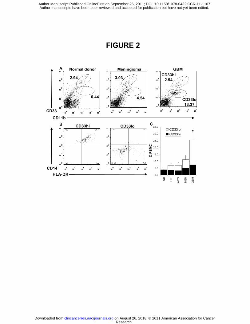
FIGURE 2
2.94CD33hi
2.943.03
A Normal donor Meningioma GBM
CD33CD11b
0.44 4.54 CD33lo13.37
B CD33hi CD33l C
10.0
15.0
20.0
25.0
30.0
35.0
% P
BM
C
CD33loCD33hi
B CD33hi CD33lo C
*
0.0
5.0
10.0N
D
PIT
AP
G
ME
N
GB
M
CD14HLA-DR
Research. on August 26, 2018. © 2011 American Association for Cancerclincancerres.aacrjournals.org Downloaded from
Author manuscripts have been peer reviewed and accepted for publication but have not yet been edited. Author Manuscript Published OnlineFirst on September 26, 2011; DOI: 10.1158/1078-0432.CCR-11-1107

Normal donor GBMA
FIGURE 3
ND flow through
FSC
CD11b+CD66+Cg
FSCSSC
B GBM - CD33lo CD11b+CD66-ND flow through
CD15CD66b
Research. on August 26, 2018. © 2011 American Association for Cancerclincancerres.aacrjournals.org Downloaded from
Author manuscripts have been peer reviewed and accepted for publication but have not yet been edited. Author Manuscript Published OnlineFirst on September 26, 2011; DOI: 10.1158/1078-0432.CCR-11-1107

Untreated PBMC fMLP PBMCA
/ml)
C
FIGURE 4
*
FSCSSC
B
2.97 32.8
ND
MEN
GBM
Arg
I (ng
MET
fMLP stimulated PBMCs
CD33 CD14 CD66
B
27.7
2.57
fMLP stimulated PBMCs
CD33hi
CD33lo
CD33CD11b HLA-DR
CD14 CD66CD15
D
Research. on August 26, 2018. © 2011 American Association for Cancerclincancerres.aacrjournals.org Downloaded from
Author manuscripts have been peer reviewed and accepted for publication but have not yet been edited. Author Manuscript Published OnlineFirst on September 26, 2011; DOI: 10.1158/1078-0432.CCR-11-1107

Am
ber
81012
D3ζ
FIGURE 5
200
250 PHA 48hrPHA 72hr
FI, CD3ζ
Cel
l num
0246
MEN
GBMPI
T
NFI
, C *
C *
0
50
100
150
200
ND MEN GBM
PHA 72hr
250300350
MLR 48hrMLR 72hr400
600
800
1000
1200
1400 PBMC
fMLP PBMC
INF-γ
(pg/
ml)
BAr
g I (
ng/m
l)C
**
*
**
050
100150200250
ND MEN GBM
MLR 72hr
0
200
400
48 72
**
600700
500
600
ml)
DPHA – 48hrs MLR – 48hrs
0100200300
400500
0
100
200
300
400
500
ND
MEN PI
TG
BM+
Arg
OH
A
ND
MEN PI
TG
BM+
Arg
OH
A
INF-
γ(p
g/m
* *
GG
BM +
GBM
+ N
O M GG
BM +
GBM
+ N
O
Research. on August 26, 2018. © 2011 American Association for Cancerclincancerres.aacrjournals.org Downloaded from
Author manuscripts have been peer reviewed and accepted for publication but have not yet been edited. Author Manuscript Published OnlineFirst on September 26, 2011; DOI: 10.1158/1078-0432.CCR-11-1107

Published OnlineFirst September 26, 2011.Clin Cancer Res Trisha R. Sippel, Jason T. White, Kamalika Nag, et al. Targeting Arginase Iwith Glioblastoma: Restoration of Cellular Immune Function by Neutrophil Degranulation and Immunosuppression in Patients
Updated version
10.1158/1078-0432.CCR-11-1107doi:
Access the most recent version of this article at:
Material
Supplementary
http://clincancerres.aacrjournals.org/content/suppl/2011/09/26/1078-0432.CCR-11-1107.DC1
Access the most recent supplemental material at:
Manuscript
Authoredited. Author manuscripts have been peer reviewed and accepted for publication but have not yet been
E-mail alerts related to this article or journal.Sign up to receive free email-alerts
Subscriptions
Reprints and
To order reprints of this article or to subscribe to the journal, contact the AACR Publications
Permissions
Rightslink site. Click on "Request Permissions" which will take you to the Copyright Clearance Center's (CCC)
.http://clincancerres.aacrjournals.org/content/early/2011/09/24/1078-0432.CCR-11-1107To request permission to re-use all or part of this article, use this link
Research. on August 26, 2018. © 2011 American Association for Cancerclincancerres.aacrjournals.org Downloaded from
Author manuscripts have been peer reviewed and accepted for publication but have not yet been edited. Author Manuscript Published OnlineFirst on September 26, 2011; DOI: 10.1158/1078-0432.CCR-11-1107