cell biology. cells and extracellular material together comprise all the tissues that make up the...
TRANSCRIPT
INTRODUCTION Cell biology

Cells and extracellular material together comprise all the tissues that make up the organs of multicellular animals. In all tissues, cells themselves are the basic structural and functional units, the smallest living parts of the body.
Animal cells are eukaryotic (Gr. eu, good, + karyon, nucleus), with distinct membrane-limitednuclei surrounded by cytoplasm containing many varied membrane-limited organelles

In contrast the small prokaryotic cells of bacteria typically have a cell wallaround the plasmalemma, lack other membranous structures including an envelope around the genetic material (DNA).

CYTOPLASMIC ORGANELLES The cell is composed of two basic parts:
cytoplasm (Gr. kytos, cell, + plasma, thing formed) and nucleus (L. nux, nut).
The outermost component of the cell, separating the cytoplasm from its extracellular environment, is the plasma membrane (plasmalemma

The plasma membrane defines the external limit of the cell, a continuum exists between the interior of the cell and extracellular macromolecules.
The plasmamembrane contains proteins called integrins that are linked to both cytoplasmic cytoskeletal filaments and extracellular matrix components.

Through these linkages there is a constant exchange of influences, in both directions, between the extracellular matrix and the cytoplasm.

The cytoplasm itself is composed of a fluidcomponent, or cytosol, in which are contained metabolically active structures, the organelles, which can be membranous (such as mitochondria) or non-membranousprotein complexes (such as ribosomes and proteasomes)

The shape and motility of eukaryotic cells are determined by components of the cytoskeleton. Other minorcytoplasmic structures are inclusions which are generally deposits of carbohydrates, lipids, or pigments

The cytosol contains hundreds of enzymes Such as those of the glycolytic pathway, that produce building blocks for larger molecules and break down small moleculesto liberate energy.
Oxygen, CO2, electrolytic ions, low molecular weight substrates, metabolites, waste products, etc all diffuse through the cytosol, either freely or bound to
proteins, passing to or leaving the organelles where they are used or produced

PLASMA MEMEBRANE

All eukaryotic cells are enveloped by a limiting membrane composed of phospholipids, cholesterol, proteins, and chains of carbohydrate linked tophospholipid and protein molecules.
The plasma, or cell, membrane functions as a selective barrier that regulates the passage of certain materials into and out of thecell and facilitates the transport of specific molecules.

PHOSPHOLIPIDS Hydrophilic Hydrophobic ends Fatty acid disolve in between Outer and inner leaflets of phospholipids
are not identical and same apply to protein attach to it ,makes it asymmetrical
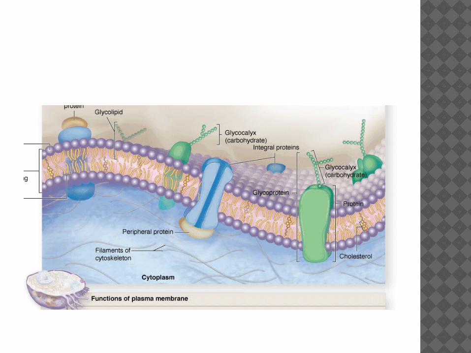

MEMBRANE PROTEINS Integral proteins are directly
incorporated within the lipid bilayer itself,
whereas peripheral proteins exhibit a looser association with one of the two membrane surfaces.

The plasma membrane is the site where materials are exchanged between the cell and its environment. Some ions, such as Na+, K+, andCa2+, cross the cell membrane by passing through integral membrane proteins.
This can involve passive diffusion through ion channels or active transport via ion pumps using energy from the breakdown of adenosine triphosphate (ATP).
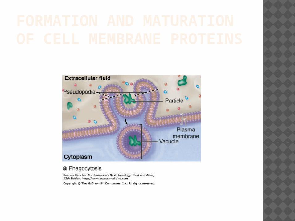
FORMATION AND MATURATION OF CELL MEMBRANE PROTEINS

Bulk uptake of material also occurs across the plasma membrane in a general process called endocytosis, which involves folding and fusion of this membrane to form vesicles which enclose the material transported.

In exocytosis a membrane-limited cytoplasmic vesicle fuses with the plasma membrane, resulting in the release of its contents into the extracellular space without compromising the integrity of the plasma membran

MITOCHONDRIA Mitochondria (Gr. mitos, thread, +
chondros, granule) are membrane-enclosed organelles with enzyme arrays specialized for aerobic respiration and production ofadenosine triphsphate (ATP).


The two mitochondrial membranes and central matrix can be seen here in the diagram and the TEM.
The outer membrane is smooth and the inner membrane, shown above, has many sharp folds called cristae which increase its surface area greatly.

Cristae are most numerous in mitochondria of highly active cells. The matrix is a gelcontaining numerous enzymes.
The inner membrane surface in contact with the matrix is studded with many multimeric protein complexes resembling globular units on short stalks.
These contain the ATP synthase complexes that generate most of the cell's ATP.

The inner membrane is folded to form a series of long infoldings called cristae, which project into the matrix and greatly increase the membrane's surface area
The number of cristae in mitochondria also corresponds to the energy needs of the cell.

RIBOSOME Ribosomes are small electron-dense
particles, about 20 x 30 nm in size. Ribosomes found in the cytosol are composed of four segments of rRNA and approximately 80different proteins.
In eukaryotic cells, the RNA molecules of both subunits are synthesized within the nucleus.
Their numerous proteins are synthesized in the cytoplasm but then enter thenucleus and associate with rRNAs.


A ribosome is made from complexes of RNAs and proteins and is therefore a ribonucleoprotein.
The ribosome is responsible for the synthesis of proteins in cells and is found in all cellular organisms. It serves to convert the instructions found in messenger RNA .

(mRNA, which itself is made from instructions in DNA) into the chains of amino-acids that make up proteins.
Prokaryotic ribosomes are around 20 nm (200 Å) in diameter and are composed of 65% rRNA and 35% ribosomal protein

ENDOPLASMIC RETICULUM The cytoplasm of eukaryotic cells
contains an anastomosing network of intercommunicating channels and sacs formed by a continuous membrane which encloses a space called a cisterna

This membrane system is called the endoplasmic reticulum (ER) .In many places the cytosolic side of the membrane is covered by polyribosomes synthesizing protein molecules which are injected into the cisternae.
This permits the distinction between the two types of endoplasmic reticulum: rough and smooth.


ROUGH ENDOPLASMIC RETICULUM Rough endoplasmic reticulum (RER) is
prominent in cells specialized for protein secretion….
such as pancreatic acinar cells (digestive enzymes), fibroblasts (collagen),
and plasma cells (immunoglobulins).
The RER consists of saclike as well as parallel stacks of flattened cisternae (Figure 2–15), limited by membranes that are continuous with the outer membrane of the nuclear envelope


ROUGH ENDOPLASMIC RETICULUM The binding site of the ribosome on the
rough endoplasmic reticulum is the translocon.
The name "rough endoplasmic reticulum" refers to the presence of polyribosomes on the cytosolic surface of this structure's membrane
The membrane of the rough endoplasmic reticulum forms large double membrane sheets that are located near, and continuous with, the outer layer of the nuclear envelope


1) Nucleolus2) Nucleus3) Ribosome (little dots)4) Vesicle5) Rough endoplasmic reticulum6) Golgi apparatus (or "Golgi body")7) Cytoskeleton8) Smooth endoplasmic reticulum9) Mitochondrion10) Vacuole11) Cytosol (fluid that contains organelles)12) Lysosome13) Centrosome14) Cell membrane


GOLGI APPARTUS

The highly dynamic Golgi apparatus, or Golgi
complex, completes posttranslational modifications and then packages and addresses proteins synthesized in the RER.
The Golgi apparatus generally shows two distinct sides structurally and functionally, which reflects the complex traffic of vesicles within cells.
Near the Golgi, the RERcan be seen budding off small transport vesicles that shuttle newly synthesized proteins to the Golgi apparatus for further processing.


LYSOSOMES Lysosomes are sites of intracellular
digestion and turnover of cellular components.
Lysosomes (Gr. lysis, solution, + soma, body) are membrane-limited vesicles thatcontain about 40 different hydrolytic enzymes and are particularly abundant in cells with great phagocytic activity (eg, macrophages, neutrophils).

Lysosomes are large, generally spherical membrane-enclosed vesicles that function as sites of intracellular digestion and are particularly numerous in cells active in various types of endocytosis.


PROTEASOMES
Proteasomes are abundant cytoplasmic protein complexes not associated with membrane, each approximately the size of the small ribosomal subunit.
They functionto degrade denatured or otherwise nonfunctional polypeptides. Proteasomes also remove proteins no longer needed by the cell and provide an important mechanismfor restricting activity of a specific protein to a certain window of time.

Whereas lysosomes digest bulk material introduced into the cell, or whole organelles andvesicles, proteasomes deal primarily with proteins as individual molecules
The proteasome is a cylindrical structure made of four stacked rings, each composed of seven proteins including proteases.

UBIQUITIN At each end of the cylinder is a
regulatory particle that contains ATPase and recognizes proteins with ubiquitin molecules attached.
Ubiquitin is an abundant cytosolic 76-amino acid protein found in all cells and is highly conserved during evolution—it has virtually the same structure from bacteria to humans.

A ubiquinated protein is recognized by the regulatory particle of proteasomes, unfolded by the ATPase using energy from ATP, and then translocated into the core particle, where it is broken into short peptides.
These peptides are transferred to the cytosol and the ubiquitin molecules are released by theregulatory particles for reuse.
The peptides may be broken down further to amino acids or they may have other specialized destinations, such as the antigen-presenting complexes of cells activating an immune response.

PROTEASOMES