zoology assignment fourth semester 2018...zoology assignment fourth semester 2018 submitted to: dr....
TRANSCRIPT

Zoology Assignment Fourth Semester 2018 Submitted To: Dr. Amrit Kaur Bansal Submitted By: Himanshu Goyal
Thyroid Gland
Location
The thyroid gland surrounds the front of the larynx and upper part of the trachea in the
neck.
Structure
● Thyroid is the largest endocrine gland and weighs about 28 gram. It is bilobed,
highly vascular organ.
● The two lobes are connected by a narrow band, the isthmus.
● The gland is composed of a irregularly rounded, about 0.1 mm wide follicles held
together by connective tissue very rich in blood capillaries.
● A follicle has a wall of cuboidal epithelium resting on a basement membrane and
is filled with a clear, gelatinous colloid.
1

● The follicular cells become columnar and bear microvilli on the free surface when
the gland is activated by thyroid stimulating hormone.
Physiology
The thyroid gland produces the hormones- thyroxine (T4) and Triiodothyronine (T3),
which regulate metabolic body processes, cellular respiration, Total energy expenditure,
growth, and maturation of tissues, and turnover of hormones substrates, and vitamins.
Thyroperoxidase (TPO) is one of the primary enzyme produced in the thyroid. It is
synthesized within the endoplasmic reticulum of thyrocyte and oxidizes iodine, thereby
facility the formation of T3 and T4.
Release of hormones into the bloodstream involve the negative feedback system of the
hypothalamic-pituitary-thyroid axis. A low metabolic rate or a decrease in serum T3
and/or T4 level signals the hypothalamus to secrete thyrotropin releasing hormone
(TRH), which travels to the anterior pituitary gland and stimulate secretion of thyroid
stimulating hormone (TSH). TSH, in turn, stimulates the thyroid gland to manufacture
and release T3 and T4 until the metabolic rate is normalised. An elevated T3 serum level
inhibits the release of TRH and TSH.
2

Parathyroid
Location
Parathyroid glands are situated in the back of the thyroid.
Structure
● The parathyroids are four small, rounded glands, two on each side.
● The cells of parathyroids are arranged in compact mass, not in follicles, like those
of thyroid.
● Healthy parathyroid glands generally weigh about 30 mg in men and 35 mg in
women.
3

Physiology
The parathyroid secrete a single hormone called parathormone (PTH), also called the
Collip’s hormone after its discoverer. It is a long peptide of 84 amino acid units.
Parathormone and thyroid hormone calcitonin work antagonistically to regulate the
calcium-phosphorus balance in the blood. The release of parathormone increases blood
calcium to normal by drying calcium from the bones into the plasma, by increasing
calcium absorption in the digestive tract, and by reducing loss of calcium in the urine. It
lowers blood phosphate by increasing elimination of phosphate in the urine. Proper
calcium-phosphorus balance is necessary for the growth of bones and teeth. Calcium is
vital for blood clotting, for muscle tone and for normal nervous activity. It is also needed
for the activities of many enzymes.
Parathyroids are under the feedback control of blood calcium level. A fall in blood
calcium level stimulates them to secrete parathormone which restores blood calcium
level. A rise in blood calcium level inhibits secretion of parathormone.
Adrenal Glands
Location
The adrenal glands, also called suprarenal glands, are paired glands placed in front of
the upper parts of the kidney.
4

Structure
● The adrenals are conical, yellowish bodies, each weighing about 5 gram.
● Each gland has two distinct regions, having different embryonic origin, structure
and function.
● The two regions are called adrenal cortex and adrenal medulla, both produce
different hormones.
● The adrenal cortex is an external firm, pale-yellowish-pink structure covered by a
fibrous capsule. It forms 80% of the gland. It is formed from mesoderm.
● The adrenal medulla is an internal soft, dark-reddish-brown tissue drive from the
ectoderm. It consists of strands of cells surrounded by blood capillaries.
5

Physiology
Adrenal Cortex:
● Mineralocorticoids: These hormones are secreted by the outer region of adrenal
cortex. They regulate mineral metabolism. Their secretion is stimulated by a fall
in the sodium concentration in the Plasma or in the volume of blood. The major
mineralocorticoid, aldosterone tends to increase the reabsorption of sodium
from urine, saliva, bile and sweat to reduce its loss from the body.
● Glucocorticoids: These hormones are secreted by the middle region of adrenal
cortex. They regulate the metabolism of Carbohydrates, proteins and fats. They
increase the blood glucose level by converting proteins and fats into
carbohydrates which are in turn converted to glucose.
● Sexcorticoids: These hormones are secreted by both the middle and inner
regions of the adrenal cortex. They include small amounts of both male and
female sex hormones. Testosterone stimulates the development of male
secondary sexual characters and oestrogen stimulates the appearance of female
secondary sexual characters.
Adrenal Medulla: Cells of adrenal medulla secrete two similar hormones: adrenaline
(epinephrine) and noradrenaline (norepinephrine). The adrenal medulla enable the
animal to face physical and emotional stress which require more energy which is
provided by increasing Heartbeat, blood pressure, respiratory rate, sugar level of blood
and blood supply of heart. The hormones also causes contraction of spleen to squeeze
out the reserve supply of blood, decrease in clotting time of blood and dilation of Pupil.
The above role of the two hormones is often called “Fight or Flight" reaction. Because of
the above role of their hormones, the adrenals are known as the “glands of emergency”.
6
Hypothalamus
Location
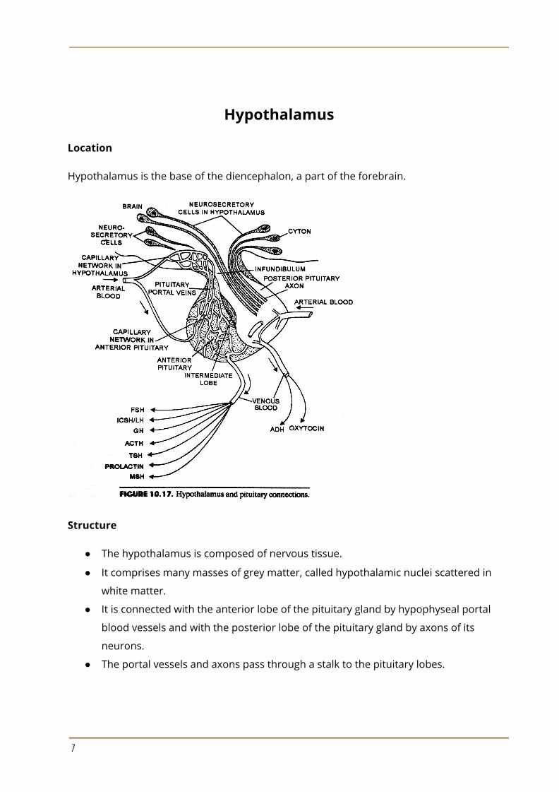
Hypothalamus is the base of the diencephalon, a part of the forebrain.
Structure
● The hypothalamus is composed of nervous tissue.
● It comprises many masses of grey matter, called hypothalamic nuclei scattered in
white matter.
● It is connected with the anterior lobe of the pituitary gland by hypophyseal portal
blood vessels and with the posterior lobe of the pituitary gland by axons of its
neurons.
● The portal vessels and axons pass through a stalk to the pituitary lobes.
7

Physiology
Neurosecretory cells of the hypothalamus secrete hormones called neurohormones.
These are short peptides, having 3 to 14 amino acids. The neurohormones are carried
by the portal blood to the anterior lobe of the pituitary gland and stimulate the
hypothalamus to release its hormones. On this account, such hypothalamic hormones
are also called releaser hormones (RH). Certain hypothalamic hormones inhibit the
secretion of some pituitary hormones. These are termed inhibitory hormones the
well-known releasing and inhibitory hormones (IH). The well known releasing and
inhibitory hormones of hypothalamus are given below:
● Thyrotropin-Releasing hormone (TRH)
● Adrenocorticotropic-Releasing Hormone
● Follicle-Stimulating hormone-Releasing hormone (FSH-RH)
● Luteinizing hormone-Releasing hormone (LH-RH)
● Growth hormone-Releasing hormone (GHRH)
● Growth hormone Release-Inhibiting Hormone (GH-RIH)
● Prolactin-Releasing hormone (PRH)
● Prolactin release-Inhibiting Hormone (PR-IH)
● Melanocyte-Stimulating hormone-Releasing Hormone (MSH-RH)
● Melanocyte-Stimulating hormone Release-Inhibiting Hormone (MSHR-IH)
8

Hypothalamus is able to control endocrine secretion because it can monitor metabolites
and hormone levels in the blood. The information so gathered and also the information
reaching from other parts of the brain is passed to the pituitary via releasing hormones.
The hypothalamic-pituitary system is of paramount importance for homeostasis as it
regulate the most major physiological activities in the body.
9