zika virus & microcephaly - hanys · microcephaly in the context of zika virus infection what...
TRANSCRIPT
Zika Virus & Microcephaly
Update for Healthcare Providers
Of Pregnant Women and Infants
April 27, 2016
Dr. Deborah Campbell, MD, FAAP Deborah Fox, MPHProfessor of Clinical Pediatrics DirectorAlbert Einstein College of Medicine Congenital Malformations RegistryChief, Division of Neonatology Center for Environmental HealthChildren's Hospital at Montefiore New York State Department of Health
2
ObjectivesAt the conclusion of this webinar, healthcare providers will be
able to:
• Describe what is known about maternal Zika virus infection
and birth outcomes
• Discuss other factors that can cause microcephaly in a fetus
• Describe how to determine if a fetus or newborn is
microcephalic
• Understand how your efforts and reporting to the Congenital
Malformations Registry will improve knowledge about Zika
virus infection and microcephaly
3
CDC Media Briefing on Wednesday 4/13/16
“It is now clear that Zika does cause microcephaly”
“An unprecedented association - Never before in history
could a bite from a mosquito result in a devastating birth
malformation”
“Still working to understand range of birth defects
associated with Zika, and time of greatest risk”
3Rasmussen SA, et al. Zika Virus and Birth Defects – Reviewing the Evidence for Causality. NEJM 2016 DOI: 10.1056/NEJMsr1604338.
4
Microcephaly in context of Zika virus (ZIKV) infection
What we know:
• Timing of ZIKV exposure associated with severe microcephaly appears to be in late 1st/early 2nd trimester, which would be consistent with the defects observed – Exposure determined by timing of symptoms or by
travel to ZIKV infected area
• ZIKV infections later in pregnancy associated with poor intrauterine growth and fetal death
Rasmussen SA. NEJM 2016 DOI: 10.1056/NEJMsr1604338.Brasil P. NEJM, March 4, 2016, DOI: 10.1056/NEJMoa1602412
5
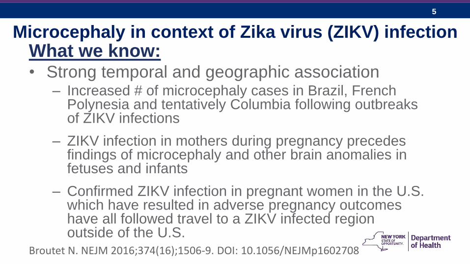
Microcephaly in context of Zika virus (ZIKV) infectionWhat we know:• Strong temporal and geographic association
– Increased # of microcephaly cases in Brazil, French Polynesia and tentatively Columbia following outbreaks of ZIKV infections
– ZIKV infection in mothers during pregnancy precedes findings of microcephaly and other brain anomalies in fetuses and infants
– Confirmed ZIKV infection in pregnant women in the U.S. which have resulted in adverse pregnancy outcomes have all followed travel to a ZIKV infected region outside of the U.S.
Broutet N. NEJM 2016;374(16);1506-9. DOI: 10.1056/NEJMp1602708
6
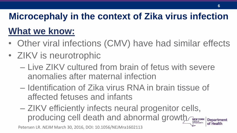
Microcephaly in the context of Zika virus infection
What we know:
• Other viral infections (CMV) have had similar effects
• ZIKV is neurotrophic
– Live ZIKV cultured from brain of fetus with severe anomalies after maternal infection
– Identification of Zika virus RNA in brain tissue of affected fetuses and infants
– ZIKV efficiently infects neural progenitor cells, producing cell death and abnormal growth
Petersen LR. NEJM March 30, 2016, DOI: 10.1056/NEJMra1602113
7
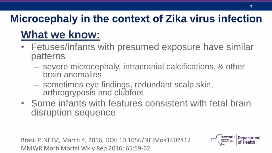
Microcephaly in the context of Zika virus infection
What we know:• Fetuses/infants with presumed exposure have similar
patterns – severe microcephaly, intracranial calcifications, & other
brain anomalies
– sometimes eye findings, redundant scalp skin, arthrogryposis and clubfoot
• Some infants with features consistent with fetal brain disruption sequence
Brasil P. NEJM, March 4, 2016, DOI: 10.1056/NEJMoa1602412MMWR Morb Mortal Wkly Rep 2016; 65:59-62.
8
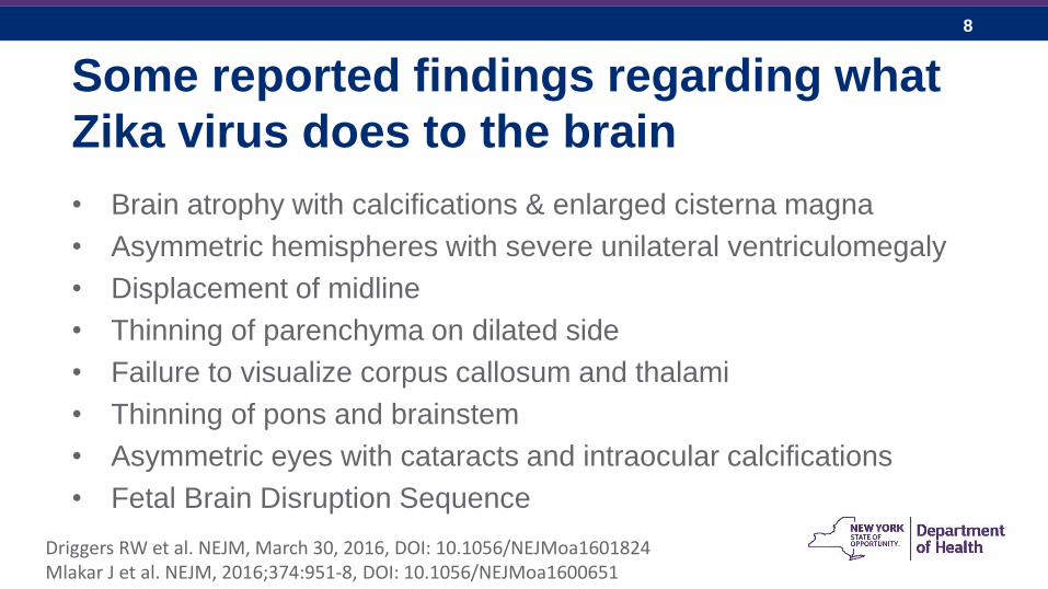
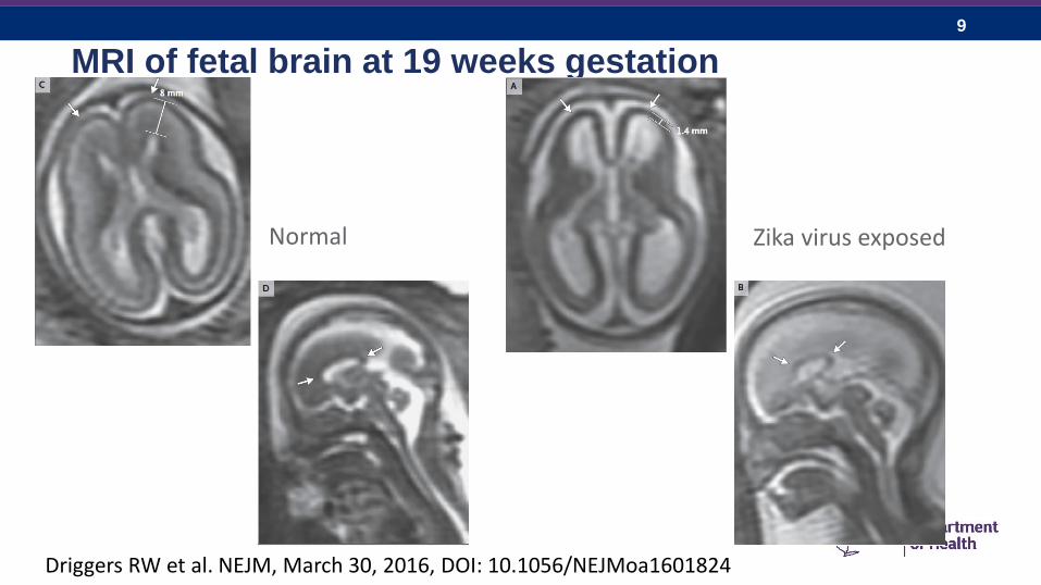
Some reported findings regarding what
Zika virus does to the brain
• Brain atrophy with calcifications & enlarged cisterna magna
• Asymmetric hemispheres with severe unilateral ventriculomegaly
• Displacement of midline
• Thinning of parenchyma on dilated side
• Failure to visualize corpus callosum and thalami
• Thinning of pons and brainstem
• Asymmetric eyes with cataracts and intraocular calcifications
• Fetal Brain Disruption Sequence
Driggers RW et al. NEJM, March 30, 2016, DOI: 10.1056/NEJMoa1601824Mlakar J et al. NEJM, 2016;374:951-8, DOI: 10.1056/NEJMoa1600651
9
MRI of fetal brain at 19 weeks gestation
Driggers RW et al. NEJM, March 30, 2016, DOI: 10.1056/NEJMoa1601824
Zika virus exposedNormal
10
Microcephaly in the context of Zika virus infection
What we don’t know• Full spectrum of defects caused by prenatal Zika virus infection
(ocular, hearing, heart, …)
• Relative and absolute risk among infants born to mothers infected at different times during pregnancy
• Other factors that might affect a woman’s risk of adverse pregnancy or birth outcomes – Co-infection with another virus
– Preexisting immune response to another flavivirus
– Genetic background of mother and fetus
– Timing and/or severity of maternal infection
• Validated birth prevalence estimates of microcephaly in Brazil or other countries in recent outbreak
11
Estimation of Prevalence – Monitoring Change
• Prevalence of microcephaly in U.S. varies from 2 to 12 cases per 10,000 live births among state-based birth defects programs
• In NYS, 110-160 congenital microcephaly cases* reported annually (4.6 – 6.7 per 10,000 live births)
• Historically, microcephaly has been inconsistently measured and reported
• Our aim is to improve surveillance of microcephaly in NYS
*Does NOT exclude cases with other diagnoses associated with abnormal head size or with other causes (in utero infections, genetic causes, teratogenic exposures, maternal radiation, etc..)
12
Enhanced microcephaly surveillance will:
• Provide better estimates of baseline risk of
microcephaly
• Permit observation of temporal and spatial patterns
• Allow identification of ZIKV associated cases and
other emerging public health threats
13
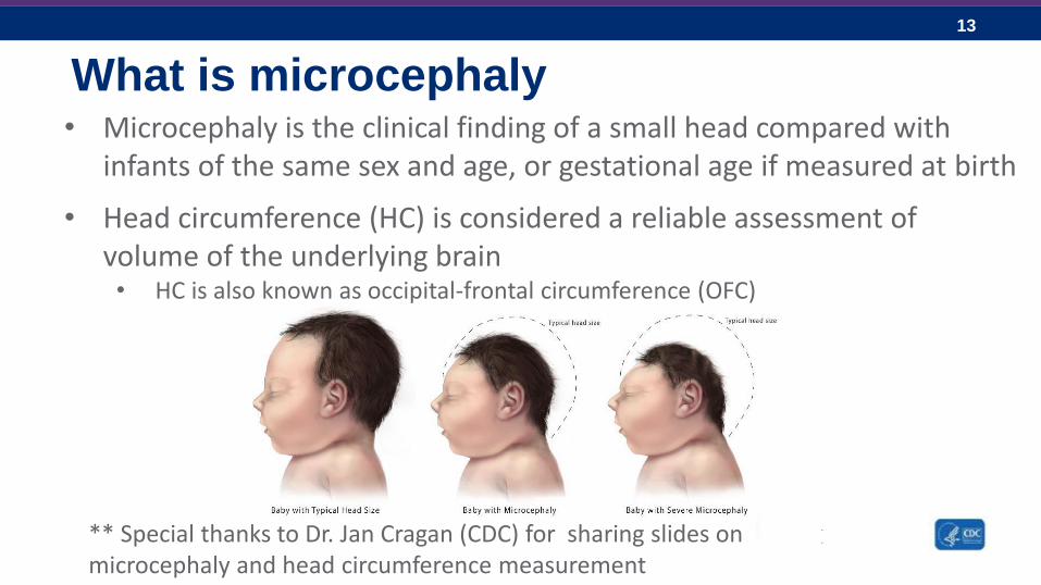
What is microcephaly
** Special thanks to Dr. Jan Cragan (CDC) for sharing slides on microcephaly and head circumference measurement
• Microcephaly is the clinical finding of a small head compared with infants of the same sex and age, or gestational age if measured at birth
• Head circumference (HC) is considered a reliable assessment of volume of the underlying brain• HC is also known as occipital-frontal circumference (OFC)
14
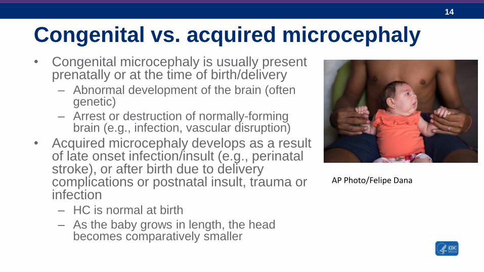
Congenital vs. acquired microcephaly• Congenital microcephaly is usually present
prenatally or at the time of birth/delivery– Abnormal development of the brain (often
genetic)
– Arrest or destruction of normally-forming brain (e.g., infection, vascular disruption)
• Acquired microcephaly develops as a result of late onset infection/insult (e.g., perinatal stroke), or after birth due to delivery complications or postnatal insult, trauma or infection– HC is normal at birth
– As the baby grows in length, the head becomes comparatively smaller
AP Photo/Felipe Dana
15
Types of microcephaly
• Disproportionate – Head is small out of proportion to
the weight and length, which may be normal for age
and sex
• Proportionate – Head size, weight and length are all
small for age and sex but proportional to each other
• “Relative” microcephaly – Head size measures within
the normal range for age and sex, but is small out of
proportion to the weight and length
16
Causes of Congenital Microcephaly
– In utero infections
• Toxoplasmosis
• Rubella
• Cytomegalovirus (CMV)
• Herpes
• Human Immunodeficiency Virus (HIV)
• Syphilis
• Zika virus
17
Causes of Congenital Microcephaly– Genetic causes
• Single gene disorders (syndromes)
• Chromosomal abnormalities, microdeletions, microduplications
• Mitochondrial mutations
– In utero ischemia/hypoxia (e.g., placental insufficiency or abruption)
– Teratogens (e.g., maternal alcohol, hydantoin)
– Radiation
– Mercury (e.g., fish and seafood)
– Maternal conditions (e.g., poorly controlled diabetes, hyperphenylalaninemia)
18
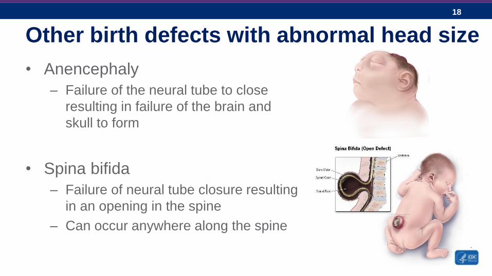
Other birth defects with abnormal head size
• Anencephaly
– Failure of the neural tube to close
resulting in failure of the brain and
skull to form
• Spina bifida
– Failure of neural tube closure resulting
in an opening in the spine
– Can occur anywhere along the spine
19
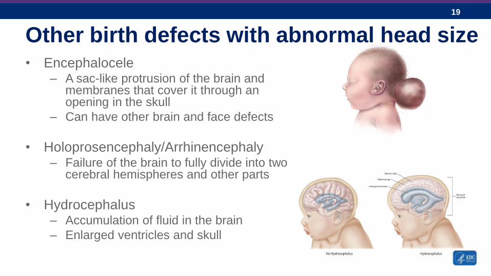
• Encephalocele– A sac-like protrusion of the brain and
membranes that cover it through an opening in the skull
– Can have other brain and face defects
• Holoprosencephaly/Arrhinencephaly– Failure of the brain to fully divide into two
cerebral hemispheres and other parts
• Hydrocephalus– Accumulation of fluid in the brain
– Enlarged ventricles and skull
Other birth defects with abnormal head size
20
Brain abnormalities that can occur with
microcephaly
• Intracranial calcifications
• Hydrocephalus ex-vacuo (extra axial fluid collection)
– Damaged brain matter shrinks and is surrounded by fluid
– Cerebral atrophy
• Hydranencephaly
– Damaged brain matter replaced by pockets of fluid
• Pachygyria, lissencephaly
– Abnormal ridges and folds (gyri) in the brain

21
Measuring HC (per World Health Organization [WHO])
• Use tape measure that cannot be stretched
• Securely wrap tape around widest possible circumference of head– 1-2 finger-widths above eyebrow on
forehead
– At the most prominent part of back of head
• Take measurement 3 times and select the largest measurement to nearest 0.1 cm
• Optimal measurement at 24-36 hours after birth when molding of head has subsided– Head shape can affect the accuracy of HC
estimate of brain volume
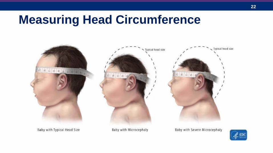
22
Measuring Head Circumference

23
The following video clip is taken from a McMaster Neonatal Research Lab presentation, “Standardized Measurement Techniques for Growth” made available to us by Dr. Christopher Fusch.
Useful teaching video on how to measuring head circumferencehttps://www.youtube.com/watch?v=LW38bgQ9vVY
24
From a McMaster Neonatal Research Lab presentation, “Standardized Measurement Techniques for Growth” made available to us by Dr. Christopher Fusch
25
25
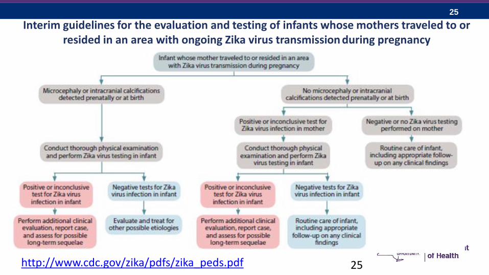
Interim guidelines for the evaluation and testing of infants whose mothers traveled to or resided in an area with ongoing Zika virus transmission during pregnancy
http://www.cdc.gov/zika/pdfs/zika_peds.pdf
26
Prenatal diagnosis of microcephaly is more
challenging• Can be detected during mid-pregnancy anomaly scan
(ultrasound) at 18-20 weeks
• May not be evident until the late 2nd or into the 3rd
trimester
• Usually present by 36 weeks
• Serial prenatal ultrasounds may be needed to detect the
development of microcephaly in utero
• Fetal MRI can be another useful imaging tool
27
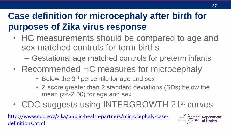
Case definition for microcephaly after birth for
purposes of Zika virus response
• HC measurements should be compared to age and sex matched controls for term births
– Gestational age matched controls for preterm infants
• Recommended HC measures for microcephaly• Below the 3rd percentile for age and sex
• Z score greater than 2 standard deviations (SDs) below the mean (z<-2.00) for age and sex
• CDC suggests using INTERGROWTH 21st curves
http://www.cdc.gov/zika/public-health-partners/microcephaly-case-definitions.html
28
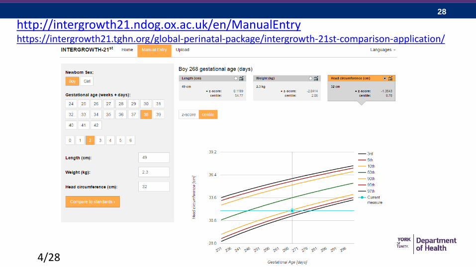
http://intergrowth21.ndog.ox.ac.uk/en/ManualEntryhttps://intergrowth21.tghn.org/global-perinatal-package/intergrowth-21st-comparison-application/
4/28/2016 28
29
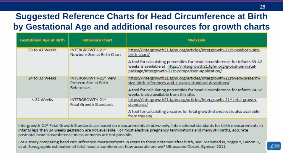
Suggested Reference Charts for Head Circumference at Birth
by Gestational Age and additional resources for growth charts
30
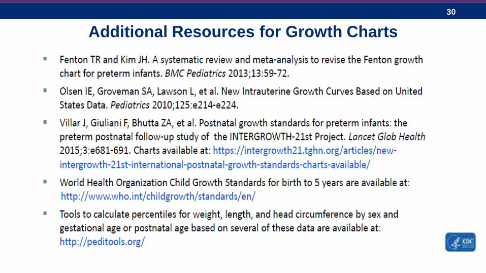
Additional Resources for Growth Charts
31
CDC establishing the US Zika Pregnancy Registry
Purpose:
– To understand more about ZIKV infection during pregnancy
– To update recommendations for clinical care of pregnant women with ZIKV infection
Who should be reported:
– All pregnant women in the US with laboratory evidence of ZIKV infection
– Infants with congenital ZIKV infection
http://www.cdc.gov/zika/hc-providers/registry.html
32
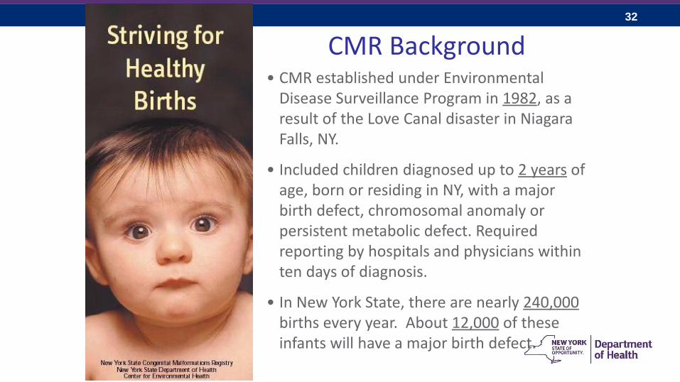
CMR Background• CMR established under Environmental
Disease Surveillance Program in 1982, as a result of the Love Canal disaster in Niagara Falls, NY.
• Included children diagnosed up to 2 years of age, born or residing in NY, with a major birth defect, chromosomal anomaly or persistent metabolic defect. Required reporting by hospitals and physicians within ten days of diagnosis.
• In New York State, there are nearly 240,000births every year. About 12,000 of these infants will have a major birth defect.
33
• In additional to reporting by hospitals and physicians,
require reporting by nurse practitioners authorized to
diagnose congenital anomalies, physicians assistants
authorized to diagnose congenital anomalies and
midwives
• Require reporting of prenatal diagnosis of birth defects
• Extend the case capture periods for certain defects,
including fetal alcohol syndrome (FAS), muscular
dystrophies, genetic conditions and heart defects
Several CMR Regulatory Changes Effective May 2016
34
Reporting congenital microcephaly in newborns• Hospitals will report congenital microcephaly diagnosed at birth
• CMR is seeking reports from other health facilities/practices:
– Birthing centers/Midwifery practices
– Home births
– Subspecialty clinics (neurology, genetics)
– Other healthcare providers
• To request forms or get additional information, email:
Currently not requesting reporting of prenatally diagnosed microcephaly
35
From CDC: For Health Care Providers
4/28/2016 35
http://www.cdc.gov/zika/hc-providers/index.html
36
Acknowledgements• Dr. Marilyn Kacica, Medical Director, Division of Family Health,
New York State Department of Health
• Dr. Marilyn Browne, Director, Birth Defects Research Section,
New York State Department of Health
• Dr. Jan Cragan & Dr. Cara Mai, Birth Defects Branch, National
Center for Birth Defects and Developmental Disabilities, Centers
for Disease Control and Prevention
• Dr. Peer Dar, Director, Division of Fetal Medicine and OBGYN
Ultrasound, Montefiore Medical Center
• McMaster Neonatal Research Lab and Dr. Christopher Fusch
37
NYSDOH Zika Testing EligibilityIdentify a patient presenting for care who meets the following criteria:
1. Pregnant woman who had possible exposure (travel to an area with Zika virus transmission while pregnant or unprotected sex with partner who traveled) OR
2. Non-pregnant woman, man, or child who develops (or developed) compatible symptoms during or within 4 weeks of travel to an area with Zika virus transmission OR
3. A person who traveled to an area with active Zika virus transmission and who presents with Guillain-Barré syndrome OR
4. Infant with microcephaly, intracranial calcifications or other abnormality whose mother had exposure during pregnancy (or born to mothers with positive or inconclusive test results)
38
NYSDOH Zika Testing Procedures
Patient needs three things:
1. Lab order (PROVIDER)
Order “Zika blood and urine PCR and Zika blood for serology”
2. Infectious Disease Requisition Form (IDR) (PROVIDER)
3. LHD authorization form (Local Health Department)
38
39
NYSDOH Zika Testing Results
• Results of Zika virus testing will be made available to providers
• Providers can access public health consultation for assistance with interpretation of results by calling the
NYSDOH Zika Information Line at:
1-888-364-4723
Weekdays between 9AM and 5PM
40
NYC- Process for Ordering Zika Virus Testing as of March 21, 2016
• Healthcare providers must now call the NYC Health Department’s Provider Access Line (PAL) to request Zikatesting
PAL:1-866-692-3641
• This is to ensure that appropriate tests are ordered and specimens are correctly collected, labeled, processed, packaged and transported
41
NYC- What to Expect When You Call
• Health Department representative will review case with you to ensure
testing criteria are met
• If testing is appropriate then the representative on the phone will:– Collect the information needed to order the correct testing
– Complete the required laboratory submission form
– Advise on the type(s) of specimen(s) to collect
• The completed lab submission form(s) for your patient(s) and
instructions for specimen collection and handling will be sent to you
within 30 minutes via email or fax