reviewarticlefac.ksu.edu.sa/sites/default/files/new_therapeutics_in_promoting... · type x are...
TRANSCRIPT

Hindawi Publishing CorporationBioMed Research InternationalVolume 2013, Article ID 789679, 10 pageshttp://dx.doi.org/10.1155/2013/789679
Review ArticleNew Therapeutics in Promoting and Modulating MandibularGrowth in Cases with Mandibular Hypoplasia
Tarek El-Bialy1 and Adel Alhadlaq2
1 7-020D Katz Group Centre for Pharmacy and Health Research, University of Alberta, Edmonton, AB, Canada T6G 2E12 College of Dentistry, King Saud University, P.O. Box 60169, Riyadh 11545, Saudi Arabia
Correspondence should be addressed to Tarek El-Bialy; [email protected]
Received 13 January 2013; Revised 10 April 2013; Accepted 18 April 2013
Academic Editor: Brian L. Foster
Copyright © 2013 T. El-Bialy and A. Alhadlaq. This is an open access article distributed under the Creative Commons AttributionLicense, which permits unrestricted use, distribution, and reproduction in any medium, provided the original work is properlycited.
Children withmandibular growth deficiencymay develop airway obstruction.The standard treatment of severe airway obstructioninvolves invasive procedures such as tracheostomy. Mandibular distraction osteogenesis has been proposed in neonates withmandibular deficiency as a treatment option to avoid tracheostomy procedure later in life. Both tracheostomy and distractionosteogenesis procedures suffer from substantial shortcomings including scarring, unpredictability, and surgical complications.Forward jaw positioning appliances have been also used to enhancemandible growth.However, the effectiveness of these appliancesis limited and lacks predictability. Current and future approaches to enhance mandibular growth, both experimental and clinicaltrials, and their effectiveness are presented and discussed.
1. Introduction
Underdeveloped mandibles can cause severe psychologicaland functional impact upon the growing child and maybe associated with life-threatening complications such asobstructive sleep apnea (OSA) due to severe airway constric-tion [1, 2].Theprevalence ofOSA in children is approximately3% [3]. Patients with OSA usually have underdevelopedmandible (mandible) [4, 5].Themortality rate due toOSAhasbeen reported to reach 15% of the affected individuals [6, 7].This increased mortality is mainly attributed to the retrudedposition of the mandible which compromises the airway [8].
When the airway is compromised because of severemandible underdevelopment, jaw-positioning appliances [9],nasopharyngeal airway [10], orthodontic plates with velarextension [11], intubation [12], tongue-lip adhesion [13],continuous positive airway pressure (CPAP) [14], tra-cheostomy [15], mandibular advancement with orthognathicsurgery/distraction osteogenesis [16, 17], or an anteriormandibular positioning device is used to manage the airwayobstruction [18, 19].
It is to be noted that tracheotomy and mandibular dis-traction osteogenesis (DO) have major limitations; theyare lifesaving and provide substantial lengthening of themandible quickly. In spite of recent advancements, thetracheostomy procedure remains a treatment with seriousand frequent side effects. These side effects include potentialtissue traumatization, injury of the laryngeal or trachealmucosa, and/or other complications (e.g., pneumothorax,hemorrhage, wound complication, tracheal stenosis, andlaryngeal stenosis) [15]. To avoid tracheostomies, distractionosteogenesis of the mandible (surgical lengthening of themandible) has recently been recommended as a viable optionfor pediatric patients with upper airway obstruction dueto mandibular deficiency [16]. This technique has beendescribed as an alternative to tracheostomy in neonates (6 to26 days of age) to improve airway and breathing [17].
Conversely, improving the airway in OSA adult patientswith the use of a removable mandibular advancement device(MAD) has recently been shown to have a success rate ofonly 54.8% [18]. Advancing themandible with oral appliancesdepends solely on patient’s compliance and has been reported

2 BioMed Research International
to be effective in short term only [19]. Nonetheless, the long-term efficacy of all above-mentioned treatment modalities isunknown.
2. Mandibular Growth
The condylar cartilage in the mandible is a secondary carti-lage. It has been shown that mechanical stimuli are necessaryfor the normal growth of this type of cartilage [20–24].Bite-jumping appliances (orthodontic/orthopedic functionalappliances) have long been used for “growth modification”of the mandible in the field of orthodontics and craniofacialorthopedics. However, the effectiveness of these applianceshas been criticized and is still considered an area of con-troversy [25]. Other currently available mechanical loadingtechniques, for example, electroforce 3200mechanical testingmachine, are not clinically applicable for severe mandibu-lar underdevelopment due to the large size of the device(mechanical testing machine) or because patients can notfit within the proposed devices (electroforce or mechanicaltesting machines) [20].
3. Current TreatmentModalities and Challenges
3.1. Bite-JumpingAppliances (Functional Appliances). Growthmodification of the mandible using functional appliances(FAs) has been used to enhance forward positioningof the mandible. They are commonly used clinically toenhancemandibular growth in patients with underdevelopedmandibles. Recent animal experiments have demonstratedsignificant increase in the endochondral ossification (boneformation within the growing cartilage) at the mandibularcondyle in response to the mandibular protrusive forces[20, 26, 27]. This forward mandibular positioning has beenhypothesized to solicit a sequence of cellular events thatlead to increased vascularization, new bone formation, andenhanced condylar growth [28]. Also, a recent clinical trialon the effectiveness of twin block functional appliance onmandibular growth in 15 boys and 19 girls ranging in age from9 years 3months to 10 years 8months at the start of treatmentshowed that its use increases the mandibular length 2.3mmmore than that of a control group [29]. Controversially, otherclinical trials of FAs therapy have demonstrated either nosubstantial growth enhancement or increased mandibulargrowth only at the initial stage, with the growth phenotype ofthe mandible returning to its original pattern afterwards [30,31]. Interestingly, FAs were reported to increase the numberof replicating mesenchymal stem cells in growing rats atboth the mandibular condyle and the glenoid fossa [32].In a related study, a correlation was demonstrated betweenthe application of FAs as a mechanical stimulator and thenumber of stem cells in mandibular condyles and the glenoidfossa [33]. These studies demonstrated that the number ofmesenchymal cells in a given locus normally determines thepotential for bone growth in that area and that the number of
mesenchymal cells in the glenoid fossa is directly correlatedwith the amount of bone produced during natural growth andmandibular advancement [32, 33]. It has been hypothesizedthat a lack of native stem cells in the mandibular condyleand glenoid fossa contributes to the underdevelopment ofthe mandible [32, 33]. Consequently, development of newtechniques to foster stem cell recruitment to the growingcondyles and the glenoid fossa becomes practical.
Although the exact mechanism of FAs involvement inpromotingmandibular growth is not fully understood, previ-ous reports have shown that FAs enhancemandibular growththrough an increase in the production of Runx2 [27], whichis a transcription factor that belongs to the Runt domaingene family. The Runx2 promotes osteoblast’s differentiationand function by transcriptionally upregulating all the majorosteoblast-specific genes, including osteocalcin, type-I col-lagen, bone sialoprotein, osteopontin, alkaline phosphatase,and collagenase-3 [34]. Also, FAs enhance Sox9 and typeII collagen expression in rats [35], a result that has beenconfirmed by immunostaining techniques [20, 26–28].
3.2. Low-Intensity Pulsed Ultrasound. Low-intensity pulsedultrasound (LIPUS) produces mechanical waves that propa-gate through biological tissues at a pulse frequency of 1.5MHzwith a pulse repetition frequency of 1 kHz.At an output powerof 30mW/cm2, LIPUS can stimulate tissue growth withoutheating [15, 16]. A daily treatment with LIPUS for 20 minuteshas been established as a favorable treatment modality in thefield of orthopedics [36–39].This treatment protocol has beenfound to stimulate bone healing after fracture in a variety ofhuman and animalmodels by promoting new vascularizationand bone formation [35–37]. Also, this technique has beenapplied successfully to promote growth and healing afterdistraction (i.e., excessive separation of bony segments) ofthe tibia in a rabbit model and after distraction of the callus[36, 39]. Daily direct application of LIPUS for 4 weeksalso stimulated mandibular bone growth in rats and rabbits[22, 23]. However, achieving similar results in monkeysrequired four months of treatment [40] and about a year inhumans (when combined with FAs) [41] (Figure 1). Such longperiods of daily application of LIPUS are challenging andhighly demanding for a clinical implementation. Thus, thedevelopment of an approach to boost the stimulatory effectof LIPUS on bone growth is necessary.
Recent reports have shown that LIPUS has anabolic effectin chondrocytes with increased stimulation when LIPUS isapplied for longer durations [42]. Furthermore, the currentconsensus in the literature is that the stimulatory effect oftherapeutic ultrasound on bone formation is dose dependent(daily treatment time) [42–44]. Angiogenesis has also beenshown to be increased with LIPUS treatment, thus improvingthe blood flow to the treatment area that is critical for bonegrowth [45, 46]. In addition, it has been shown that LIPUSpromotes stem cell expansion and differentiation [47–49].Indeed, the application of ultrasound in stem cell expansionand differentiation as well as in bone mineralization andregeneration has gained considerable attention in recent years[50, 51].

BioMed Research International 3
(a) (b)
Figure 1: A hemifacial microsomia patient treated with 20 minutes LIPUS per day for a year with a hybrid functional appliance. (a) Beforeand (b) after treatment.
The mechanism of mandibular growth enhancement byLIPUS is not fully understood. The hypothesized mecha-nism of action of LIPUS to stimulate mandibular growthis through increase in vascular endothelial growth factor(VEGF) and Runx2 in bone healing since these have beenshown to be correlated to increased mandibular growth[46]. LIPUS is known to increase the expression of VEGFand Runx2 in bone healing [45]. Also, LIPUS has beenshown to increase the expression of osteocalcin, Runx2,and bone sialoprotein in stem cells [52]. In addition, theexpression of Runx2, Msx2, Dlx5, osterix, bone sialoprotein,and bone morphogenetic protein-2 has been shown to beenhanced in MG-63 osteoblasts [53]. Sox9 is known to bea crucial molecule in stem cell proliferation, condensation,and chondrocyte differentiation [54–56]. Runx2 is involvedin chondrocyte differentiation, whereas VEGF and collagentype X are involved in endochondral ossification [46, 54].Integrins may act as mechanotransducers that can transformacoustic pulsed energy into intracellular biochemical signalsthat subsequently induce cell proliferation [57]. Although themechanisms are still to be uncovered, current knowledgeindicates that FAs and LIPUS have a potential synergisticeffect in enhancing mandibular growth through mutualupregulation of Sox9, Runx2, and type-II collagen. Thissynergistic effect may be further increased by neovascular-ization produced by local application of bone marrow stemcells to mandibular condyles [58–64]. A critical need stillexists for an optimized and effective technique to promotemandibular growth in a reasonable period of time (2-3months in humans) for clinical application. Yet, it is to betested the effect of LIPUS on increase of Sox9, Runx2, VEGF,and Type-II collagen in mandibular condyles treated byLIPUS. It is possible that once LIPUS parameters are optimize(Frequency, intensity, and treatment time), they may be used
clinically with minimum compliance of growing childrenwith underdeveloped mandibles.
Growth hormone: growth hormone (GH) is an anteriorpituitary hormone that induces general growth includingbone [65–69]. It has been reported that systemic admin-istration of GH enhances bone formation in animals [68].Also, GH plays an important role not only in skeletal growthand development in young people but also in regulatingbone remodeling throughout life [70]. Cell surface receptorsfor GH have been reported to be present in the temporo-mandibular joint (TMJ) [71].
Children undergoing GH therapy for short stature orisolated GH deficiency (who usually have normal jaw size)can experience a burst in jaw growthwhile on theGH therapy[72, 73].
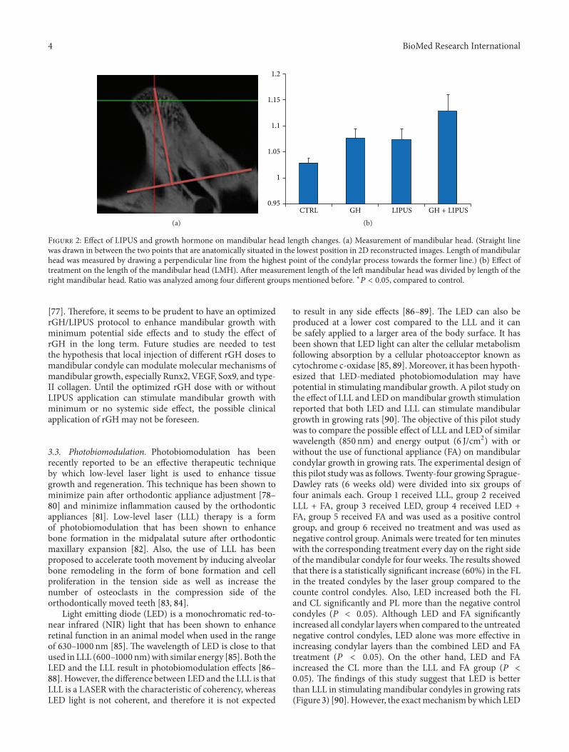
In spite of the potential side effects of GH administrationsuch as inducing body weight gain [74] and upregulation ofproto-oncogenes like C-jun in liver [75], kidney, and othervital tissues [76], there has been an attempt to enhancemandibular growth by local injection of recombinant growthhormone (rGH) into the posterior attachment of mandibularcondyle of growing rats with or without LIPUS application[77]. The hypothesized mechanism of action of local rGHapplication is to increase endochondoral bone formationin the mandibular condyles without possible side effectof systemic use of rGH. The findings indicated that localrGH injection into mandibular condyles in rats increasedmandibular growth compared to the control group.The studyconcluded that the used rGH dose does not have synergisticeffect in combination with LIPUS application in enhancingmandibular bone volume or mandibular surface area whilethe combined treatment increased mandibular head lengthcompared to either treatment alone (Figures 2(a) and 2(b)).Also, local injection of rGH increased C-jun in the liver
4 BioMed Research International
(a)
1.2
1.15
1.1
1.05
1
0.95CTRL GH LIPUS GH + LIPUS
(b)
Figure 2: Effect of LIPUS and growth hormone on mandibular head length changes. (a) Measurement of mandibular head. (Straight linewas drawn in between the two points that are anatomically situated in the lowest position in 2D reconstructed images. Length of mandibularhead was measured by drawing a perpendicular line from the highest point of the condylar process towards the former line.) (b) Effect oftreatment on the length of the mandibular head (LMH). After measurement length of the left mandibular head was divided by length of theright mandibular head. Ratio was analyzed among four different groups mentioned before. ∗𝑃 < 0.05, compared to control.
[77]. Therefore, it seems to be prudent to have an optimizedrGH/LIPUS protocol to enhance mandibular growth withminimum potential side effects and to study the effect ofrGH in the long term. Future studies are needed to testthe hypothesis that local injection of different rGH doses tomandibular condyle can modulate molecular mechanisms ofmandibular growth, especially Runx2, VEGF, Sox9, and type-II collagen. Until the optimized rGH dose with or withoutLIPUS application can stimulate mandibular growth withminimum or no systemic side effect, the possible clinicalapplication of rGH may not be foreseen.
3.3. Photobiomodulation. Photobiomodulation has beenrecently reported to be an effective therapeutic techniqueby which low-level laser light is used to enhance tissuegrowth and regeneration. This technique has been shown tominimize pain after orthodontic appliance adjustment [78–80] and minimize inflammation caused by the orthodonticappliances [81]. Low-level laser (LLL) therapy is a formof photobiomodulation that has been shown to enhancebone formation in the midpalatal suture after orthodonticmaxillary expansion [82]. Also, the use of LLL has beenproposed to accelerate tooth movement by inducing alveolarbone remodeling in the form of bone formation and cellproliferation in the tension side as well as increase thenumber of osteoclasts in the compression side of theorthodontically moved teeth [83, 84].
Light emitting diode (LED) is a monochromatic red-to-near infrared (NIR) light that has been shown to enhanceretinal function in an animal model when used in the rangeof 630–1000 nm [85]. The wavelength of LED is close to thatused in LLL (600–1000 nm)with similar energy [85]. Both theLED and the LLL result in photobiomodulation effects [86–88]. However, the difference between LED and the LLL is thatLLL is a LASER with the characteristic of coherency, whereasLED light is not coherent, and therefore it is not expected
to result in any side effects [86–89]. The LED can also beproduced at a lower cost compared to the LLL and it canbe safely applied to a larger area of the body surface. It hasbeen shown that LED light can alter the cellular metabolismfollowing absorption by a cellular photoacceptor known ascytochrome c-oxidase [85, 89]. Moreover, it has been hypoth-esized that LED-mediated photobiomodulation may havepotential in stimulating mandibular growth. A pilot study onthe effect of LLL and LED onmandibular growth stimulationreported that both LED and LLL can stimulate mandibulargrowth in growing rats [90]. The objective of this pilot studywas to compare the possible effect of LLL and LED of similarwavelength (850 nm) and energy output (6 J/cm2) with orwithout the use of functional appliance (FA) on mandibularcondylar growth in growing rats. The experimental design ofthis pilot study was as follows. Twenty-four growing Sprague-Dawley rats (6 weeks old) were divided into six groups offour animals each. Group 1 received LLL, group 2 receivedLLL + FA, group 3 received LED, group 4 received LED +FA, group 5 received FA and was used as a positive controlgroup, and group 6 received no treatment and was used asnegative control group. Animals were treated for ten minuteswith the corresponding treatment every day on the right sideof themandibular condyle for four weeks.The results showedthat there is a statistically significant increase (60%) in the FLin the treated condyles by the laser group compared to thecounte control condyles. Also, LED increased both the FLand CL significantly and PL more than the negative controlcondyles (𝑃 < 0.05). Although LED and FA significantlyincreased all condylar layers when compared to the untreatednegative control condyles, LED alone was more effective inincreasing condylar layers than the combined LED and FAtreatment (𝑃 < 0.05). On the other hand, LED and FAincreased the CL more than the LLL and FA group (𝑃 <0.05). The findings of this study suggest that LED is betterthan LLL in stimulating mandibular condyles in growing rats(Figure 3) [90]. However, the exactmechanism bywhich LED

BioMed Research International 5
050
100150200250300350400450
Lase
r(s
elf c
ntrl)
Lase
r (Tx
)
Lase
r+FA
(sel
f cnt
rl)
LED
(sel
f cnt
rl)
LED
(Tx)
LED+
FA(s
elf c
ntrl) FA
Neg
ativ
eco
ntro
l
LED+
FA(T
x)
Lase
r+FA
(Tx)
(a) Fibrous layer surface area in 𝜇m2
050
100150200250
Lase
r(s
elf c
ntrl)
Lase
r (Tx
)
Lase
r+FA
(sel
f cnt
rl)
LED
(sel
f cnt
rl)
LED
(Tx)
LED+
FA(s
elf c
ntrl) FA
Neg
ativ
eco
ntro
l
LED+
FA(T
x)
Lase
r+FA
(Tx)
(b) Proliferative layer surface area in 𝜇m2
Lase
r(s
elf c
ntrl)
Lase
r (Tx
)
Lase
r+FA
(sel
f cnt
rl)
LED
(sel
f cnt
rl)
LED
(Tx)
LED+
FA(s
elf c
ntrl) FA
Neg
ativ
eco
ntro
l
LED+
FA(T
x)
Lase
r+FA
(Tx)
020406080
100120140160
(c) Hyoertrophic layer surface area in 𝜇m2La
ser
(sel
f cnt
rl)
Lase
r (Tx
)
Lase
r+FA
(sel
f cnt
rl)
LED
(sel
f cnt
rl)
LED
(Tx)
LED+
FA(s
elf c
ntrl) FA
Neg
ativ
eco
ntro
l
LED+
FA(T
x)
Lase
r+FA
(Tx)
0
100
200
300
400
(d) Chondrocyte layer surface area in 𝜇m2
Figure 3: Surface area of mandibular condylar layers. (a) Fibrous, (b) proliferative, (c) hypertrophic, and (d) cartilaginous layers in ratstreated by LED, laser with or without functional appliance (FA). It can be seen that LED increases surface areas in (a), (b), and (d) comparedto control and almost similar to control in (c).
or LLL stimulate mandibular growth is yet to be understoodfully so that an optimum technique can be developed usingphotobiomodulation. Future studies are needed to test thehypothesis that LED treatment to mandibular condyle canmodulate molecular mechanisms of mandibular growth,especially Runx2, VEGF, Sox9, and type-II collagen.
Photobiomodulation for stimulation of mandibularcondylar growth may be close to application in the clinicbecause clinical trials using LED in patients undergoingorthodontic treatment have identified enhanced toothmovement and minimal side effects, such as root resorption.
3.4. Gene Therapy. Gene therapy involves either physical orchemical transfer of genetic material in the host cell, [82].Gene therapy involves using plasmid DNA alone (nakedDNA) [91, 92] or associated with gene carriers or vectors suchas nonviral vectors (liposomes or a polymer matrix) [93].Liposomes have proven sufficient for gene transfer into chon-drocytes, and they have several advantages over adenovirusvectors including ease of preparation, lack of limitations onthe size of the DNA, and minimum immunological reaction[94]. However, in vivo or clinical application is yet to beproven. Viral vectors carrying vascular endothelial growthfactor (rAAV-VEGF) have been shown to stimulatemandibu-lar growth in vivo in rats [95]. Yet, more research is neededto optimize the technique and detailed toxicity evaluationof viral and nonviral vectors (both local and systemic), andtesting optimized techniques in higher animals before clinicaltrials can be conducted. The hypothesis underlying localinjection of vector-loaded VEGF into mandibular condyles
is that this VEGF can modulate mandibular growth throughadded VEGF effect that has been shown to be correlated tomandibular growth stimulation [27, 28]. VEGFmay stimulatemandibular growth through two mechanisms: (1) throughstimulation of endochondral bone growth and (2) throughrecruitment of new replicatingmesenchymal stem cells whichis correlated to mandibular growth [32, 33]. It seems that theroad towards clinical application of gene therapy to enhancemandibular condylar growth is long compared to othermodalities like LIPUS or LED. Many questions remain to beanswered regarding the safety, optimization, and mechanismunderlying gene therapy, which is also more invasive andcurrently less accepted than LIPUS or LED.
4. Syndromic Mandibular Hypoplasia andTreatment Possibilities
Syndromic mandibular hypoplasia, as in hemifacial micro-somia (HFM), is distinct from symmetrical mandibularhypoplasia. HFM is a congenital anomaly that is presented byasymmetric facial structures in which themandible and over-lying structures fail to develop normally. HFM is also knownas otomandibular dysostosis, [96] first and second branchialarch syndrome, [97, 98] oculo-auriculovertebral dysplasia,[99] Goldenhar syndrome, [100, 101] lateral facial dysplasia;[102] and craniofacial microsomia [103, 104]. The prevalenceof HFMhas been previously reported as 1 in 3000 or 1 in 5600births [105–108]. Males are more affected than females [109],and it has been reported that the right side is affected morethan the left side (3 : 2 ratio) [110]. At present, the underlying

6 BioMed Research International
cause of HFM remains unknown. It has been hypothesizedthat HFM results from a developmental abnormality relatedto hemorrhage and rupture of the stapedial artery (a smallblood vessel near the ear), as supported by mouse studies[109–111]. It is to be noted that small animal models of HFMcannot be extrapolated to humans, as there is no publishedreports that indicate that hypothesized etiology of humanHFM is similar to those in lower animal models. Pruzanskyclassification is the most known HFM classification usedby clinicians and researchers [112–114]. Treatment of HFMdepends on each case severity and patient’s age. Treatment ofHFM may include orthodontic hybrid functional appliancesin less severe cases and/or surgical intervention utilizingorthognathic surgery or distraction osteogenesis [115]. Sincethe etiology of HFM is not fully understood, the use ofthe new proposed techniques may not be fully applicable tosyndromic cases like HFM.
5. Current Challenges in Available TechniquesUsed to Enhance Mandibular Growth
As noted above, although LIPUS, LED, GH, or gene therapymay be future techniques that may be used one day formandibular stimulation in patients with underdevelopedmandibles, the following challenges are foreseen for thesetechniques. While optimized LIPUS treatment is hypothe-sized to be dose (treatment-time) dependent, it is a chal-lenge to use LIPUS daily for more than 20 minutes perday to mandibular condyles, especially in growing chil-dren. From the preliminary study that showed a proof ofprinciple that local rGH injection can enhance mandibulargrowth, possible increase in rGH dose might bring a riskof systemic unwanted effect(s). While LED treatment togrowing mandibles show promising effect, the underlyingmechanisms that are involved in LED-mediated mandibulargrowth stimulation are not known; hence, possible opti-mized technique of LED application is not known or cannotbe hypothesized. Finally, it seems to be very early forproposing genetherapy for human mandibular growth dueto the following challenges. It is not known whether locallyinjected vector-loaded genes have possible systemic effect ornot. Although nonviral vectors have been investigated forpossible future use in humans, it is not known the possibleside effects of these nonviral vectors. It is also not knownthe optimum dose of each vector or vector-loaded geneconcentration that can enhance mandibular growth withoutinducing unnecessary overgrowth of the mandibles or induc-ing neoplastic growth. With these challenges, future researchmay be directed towards uncovering these mechanisms andstudying possible side effects as well as optimized techniquesin mandibular growth stimulation.
Although shown to be a clinically acceptable treat-ment modality, bite-jumping appliances (functional appli-ances (Fas)) alone may not be fully effective in stimulatingmandibular growth to the level that they can substitutesurgical repositioning of the mandible in severe mandibulardeficiency cases. LIPUS can stimulate mandibular growthin growing animals and in humans; however, an optimized
technique to shorten treatment time requires further inves-tigations in lower and higher animals before any clinicaltrials may be proposed. An optimized technique that utilizeslocal rGH administration with or without LIPUS is worthinvestigation to stimulate mandibular growth withminimumpotential side effects. Gene therapy as well as LLL or LEDseems to be promising approaches in stimulatingmandibulargrowth. However, detailed toxicity investigations of thesetechniques are required before potential clinical trials can beperformed.
Acknowledgment
This work was sponsored by King Saud University, SaudiArabia.
References
[1] C. Sunitha and S. A. Kumar, “Obstructive sleep apnea and itsmanagement,” Indian Journal of Dental Research, vol. 21, no. 1,pp. 119–124, 2010.
[2] E. T. Chang and G. M. Shiao, “Craniofacial abnormalities inChinese patients with obstructive and positional sleep apnea,”Sleep Medicine, vol. 9, no. 4, pp. 403–410, 2008.
[3] S. J. Chang and K. Y. Chae, “Obstructive sleep apnea syn-drome in children: epidemiology, pathophysiology, diagnosisand sequelae,” Korean Journal of Pediatrics, vol. 53, no. 10, pp.863–871, 2010.
[4] N. Higurashi, M. Kikuchi, S. Miyazaki, and Y. Itasaka, “Com-parison of Ricketts analysis and Downs-Northwestern analysisfor the evaluation of obstructive sleep apnea cephalograms,”Psychiatry andClinical Neurosciences, vol. 55, no. 3, pp. 259–260,2001.
[5] B. B. Vieira, C. E. Itikawa, L. A. de Almeida et al., “Cephalo-metric evaluation of facial pattern and hyoid bone position inchildren with obstructive sleep apnea syndrome,” InternationalJournal of Pediatric Otorhinolaryngology, vol. 75, no. 3, pp. 383–386, 2011.
[6] P. Lavie, P. Herer, R. Peled et al., “Mortality in sleep apneapatients: a multivariate analysis of risk factors,” Sleep, vol. 18, no.3, pp. 149–157, 1995.
[7] P. Lavie, P. Herer, and L. Lavie, “Mortality risk factors in sleepapnoea: amatched case-control study,” Journal of Sleep Research,vol. 16, no. 1, pp. 128–134, 2007.
[8] R. B. Cohen, “Obstructive sleep apnea: a mandibular position-ing device for treatment and diagnosis of an obstruction site,”Compendium of Continuing Education in Dentistry, vol. 16, no.6, pp. 618–629, 1995.
[9] M. P. Villa, E. Bernkopf, J. Pagani, V. Broia, M. Montesano,and R. Ronchetti, “Randomized controlled study of an oraljaw-positioning appliance for the treatment of obstructive sleepapnea in children with malocclusion,” American Journal ofRespiratory and Critical Care Medicine, vol. 165, no. 1, pp. 123–127, 2002.
[10] H. Huo, W. Y. Li, P. Shen, and J. H. Liu, “One night treatment ofobstructive sleep apnea and hypopnea syndromewith nasopha-ryngeal airway,” Zhonghua Er Bi Yan Hou Tou Jing Wai Ke ZaZhi, vol. 45, no. 5, pp. 382–386, 2010.
[11] W. Hochban and B. Hoch, “Obstructive sleep apnoea inchildren: an interdisciplinary approach with special regard to

BioMed Research International 7
craniofacial disorders,” Pneumologie, vol. 52, no. 3, pp. 147–153,1998.
[12] R. M. Corso, E. Piraccini, M. Calli et al., “Obstructive sleepapnea is a risk factor for difficult endotracheal intubation,”Minerva Anestesiologica, vol. 77, no. 1, pp. 99–100, 2011.
[13] G. Freed, M. A. Pearlman, A. S. Brown, and L. R. Barot,“Polysomnographic indications for surgical intervention inPierre Robin sequence: acute airway management and follow-up studies after repair and take-down of tongue-lip adhesion,”Cleft Palate Journal, vol. 25, no. 2, pp. 151–155, 1988.
[14] K. A. Waters, F. M. Everett, J. W. Bruderer, and C. E. Sullivan,“Obstructive sleep apnea: the use of nasal CPAP in 80 children,”American Journal of Respiratory and Critical Care Medicine, vol.152, no. 2, pp. 780–785, 1995.
[15] B. Kremer, A. I. Botos-Kremer, H. E. Eckel, and G. Schlondorff,“Indications, complications, and surgical techniques for pedi-atric tracheostomies—an update,” Journal of Pediatric Surgery,vol. 37, no. 11, pp. 1556–1562, 2002.
[16] M. Miloro, “Mandibular distraction osteogenesis for pediatricairway management,” Journal of Oral andMaxillofacial Surgery,vol. 68, no. 7, pp. 1512–1523, 2010.
[17] A. Denny and B. Kalantarian, “Mandibular distraction inneonates: a strategy to avoid tracheostomy,” Plastic and Recon-structive Surgery, vol. 109, no. 3, pp. 896–904, 2002.
[18] C. Tison, S. Sebille-Elhage, and J. Ferri, “Mandibular advance-ment device: a 5-year long experience in obstructive sleepapnea/hypopnea syndrome,” Revue de Stomatologie et deChirurgie Maxillo-Faciale, vol. 112, no. 2, pp. 80–86, 2011.
[19] B. Lam, K. Sam, J. C. M. Lam, A. Y. K. Lai, C. L. Lam, and M. S.M. Ip, “The efficacy of oral appliances in the treatment of severeobstructive sleep apnea,” Sleep and Breathing, vol. 15, no. 2, pp.195–201, 2011.
[20] T. Sobue, W. C. Yeh, A. Chhibber et al., “Murine TMJ loadingcauses increased proliferation and chondrocyte maturation,”Journal of Dental Research, vol. 90, no. 4, pp. 512–516, 2011.
[21] A. B. Rabie andU.Hagg, “Factors regulatingmandibular condy-lar growth,” American Journal of Orthodontics and DentofacialOrthopedics, vol. 122, pp. 401–409, 2002.
[22] R. Oyonarte, M. Zarate, and F. Rodriguez, “Low-intensitypulsed ultrasound stimulation of condylar growth in rats,”AngleOrthodontist, vol. 79, no. 5, pp. 964–970, 2009.
[23] T. El-Bialy, I. El-Shamy, and T. M. Graber, “Growth modifi-cation of the rabbit mandible using therapeutic ultrasound:is it possible to enhance functional appliance results?” AngleOrthodontist, vol. 73, no. 6, pp. 631–639, 2003.
[24] T. Peltomaki, S. Kylamarkula, H. Vinkka-Puhakka, M. Rintala,T. Kantomaa, and O. Ronning, “Tissue-separating capacity ofgrowth cartilages,” European Journal of Orthodontics, vol. 19, no.5, pp. 473–481, 1997.
[25] G. Shen and M. A. Darendeliler, “Cephalometric evaluationof condylar and mandibular growth modification: a review,”Orthodontics&Craniofacial Research, vol. 9, no. 1, pp. 2–9, 2006.
[26] G. Shen, A. B. Rabie, U. Hagg, and R. J. Chen, “Neovas-cularization in condylar cartilage in response to mandibularprotrusion,” Chinese Journal of Dental Research, vol. 6, pp. 28–38, 2003.
[27] A. B. M. Rabie, L. Shum, and A. Chayanupatkul, “VEGF andbone formation in the glenoid fossa during forwardmandibularpositioning,” American Journal of Orthodontics and DentofacialOrthopedics, vol. 122, no. 2, pp. 202–209, 2002.
[28] A. B. M. Rabie, F. Y. C. Leung, A. Chayanupatkul, and U. Hagg,“The correlation between neovascularization and bone forma-tion in the condyle during forward mandibular positioning,”Angle Orthodontist, vol. 72, no. 5, pp. 431–438, 2002.
[29] A. Sidlauskas, “Clinical effectiveness of the Twin block appli-ance in the treatment of Class II Division 1 malocclusion,”Stomatologija, vol. 7, no. 1, pp. 7–10, 2005.
[30] X. Du, U. Hagg, and A. B. M. Rabie, “Effects of headgear Herbstandmandibular step-by-step advancement versus conventionalHerbst appliance and maximal jumping of the mandible,”European Journal of Orthodontics, vol. 24, no. 2, pp. 167–174,2002.
[31] M. Bendeus, U. Hagg, and B. Rabie, “Growth and treatmentchanges in patients treatedwith a headgear-activator appliance,”American Journal of Orthodontics and Dentofacial Orthopedics,vol. 121, no. 4, pp. 376–384, 2002.
[32] A. B. M. Rabie, L. Wong, and M. Tsai, “Replicating mesenchy-mal cells in the condyle and the glenoid fossa during mandibu-lar forward positioning,” American Journal of Orthodontics andDentofacial Orthopedics, vol. 123, no. 1, pp. 49–57, 2003.
[33] A. B. M. Rabie, L. Wong, and U. Hagg, “Correlation of replicat-ing cells and osteogenesis in the glenoid fossa during stepwiseadvancement,” American Journal of Orthodontics and Dentofa-cial Orthopedics, vol. 123, no. 5, pp. 521–526, 2003.
[34] G. H. Tang and A. B. M. Rabie, “Runx2 regulates endochondralossification in condyle during mandibular advancement,” Jour-nal of Dental Research, vol. 84, no. 2, pp. 166–171, 2005.
[35] A. B.M. Rabie, T. T. She, andV. R. Harley, “Forwardmandibularpositioning up-regulates SOX9 and type II collagen expressionin the glenoid fossa,” Journal of Dental Research, vol. 82, no. 9,pp. 725–730, 2003.
[36] J. D. Heckman, J. P. Ryaby, J. McCabe, J. J. Frey, and R.F. Kilcoyne, “Acceleration of tibial fracture-healing by non-invasive, low-intensity pulsed ultrasound,” Journal of Bone andJoint Surgery. American, vol. 76, no. 1, pp. 26–34, 1994.
[37] T. K. Kristiansen, J. P. Ryaby, J. McCabe, J. J. Frey, and L. R.Roe, “Accelerated healing of distal radial fractures with the useof specific, low-intensity ultrasound: a multicenter, prospective,randomized, double- blind, placebo-controlled study,” Journalof Bone and Joint Surgery. American, vol. 79, no. 7, pp. 961–973,1997.
[38] A. Shimazaki, K. Inui, Y. Azuma, N. Nishimura, and Y. Yamano,“Low-intensity pulsed ultrasound accelerates bone maturationin distraction osteogenesis in rabbits,” Journal of Bone and JointSurgery. British, vol. 82, no. 7, pp. 1077–1082, 2000.
[39] E. Mayr, A. Laule, G. Suger, A. Ruter, and L. Claes, “Radio-graphic results of callus distraction aided by pulsed low-intensity ultrasound,” Journal of Orthopaedic Trauma, vol. 15,no. 6, pp. 407–414, 2001.
[40] T. El-Bialy, A. Hassan, T. Albaghdadi, H. A. Fouad, andA. R. Maimani, “Growth modification of the mandible withultrasound in baboons: a preliminary report,”American Journalof Orthodontics and Dentofacial Orthopedics, vol. 130, no. 4, pp.e7–e14, 2006.
[41] T. El-Bialy, A. Hasan, A. Alyamani, and T. Albaghdadi, “Treat-ment of hemifacial microsomia by therapeutic ultrasound andhybrid functional appliance. A non-surgical approach,” OpenAccess Journal of Clinical Trials, vol. 2, pp. 29–36, 2010.
[42] D. Schumann, R. Kujat, J. Zellner et al., “Treatment of humanmesenchymal stem cells with pulsed low intensity ultrasoundenhances the chondrogenic phenotype in vitro,” Biorheology,vol. 43, no. 3-4, pp. 431–443, 2006.
8 BioMed Research International
[43] T. El-Bialy, T. J. Royston, R. L. Magin, C. A. Evans, A. E. M.Zaki, and L. A. Frizzell, “The effect of pulsed ultrasound onmandibular distraction,” Annals of Biomedical Engineering, vol.30, no. 10, pp. 1251–1261, 2002.
[44] C. W. Chan, L. Qin, K. M. Lee, W. H. Cheung, J. C. Y. Cheng,and K. S. Leung, “Dose-dependent effect of low-intensitypulsed ultrasound on callus formation during rapid distractionosteogenesis,” Journal of Orthopaedic Research, vol. 24, no. 11,pp. 2072–2079, 2006.
[45] S. R. Young andM.Dyson, “The effect of therapeutic ultrasoundon angiogenesis,” Ultrasound in Medicine and Biology, vol. 16,no. 3, pp. 261–269, 1990.
[46] J. Dai and A. B. M. Rabie, “VEGF: an essential mediator of bothangiogenesis and endochondral ossification,” Journal of DentalResearch, vol. 86, no. 10, pp. 937–950, 2007.
[47] W. T. Ang, C. Scurtescu, W. Hoy, T. El-Bialy, Y. Y. Tsui, and J.Chen, “Design and implementation of therapeutic ultrasoundgenerating circuit for dental tissue formation and tooth-roothealing,” IEEE Transactions on Biomedical Circuits and Systems,vol. 4, no. 1, pp. 49–61, 2010.
[48] T. A. Aldosary, H. Uludag, M. Doschak, J. Chen, Y. Tsui,and T. EL-Bialy, “Effect of ultrasound on human umbilicalcord perivascular-stem cell expansion,” in Proceedings of theInternational Association for Dental Research (IADR) Meeting,Toronto, Canada, July 2008, Abstract no. 873.
[49] S. Marvel, S. Okrasinski, S. H. Bernacki, E. Loboa, and P. A.Dayton, “The development and validation of a lipus systemwithpreliminary observations of ultrasonic effects on human adultstem cells,” IEEE Transactions on Ultrasonics, Ferroelectrics, andFrequency Control, vol. 57, no. 9, pp. 1977–1984, 2010.
[50] A. Suzuki, T. Takayama, N. Suzuki, M. Sato, T. Fukuda,and K. Ito, “Daily low-intensity pulsed ultrasound-mediatedosteogenic differentiation in rat osteoblasts,” Acta Biochimica etBiophysica Sinica, vol. 41, no. 2, pp. 108–115, 2009.
[51] K. Nishizawa, S. Imai, T. Mimura et al., “In-advance trans-medullary stimulation of bone marrow enhances spontaneousrepair of full-thickness articular cartilage defects in rabbits,”Celland Tissue Research, vol. 341, no. 3, pp. 371–379, 2010.
[52] T. Jiang, T. Xu, F. Gu, A. Chen, Z. Xiao, and D. Zhang,“Osteogenic effect of low intensity pulsed ultrasound on ratadipose-derived stem cells in vitro,” Journal of Huazhong Uni-versity of Science and Technology. Medical sciences, vol. 32, no. 1,pp. 75–81, 2012.
[53] J. J. Leskinen, H. M. Karjalainen, A. Olkku, K. Hynynen,A. Mahonen, and M. J. Lammi, “Genome-wide microarrayanalysis of MG-63 osteoblastic cells exposed to ultrasound,”Biorheology, vol. 45, no. 3-4, pp. 345–354, 2008.
[54] F. Las Heras, H. K. Gahunia, and K. P. Pritzker, “Articularcartilage development: a molecular perspective,” OrthopedicClinics of North America, vol. 43, no. 2, pp. 155–171, 2012.
[55] N. Z. Mostafa, H. Uludag, D. N. Dederich, M. R. Doschak, andT. El-Bialy, “Anabolic effects of low-intensity pulsed ultrasoundon human gingival fibroblasts,”Archives of Oral Biology, vol. 54,no. 8, pp. 743–748, 2009.
[56] T. El-Bialy, H. Uludag, N. Jomha, and S. F. Badylak, “In vivoultrasound-assisted tissue-engineered mandibular condyle: apilot study in rabbits,” Tissue Engineering, Part C, vol. 16, no.6, pp. 1315–1323, 2010.
[57] S. Zhou, A. Schmelz, T. Seufferlein, Y. Li, J. Zhao, and M.G. Bachem, “Molecular mechanisms of low intensity pulsedultrasound in human skin fibroblasts,” Journal of BiologicalChemistry, vol. 279, no. 52, pp. 54463–54469, 2004.
[58] T. Matsumoto, G. M. Cooper, B. Gharaibeh et al., “Cartilagerepair in a rat model of osteoarthritis through intraarticulartransplantation of muscle-derived stem cells expressing bonemorphogenetic protein 4 and soluble Flt-1,” Arthritis andRheumatism, vol. 60, no. 5, pp. 1390–1405, 2009.
[59] M. Pei, F. He, B. M. Boyce, and V. L. Kish, “Repair of full-thickness femoral condyle cartilage defects using allogeneicsynovial cell-engineered tissue constructs,” Osteoarthritis andCartilage, vol. 17, no. 6, pp. 714–722, 2009.
[60] B. Grigolo, G. Lisignoli, G. Desando et al., “Osteoarthritistreated with mesenchymal stem cells on Hyaluronan-basedscaffold in rabbit,” Tissue Engineering, Part C, vol. 15, no. 4, pp.647–658, 2009.
[61] F. S. Toghraie, N. Chenari, M. A. Gholipour et al., “Treatmentof osteoarthritis with infrapatellar fat pad derivedmesenchymalstem cells in Rabbit,” Knee, vol. 18, no. 2, pp. 71–75, 2011.
[62] J. M. Murphy, D. J. Fink, E. B. Hunziker, and F. P. Barry, “Stemcell therapy in a caprine model of osteoarthritis,” Arthritis andRheumatism, vol. 48, no. 12, pp. 3464–3474, 2003.
[63] C. Hamou, M. J. Callaghan, H. Thangarajah et al., “Mesenchy-mal stem cells can participate in ischemic neovascularization,”Plastic and Reconstructive Surgery, vol. 123, no. 2, supplement,pp. 45S–55S, 2009.
[64] K. Chen, C. Man, B. Zhang, J. Hu, and S. S. Zhu, “Effect of invitro chondrogenic differentiation of autologous mesenchymalstem cells on cartilage and subchondral cancellous bone repairin osteoarthritis of temporomandibular joint,” InternationalJournal of Oral andMaxillofacial Surgery, vol. 42, no. 2, pp. 240–248, 2012.
[65] C. Ohlsson, B. A. Bengtsson, O. G. P. Isaksson, T. T. Andreassen,and M. C. Slootweg, “Growth hormone and bone,” EndocrineReviews, vol. 19, no. 1, pp. 55–79, 1998.
[66] A. Giustina, G. Mazziotti, and E. Canalis, “Growth hor-mone, insulin-like growth factors, and the skeleton,” EndocrineReviews, vol. 29, no. 5, pp. 535–559, 2008.
[67] O. G. P. Isaksson, J. O. Jansson, and I. A. M. Gause, “Growthhormone stimulates longitudinal bone growth directly,” Science,vol. 216, no. 4551, pp. 1237–1239, 1982.
[68] E. Hedner, A. Linde, and A. Nilsson, “Systemically and locallyadministered growth hormone stimulates bone healing in com-bination with osteopromotive membranes: an experimentalstudy in rats,” Journal of Bone and Mineral Research, vol. 11, no.12, pp. 1952–1960, 1996.
[69] G. Johannsson, T. Rosen, I. Bosaeus, L. Sjostrom, and B. A.Bengtsson, “Two years of growth hormone (GH) treatmentincreases bone mineral content and density in hypopituitarypatients with adult-onset GH deficiency,” Journal of ClinicalEndocrinology and Metabolism, vol. 81, no. 8, pp. 2865–2873,1996.
[70] A. M. Parfitt, “Growth hormone and adult bone remodeling,”Clinical Endocrinology, vol. 35, pp. 467–470, 1991.
[71] V. Visnapuu, T. Peltomaki, O. Ronning, T. Vahlberg, and H.Helenius, “Growth hormone and insulin-like growth factor Ireceptors in the temporomandibular joint of the rat,” Journal ofDental Research, vol. 80, no. 10, pp. 1903–1907, 2001.
[72] R. van Erum, M. Mulier, C. Cards, G. Verbeke, and F. deZegher, “Craniofacial growth in short children born small forgestational age: effect of growth hormone treatment,” Journal ofDental Research, vol. 76, no. 9, pp. 1579–1586, 1997.
[73] C. M. Forsberg, L. Krekmanova, and G. Dahllof, “The effectof growth hormone therapy on mandibular and cranial base

BioMed Research International 9
development in children treated with total body irradiation,”European Journal of Orthodontics, vol. 24, no. 3, pp. 285–292,2002.
[74] G. M. Farris, G. K. Miller, G. K. Wollenberg, S. Molon-Noblot,C. Chan, and S. Prahalada, “Recombinant rat andmouse growthhormones: risk assessment of carcinogenic potential in 2-yearbioassays in rats and mice,” Toxicological Sciences, vol. 97, no. 2,pp. 548–561, 2007.
[75] Y. Murakami, M. Satake, Y. Yamaguchi-Iwai, M. Sakai, M.Muramatsu, and Y. Ito, “The nuclear protooncogenes c-junand c-fos as regulators of DNA replication,” Proceedings of theNational Academy of Sciences of the United States of America,vol. 88, no. 9, pp. 3947–3951, 1991.
[76] P. Rotwein, A. M. Granowski, andM. J.Thomas, “Rapid nuclearactions of growth hormone,”Hormone Research, vol. 42, no. 4-5,pp. 170–175, 1994.
[77] I. Khan,A. EL-Kadi, andT. El-Bialy, “Effects of growth hormoneand ultrasound on mandibular growth in rats: microCT andtoxicity analyses,” Archives of Oral Biology. In press.
[78] H. M. Lim, K. K. K. Lew, and D. K. L. Tay, “A clinicalinvestigation of the efficacy of low level laser therapy inreducing orthodontic postadjustment pain,” American Journalof Orthodontics and Dentofacial Orthopedics, vol. 108, no. 6, pp.614–622, 1995.
[79] D. Turhani, M. Scheriau, D. Kapral, T. Benesch, E. Jonke, andH. P. Bantleon, “Pain relief by single low-level laser irradiationin orthodontic patients undergoing fixed appliance therapy,”American Journal of Orthodontics and Dentofacial Orthopedics,vol. 130, no. 3, pp. 371–377, 2006.
[80] K. Fujiyama, T. Deguchi, T. Murakami, A. Fujii, K. Kushima,and T. Takano-Yamamoto, “Clinical effect of CO
2laser in
reducing pain in orthodontics,” Angle Orthodontist, vol. 78, no.2, pp. 299–303, 2008.
[81] M. T. J. Rodrigues, M. S. Ribeiro, E. B. Grtoth, and D. M.Zezell, “Evaluation of effects of laser therapy on oral ulcerationinduced by fixed orthodontic appliances,” Lasers in Surgery andMedicine, vol. 30, supplement 14, p. 15, 2002.
[82] S. Saito and N. Shimizu, “Stimulatory effects of low-power laserirradiation on bone regeneration in midpalatal suture duringexpansion in the rat,” American Journal of Orthodontics andDentofacial Orthopedics, vol. 111, no. 5, pp. 525–532, 1997.
[83] K. Kawasaki and N. Shimizu, “Effects of low-energy laserirradiation on bone remodelling during experimental toothmovement in rats,” Lasers in Surgery and Medicine, vol. 26, pp.282–291, 2000.
[84] X. Sun, X. Zhu, C. Xu, N. Ye, and H. Zhu, “Effects of low energylaser on tooth movement and remodeling of alveolar bone inrabbits,”Hua Xi KouQiang Yi Xue Za Zhi, vol. 19, no. 5, pp. 290–293, 2001.
[85] T. I. Karu, L. V. Pyatibrat, S. F. Kolyakov, and N. I. Afanasyeva,“Absorption measurements of a cell monolayer relevant tophototherapy: reduction of cytochrome c oxidase under near IRradiation,” Journal of Photochemistry and Photobiology B, vol. 81,no. 2, pp. 98–106, 2005.
[86] M. Khadra, H. J. Rønold, S. P. Lyngstadaas, J. E. Ellingsen,and H. R. Haanæs, “Low-level laser therapy stimulates bone-implant interaction: an experimental study in rabbits,” ClinicalOral Implants Research, vol. 15, no. 3, pp. 325–332, 2004.
[87] J. T. Eells, M. T. T. Wong-Riley, J. VerHoeve et al., “Mito-chondrial signal transduction in accelerated wound and retinalhealing by near-infrared light therapy,” Mitochondrion, vol. 4,no. 5-6, pp. 559–567, 2004.
[88] H. T. Whelan, R. L. Smits Jr., E. V. Buchman et al., “Effectof NASA light-emitting diode irradiation on wound healing,”Journal of Clinical Laser Medicine and Surgery, vol. 19, no. 6, pp.305–314, 2001.
[89] M. T. T. Wong-Riley, H. L. Liang, J. T. Eells et al., “Photo-biomodulation directly benefits primary neurons functionallyinactivated by toxins: role of cytochrome c oxidase,” Journal ofBiological Chemistry, vol. 280, no. 6, pp. 4761–4771, 2005.
[90] A. Ebrahim, J. Yeung, A. Habib, T. EL-Bialy, and J. M. AL-Qahtani, “Histomorphometric analysis: effect of laser and LEDonmandibular growth,” in Proceedings of the Annual Meeting &Exhibition of the AADR, Tampa, Fla, USA, March 2012, Poster #954.
[91] J. A.Wolff, R.W.Malone, P.Williams et al., “Direct gene transferintomousemuscle in vivo,” Science, vol. 247, no. 4949, pp. 1465–1468, 1990.
[92] H. Herweijer and J. A. Wolff, “Progress and prospects: nakedDNA gene transfer and therapy,” Gene Therapy, vol. 10, no. 6,pp. 453–458, 2003.
[93] T. Niidome and L. Huang, “Gene therapy progress andprospects: nonviral vectors,” Gene Therapy, vol. 9, no. 24, pp.1647–1652, 2002.
[94] J. Park, J. Ries, K. Gelse et al., “Bone regeneration in critical sizedefects by cell-mediated BMP-2 gene transfer: a comparison ofadenoviral vectors and liposomes,”GeneTherapy, vol. 10, no. 13,pp. 1089–1098, 2003.
[95] J. Dai and A. B. M. Rabie, “Gene therapy to enhance condylargrowth using rAAV-VEGF,” Angle Orthodontist, vol. 78, no. 1,pp. 89–94, 2008.
[96] J. J. Francois and L. Haustrate, “Anomalies colobomateusesdu globe oculaire et syndrome du premier arc,” Annalesd’Oculistique, vol. 187, pp. 340–368, 1954.
[97] R. B. Stark and D. E. Saunders, “The first branchial syndrome.The oral-mandibular-auricular syndrome,” Plastic and Recon-structive Surgery, vol. 29, pp. 229–239, 1962.
[98] W. C. Grabb, “The first and second branchial arch syndrome,”Plastic and Reconstructive Surgery, vol. 36, no. 5, pp. 485–508,1965.
[99] R. J. Gorlin, K. L. Jue, U. Jacobsen, and E. Goldschmidt,“Oculoauriculovertebral dysplasia,” The Journal of Pediatrics,vol. 63, no. 5, pp. 991–999, 1963.
[100] M.Goldenhar, “Associationmalformatives de l’oeil et de l’oreille,en particulier le syndrome dermoide epibulbaire-appendicesauriculaires-fistula auris congenita et ses relations avec ladysotose mandibulo-faciale,” Journal of Human Genetics, vol. 1,pp. 243–282, 1952.
[101] R. J. Gorlin, J. J. Pindborg, andM.M.Cohen Jr., Syndromes of theHead andNeck,McGraw-Hill, NewYork, NY,USA, 2nd edition,1976.
[102] R. B. Ross, “Lateral facial dysplasia. (First and second branchialarch syndrome, hemifacialmicrosomia),”BirthDefects: OriginalArticle Series, vol. 11, no. 7, pp. 51–59, 1975.
[103] J. M. Converse, P. J. Coccardo, M. H. Becker, and D. Wood-Smith, “Clinical aspects of craniofacial microsomia,” in Sym-posium on Diagnosis and Treatment of Craniofacial Anomalies,J. M. Converse, J. G. McCarthy, and D. Wood-Smith, Eds., pp.461–475, CV Mosby, St. Louis, Mo, USA, 1979.
[104] J. E. Horgan, B. L. Padwa, R. A. LaBrie, and J. B. Mul-liken, “OMENS-Plus: analysis of craniofacial and extracranio-facial anomalies in hemifacial microsomia,” The Cleft Palate-Craniofacial Journal, vol. 32, no. 5, pp. 405–412, 1995.
10 BioMed Research International
[105] M. M. Cohen Jr., “Perspectives on craniofacial asymmetry. I.The biology of asymmetry,” International Journal of Oral andMaxillofacial Surgery, vol. 24, no. 1, part 1, pp. 2–7, 1995.
[106] M. M. Cohen Jr., “Perspectives on craniofacial asymmetry. II.Asymmetric embryopathies,” International Journal of Oral andMaxillofacial Surgery, vol. 24, no. 1, part 1, pp. 8–12, 1995.
[107] M. M. Cohen Jr., “Perspectives on craniofacial asymmetry. III.Common and/or well-known causes of asymmetry,” Interna-tional Journal of Oral and Maxillofacial Surgery, vol. 24, no. 2,pp. 127–133, 1995.
[108] M. M. Cohen Jr., “Perspectives on craniofacial asymmetry. IV.Hemi-asymmetries,” International Journal of Oral and Maxillo-facial Surgery, vol. 24, no. 2, pp. 134–141, 1995.
[109] R. R. J. Cousley and D. J. Wilson, “Hemifacial microsomia:developmental consequence of perturbation of the auriculofa-cial cartilagemodel?”American Journal ofMedical Genetics, vol.42, no. 4, pp. 461–466, 1992.
[110] R. R. Wang and C. J. Andres, “Hemifacial microsomia andtreatment options for auricular replacement: a review of theliterature,”The Journal of Prosthetic Dentistry, vol. 82, no. 2, pp.197–204, 1999.
[111] L. K. Robinson, H. E. Hoyme, D. K. Edwards, and K. L.Jones, “Vascular pathogenesis of unilateral craniofacial defects,”Journal of Pediatrics, vol. 111, no. 2, pp. 236–239, 1987.
[112] S. Pruzansky, “Not all dwarfed mandibles are alike,” BirthDefects: Original Article Series, vol. 5, pp. 120–129, 1969.
[113] L. T. Murray and J. E. Murray, “Asymmetries of the lower partof the face,” in Symposium on Reconstruction of Jaw Deformities,L. A. Whitaker and P. Randall, Eds., pp. 171–196, CV Mosby, St.Louis, Mo, USA, 1978.
[114] L. B. Kaban, J. B. Mulliken, and J. E. Murray, “Three-dimensional approach to analysis and treatment of hemifacialmicrosomia,” Cleft Palate Journal, vol. 18, no. 2, pp. 90–99, 1981.
[115] C. Moulin-Romsee, A. Verdonck, J. Schoenaers, and C. Carels,“Treatment of hemifacial microsomia in a growing child: theimportance of co-operation between the orthodontist and themaxillofacial surgeon,” Journal of Orthodontics, vol. 31, no. 3, pp.190–200, 2004.