working with molecular genetics chpt. 3: isolating …ross/workmg/isolat_analyz_genes_chpt3.pdf ·...
TRANSCRIPT
Working with Molecular Genetics Chpt. 3: Isolating and Analyzing Genes
CHAPTER 3ISOLATING AND ANALYZING GENES
Recombinant DNA, Polymerase Chain Reaction and Applications to Eukaryotic GeneStructure and Function
The first two chapters covered many important aspects of genes, such as how they function ininheritance, how they code for protein (in general terms) and their chemical nature. All this waslearned without having a single gene purified. A full understanding of a gene, or the entire set ofgenes in a genome, requires that they be isolated and then studied intensively. Once a gene is “inhand”, in principal one can determine both its biochemical structures and its function(s) in anorganism. One of the goals of biochemistry and molecular genetics is to assign particular functionsto individual or composite structures. This chapter covers some of the techniques commonly usedto isolate genes and illustrates some of the analyses that can be done on isolated genes.
Methods to purify some abundant proteins were developed early in the 20th century, andsome of the experiments on the fine structure of the gene (colinearity of gene and protein for trpAand tryptophan synthase) used microbial genetics and proteins sequencing. However, methods toisolate genes were not developed until the 1960’s, and the were applicable to only a few genes.
All this changed in the late 1970’s with the development of recombinant DNA technology, ormolecular cloning. This technique enabled researchers to isolate any gene from any organism fromwhich one could isolate intact DNA (or RNA). The full potential to provide access to all genes oforganisms is now being realized as full genomes are sequenced. One of the by-products of theintense investigation of individual DNA molecules after the advent of recombinant DNA was aprocedure to isolate any DNA for which one knows the sequence. This technique, called thepolymerase chain reaction (PCR), is far easier than traditional molecular cloning methods, and it hasbecome a staple of many laboratories in the life sciences. After covering the basic techniques inrecombinant DNA technology and PCR, their application to studies of eukaryotic gene structureand function will be discussed.
Like many advances in molecular genetics, recombinant DNA technology has its roots inbacterial genetics.
Transducing phage
The first genes isolated were bacterial genes that could be picked up by bacteriophage. Byisolating these hybrid bacteriophage, the DNA for the bacterial gene could be recovered in a highlyenriched form. This is the basic principal behind recombinant DNA technology.
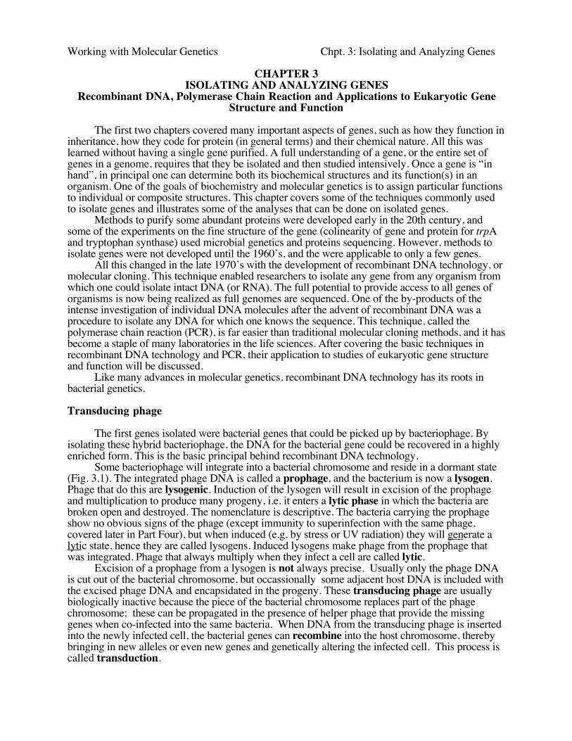
Some bacteriophage will integrate into a bacterial chromosome and reside in a dormant state(Fig. 3.1). The integrated phage DNA is called a prophage, and the bacterium is now a lysogen.Phage that do this are lysogenic. Induction of the lysogen will result in excision of the prophageand multiplication to produce many progeny, i.e. it enters a lytic phase in which the bacteria arebroken open and destroyed. The nomenclature is descriptive. The bacteria carrying the prophageshow no obvious signs of the phage (except immunity to superinfection with the same phage,covered later in Part Four), but when induced (e.g. by stress or UV radiation) they will generate alytic state, hence they are called lysogens. Induced lysogens make phage from the prophage thatwas integrated. Phage that always multiply when they infect a cell are called lytic.
Excision of a prophage from a lysogen is not always precise. Usually only the phage DNAis cut out of the bacterial chromosome, but occassionally some adjacent host DNA is included withthe excised phage DNA and encapsidated in the progeny. These transducing phage are usuallybiologically inactive because the piece of the bacterial chromosome replaces part of the phagechromosome; these can be propagated in the presence of helper phage that provide the missinggenes when co-infected into the same bacteria. When DNA from the transducing phage is insertedinto the newly infected cell, the bacterial genes can recombine into the host chromosome, therebybringing in new alleles or even new genes and genetically altering the infected cell. This process iscalled transduction.
Working with Molecular Genetics Chpt. 3: Isolating and Analyzing Genes
Figure 3.1. Transfer of bacterial genes by transduction: A lac+ transducing phage can convert alac- strain to lac+ by infection (and subsequent crossing over).
Note that the transducing phage are carrying one or a small number of bacterial genes. This isa way of isolating the genes. The bacterial gene in the transducing phage has been separated fromthe other 4000 bacterial genes (in E. coli). By isolating large numbers of the transducing phage, thephage DNA, including the bacterial genes, can be obtained in large quantities for biochemicalinvestigation. One can isolate µg or mg quantities of a single DNA molecule, which allows forprecise structural determination and detailed investigation.
A generalized transducing phage can integrate at many different locations on the bacterialchromosome. Imprecise excision from any of those locations generates a particular transducingphage, carrying a short sections of the bacterial genome adjacent to the integration site. Thus ageneralized transducing phage such as P1 can pick up many different parts of the E. coli genome.
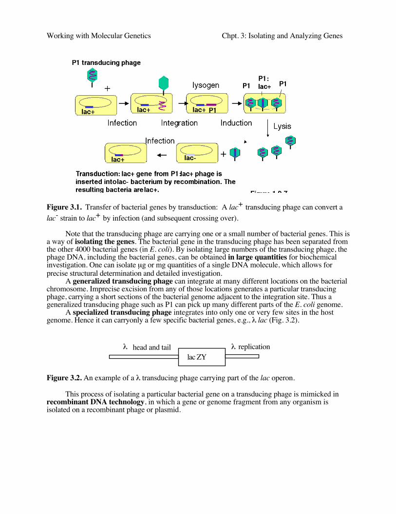
A specialized transducing phage integrates into only one or very few sites in the hostgenome. Hence it can carryonly a few specific bacterial genes, e.g., λ lac (Fig. 3.2).
lac ZYλ replicationλ head and tail
Figure 3.2. An example of a λ transducing phage carrying part of the lac operon.
This process of isolating a particular bacterial gene on a transducing phage is mimicked inrecombinant DNA technology, in which a gene or genome fragment from any organism isisolated on a recombinant phage or plasmid.
Working with Molecular Genetics Chpt. 3: Isolating and Analyzing Genes
Overview of Recombinant DNA Technology
Recombinant DNA technology utilizes the power of microbiological selection andscreening procedures to allow investigators to isolate a gene that represents as little as 1 part in amillion of the genetic material in an organism. The DNA from the organism of interest is dividedinto small pieces that are then placed into individual cells (usually bacterial). These can then beseparated as individual colonies on plates, and they can be screened through rapidly to find the geneof interest. This process is called molecular cloning.
Joining DNA in vitro to form recombinant molecules
Restriction endonucleases cut at defined sequences of (usually) 4 or 6 bp. This allows theDNA of interest to be cut at specific locations. The physiological function of restrictionendonucleases is to serve as part of system to protect bacteria from invasion by viruses or otherorganisms. (See Chapter 7)
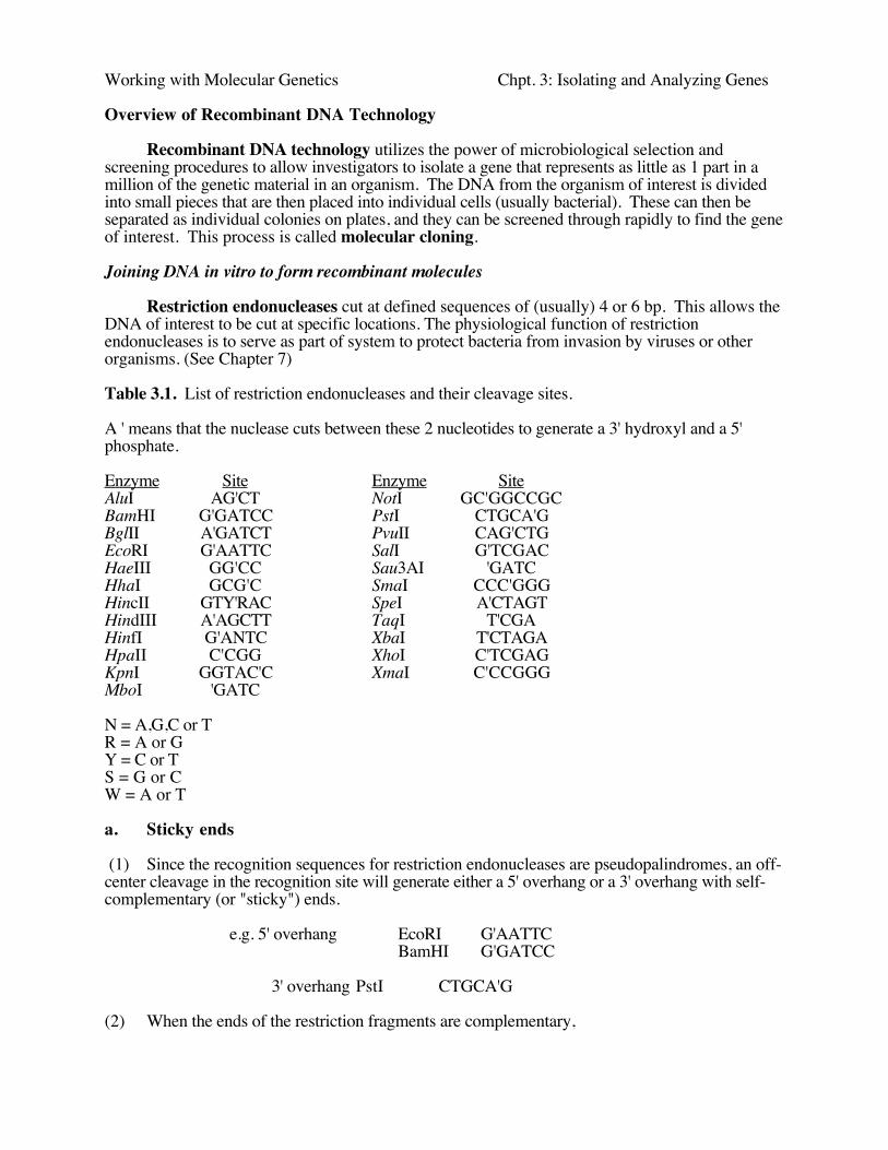
Table 3.1. List of restriction endonucleases and their cleavage sites.
A ' means that the nuclease cuts between these 2 nucleotides to generate a 3' hydroxyl and a 5'phosphate.
Enzyme Site Enzyme SiteAluI AG'CT NotI GC'GGCCGCBamHI G'GATCC PstI CTGCA'GBglII A'GATCT PvuII CAG'CTGEcoRI G'AATTC SalI G'TCGACHaeIII GG'CC Sau3AI 'GATCHhaI GCG'C SmaI CCC'GGGHincII GTY'RAC SpeI A'CTAGTHindIII A'AGCTT TaqI T'CGAHinfI G'ANTC XbaI T'CTAGAHpaII C'CGG XhoI C'TCGAGKpnI GGTAC'C XmaI C'CCGGGMboI 'GATC
N = A,G,C or TR = A or GY = C or TS = G or CW = A or T
a. Sticky ends
(1) Since the recognition sequences for restriction endonucleases are pseudopalindromes, an off-center cleavage in the recognition site will generate either a 5' overhang or a 3' overhang with self-complementary (or "sticky") ends.
e.g. 5' overhang EcoRI G'AATTCBamHI G'GATCC
3' overhang PstI CTGCA'G
(2) When the ends of the restriction fragments are complementary,
Working with Molecular Genetics Chpt. 3: Isolating and Analyzing Genes
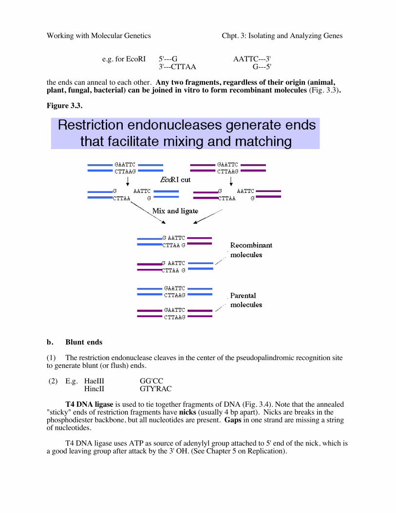
e.g. for EcoRI 5'---G AATTC---3'3'---CTTAA G---5'
the ends can anneal to each other. Any two fragments, regardless of their origin (animal,plant, fungal, bacterial) can be joined in vitro to form recombinant molecules (Fig. 3.3).
Figure 3.3.
b. Blunt ends
(1) The restriction endonuclease cleaves in the center of the pseudopalindromic recognition siteto generate blunt (or flush) ends.
(2) E.g. HaeIII GG'CCHincII GTY'RAC
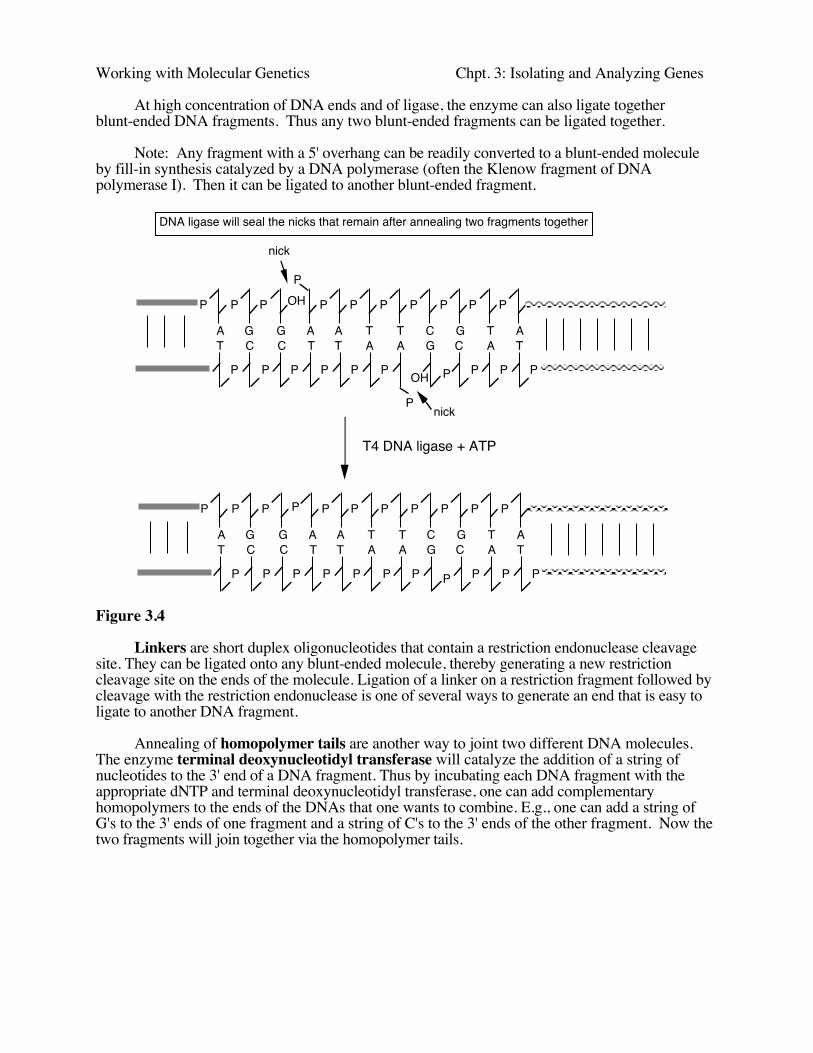
T4 DNA ligase is used to tie together fragments of DNA (Fig. 3.4). Note that the annealed"sticky" ends of restriction fragments have nicks (usually 4 bp apart). Nicks are breaks in thephosphodiester backbone, but all nucleotides are present. Gaps in one strand are missing a stringof nucleotides.
T4 DNA ligase uses ATP as source of adenylyl group attached to 5' end of the nick, which isa good leaving group after attack by the 3' OH. (See Chapter 5 on Replication).
Working with Molecular Genetics Chpt. 3: Isolating and Analyzing Genes
At high concentration of DNA ends and of ligase, the enzyme can also ligate togetherblunt-ended DNA fragments. Thus any two blunt-ended fragments can be ligated together.
Note: Any fragment with a 5' overhang can be readily converted to a blunt-ended moleculeby fill-in synthesis catalyzed by a DNA polymerase (often the Klenow fragment of DNApolymerase I). Then it can be ligated to another blunt-ended fragment.
P P P
P
P P P P P P P
P P P P P P
P
P P P P
A G G A A T T C G T AT C C T T A A G C A T
OH
OH
nick
nick
P P P P P P P P P P P
P P P P P P P P P P P
A G G A A T T C G T AT C C T T A A G C A T
T4 DNA ligase + ATP
DNA ligase will seal the nicks that remain after annealing two fragments together
Figure 3.4
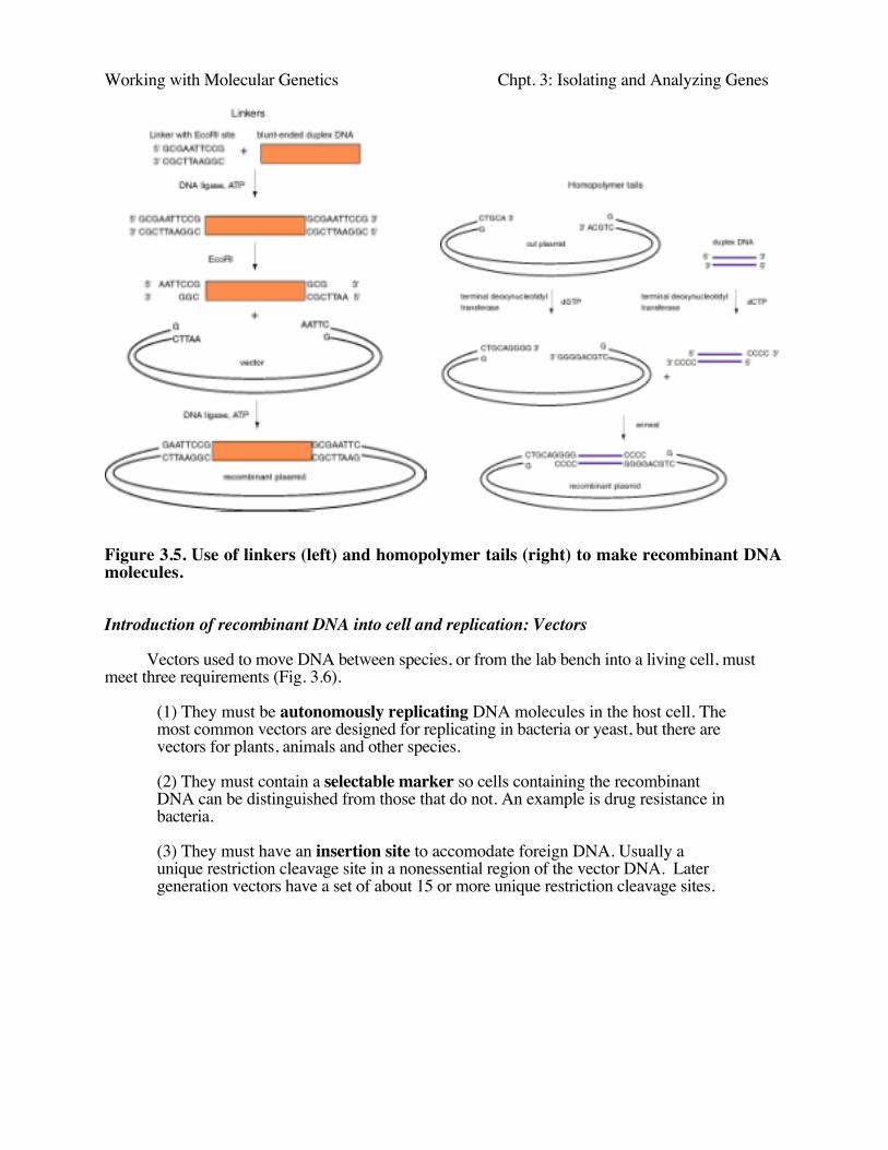
Linkers are short duplex oligonucleotides that contain a restriction endonuclease cleavagesite. They can be ligated onto any blunt-ended molecule, thereby generating a new restrictioncleavage site on the ends of the molecule. Ligation of a linker on a restriction fragment followed bycleavage with the restriction endonuclease is one of several ways to generate an end that is easy toligate to another DNA fragment.
Annealing of homopolymer tails are another way to joint two different DNA molecules.The enzyme terminal deoxynucleotidyl transferase will catalyze the addition of a string ofnucleotides to the 3' end of a DNA fragment. Thus by incubating each DNA fragment with theappropriate dNTP and terminal deoxynucleotidyl transferase, one can add complementaryhomopolymers to the ends of the DNAs that one wants to combine. E.g., one can add a string ofG's to the 3' ends of one fragment and a string of C's to the 3' ends of the other fragment. Now thetwo fragments will join together via the homopolymer tails.
Working with Molecular Genetics Chpt. 3: Isolating and Analyzing Genes
Figure 3.5. Use of linkers (left) and homopolymer tails (right) to make recombinant DNAmolecules.
Introduction of recombinant DNA into cell and replication: Vectors
Vectors used to move DNA between species, or from the lab bench into a living cell, mustmeet three requirements (Fig. 3.6).
(1) They must be autonomously replicating DNA molecules in the host cell. Themost common vectors are designed for replicating in bacteria or yeast, but there arevectors for plants, animals and other species.
(2) They must contain a selectable marker so cells containing the recombinantDNA can be distinguished from those that do not. An example is drug resistance inbacteria.
(3) They must have an insertion site to accomodate foreign DNA. Usually aunique restriction cleavage site in a nonessential region of the vector DNA. Latergeneration vectors have a set of about 15 or more unique restriction cleavage sites.
Working with Molecular Genetics Chpt. 3: Isolating and Analyzing Genes
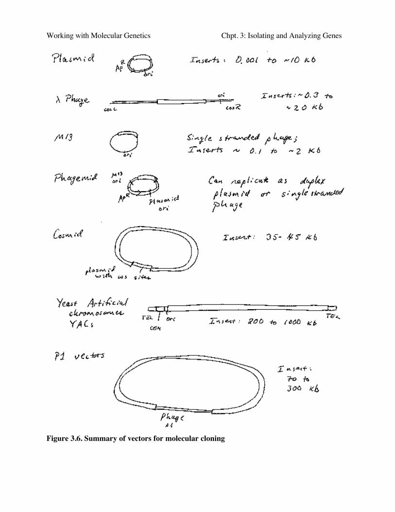
Figure 3.6. Summary of vectors for molecular cloning
Working with Molecular Genetics Chpt. 3: Isolating and Analyzing Genes
Plasmid vectors
Plasmids are autonomously replicating circular DNA molecules found in bacteria.They have their own origin of replication, and they replicate independently of the origins on the"host" chromosome. Replication is usually dependent on host functions, such as DNApolymerases, but regulation of plasmid replication is distinct from that of the host chromosome.Plamsids, such as the sex-factor F, can be very large (94 kb), but others can be small (2-4 kb).Plasmids do not encode an essential function to the bacterium, which distinguishes them fromchromosomes.
Plasmids can be present in a single copy, such as F, or in multiple copies, like those used asmost cloning vectors, such as pBR322, pUC, and pBluescript.
In nature, plasmids provide carry some useful function, such as transfer (F), or antibioticresistance. This is what keeps the plasmids in a population. In the absence of selection, plasmidsare lost from bacteria.
The antibiotic resistance genes on plasmids are often carried within, or are derived from,transposons, a types of transposable element. These are DNA segments that are capable of"jumping" or moving to new locations (see Chapter 9).
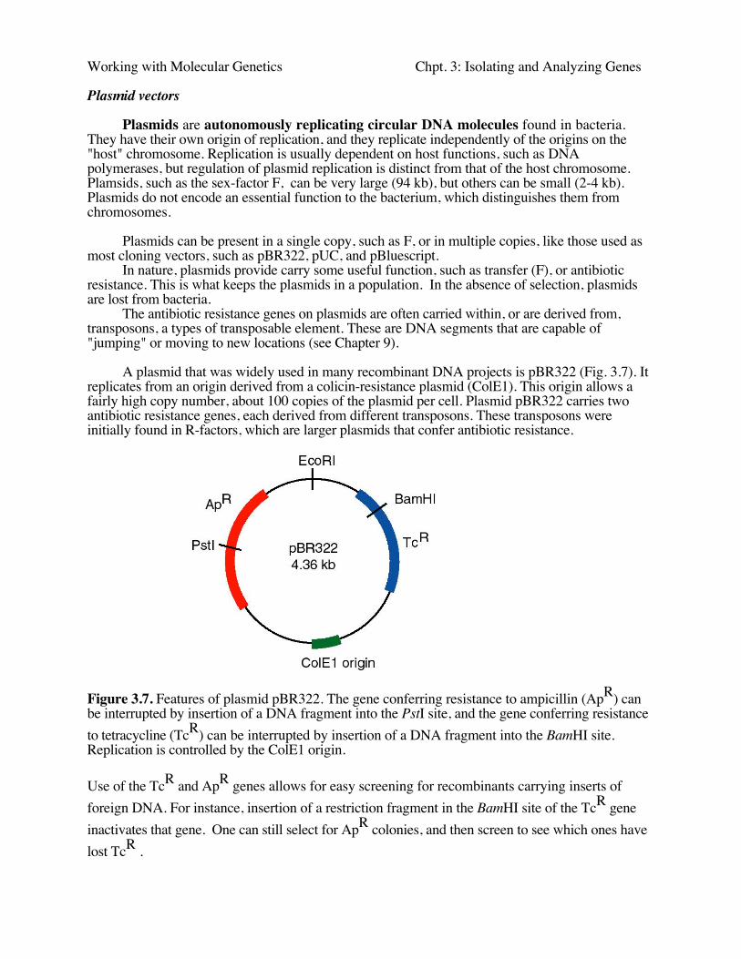
A plasmid that was widely used in many recombinant DNA projects is pBR322 (Fig. 3.7). Itreplicates from an origin derived from a colicin-resistance plasmid (ColE1). This origin allows afairly high copy number, about 100 copies of the plasmid per cell. Plasmid pBR322 carries twoantibiotic resistance genes, each derived from different transposons. These transposons wereinitially found in R-factors, which are larger plasmids that confer antibiotic resistance.
Figure 3.7. Features of plasmid pBR322. The gene conferring resistance to ampicillin (ApR) canbe interrupted by insertion of a DNA fragment into the PstI site, and the gene conferring resistanceto tetracycline (TcR) can be interrupted by insertion of a DNA fragment into the BamHI site.Replication is controlled by the ColE1 origin.
Use of the TcR and ApR genes allows for easy screening for recombinants carrying inserts offoreign DNA. For instance, insertion of a restriction fragment in the BamHI site of the TcR geneinactivates that gene. One can still select for ApR colonies, and then screen to see which ones havelost TcR .
Working with Molecular Genetics Chpt. 3: Isolating and Analyzing Genes
Question 3.1. What effects on drug resistance are seen when you use the EcoRI or PstIsites in pBR322 for inserting foreign DNA?
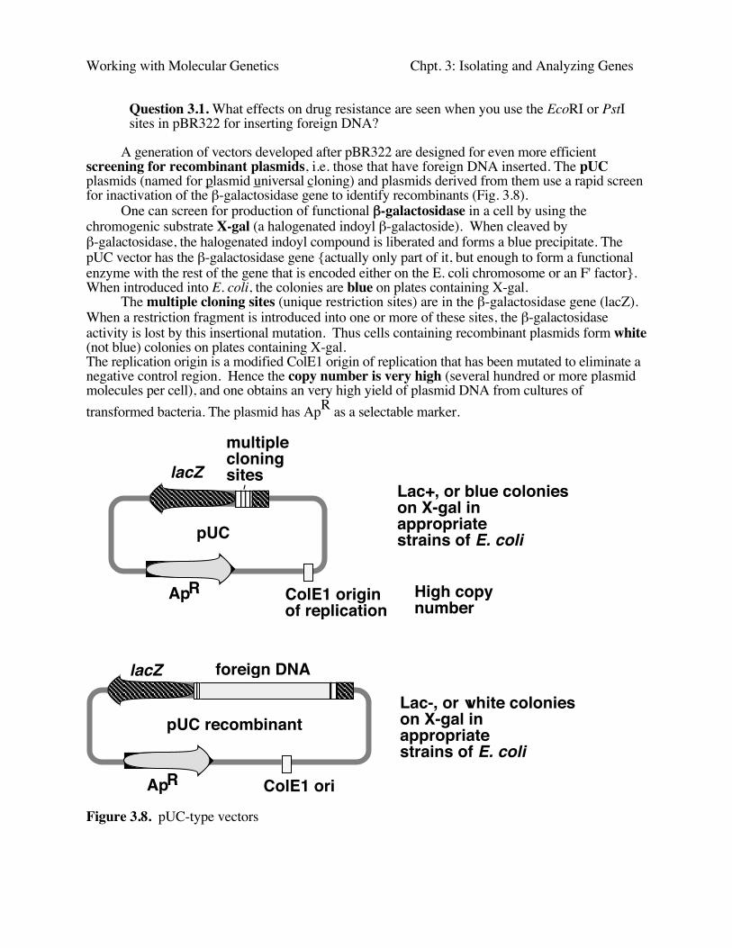
A generation of vectors developed after pBR322 are designed for even more efficientscreening for recombinant plasmids, i.e. those that have foreign DNA inserted. The pUCplasmids (named for plasmid universal cloning) and plasmids derived from them use a rapid screenfor inactivation of the β-galactosidase gene to identify recombinants (Fig. 3.8).
One can screen for production of functional β-galactosidase in a cell by using thechromogenic substrate X-gal (a halogenated indoyl β-galactoside). When cleaved byβ-galactosidase, the halogenated indoyl compound is liberated and forms a blue precipitate. ThepUC vector has the β-galactosidase gene {actually only part of it, but enough to form a functionalenzyme with the rest of the gene that is encoded either on the E. coli chromosome or an F' factor}.When introduced into E. coli, the colonies are blue on plates containing X-gal.
The multiple cloning sites (unique restriction sites) are in the β-galactosidase gene (lacZ).When a restriction fragment is introduced into one or more of these sites, the β-galactosidaseactivity is lost by this insertional mutation. Thus cells containing recombinant plasmids form white(not blue) colonies on plates containing X-gal.The replication origin is a modified ColE1 origin of replication that has been mutated to eliminate anegative control region. Hence the copy number is very high (several hundred or more plasmidmolecules per cell), and one obtains an very high yield of plasmid DNA from cultures oftransformed bacteria. The plasmid has ApR as a selectable marker.
Figure 3.8. pUC-type vectors
ColE1 originof replication
lacZmultiplecloning sites
ApR
pUC
ColE1 ori
lacZ
ApR
pUC recombinant
Lac+, or blue colonieson X-gal in appropriatestrains of E. coli
Lac-, or white colonieson X-gal in appropriatestrains of E. coli
foreign DNA
High copy number
Working with Molecular Genetics Chpt. 3: Isolating and Analyzing Genes
Introduction of a recombinant DNA molecule into a host cell
Introduction into CaCl2 treated E. coli: transformation
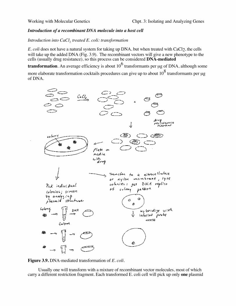
E. coli does not have a natural system for taking up DNA, but when treated with CaCl2, the cellswill take up the added DNA (Fig. 3.9). The recombinant vectors will give a new phenotype to thecells (usually drug resistance), so this process can be considered DNA-mediatedtransformation. An average efficiency is about 106 transformants per µg of DNA, although somemore elaborate transformation cocktails procedures can give up to about 108 transformants per µgof DNA.
Figure 3.9. DNA-mediated transformation of E. coli.
Usually one will transform with a mixture of recombinant vector molecules, most of whichcarry a different restriction fragment. Each transformed E. coli cell will pick up only one plasmid
Working with Molecular Genetics Chpt. 3: Isolating and Analyzing Genes
molecule, so the complex mixture of plasmids in the ligation mix has been separated into apopulation of transformed bacteria (Fig. 3.9). The bacterial cells are then plated at a sufficientlylow density that individual colonies can be identified. Each colony (or transformant) carries asingle plasmid, so as one screens the colonies, one is actually screening through individual DNAmolecules. A colony is a visible group of bacterial cells on a plate, all of which are derived from asingle bacterial cell. A group of identical cells derived from a single cell is called a clone. Sinceeach clone carries a single type of recombinant DNA molecule, the process is called molecularcloning.
Phage vectors for more efficient introduction of DNA into bacteria.
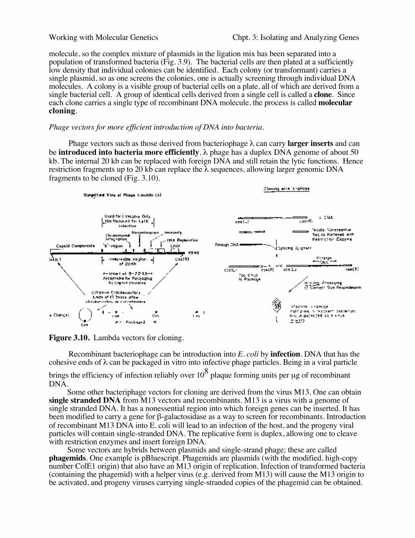
Phage vectors such as those derived from bacteriophage λ can carry larger inserts and canbe introduced into bacteria more efficiently. λ phage has a duplex DNA genome of about 50kb. The internal 20 kb can be replaced with foreign DNA and still retain the lytic functions. Hencerestriction fragments up to 20 kb can replace the λ sequences, allowing larger genomic DNAfragments to be cloned (Fig. 3.10).
Figure 3.10. Lambda vectors for cloning.
Recombinant bacteriophage can be introduction into E. coli by infection. DNA that has thecohesive ends of λ can be packaged in vitro into infective phage particles. Being in a viral particlebrings the efficiency of infection reliably over 108 plaque forming units per µg of recombinantDNA.
Some other bacteriphage vectors for cloning are derived from the virus M13. One can obtainsingle stranded DNA from M13 vectors and recombinants. M13 is a virus with a genome ofsingle stranded DNA. It has a nonessential region into which foreign genes can be inserted. It hasbeen modified to carry a gene for β-galactosidase as a way to screen for recombinants. Introductionof recombinant M13 DNA into E. coli will lead to an infection of the host, and the progeny viralparticles will contain single-stranded DNA. The replicative form is duplex, allowing one to cleavewith restriction enzymes and insert foreign DNA.
Some vectors are hybrids between plasmids and single-strand phage; these are calledphagemids. One example is pBluescript. Phagemids are plasmids (with the modified, high-copynumber ColE1 origin) that also have an M13 origin of replication. Infection of transformed bacteria(containing the phagemid) with a helper virus (e.g. derived from M13) will cause the M13 origin tobe activated, and progeny viruses carrying single-stranded copies of the phagemid can be obtained.
Working with Molecular Genetics Chpt. 3: Isolating and Analyzing Genes
Hence one can easily obtain either double- or single-stranded forms of thes plasmids. {The "blue"comes from the blue-white screening for recombinants that can be done when the multiple cloningsites are in the β-galactosidase gene. The "script" refers to the ability to make RNA copies of eitherstrand in vitro with phage RNA polymerases.}
Vectors designed to carry larger inserts
Fragments even larger than those carried in λ vectors are useful for studies of longersegments of chromosomes or whole genomes. Several vectors have been designed for cloning thesevery large fragments, 50 to 400 kb.
Cosmids are plasmids that have the cohesive ends of λ phage. They can be packaged in vitrointo infective phage particles to give a more efficient delivery of the DNA into the cells. They cancarry about 35 to 45 kb inserts (Fig. 3.6).
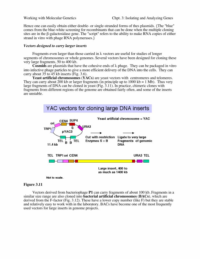
Yeast artificial chromosomes (YACs) are yeast vectors with centromeres and telomeres.They can carry about 200 kb or larger fragments (in principle up to 1000 kb = 1 Mb). Thus verylarge fragments of DNA can be cloned in yeast (Fig. 3.11). In practice, chimeric clones withfragments from different regions of the genome are obtained fairly often, and some of the insertsare unstable.
Figure 3.11
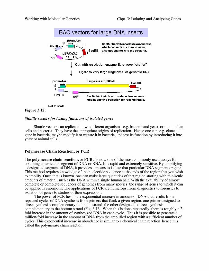
Vectors derived from bacteriophage P1 can carry fragments of about 100 kb. Fragments in asimilar size range are also cloned into bacterial artificial chromosomes (BACs), which arederived from the F-factor (Fig. 3.12). These have a lower copy number (like F) but they are stableand relatively easy to work with in the laboratory. BACs have become one of the most frequentlyused vectors for large inserts in genome projects.
Working with Molecular Genetics Chpt. 3: Isolating and Analyzing Genes
Figure 3.12.
Shuttle vectors for testing functions of isolated genes
Shuttle vectors can replicate in two different organisms, e.g. bacteria and yeast, or mammaliancells and bacteria. They have the appropriate origins of replication. Hence one can, e.g. clone agene in bacteria, maybe modify it or mutate it in bacteria, and test its function by introducing it intoyeast or animal cells.
Polymerase Chain Reaction, or PCR
The polymerase chain reaction, or PCR, is now one of the most commonly used assays forobtaining a particular segment of DNA or RNA. It is rapid and extremely sensitive. By amplifyinga designated segment of DNA, it provides a means to isolate that particular DNA segment or gene.This method requires knowledge of the nucleotide sequence at the ends of the region that you wishto amplify. Once that is known, one can make large quantities of that region starting with minisculeamounts of material, such as the DNA within a single human hair. With the availability of almostcomplete or complete sequences of genomes from many species, the range of genes to which it canbe applied is enormous. The applications of PCR are numerous, from diagnostics to forensics toisolation of genes to studies of their expression.
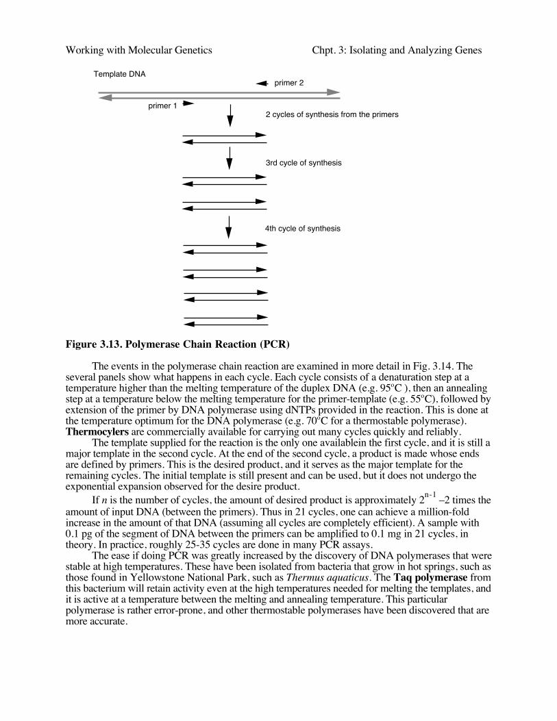
The power of PCR lies in the exponential increase in amount of DNA that results fromrepeated cycles of DNA synthesis from primers that flank a given region, one primer designed todirect synthesis complementary to the top strand, the other designed to direct synthesiscomplementary to the bottom strand (Fig. 3.13. When this is done repeatedly, there is roughly a 2-fold increase in the amount of synthesized DNA in each cycle. Thus it is possible to generate amillion-fold increase in the amount of DNA from the amplified region with a sufficient number ofcycles. This exponential increase in abundance is similar to a chemical chain reaction, hence it iscalled the polymerase chain reaction.
Working with Molecular Genetics Chpt. 3: Isolating and Analyzing GenesPolymerase Chain Reaction = PCR
2 cycles of synthesis from the primers
3rd cycle of synthesis
4th cycle of synthesis
Template DNA
primer 1
primer 2
Figure 3.13. Polymerase Chain Reaction (PCR)
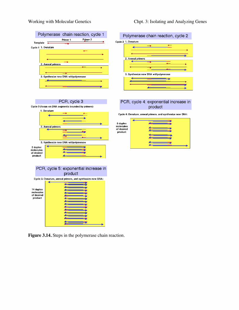
The events in the polymerase chain reaction are examined in more detail in Fig. 3.14. Theseveral panels show what happens in each cycle. Each cycle consists of a denaturation step at atemperature higher than the melting temperature of the duplex DNA (e.g. 95oC ), then an annealingstep at a temperature below the melting temperature for the primer-template (e.g. 55oC), followed byextension of the primer by DNA polymerase using dNTPs provided in the reaction. This is done atthe temperature optimum for the DNA polymerase (e.g. 70oC for a thermostable polymerase).Thermocylers are commercially available for carrying out many cycles quickly and reliably.
The template supplied for the reaction is the only one availablein the first cycle, and it is still amajor template in the second cycle. At the end of the second cycle, a product is made whose endsare defined by primers. This is the desired product, and it serves as the major template for theremaining cycles. The initial template is still present and can be used, but it does not undergo theexponential expansion observed for the desire product.
If n is the number of cycles, the amount of desired product is approximately 2n-1 –2 times theamount of input DNA (between the primers). Thus in 21 cycles, one can achieve a million-foldincrease in the amount of that DNA (assuming all cycles are completely efficient). A sample with0.1 pg of the segment of DNA between the primers can be amplified to 0.1 mg in 21 cycles, intheory. In practice, roughly 25-35 cycles are done in many PCR assays.
The ease if doing PCR was greatly increased by the discovery of DNA polymerases that werestable at high temperatures. These have been isolated from bacteria that grow in hot springs, such asthose found in Yellowstone National Park, such as Thermus aquaticus. The Taq polymerase fromthis bacterium will retain activity even at the high temperatures needed for melting the templates, andit is active at a temperature between the melting and annealing temperature. This particularpolymerase is rather error-prone, and other thermostable polymerases have been discovered that aremore accurate.
Working with Molecular Genetics Chpt. 3: Isolating and Analyzing Genes
Figure 3.14. Steps in the polymerase chain reaction.
Working with Molecular Genetics Chpt. 3: Isolating and Analyzing Genes
cDNA clones are copies of mRNAs
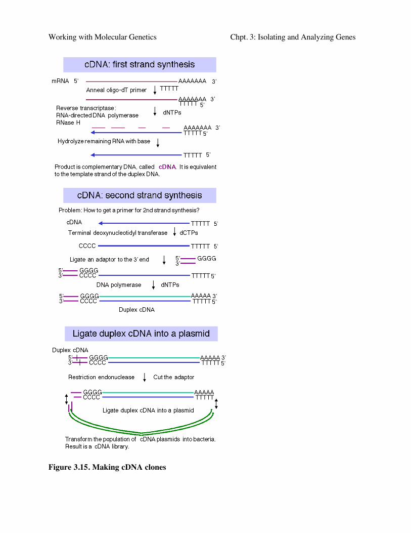
Construction of cDNA clones involves the synthesis of complementary DNA from mRNAand then inserting a duplex copy of that into a cloning vector, followed by transformation ofbacteria (Fig. 3.15).
a. First strand synthesis:
First, one anneals an oligo dT primer onto the 3' polyA tail of a population of mRNAs. Thenreverse transcriptase will begin DNA synthesis at the primer, using dNTPs supplied in the reaction,and copy the mRNA into complementary DNA, abbreviated cDNA.
The mRNA is degraded by the RNase H activity associated with reverse transcriptase and bysubsequent treatment with alkali.
b. Second strand synthesis:
For the primer to make the second strand of DNA (equivalent in sequence to the originalmRNA), one can utilize a transient hairpin at the end of the cDNA. (The basis for its formation isnot certain.) In other schemes, one generates a primer binding site and uses a primer directed tothat site; one way to do this is by homopolymer tailing of the cDNA followed by use of acomplementary primer. Random primers can also be used for second strand synthesis; althoughthis precludes the generation of a full-length cDNA (i.e. a copy of the entire mRNA). However, itis rare to generate duplex copies of the entire mRNA by any means.
DNA polymerase (e.g. Klenow polymerase) is used to synthesize the second strand,complementary to the cDNA. The product is duplex cDNA.
If the hairpin was used to prime second strand synthesis, it must be opened by a single-strandspecific nuclease such as S1.
c. Insertion of the duplex cDNA into a cloning vector:
One method is to use terminal deoxynucleotidyl transferase to add a homopolymer such aspoly-dC to the ends of the duplex cDNA and a complementary homopolymer such as poly-dG tothe vector.
An alternative approach is to use linkers; these can be employed such that a linker carrying acleavage site for one restriction endonuclease is on the 5' end of the duplex cDNA and a linkercarrying a cleavage site for a different restriction endonuclease is on the 3' end. (In this context, 5’and 3’ refer to the nontemplate, or "top" strand.) This allows "forced" cloning into the vector, andone has initial information about orientation, based on proximity to one cleavage site or the other.
The cDNA and vector are joined at the ends, using DNA ligase, to form recombinant cDNAplasmids (or phage).
d. The ligated cDNA plasmids are then transformed into E. coli. The resulting set oftransformants is a library of cDNA clones.
Working with Molecular Genetics Chpt. 3: Isolating and Analyzing Genes
Figure 3.15. Making cDNA clones
Working with Molecular Genetics Chpt. 3: Isolating and Analyzing Genes
Screening methods for cDNA clones
a. Brute force examination of individual cDNA plasmids.
If the mRNA is highly abundant in a given tissue, then many of the cDNA clones will becopies of that mRNA. One can examine DNA from individual clones and test for characteristicrestriction cleavage patterns or a particular sequence. This was a common approach for screeningcDNAs in the early days of recombinant DNA technology.
Starting in the mid-1990’s, cooperative efforts from corporations (such as Merck) andpublicly funded genome centers (such as at Washington University) have generated the sequence ofindividual clones from large cDNA libraries from many tissues from human, mouse, and rat. Otherconsortia have sequenced cDNA libraries from other species. Each sequence is called an“expressed sequence tag” or EST. These are now a major source of partially or fully characterizedcDNA clones. Hundreds of thousands of ESTs are available, and contain at part of the DNAsequence from many, if not most, human genes. The web site for NCBI(http://www.ncbi.nlm.nih.gov) is an excellent resource for examining the ESTs.
b. Hybridization with a gene-specific probe.
If the sequence of the desired cDNA is known, or if the sequence from homologs fromrelated species is known, one can use synthetic oligonucleotides (or other source of the diagnosticsequence) as a radiolabeled hybridization probe to identify the cDNA of interest.
If the amino acid sequence has been determined for all or even just parts of the proteinproduct of the gene of interest, then one can chemically synthesize oligonucleotides based on thegenetic code for those amino acids. The oligonucleotides need to be at least 18 nucleotides orlonger (so that they will anneal to specific sites in the genome), and because the genetic code isdegenerate (more than one codon per amino acid; discussed in Part Two), they have to bedegenerate as well. The oligonucleotides can be used directly as hybridization probes, although it isbecoming more common to amplify the region between two oligonucleotides using the polymerasechain reaction, and to use that amplification product as a labeled probe.
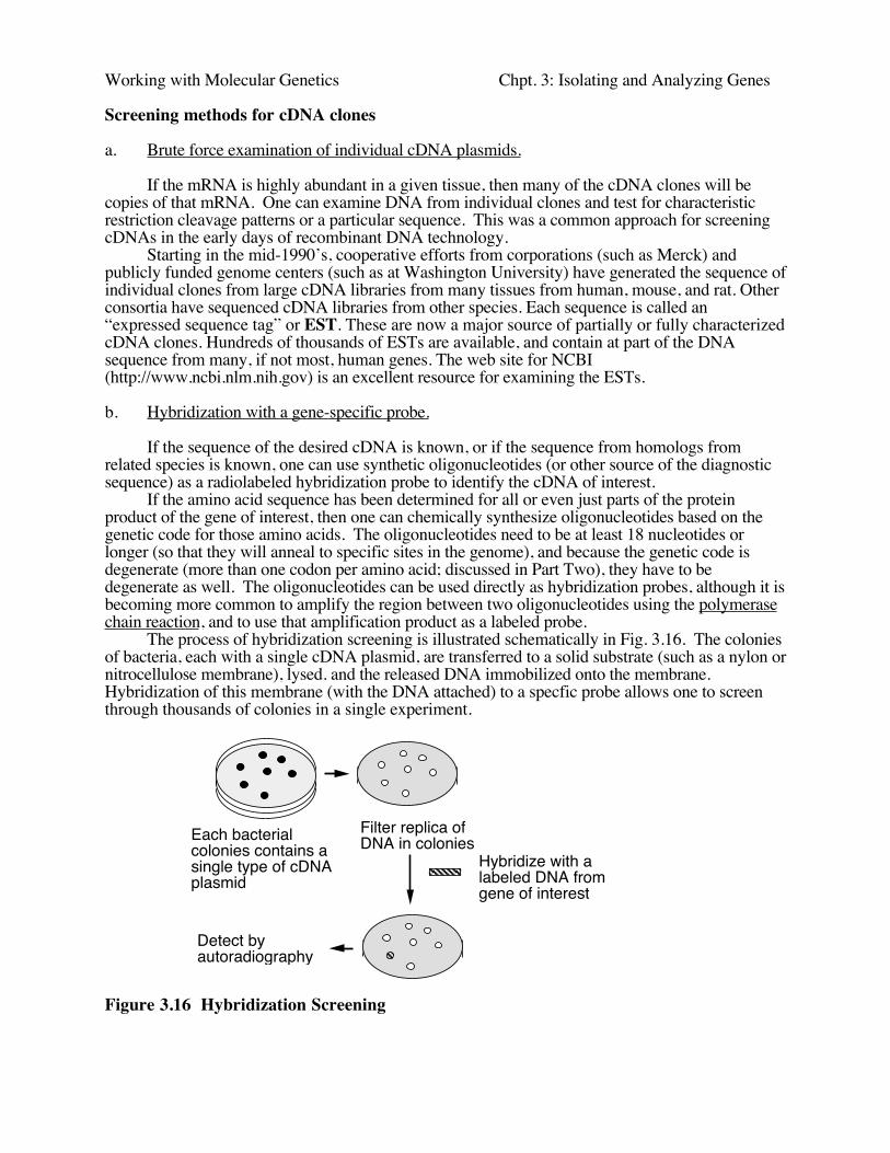
The process of hybridization screening is illustrated schematically in Fig. 3.16. The coloniesof bacteria, each with a single cDNA plasmid, are transferred to a solid substrate (such as a nylon ornitrocellulose membrane), lysed. and the released DNA immobilized onto the membrane.Hybridization of this membrane (with the DNA attached) to a specfic probe allows one to screenthrough thousands of colonies in a single experiment.
Each bacterial colonies contains a single type of cDNA plasmid
Filter replica of DNA in colonies
Hybridize with a labeled DNA from gene of interest
Detect by autoradiography
Figure 3.16 Hybridization Screening
Working with Molecular Genetics Chpt. 3: Isolating and Analyzing Genes
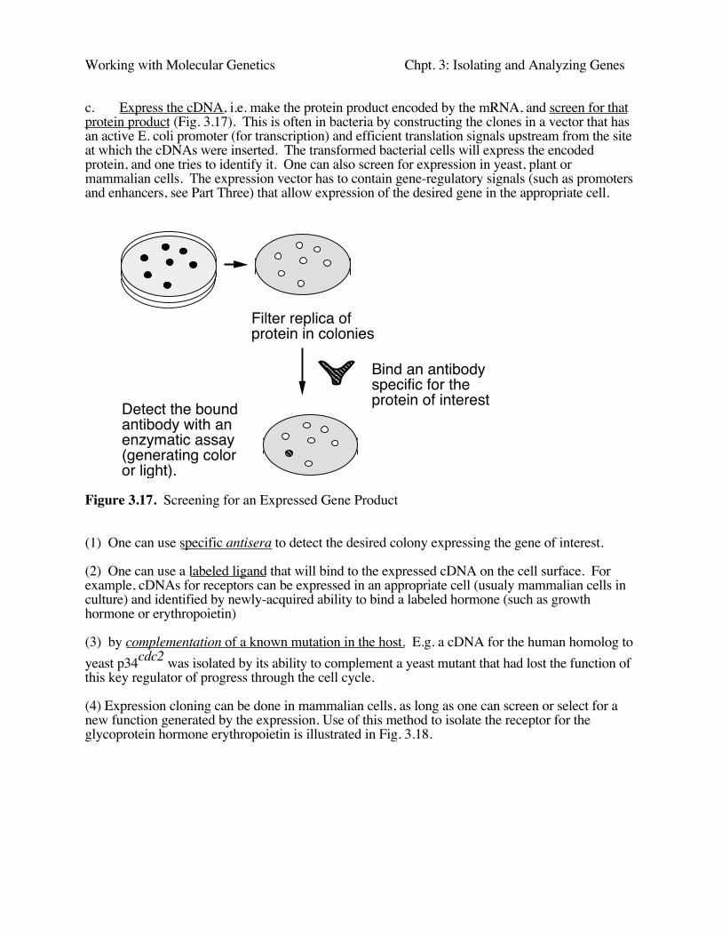
c. Express the cDNA, i.e. make the protein product encoded by the mRNA, and screen for thatprotein product (Fig. 3.17). This is often in bacteria by constructing the clones in a vector that hasan active E. coli promoter (for transcription) and efficient translation signals upstream from the siteat which the cDNAs were inserted. The transformed bacterial cells will express the encodedprotein, and one tries to identify it. One can also screen for expression in yeast, plant ormammalian cells. The expression vector has to contain gene-regulatory signals (such as promotersand enhancers, see Part Three) that allow expression of the desired gene in the appropriate cell.
Filter replica of protein in colonies
Bind an antibody specific for the protein of interestDetect the bound
antibody with an enzymatic assay (generating color or light).
Figure 3.17. Screening for an Expressed Gene Product
(1) One can use specific antisera to detect the desired colony expressing the gene of interest.
(2) One can use a labeled ligand that will bind to the expressed cDNA on the cell surface. Forexample, cDNAs for receptors can be expressed in an appropriate cell (usualy mammalian cells inculture) and identified by newly-acquired ability to bind a labeled hormone (such as growthhormone or erythropoietin)
(3) by complementation of a known mutation in the host. E.g. a cDNA for the human homolog toyeast p34cdc2 was isolated by its ability to complement a yeast mutant that had lost the function ofthis key regulator of progress through the cell cycle.
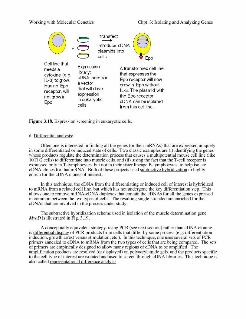
(4) Expression cloning can be done in mammalian cells, as long as one can screen or select for anew function generated by the expression. Use of this method to isolate the receptor for theglycoprotein hormone erythropoietin is illustrated in Fig. 3.18.
Working with Molecular Genetics Chpt. 3: Isolating and Analyzing Genes
Figure 3.18. Expression screening in eukaryotic cells.
d. Differential analysis:
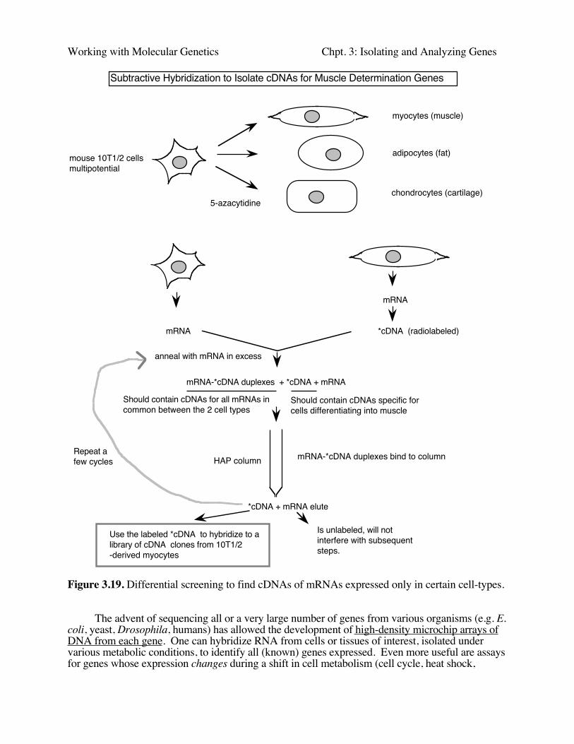
Often one is interested in finding all the genes (or their mRNAs) that are expressed uniquelyin some differentiated or induced state of cells. Two classic examples are (i) identifying the geneswhose products regulate the determination process that causes a multipotential mouse cell line (like10T1/2 cells) to differentiate into muscle cells, and (ii) ,using the fact that the T-cell receptor isexpressed only in T-lymphocytes, but not in their sister lineage B-lymphocytes, to help isolatecDNA clones for that mRNA. Both of these projects used subtractive hybridization to highlyenrich for the cDNA clones of interest.
In this technique, the cDNA from the differentiating or induced cell of interest is hybridizedto mRNA from a related cell line, but which has not undergone the key differentiation step. Thisallows one to remove mRNA-cDNA duplexes that contain the cDNAs for all the genes expressedin common between the two types of cells. The resulting single-stranded are enriched for thecDNAs that are involved in the process under study.
The subtractive hybridization scheme used in isolation of the muscle determination geneMyoD is illustrated in Fig. 3.19.
A conceptually equivalent strategy, using PCR (see next section) rather than cDNA cloning,is differential display of PCR products from cells that differ by some process (e.g. differentiation,induction, growth arrest versus stimulation, etc.). In this technique, one uses several sets of PCRprimers annealed to cDNA to mRNA from the two types of cells that are being compared. The setsof primers are empirically designed to allow many regions of cDNA to be amplified. Theamplification products are resolved (or displayed) on polyacrylamide gels, and the products specificto the cell type of interest are isolated and used to screen through cDNA libraries. This technique isalso called representational difference analysis.
Working with Molecular Genetics Chpt. 3: Isolating and Analyzing Genes
Subtractive Hybridization to Isolate cDNAs for Muscle Determination Genes
mouse 10T1/2 cellsmultipotential
myocytes (muscle)
adipocytes (fat)
chondrocytes (cartilage)5-azacytidine
mRNA
*cDNA (radiolabeled)mRNA
anneal with mRNA in excess
mRNA-*cDNA duplexes + *cDNA + mRNA
HAP column mRNA-*cDNA duplexes bind to column
*cDNA + mRNA elute
Use the labeled *cDNA to hybridize to a library of cDNA clones from 10T1/2 -derived myocytes
Should contain cDNAs for all mRNAs in common between the 2 cell types
Should contain cDNAs specific for cells differentiating into muscle
Is unlabeled, will not interfere with subsequent steps.
Repeat a few cycles
Figure 3.19. Differential screening to find cDNAs of mRNAs expressed only in certain cell-types.
The advent of sequencing all or a very large number of genes from various organisms (e.g. E.coli, yeast, Drosophila, humans) has allowed the development of high-density microchip arrays ofDNA from each gene. One can hybridize RNA from cells or tissues of interest, isolated undervarious metabolic conditions, to identify all (known) genes expressed. Even more useful are assaysfor genes whose expression changes during a shift in cell metabolism (cell cycle, heat shock,
Working with Molecular Genetics Chpt. 3: Isolating and Analyzing Genes
hormonal induction, etc.) or as a result of mutation of some other gene (e.g. a gene encoding atranscription factor of interest). This powerful new technology is being used more and more toexamine global effects on gene expression.
For a description (and movie) of the Affymetrix GeneChip, go tohttp://www.affymetrix.com/technology/index.html
Genomic DNA clones
Clones of genomic DNA, containing individual fragments of chromosomal DNA, are neededfor many purposes. Some examples include:
to obtain detailed structures of genes, to identify regulatory regions, i.e. DNA sequences needed for correct expression of the gene, to map and analyze alterations to the genome, e.g. the isolate genes that when mutated cause a
hereditary disease, to direct alterations in the genome, e.g. by homologous recombination to replace a wild-type
allele with a mutant one (to test function of the gene in mouse) or vice versa (to cure ahereditary disease, perhaps eventually in humans).
Construction of libraries of genomic DNA fragments in cloning vectors
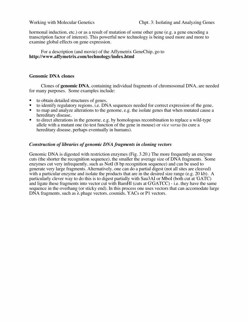
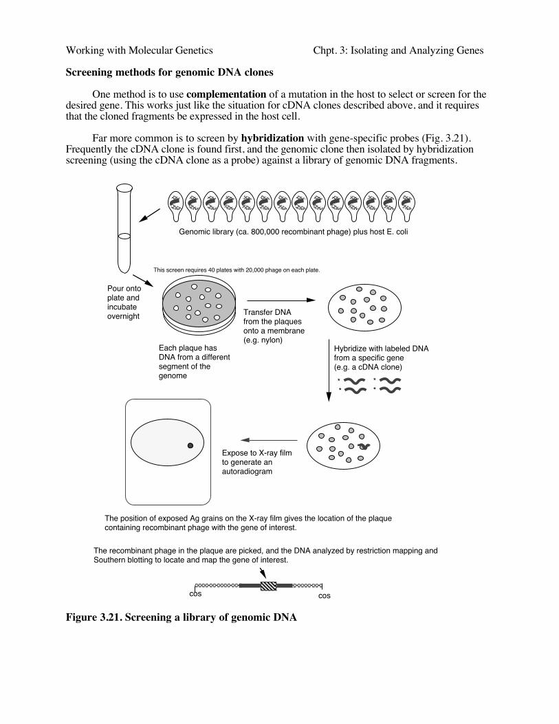
Genomic DNA is digested with restriction enzymes (Fig. 3.20.) The more frequently an enzymecuts (the shorter the recognition sequence), the smaller the average size of DNA fragments. Someenzymes cut very infrequently, such as NotI (8 bp recognition sequence) and can be used togenerate very large fragments. Alternatively, one can do a partial digest (not all sites are cleaved)with a particular enzyme and isolate the products that are in the desired size range (e.g. 20 kb). Aparticularly clever way to do this is to digest partially with Sau3AI or MboI (both cut at 'GATC)and ligate these fragments into vector cut with BamHI (cuts at G'GATCC) - i.e. they have the samesequence in the overhang (or sticky end). In this process one uses vectors that can accomodate largeDNA fragments, such as λ phage vectors, cosmids, YACs or P1 vectors.
Working with Molecular Genetics Chpt. 3: Isolating and Analyzing Genes
Total nuclear DNA from an organism with, e.g., 3 billion bp in a haploid genome
Partially digest with restriction endonuclease,select ca. 20,000 bp fragments
Construction of a Library of Genomic DNA
Mix genomic DNA fragments with a DNA from a cloning vector (e.g. lambda) and ligate
RE RE
RE RE
cos cos
cos
cos... ...
Package the concatameric DNA into phage particles in vitro
Need about 800,000 independent recombinant phage (each carrying a different segment of the genomic DNA) to have a 99% probability of having all the genomic DNA (3 billion bp) somewhere in the library, assuming all segments are capable of being propagated in lambda.
This collection of recombinant phage is called a library of genomic DNA.
Figure 3.20. Construction of a library of genomic DNA
Working with Molecular Genetics Chpt. 3: Isolating and Analyzing Genes
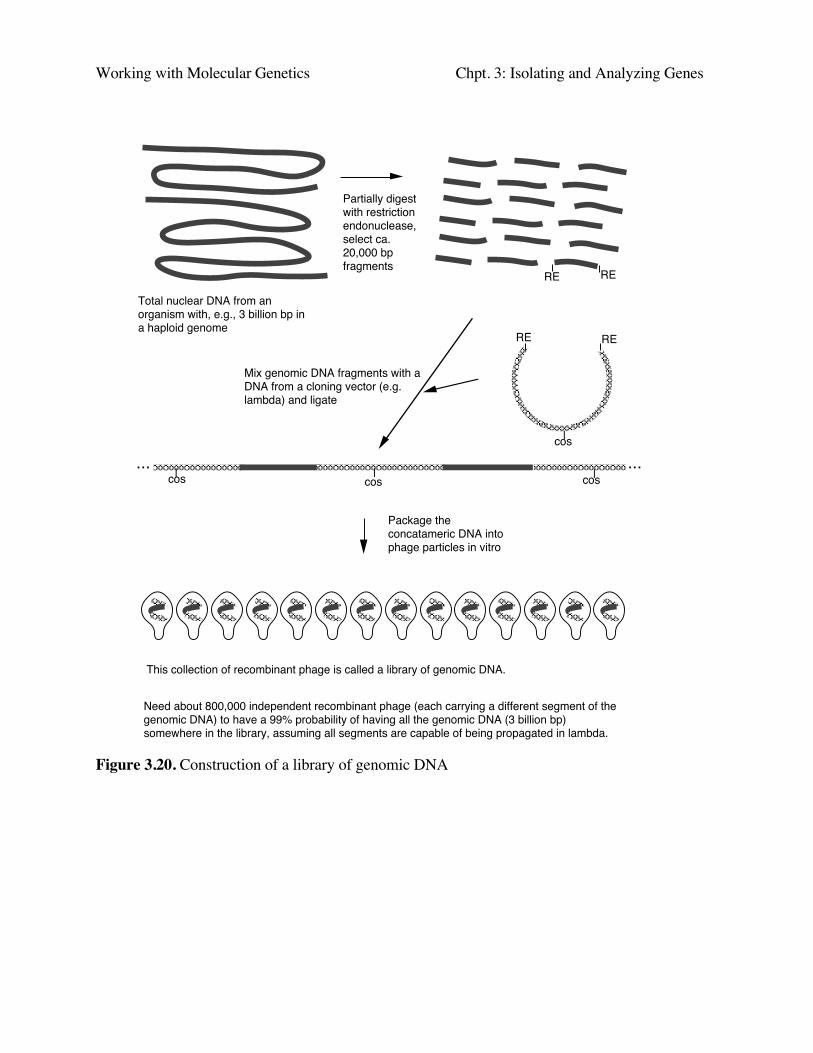
Screening methods for genomic DNA clones
One method is to use complementation of a mutation in the host to select or screen for thedesired gene. This works just like the situation for cDNA clones described above, and it requiresthat the cloned fragments be expressed in the host cell.
Far more common is to screen by hybridization with gene-specific probes (Fig. 3.21).Frequently the cDNA clone is found first, and the genomic clone then isolated by hybridizationscreening (using the cDNA clone as a probe) against a library of genomic DNA fragments.
Genomic library (ca. 800,000 recombinant phage) plus host E. coli
Pour onto plate and incubate overnight Transfer DNA
from the plaques onto a membrane (e.g. nylon)
Hybridize with labeled DNA from a specific gene(e.g. a cDNA clone)
Each plaque has DNA from a differentsegment of the genome
**
**
*Expose to X-ray filmto generate an autoradiogram
The position of exposed Ag grains on the X-ray film gives the location of the plaque containing recombinant phage with the gene of interest.
This screen requires 40 plates with 20,000 phage on each plate.
Screening a library of genomic DNA
The recombinant phage in the plaque are picked, and the DNA analyzed by restriction mapping and Southern blotting to locate and map the gene of interest.
coscos
Figure 3.21. Screening a library of genomic DNA
Working with Molecular Genetics Chpt. 3: Isolating and Analyzing Genes
Eukaryotic gene structure
Much can be learned about any gene after it has been isolated by recombinant DNA techniques.The structure of coding and noncoding regions, the DNA sequence, and more can be deduced. Thisis true for bacterial and viral genes, as well as eukaryotic cellular genes. The next sections of thischapter will focus on analysis of eukaryotic genes, showing the power of examining purified copiesof genes.
Split genes and introns
Precursors to mRNA longer than mRNA
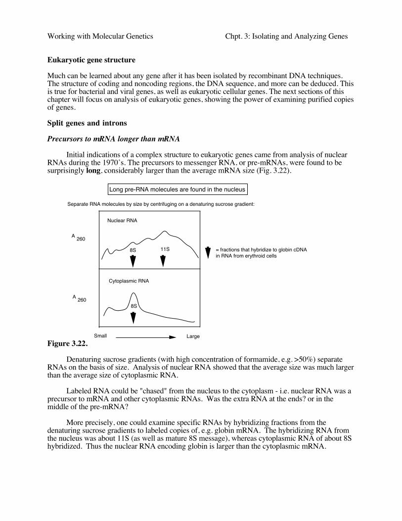
Initial indications of a complex structure to eukaryotic genes came from analysis of nuclearRNAs during the 1970’s. The precursors to messenger RNA, or pre-mRNAs, were found to besurprisingly long, considerably larger than the average mRNA size (Fig. 3.22).
Separate RNA molecules by size by centrifuging on a denaturing sucrose gradient:
260A
260A
Small Large
Nuclear RNA
Cytoplasmic RNA
8S 11S
8S
= fractions that hybridize to globin cDNA in RNA from erythroid cells
Long pre-RNA molecules are found in the nucleus
Figure 3.22.
Denaturing sucrose gradients (with high concentration of formamide, e.g. >50%) separateRNAs on the basis of size. Analysis of nuclear RNA showed that the average size was much largerthan the average size of cytoplasmic RNA.
Labeled RNA could be "chased" from the nucleus to the cytoplasm - i.e. nuclear RNA was aprecursor to mRNA and other cytoplasmic RNAs. Was the extra RNA at the ends? or in themiddle of the pre-mRNA?
More precisely, one could examine specific RNAs by hybridizing fractions from thedenaturing sucrose gradients to labeled copies of, e.g. globin mRNA. The hybridizing RNA fromthe nucleus was about 11S (as well as mature 8S message), whereas cytoplasmic RNA of about 8Shybridized. Thus the nuclear RNA encoding globin is larger than the cytoplasmic mRNA.
Working with Molecular Genetics Chpt. 3: Isolating and Analyzing Genes
Visualization of mRNA-DNA heteroduplexes revealed extra sequences internal to the mRNA-coding segments
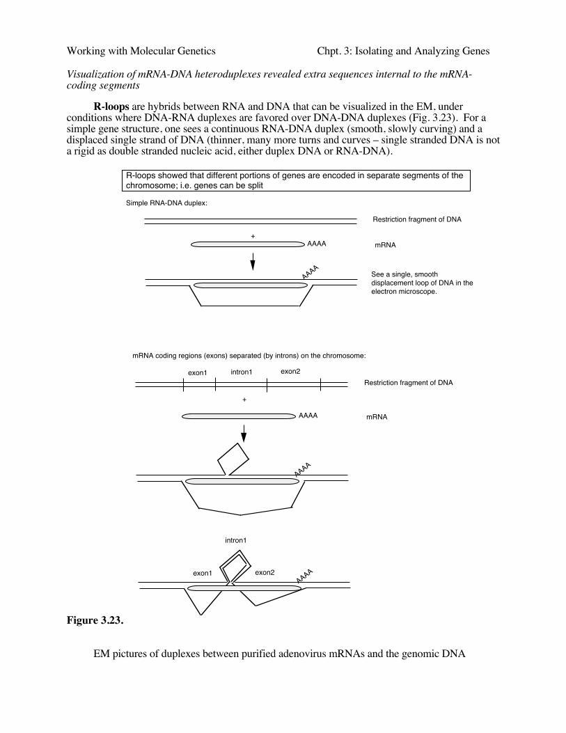
R-loops are hybrids between RNA and DNA that can be visualized in the EM, underconditions where DNA-RNA duplexes are favored over DNA-DNA duplexes (Fig. 3.23). For asimple gene structure, one sees a continuous RNA-DNA duplex (smooth, slowly curving) and adisplaced single strand of DNA (thinner, many more turns and curves – single stranded DNA is nota rigid as double stranded nucleic acid, either duplex DNA or RNA-DNA).
Simple RNA-DNA duplex:
Restriction fragment of DNA
mRNAAAAA
AAAA
+
See a single, smooth displacement loop of DNA in the electron microscope.
mRNA coding regions (exons) separated (by introns) on the chromosome:
Restriction fragment of DNA
mRNAAAAA
+
AAAA
exon1 intron1 exon2
AAAA
intron1
exon1 exon2
R-loops showed that different portions of genes are encoded in separate segments of the chromosome; i.e. genes can be split
Figure 3.23.
EM pictures of duplexes between purified adenovirus mRNAs and the genomic DNA
Working with Molecular Genetics Chpt. 3: Isolating and Analyzing Genes
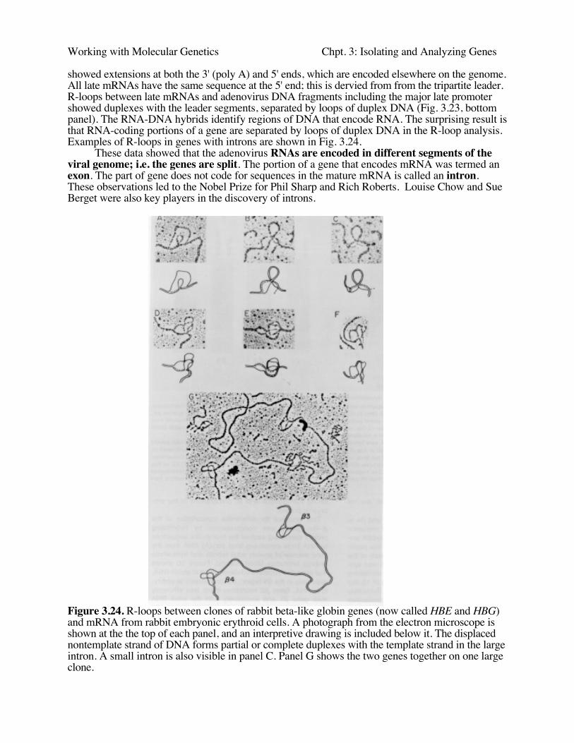
showed extensions at both the 3' (poly A) and 5' ends, which are encoded elsewhere on the genome.All late mRNAs have the same sequence at the 5' end; this is dervied from from the tripartite leader.R-loops between late mRNAs and adenovirus DNA fragments including the major late promotershowed duplexes with the leader segments, separated by loops of duplex DNA (Fig. 3.23, bottompanel). The RNA-DNA hybrids identify regions of DNA that encode RNA. The surprising result isthat RNA-coding portions of a gene are separated by loops of duplex DNA in the R-loop analysis.Examples of R-loops in genes with introns are shown in Fig. 3.24.
These data showed that the adenovirus RNAs are encoded in different segments of theviral genome; i.e. the genes are split. The portion of a gene that encodes mRNA was termed anexon. The part of gene does not code for sequences in the mature mRNA is called an intron.These observations led to the Nobel Prize for Phil Sharp and Rich Roberts. Louise Chow and SueBerget were also key players in the discovery of introns.
Figure 3.24. R-loops between clones of rabbit beta-like globin genes (now called HBE and HBG)and mRNA from rabbit embryonic erythroid cells. A photograph from the electron microscope isshown at the the top of each panel, and an interpretive drawing is included below it. The displacednontemplate strand of DNA forms partial or complete duplexes with the template strand in the largeintron. A small intron is also visible in panel C. Panel G shows the two genes together on one largeclone.
Working with Molecular Genetics Chpt. 3: Isolating and Analyzing Genes
Interruptions in cellular genes were discovered subsequently, in the late 1970's, in globingenes, immunoglobulin genes and others. We now realize that mostgenes in complex eukaryotesare split by multiple introns.
Exons are more conserved than introns (in most cases), since alterations in protein-codingregions that alter or decrease function are selected against, whereas many sequences in introns canbe altered without affecting the function of the gene product. Important sequences in introns (suchas splice junctions, the branch point, and occassionally enhancers) are covered in some detail in PartThree.
Differences in restiction maps between cDNA and genomic clones reveal introns
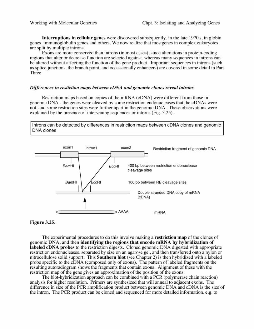
Restriction maps based on copies of the mRNA (cDNA) were different from those ingenomic DNA - the genes were cleaved by some restriction endonucleases that the cDNAs werenot, and some restriction sites were further apart in the genomic DNA. These observations wereexplained by the presence of intervening sequences or introns (Fig. 3.25).
Restriction fragment of genomic DNA
mRNAAAAA
exon1 intron1 exon2
BamHI EcoRI
BamHI EcoRI
400 bp between restriction endonucleasecleavage sites
100 bp between RE cleavage sites
Double stranded DNA copy of mRNA(cDNA)
Introns can be detected by differences in restriction maps between cDNA clones and genomic DNA clones
Figure 3.25.
The experimental procedures to do this involve making a restriction map of the clones ofgenomic DNA, and then identifying the regions that encode mRNA by hybridization oflabeled cDNA probes to the restriction digests. Cloned genomic DNA digested with appropriaterestriction endonucleases, separated by size on an agarose gel, and then transferred onto a nylon ornitrocellulose solid support. This Southern blot (see Chapter 2) is then hybridized with a labeledprobe specific to the cDNA (composed only of exons). The pattern of labeled fragments on theresulting autoradiogram shows the fragments that contain exons. Alignment of these with therestriction map of the gene gives an approximation of the position of the exons.
The blot-hybridization approach can be combined with a PCR (polymerase chain reaction)analysis for higher resolution. Primers are synthesized that will anneal to adjacent exons. Thedifference in size of the PCR amplification product between genomic DNA and cDNA is the size ofthe intron. The PCR product can be cloned and sequenced for more detailed information, e.g. to

Working with Molecular Genetics Chpt. 3: Isolating and Analyzing Genes
precisely define the exon/intron junctions.
Subsequently, the nucleotide sequence of exonic regions and preferably the entire gene isdetermined. The presence of introns were confirmed and their locations defined precisely in DNAsequences of isolated clones of the genes.
Types of exons
Eukaryotic genes are a combination of introns and exons. However, not all exons do the samething (Fig. 3.26). In particular, the protein-coding regions or genes are a subset of the sequences inexons. Exons include both the untranslated regions and the protein-coding, translated regions.Introns are the segments of genes that are present in the primary transcript (or precursor RNA) butare removed by splicing in the production of mature RNA. Methods used to detect coding regionswill not find all exons.
Figure 3.26. Types of exons
Multiple, large introns can make some eukaryotic genes very large
Eukaryotic genes can be split into many (>60), sometimes very small exons (e.g. <60 bp,coding for <20 amino acids), separated by very large introns (as large as >100kb), resulting in someenormous genes (>500 kb). E.g. the DMD gene (which when mutated can cause Duchenne'smuscular dystrophy) is almost 1 Mb, about 1/4 the size of the E. coli chromosome!
The average size of genes from more complex organisms is considerably larger than those ofsimpler ones, but the avg. size of mRNA is about the same, reflecting the presence of more andlarger introns in the more complex organisms.
tRNA and rRNA genes also contain introns
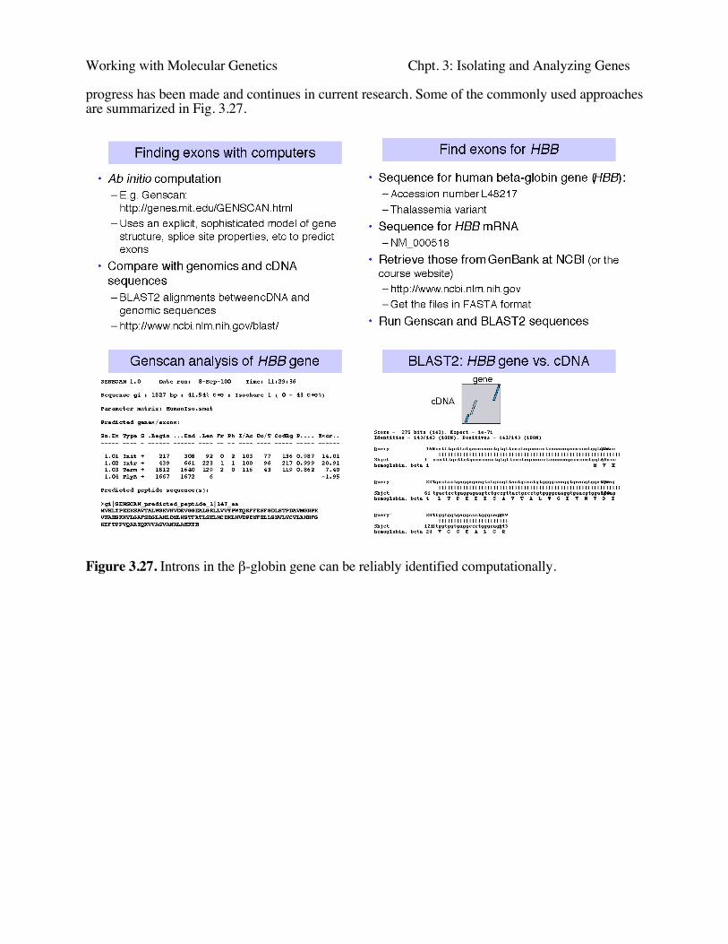
Finding exons in long genomic sequences using computer programs
Far more exons and introns have been discovered (or more accurately, predicted) throught theanalysis of genomic DNA sequences than could ever be discovered by direct experimentation. Thedifferent types of exons, the enormous length of introns, and other factors have complicated the taskof finding reliable diagnostic signatures for exons in genomic sequences. However, considerable
Working with Molecular Genetics Chpt. 3: Isolating and Analyzing Genes
progress has been made and continues in current research. Some of the commonly used approachesare summarized in Fig. 3.27.
Figure 3.27. Introns in the β-globin gene can be reliably identified computationally.

Working with Molecular Genetics Chpt. 3: Isolating and Analyzing Genes
Introns are removed by splicing RNA precursors
exon1 intron1 exon2 exon3intron2
AAAA
AAAA
cap
cap
Gene:duplex DNA
Primary transcript:single stranded RNA
Precursor to mRNA
mRNA
Protein
transcription
5' and 3' end processing
splicing
translation
Introns are removed from pre-mRNA to generate mRNA
Figure 3.28. Introns are removed from pre-mRNA to generate mRNA.
Working with Molecular Genetics Chpt. 3: Isolating and Analyzing Genes
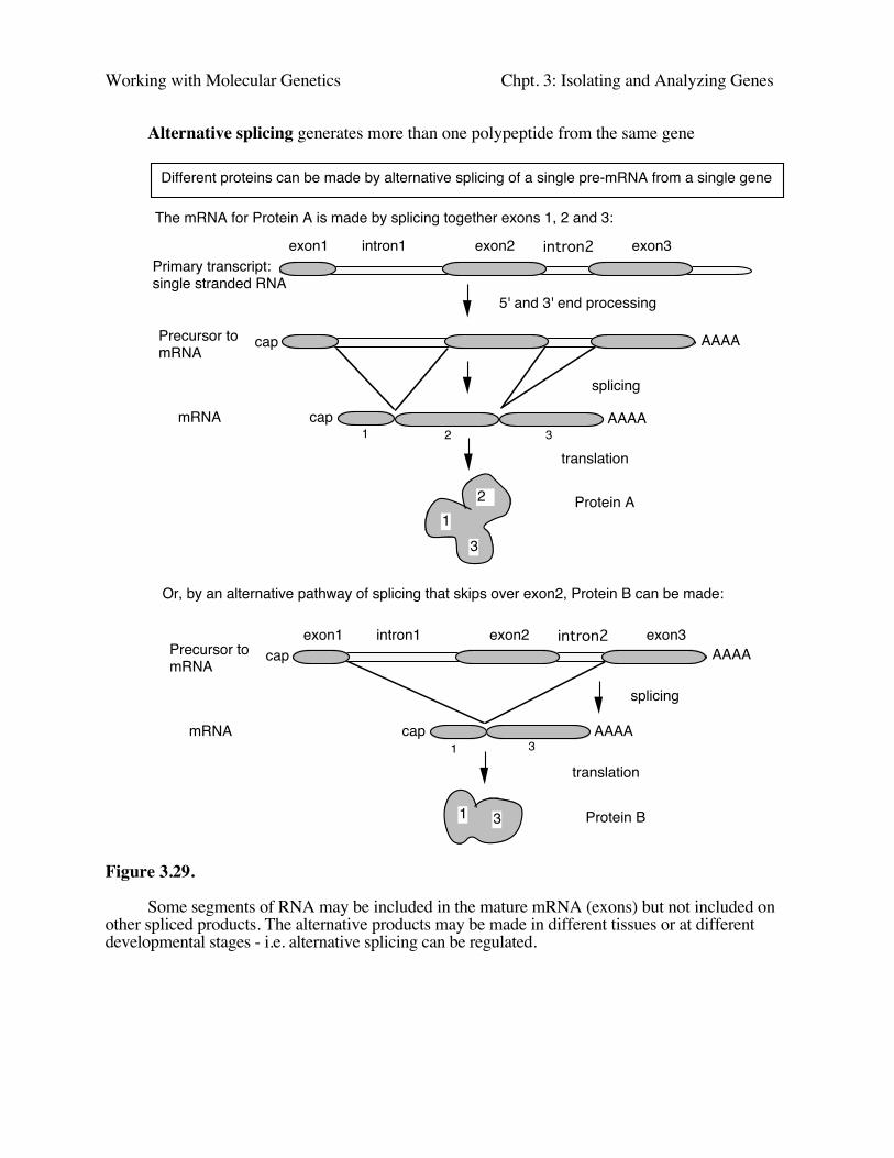
Alternative splicing generates more than one polypeptide from the same gene
AAAA
AAAA
cap
cap
Primary transcript:single stranded RNA
Precursor to mRNA
mRNA
Protein A
5' and 3' end processing
splicing
translation
Different proteins can be made by alternative splicing of a single pre-mRNA from a single gene
12
3
AAAA
AAAA
cap
cap
Precursor to mRNA
mRNA
Protein B
splicing
translation
exon1 intron1 exon2 exon3intron2
exon1 intron1 exon2 exon3intron2
Or, by an alternative pathway of splicing that skips over exon2, Protein B can be made:
31
31
1 2 3
The mRNA for Protein A is made by splicing together exons 1, 2 and 3:
Figure 3.29.
Some segments of RNA may be included in the mature mRNA (exons) but not included onother spliced products. The alternative products may be made in different tissues or at differentdevelopmental stages - i.e. alternative splicing can be regulated.
Working with Molecular Genetics Chpt. 3: Isolating and Analyzing Genes
Split genes may enhance the rate of evolution
Many exons encode a unit very close to a protein domain, e.g. the exons of leghemoglobin, orthe variable and constant regions of immunoglobulins, or domains (e.g. "kringle") in EGFprecursor that are also found in part of the LDL receptor. The exon organization tends to be wellconserved in highly divergent species. Introns tend to occur between those portions of genes thatencode structural domains of proteins.
Duplication of the exons encoding structural domains and subsequent recombination can leadto more rapid evolution of a new protein, essentially using the parts from earlier evolved genes.Analogous to building a house from prefabricated parts, as opposed to one nail and one board at atime - start with preassembled walls, roof joists etc.
However, the relationship between exons and structural domains of proteins is not exact, andsome exon-intron boundaries vary (a little) in genes for different species. A different model holdsthat the introns are transposable elements (some certainly are - see later). They can insert anywherein a gene, but they are least disruptive at domain boundaries, and these latter insertions are morelikely to be fixed in a population than insertions into the middle of a region encoding a domain. Sothe results after long years of evolution is that the introns tend to be between region codingdomains, but the gene was originally intact, not assembled from discrete exons.

Working with Molecular Genetics Chpt. 3: Isolating and Analyzing Genes
Multi-gene families and gene clusters
Many eukaryotic genes are found in multiple copies. Some of them are developmentallyregulated, such as HOX gene clusters and globin gene clusters .
Developmental switches in human hemoglobins
Human β-globin
Human α-globin
ε γ γ ψη δ βG A
ζ2 ζ1 α2ψα1 α1 θ
Embryonic Fetal Adult
0 20 40 60 80 kb
Chromosome 16
Chromosome 11LCR
HS-40ψα2
Hb Gower-1 ζ ε2 2
Hb Gower-2 α ε2 2
Hb Portland ζ γ2 2
HbF α γ2 2
HbA2 α δ 2 2
HbA α β2 2
Figure 3.30.
A multigene family contains multiple genes of similar sequence encoding similar proteins;e.g. globin genes (Fig. 3.30). Globin genes are expressed at different times of development. Theorder of developmental expression is the same as their order along the chromosome, e.g. the ε-globin gene is expressed in early embryonic red cells, the γ-globin gene is expressed at a high levelin fetal red cells, and the β-globin gene is expressed in red cells after birth. As we will see later, thiscorrelates with their distance from a dominant control element at the 5' end of the cluster, the LocusControl Region.
The order of HOX genes is also aligned with their spatial expression in the embryo. This isanother example of alignment between chromosomal position and regulation of expression.
Other multi-gene families include those encoding histones, immunoglobulins, actins, cyclins,cyclin-dependent protein kinases, and rRNAs. Some of these families are linked in gene clusters,but others are dispersed around the genome. Having multiple copies of genes may be more the rulethan the exception in eukaryotic genomes.
Working with Molecular Genetics Chpt. 3: Isolating and Analyzing Genes
Experimental techniques that reveal multigene families include the following.
Purification and analysis of a particular kind of protein, e.g. hemoglobins, immunoglobulins,and many enzymes, may reveal heterogeneity. Further purification (via chromatography andelectrophoresis) and sequencing can show that the observed heterogeneity is a result of related butnot identical proteins, and one deduces that these similar proteins are encoded by multiple geneswith similar sequences, i.e. a multigene family.
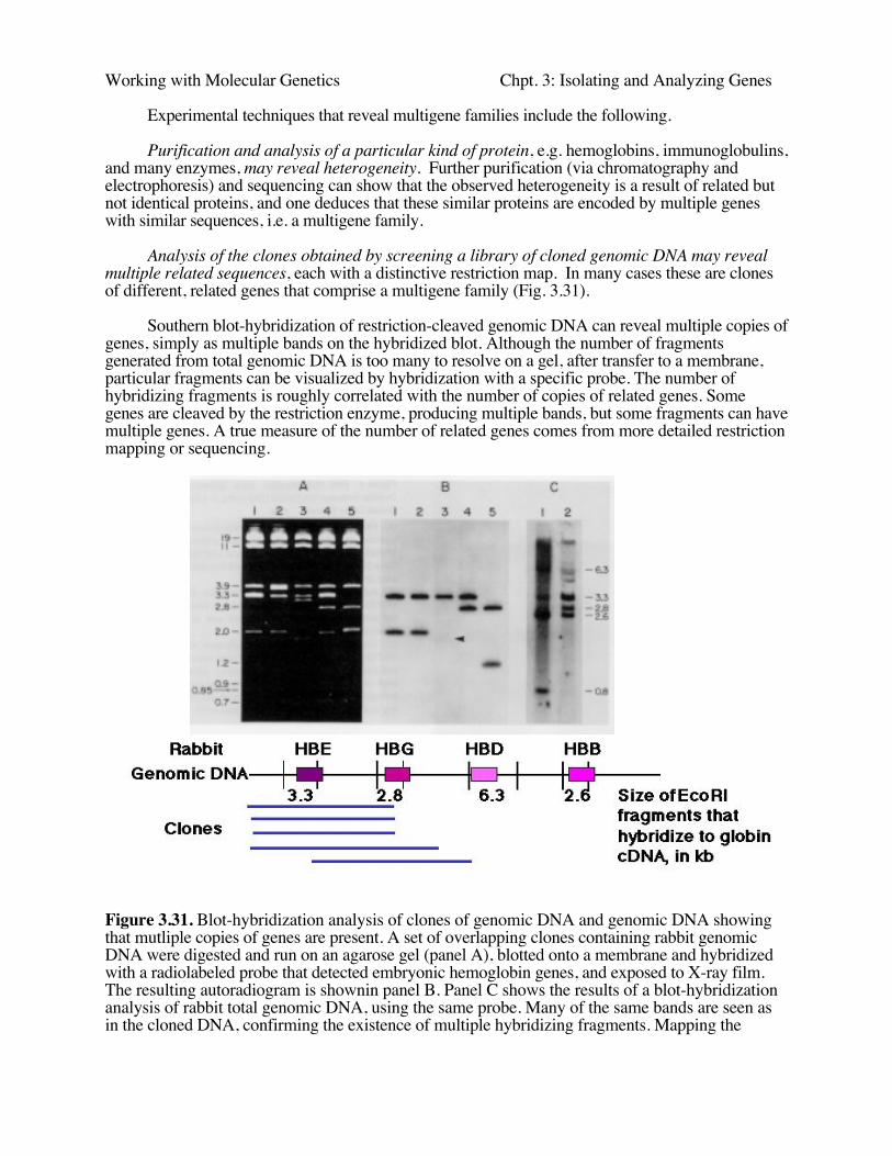
Analysis of the clones obtained by screening a library of cloned genomic DNA may revealmultiple related sequences, each with a distinctive restriction map. In many cases these are clonesof different, related genes that comprise a multigene family (Fig. 3.31).
Southern blot-hybridization of restriction-cleaved genomic DNA can reveal multiple copies ofgenes, simply as multiple bands on the hybridized blot. Although the number of fragmentsgenerated from total genomic DNA is too many to resolve on a gel, after transfer to a membrane,particular fragments can be visualized by hybridization with a specific probe. The number ofhybridizing fragments is roughly correlated with the number of copies of related genes. Somegenes are cleaved by the restriction enzyme, producing multiple bands, but some fragments can havemultiple genes. A true measure of the number of related genes comes from more detailed restrictionmapping or sequencing.
Figure 3.31. Blot-hybridization analysis of clones of genomic DNA and genomic DNA showingthat mutliple copies of genes are present. A set of overlapping clones containing rabbit genomicDNA were digested and run on an agarose gel (panel A), blotted onto a membrane and hybridizedwith a radiolabeled probe that detected embryonic hemoglobin genes, and exposed to X-ray film.The resulting autoradiogram is shownin panel B. Panel C shows the results of a blot-hybridizationanalysis of rabbit total genomic DNA, using the same probe. Many of the same bands are seen asin the cloned DNA, confirming the existence of multiple hybridizing fragments. Mapping the
Working with Molecular Genetics Chpt. 3: Isolating and Analyzing Genes
fragments showed that they represented separate genes.
Keeping multigene families homogeneous
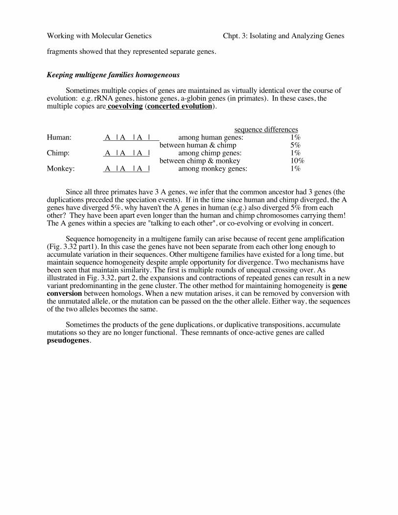
Sometimes multiple copies of genes are maintained as virtually identical over the course ofevolution: e.g. rRNA genes, histone genes, a-globin genes (in primates). In these cases, themultiple copies are coevolving (concerted evolution).
sequence differencesHuman: A | A | A | among human genes: 1%
between human & chimp 5%Chimp: A | A | A | among chimp genes: 1%
between chimp & monkey 10%Monkey: A | A | A | among monkey genes: 1%
Since all three primates have 3 A genes, we infer that the common ancestor had 3 genes (theduplications preceded the speciation events). If in the time since human and chimp diverged, the Agenes have diverged 5%, why haven't the A genes in human (e.g.) also diverged 5% from eachother? They have been apart even longer than the human and chimp chromosomes carrying them!The A genes within a species are "talking to each other", or co-evolving or evolving in concert.
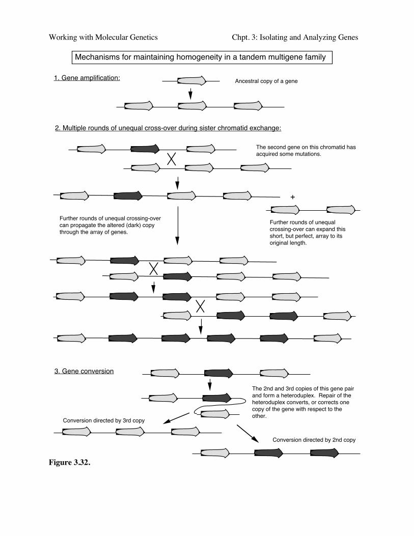
Sequence homogeneity in a multigene family can arise because of recent gene amplification(Fig. 3.32 part1). In this case the genes have not been separate from each other long enough toaccumulate variation in their sequences. Other multigene families have existed for a long time, butmaintain sequence homogeneity despite ample opportunity for divergence. Two mechanisms havebeen seen that maintain similarity. The first is multiple rounds of unequal crossing over. Asillustrated in Fig. 3.32, part 2, the expansions and contractions of repeated genes can result in a newvariant predominanting in the gene cluster. The other method for maintaining homogeneity is geneconversion between homologs. When a new mutation arises, it can be removed by conversion withthe unmutated allele, or the mutation can be passed on the the other allele. Either way, the sequencesof the two alleles becomes the same.
Sometimes the products of the gene duplications, or duplicative transpositions, accumulatemutations so they are no longer functional. These remnants of once-active genes are calledpseudogenes.
Working with Molecular Genetics Chpt. 3: Isolating and Analyzing Genes
Mechanisms for maintaining homogeneity in a tandem multigene family
1. Gene amplification:
2. Multiple rounds of unequal cross-over during sister chromatid exchange:
The second gene on this chromatid has acquired some mutations.
+
Further rounds of unequal crossing-over can expand this short, but perfect, array to its original length.
Further rounds of unequal crossing-over can propagate the altered (dark) copy through the array of genes.
Ancestral copy of a gene
3. Gene conversion
The 2nd and 3rd copies of this gene pair and form a heteroduplex. Repair of the heteroduplex converts, or corrects one copy of the gene with respect to the other.
Conversion directed by 3rd copy
Conversion directed by 2nd copy
Figure 3.32.
Working with Molecular Genetics Chpt. 3: Isolating and Analyzing Genes
Functional analysis of isolated genes
Gene expression
"Northern blots" or RNA blot-hybridization
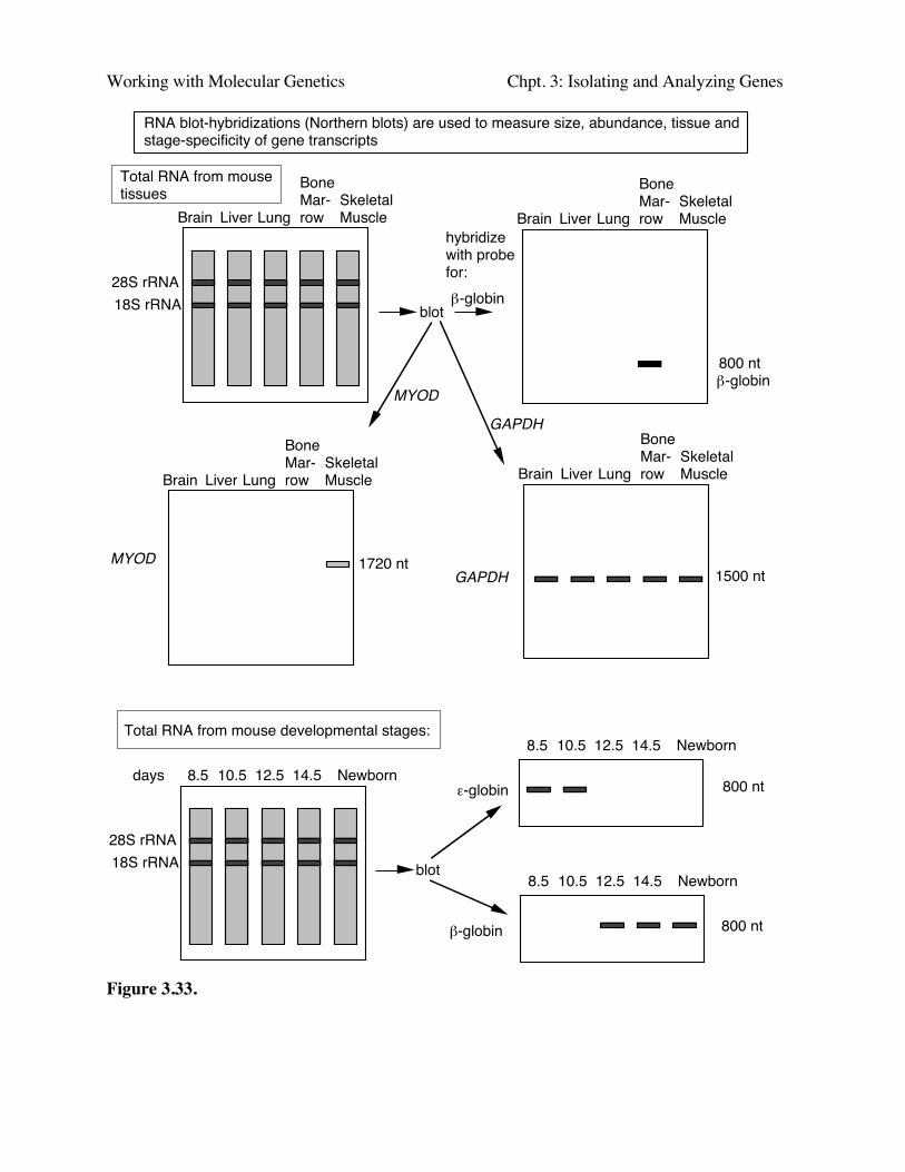
In the reverse of Southern blot-hybridizations, one can separate RNAs by size on adenaturing agarose gel, and transfer them to nylon or other appropriate solid support. LabeledDNA can then be used to visualize the corresponding mRNA (Fig. 3.33). Ed Southern initiallyused labeled rRNA to find the complementary regions in immobilized, digested DNA, so this"reverse" of Southern blot-hybridizations, i.e. using a labeled DNA probe to hybridize toimmobilized RNA, is often referred to as "Northern" blot-hybridizations.
One can hybridize a labeled DNA clone to a panel of RNA samples from a wide variety oftissues to determine in what tissues a particular cloned gene is expressed (top panel of Fig. 3.33.More precisely, this technique reveals the tissues in which the genes is transcribed into stable RNA.The results allow one to determine the tissue specificity of expression, e.g. a gene may only beexpressed in liver, or only in erythroid cells (e.g. the β-globin gene). This helps give some generalidea of the possible function of the gene, since it should reflect the function of that tissue. Othergenes are expressed in almost all cells or tissue types (such as GAPDH); these are referred to ashousekeeping genes. They are involved in functions common to all cells, such as basic energymetabolism, cell structure, etc. The relative amounts of RNA in the different lanes can be directlycompared to see, e.g., which tissues express the gene most abundantly.
One can hybridize a labeled DNA clone to a panel of RNA samples from a progressivestages of development to determine the developmental stage when during development aparticular cloned gene is expressed as RNA (bottom panel of Fig. 3.33). For instance, a geneproduct may be required for determination decisions early in development, and only be expressed inearly embryos.
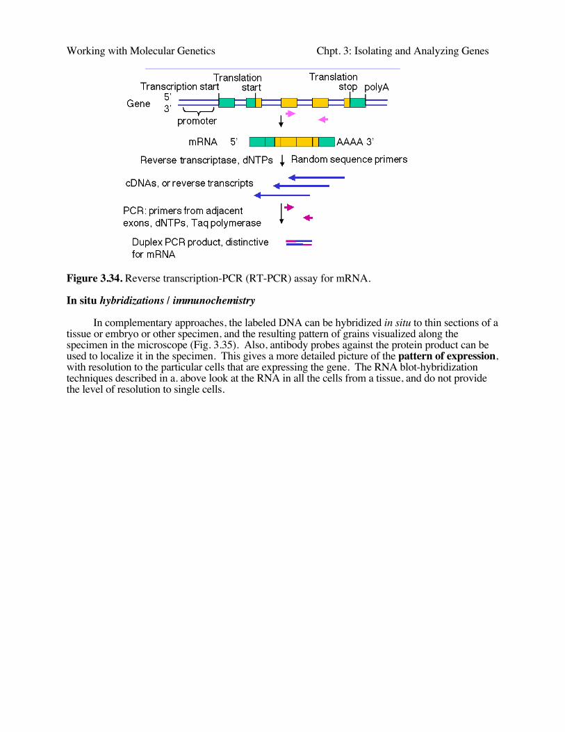
Once the DNA sequence of the gene of interest is known, and its intron-exon structuredetermined, highly sensitive RT-PCR assays can be designed (Fig. 3.34). The RNA from the cellor tissue of interest is copied into cDNA using reverse transcriptase and dNTPs, and then primersare annealed for PCR. Ideally, the primers are in different exons so that the product of amplifyingthe cDNA will be smaller than the product of amplifying the genomic DNA.
Working with Molecular Genetics Chpt. 3: Isolating and Analyzing Genes
RNA blot-hybridizations (Northern blots) are used to measure size, abundance, tissue and stage-specificity of gene transcripts
28S rRNA18S rRNA
Total RNA from mouse tissues
Brain Liver LungSkeletalMuscle
BoneMar-row Brain Liver Lung
SkeletalMuscle
BoneMar-row
Brain Liver LungSkeletalMuscle
BoneMar-row Brain Liver Lung
SkeletalMuscle
BoneMar-row
blot
hybridizewith probefor:
800 nt
1720 nt1500 nt
28S rRNA18S rRNA
Total RNA from mouse developmental stages:
8.5 10.5 12.5 Newborn14.5
blot
days
8.5 10.5 12.5 Newborn14.5
8.5 10.5 12.5 Newborn14.5
800 nt
800 ntβ-globin
ε-globin
β-globin
MYOD
GAPDH
β-globin
MYODGAPDH
Figure 3.33.
Working with Molecular Genetics Chpt. 3: Isolating and Analyzing Genes
Figure 3.34. Reverse transcription-PCR (RT-PCR) assay for mRNA.
In situ hybridizations / immunochemistry
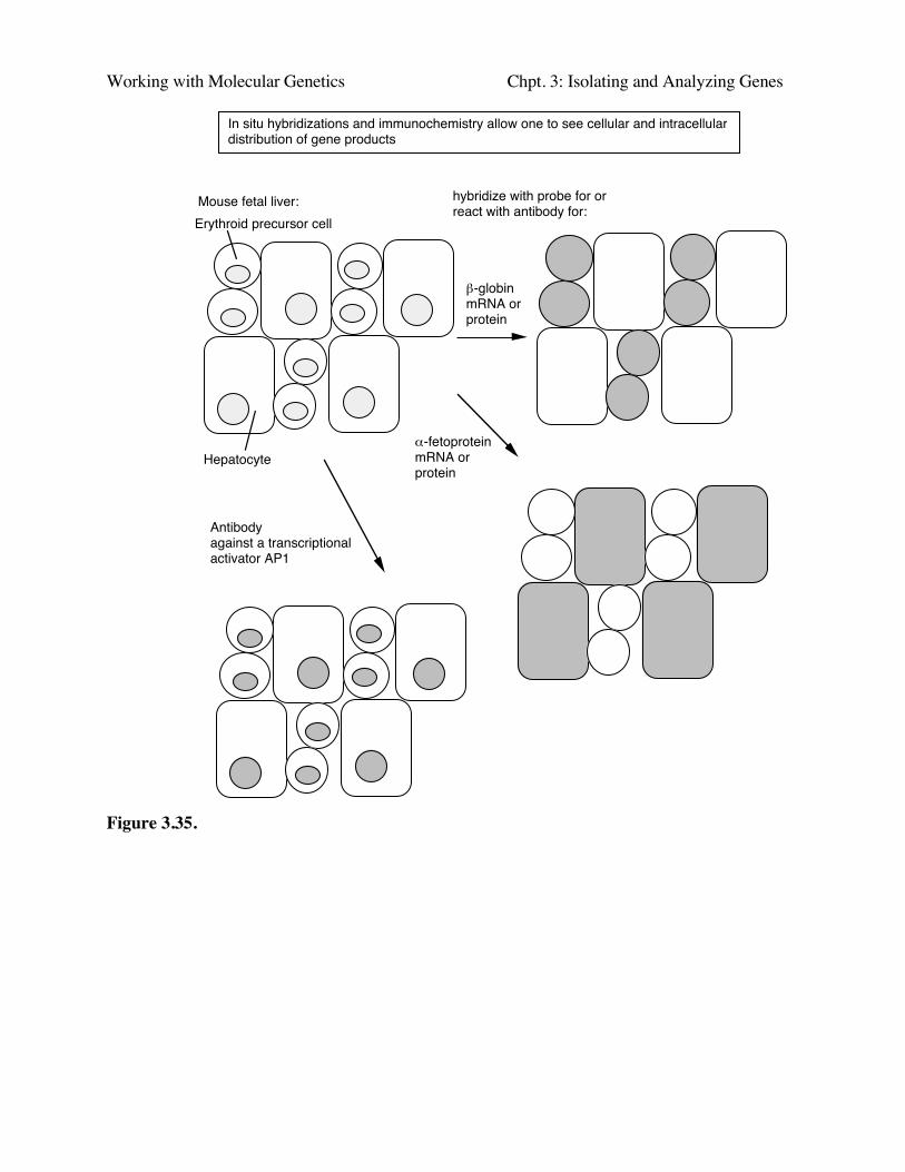
In complementary approaches, the labeled DNA can be hybridized in situ to thin sections of atissue or embryo or other specimen, and the resulting pattern of grains visualized along thespecimen in the microscope (Fig. 3.35). Also, antibody probes against the protein product can beused to localize it in the specimen. This gives a more detailed picture of the pattern of expression,with resolution to the particular cells that are expressing the gene. The RNA blot-hybridizationtechniques described in a. above look at the RNA in all the cells from a tissue, and do not providethe level of resolution to single cells.
Working with Molecular Genetics Chpt. 3: Isolating and Analyzing Genes
Hepatocyte
Erythroid precursor cell
In situ hybridizations and immunochemistry allow one to see cellular and intracellular distribution of gene products
Mouse fetal liver: hybridize with probe for or react with antibody for:
β-globinmRNA or protein
α-fetoproteinmRNA or protein
Antibodyagainst a transcriptionalactivator AP1
Figure 3.35.
Working with Molecular Genetics Chpt. 3: Isolating and Analyzing Genes
Microarrays
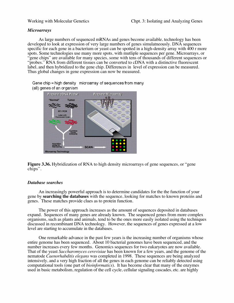
As large numbers of sequenced mRNAs and genes become available, technology has beendeveloped to look at expression of very large numbers of genes simulatneously. DNA sequencesspecific for each gene in a bacterium or yeast can be spotted in a high-density array with 400 r morespots. Some technologies use many more spots, with mutliple sequences per gene. Microarrays, or“gene chips” are available for many species, some with tens of thousands of different sequences or“probes.” RNA from different tissues can be converted to cDNA with a distinctive fluorescentlabel, and then hybridized to the gene chip. Differences in level of expression can be measured.Thus global changes in gene expression can now be measured.
Figure 3.36. Hybridization of RNA to high density microarrays of gene sequences, or “genechips”.
Database searches
An increasingly powerful approach is to determine candidates for the the function of yourgene by searching the databases with the sequence, looking for matches to known proteins andgenes. These matches provide clues as to protein function.
The power of this approach increases as the amount of sequences deposited in databasesexpand. Sequences of many genes are already known. The sequenced genes from more complexorganisms, such as plants and animals, tend to be the ones more easily isolated using the techniquesdiscussed in recombinant DNA technology. However, the sequences of genes expressed at a lowlevel are starting to accumulate in the databases.
One remarkable advance in the past few years is the increasing number of organisms whoseentire genome has been sequenced. About 10 bacterial genomes have been sequenced, and thenumber increases every few months. Genomics sequences for two eukaryotes are now available.That of the yeast Saccharomyces cerevisiae has been known for a few years, and the genome of thenematode Caenorhabditis elegans was completed in 1998. These sequences are being analyzedintensively, and a very high fraction of all the genes in each genome can be reliably detected usingcomputational tools (one part of bioinformatics). It has become clear that many of the enzymesused in basic metabolism, regulation of the cell cycle, cellular signaling cascades, etc. are highly
Working with Molecular Genetics Chpt. 3: Isolating and Analyzing Genes
conserved across a broad phylogenetic spectrum. Thus it is common to find significant sequencematches in the genomes of model organisms when they are queried by the sequence of a previouslyunknown gene, e.g. from humans or mouse. The function already established for that gene inworms or yeast is a highly reliable guide to the function of the homologous gene in humans. Theworm C. elegans is multicellular, and fate of each of its cells during development has been mapped.Thus it is possible that many functions involved in cellular interactions and cell-cell signaling willbe conserved in this species, thus expanding the list of potential targets for a search in thedatabases.
This potential is being realized as working draft sequences of the human and mouse genomesare being analyzed. Within these data is a good approximation of sequences from virtually allhuman and mouse genes. Random clones have been partially sequenced from libraries of cDNAsfrom various human tissues, normalized to remove much of the products of abundant mRNAs andthus increasing the frequency of products of rare mRNAs. These sequences from the ends of thecDNA clones are called expressed sequence tags, or ESTs. The name is derived from the fact thatsince they are in cDNA libraries, they are obviously expressed at the level of mRNA, and some areused as tags in generating high-resolution maps of human chromosome. Hundreds of thousandsof these have now been sequenced in collaborative efforts between pharmaceutical companies, othercompanies and universities. The database dbEST records all those in the public domain, and it is astrong complement to the databases recording all known sequences of genes. Many different partsof the same, or highly related, cDNAs, are recorded as separate entries in dbEST. Projects areunderway to group all the sequences from the same (or highly related) gene into a a unifiedsequence. One example is the Unigene project at NCBI. The number of entries grows continually,but in the summer of 1998 there are about 50,000 entries, each representing about one gene. Thenumber is higher now. Current estimates of the number of human genes are around 30,000, so it ispossible that some UniGene clusters represent only parts of genes, and some genes match morethan one cluster.
Very efficient search engines have been designed for handling queries to thesedatabases, and several are freely available over the World Wide Web. One of the most popular anduseful sites for this and related activities is maintained by the National Center for BiotechnologyInformation (http://www.ncbi.nlm.nih.gov/). Their Entrez browser provides integrated access tosequence, mapping and some functional information, PubMed provides access to abstracts ofpapers in journals in the National Library of Medicine, and the BLAST server allows rapid searchesthrough various sequence databases. dbEST and the Unigene collection are maintained here, manygenome maps are available, and three-dimensional structures of proteins and nucleic acids areavailable.
Make the protein product and analyze it
It is often possible to express the gene and make the encoded protein in large amounts. Theprotein can be purified and assayed for various enzymatic or other activities. Hypotheses for suchactivities may come from database searches.
Directed mutation
The previously describe approaches give some idea about gene function, but they do notfirmly establish those functions. Indeed, this is a modern problem of trying to assign a function toan isolated gene. Several “reverse genetic” approaches can now be taken to tackle this problem.The most powerful approach to determining the physiological role(s) of a gene product is tomutate the gene in an appropriate organism and search for an altered phenotype.
The easiest experiment to do, but sometimes most difficult to interpret, is a gain of function
Working with Molecular Genetics Chpt. 3: Isolating and Analyzing Genes
assay. In this case, one forces expression of the gene in a transgenic organism, which often alreadyhas a wild type copy of the gene. One can look for a phenotype resulting from over-expression intissues where it is normally expressed, or ectopic expression in tissues where it is normally silent.
In some organisms, it is possible to engineer a loss of function of the gene. The mosteffective method is to use homologous recombination to replace the wild type gene with oneengineered to have no function. This knock-out mutation will prevent expression of theendogenous gene and one can see the effects on the whole organism. Unfortunately, the efficiencyof homologous recombination is low in many organisms and cell lines, so this is not alwaysfeasible. Other methods for knocking out expression are being developed, although the mechanismfor their effect (when successful) is still being studied. In some cases, one can block expression ofthe endogenous gene by forcing production of antisense RNA. Another method that is effective insome, but currently not all organisms, is the use of double-stranded, interfering RNA (RNAi).Duplex RNAs less than 30 nucleotide pairs long from the gene of interest can prevent expressionof genes in worms, flies, and plants. Some success in mammals was recently reported.
Another way to generate a loss-of-function phenotype is to express dominant negativealleles of the gene. These mutant alleles encode stable proteins that form an aberrant structure thatprevents functioning of the endogenous protein. This usually requires some protein-proteininteraction (e.g. homodimers or heterodimers).
Localization on a genetic map
Sometimes the gene you have isolated maps to a region on a chromosome with a knownfunction. Of course, many genes are probably located in that region, so it is critical to show that acandidate gene really is the one that when mutated causes an altered phenotype. This can be doneby showing that a wild type copy of the candidate gene will restore a normal phenotype to themutant. If a marker is known to be very tightly linked to the candidate gene, one can test whetherthis marker is always in linkage disequilibrium with the determinant of the mutant phenotype, i.e. ina large number of crosses, the marker for the candidate gene and the mutant phenotype neverseparated by recombination.
The mapping is often done with gene-specific probes for in situ hybridizations to mitoticchromosomes. One then aligns the hybridization pattern with the chromosome banding patterns tomap the isolated gene. Another method is to hybridize to a panel of DNAs from hybrid cells thatcontain only part of the chromosomal complement of the genome of interest. This is particularlypowerful with radiation hybrid panels.
Working with Molecular Genetics Chpt. 3: Isolating and Analyzing Genes
QUESTIONSCHAPTER 3
ISOLATION AND ANALYSIS OF GENES
3.2 Altering the ends of DNA fragments for ligation into vectors.(Adapted from POB)
a) Draw the structure of the end of a linear DNA fragment that was generated bydigesting with the restriction endonuclease EcoRI. Include those sequences remaining from theEcoRI recognition sequence.
b) Draw the structure resulting from the reaction of this end sequence with DNApolymerase I and the four deoxynucleoside triphosphates.
c) Draw the sequence produced at the junction if two ends with the structure derived in (b)are ligated.
d) Design two different short synthetic DNA fragments that would permit ligation ofstructure (a) with a DNA fragment produced by a PstI restriction digest. In one of these syntheticfragments, design the sequence so that the final junction contains the recognition sequences forboth EcoRI and PstI. Design the sequence of the other fragment so that neither the EcoRI nor thePstI sequence appears in the junction.
3.3. What properties are required of vectors used in molecular cloning of DNA?
3.4. A student ligated a BamHI fragment containing a gene of interest to a pUC vector digestedwith BamHI, transformed E. coli with the mixture of ligation products and plated the cells on platescontaining the antibiotic ampicillin and the chromogenic substrate X-gal. Which colonies shouldthe student pick to find the ones containing the recombinant plasmid (with the gene of interest inpUC)?
3.5. Starting with an isolated mRNA, one wishes to make a double stranded copy of the mRNAand insert it at the PstI site of pBR322 via G-C homopolymer tailing. One then transforms E. coliwith this recombinant plasmid, selecting for tetracycline resistance. What are the four enzymaticsteps used in preparing the cDNA insert? Name the enzymes and describe the intermediates.
3.6 A researcher needs to isolate a cDNA clone of giraffe actin mRNA, and she knows the size(Mr = 42,000) and partial amino acid sequence of giraffe actin protein and has specific antibodiesagainst giraffe actin. After constructing a bank of cDNA plasmids from total mRNA of giraffefibroblasts (dG-dC tailed into the PstI site of pBR322), what methods of screening the bank couldbe used to identify the actin cDNA clone?
Working with Molecular Genetics Chpt. 3: Isolating and Analyzing Genes
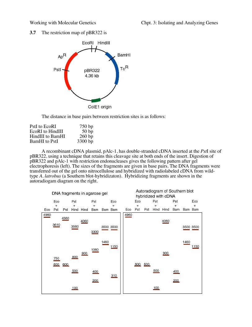
3.7 The restriction map of pBR322 is
The distance in base pairs between restriction sites is as follows:
PstI to EcoRI 750 bpEcoRI to HindIII 50 bpHindIII to BamHI 260 bpBamHI to PstI 3300 bp
A recombinant cDNA plasmid, pAlc-1, has double-stranded cDNA inserted at the PstI site ofpBR322, using a technique that retains this cleavage site at both ends of the insert. Digestion ofpBR322 and pAlc-1 with restriction endonucleases gives the following pattern after gelelectrophoresis (left). The sizes of the fragments are given in base pairs. The DNA fragments weretransferred out of the gel onto nitrocellulose and hybridized with radiolabeled cDNA from wild-type A. latrobus (a Southern blot-hybridizaton). Hybridizing fragments are shown in theautoradiogam diagram on the right.
Working with Molecular Genetics Chpt. 3: Isolating and Analyzing Genes
a) What is the size of the cDNA insert?
b) What two restriction endonucleases cleave within the cDNA insert?
c) For those two restriction endonucleases, each DNA fragment in the single digest is cutby PstI into two DNA fragments in the double digest (i.e. the restriction endonuclease plus PstI).Determine which fragments each single digest fragment is cut into, and use this information toconstruct a map.
d) Draw a restriction map for pAlc-1, showing sites for PstI, EcoRI, BamHI and HindIII.Indicate the distance between sites and show the cDNA insert clearly.
3.8. You isolate and clone a KpnI fragment from A. latrobus genomic DNA that encodes themRNA cloned in pAlc-1 (as analyzed in question 3.7). The restriction map of the genomicfragment is
KpnI * EcoRI
BamHI SalI
HindIII
SalI
KpnI* * *500 400 100 300 600 400
Each fragment that hybridizes to pAlc-1 is indicated by an asterisk. What does this map,especially when compared to that in problem 3.7, tell you about the structure of the gene? Be asquantitative as possible.
3.9. Some particular enzyme is composed of a polypeptide chain of 192 amino acids. The genethat encodes it has 1,440 nucleotide pairs. Explain the relationship between the number of aminoacids in this polypeptide and the number of nucleotide pairs in its gene.
3.10. When viewed in the electron microscope, a hybrid between a cloned giraffe actin gene(genomic DNA) and mature actin mRNA looks like this:
What can you conclude about actin gene structure in the giraffe?
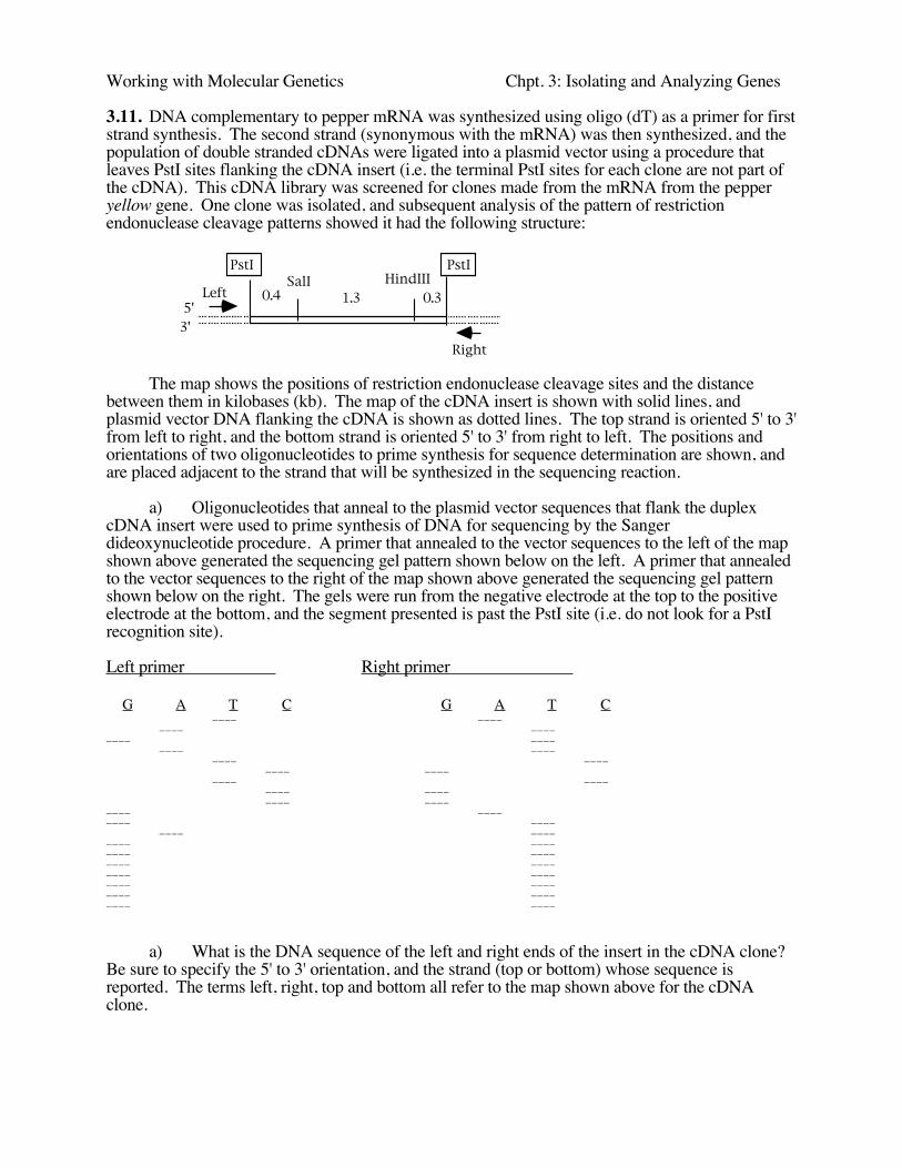
Working with Molecular Genetics Chpt. 3: Isolating and Analyzing Genes
3.11. DNA complementary to pepper mRNA was synthesized using oligo (dT) as a primer for firststrand synthesis. The second strand (synonymous with the mRNA) was then synthesized, and thepopulation of double stranded cDNAs were ligated into a plasmid vector using a procedure thatleaves PstI sites flanking the cDNA insert (i.e. the terminal PstI sites for each clone are not part ofthe cDNA). This cDNA library was screened for clones made from the mRNA from the pepperyellow gene. One clone was isolated, and subsequent analysis of the pattern of restrictionendonuclease cleavage patterns showed it had the following structure:
PstI PstI
0.4 1.3 0.3SalI HindIII
5'3'
Left
Right
The map shows the positions of restriction endonuclease cleavage sites and the distancebetween them in kilobases (kb). The map of the cDNA insert is shown with solid lines, andplasmid vector DNA flanking the cDNA is shown as dotted lines. The top strand is oriented 5' to 3'from left to right, and the bottom strand is oriented 5' to 3' from right to left. The positions andorientations of two oligonucleotides to prime synthesis for sequence determination are shown, andare placed adjacent to the strand that will be synthesized in the sequencing reaction.
a) Oligonucleotides that anneal to the plasmid vector sequences that flank the duplexcDNA insert were used to prime synthesis of DNA for sequencing by the Sangerdideoxynucleotide procedure. A primer that annealed to the vector sequences to the left of the mapshown above generated the sequencing gel pattern shown below on the left. A primer that annealedto the vector sequences to the right of the map shown above generated the sequencing gel patternshown below on the right. The gels were run from the negative electrode at the top to the positiveelectrode at the bottom, and the segment presented is past the PstI site (i.e. do not look for a PstIrecognition site).
Left primer Right primer
G A T C G A T C____ ____
____ ________ ____
____ ________ ____
____ ________ ____
____ ________ ____
____ ________ ____
____ ________ ________ ________ ________ ________ ________ ________ ____
a) What is the DNA sequence of the left and right ends of the insert in the cDNA clone?Be sure to specify the 5' to 3' orientation, and the strand (top or bottom) whose sequence isreported. The terms left, right, top and bottom all refer to the map shown above for the cDNAclone.
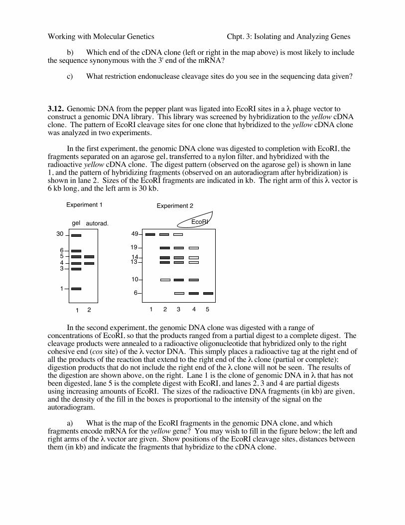
Working with Molecular Genetics Chpt. 3: Isolating and Analyzing Genes
b) Which end of the cDNA clone (left or right in the map above) is most likely to includethe sequence synonymous with the 3' end of the mRNA?
c) What restriction endonuclease cleavage sites do you see in the sequencing data given?
3.12. Genomic DNA from the pepper plant was ligated into EcoRI sites in a λ phage vector toconstruct a genomic DNA library. This library was screened by hybridization to the yellow cDNAclone. The pattern of EcoRI cleavage sites for one clone that hybridized to the yellow cDNA clonewas analyzed in two experiments.
In the first experiment, the genomic DNA clone was digested to completion with EcoRI, thefragments separated on an agarose gel, transferred to a nylon filter, and hybridized with theradioactive yellow cDNA clone. The digest pattern (observed on the agarose gel) is shown in lane1, and the pattern of hybridizing fragments (observed on an autoradiogram after hybridization) isshown in lane 2. Sizes of the EcoRI fragments are indicated in kb. The right arm of this λ vector is6 kb long, and the left arm is 30 kb.
30
6543
1
1 2
Experiment 1 Experiment 2
49
191413
10
6
1 2 3 4 5
gel autorad. EcoRI
In the second experiment, the genomic DNA clone was digested with a range ofconcentrations of EcoRI, so that the products ranged from a partial digest to a complete digest. Thecleavage products were annealed to a radioactive oligonucleotide that hybridized only to the rightcohesive end (cos site) of the λ vector DNA. This simply places a radioactive tag at the right end ofall the products of the reaction that extend to the right end of the λ clone (partial or complete);digestion products that do not include the right end of the λ clone will not be seen. The results ofthe digestion are shown above, on the right. Lane 1 is the clone of genomic DNA in λ that has notbeen digested, lane 5 is the complete digest with EcoRI, and lanes 2, 3 and 4 are partial digestsusing increasing amounts of EcoRI. The sizes of the radioactive DNA fragments (in kb) are given,and the density of the fill in the boxes is proportional to the intensity of the signal on theautoradiogram.
a) What is the map of the EcoRI fragments in the genomic DNA clone, and whichfragments encode mRNA for the yellow gene? You may wish to fill in the figure below; the left andright arms of the λ vector are given. Show positions of the EcoRI cleavage sites, distances betweenthem (in kb) and indicate the fragments that hybridize to the cDNA clone.
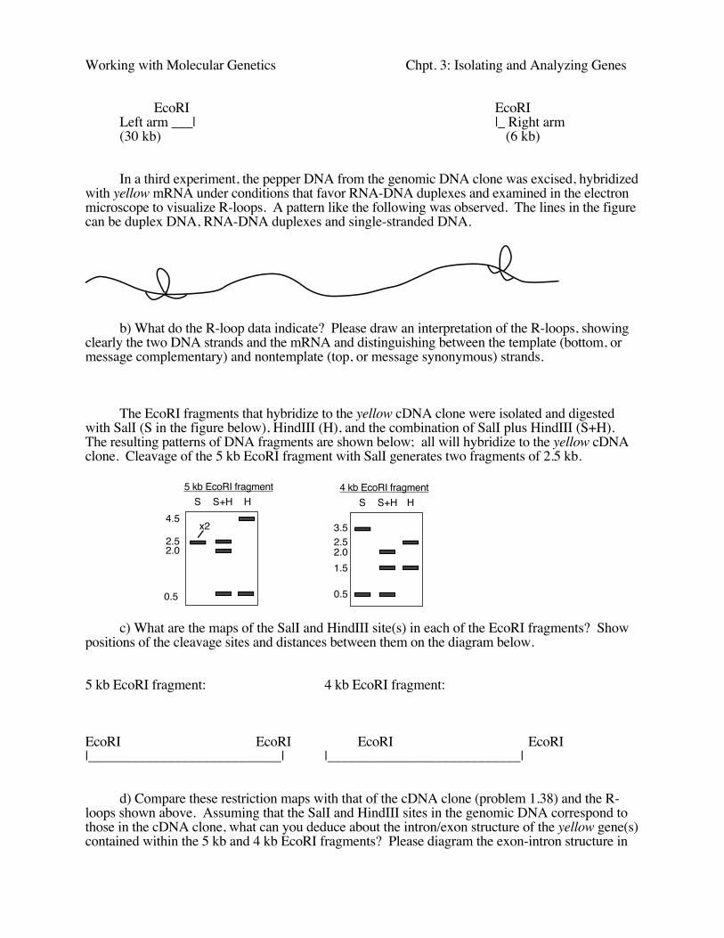
Working with Molecular Genetics Chpt. 3: Isolating and Analyzing Genes
EcoRI EcoRILeft arm ___| |_ Right arm(30 kb) (6 kb)
In a third experiment, the pepper DNA from the genomic DNA clone was excised, hybridizedwith yellow mRNA under conditions that favor RNA-DNA duplexes and examined in the electronmicroscope to visualize R-loops. A pattern like the following was observed. The lines in the figurecan be duplex DNA, RNA-DNA duplexes and single-stranded DNA.
b) What do the R-loop data indicate? Please draw an interpretation of the R-loops, showingclearly the two DNA strands and the mRNA and distinguishing between the template (bottom, ormessage complementary) and nontemplate (top, or message synonymous) strands.
The EcoRI fragments that hybridize to the yellow cDNA clone were isolated and digestedwith SalI (S in the figure below), HindIII (H), and the combination of SalI plus HindIII (S+H).The resulting patterns of DNA fragments are shown below; all will hybridize to the yellow cDNAclone. Cleavage of the 5 kb EcoRI fragment with SalI generates two fragments of 2.5 kb.
S S+H H4.5
2.52.0
0.5
5 kb EcoRI fragment 4 kb EcoRI fragmentS S+H H
3.52.52.0
0.5
x2
1.5
c) What are the maps of the SalI and HindIII site(s) in each of the EcoRI fragments? Showpositions of the cleavage sites and distances between them on the diagram below.
5 kb EcoRI fragment: 4 kb EcoRI fragment:
EcoRI EcoRI EcoRI EcoRI|___________________________| |___________________________|
d) Compare these restriction maps with that of the cDNA clone (problem 1.38) and the R-loops shown above. Assuming that the SalI and HindIII sites in the genomic DNA correspond tothose in the cDNA clone, what can you deduce about the intron/exon structure of the yellow gene(s)contained within the 5 kb and 4 kb EcoRI fragments? Please diagram the exon-intron structure in
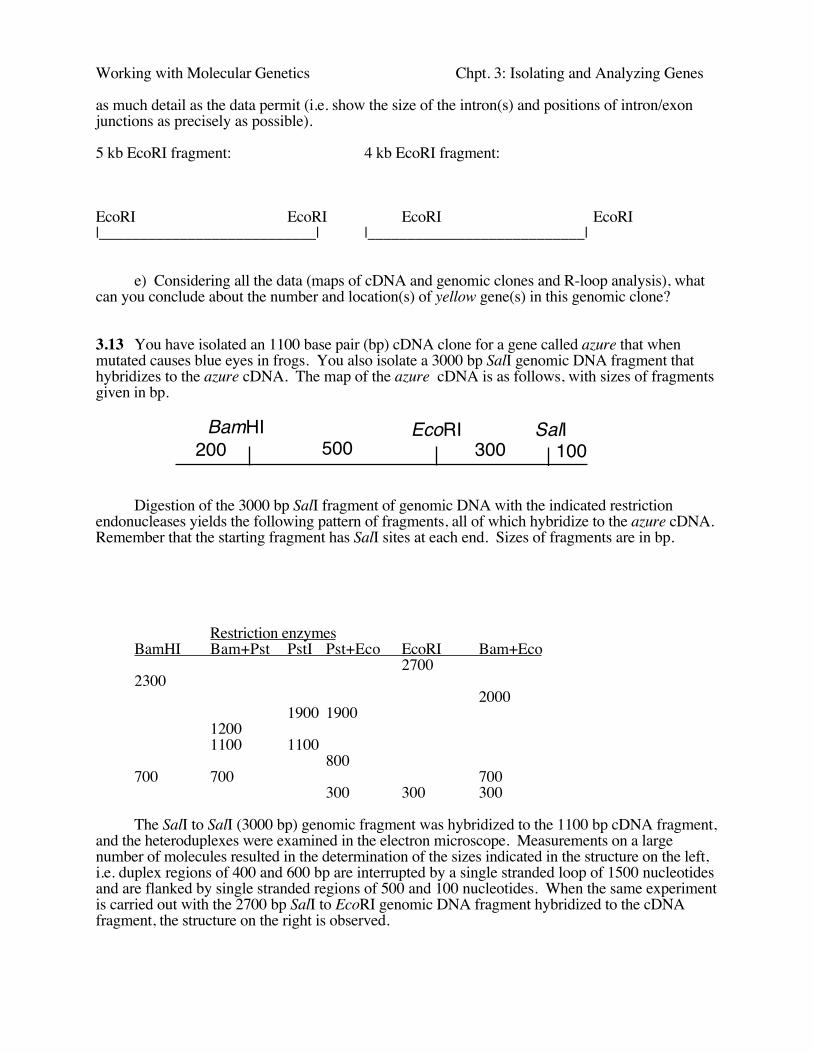
Working with Molecular Genetics Chpt. 3: Isolating and Analyzing Genes
as much detail as the data permit (i.e. show the size of the intron(s) and positions of intron/exonjunctions as precisely as possible).
5 kb EcoRI fragment: 4 kb EcoRI fragment:
EcoRI EcoRI EcoRI EcoRI|___________________________| |___________________________|
e) Considering all the data (maps of cDNA and genomic clones and R-loop analysis), whatcan you conclude about the number and location(s) of yellow gene(s) in this genomic clone?
3.13 You have isolated an 1100 base pair (bp) cDNA clone for a gene called azure that whenmutated causes blue eyes in frogs. You also isolate a 3000 bp SalI genomic DNA fragment thathybridizes to the azure cDNA. The map of the azure cDNA is as follows, with sizes of fragmentsgiven in bp.
BamHI EcoRI SalI200 500 300 100
Digestion of the 3000 bp SalI fragment of genomic DNA with the indicated restrictionendonucleases yields the following pattern of fragments, all of which hybridize to the azure cDNA.Remember that the starting fragment has SalI sites at each end. Sizes of fragments are in bp.
Restriction enzymesBamHI Bam+Pst PstI Pst+Eco EcoRI Bam+Eco
27002300
20001900 1900
12001100 1100
800700 700 700
300 300 300
The SalI to SalI (3000 bp) genomic fragment was hybridized to the 1100 bp cDNA fragment,and the heteroduplexes were examined in the electron microscope. Measurements on a largenumber of molecules resulted in the determination of the sizes indicated in the structure on the left,i.e. duplex regions of 400 and 600 bp are interrupted by a single stranded loop of 1500 nucleotidesand are flanked by single stranded regions of 500 and 100 nucleotides. When the same experimentis carried out with the 2700 bp SalI to EcoRI genomic DNA fragment hybridized to the cDNAfragment, the structure on the right is observed.
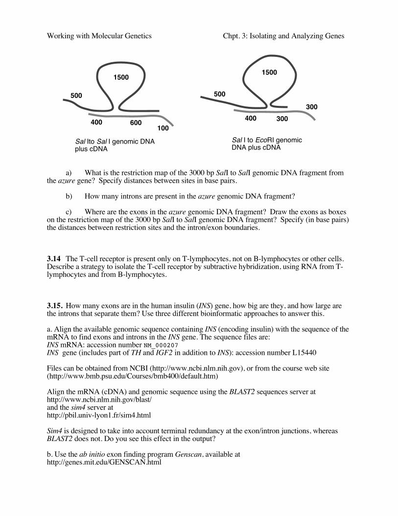
Working with Molecular Genetics Chpt. 3: Isolating and Analyzing Genes
a) What is the restriction map of the 3000 bp SalI to SalI genomic DNA fragment fromthe azure gene? Specify distances between sites in base pairs.
b) How many introns are present in the azure genomic DNA fragment?
c) Where are the exons in the azure genomic DNA fragment? Draw the exons as boxeson the restriction map of the 3000 bp SalI to SalI genomic DNA fragment? Specify (in base pairs)the distances between restriction sites and the intron/exon boundaries.
3.14 The T-cell receptor is present only on T-lymphocytes, not on B-lymphocytes or other cells.Describe a strategy to isolate the T-cell receptor by subtractive hybridization, using RNA from T-lymphocytes and from B-lymphocytes.
3.15. How many exons are in the human insulin (INS) gene, how big are they, and how large arethe introns that separate them? Use three different bioinformatic approaches to answer this.
a. Align the available genomic sequence containing INS (encoding insulin) with the sequence of themRNA to find exons and introns in the INS gene. The sequence files are:INS mRNA: accession number NM_000207INS gene (includes part of TH and IGF2 in addition to INS): accession number L15440
Files can be obtained from NCBI (http://www.ncbi.nlm.nih.gov), or from the course web site(http://www.bmb.psu.edu/Courses/bmb400/default.htm)
Align the mRNA (cDNA) and genomic sequence using the BLAST2 sequences server athttp://www.ncbi.nlm.nih.gov/blast/and the sim4 server athttp://pbil.univ-lyon1.fr/sim4.html
Sim4 is designed to take into account terminal redundancy at the exon/intron junctions, whereasBLAST2 does not. Do you see this effect in the output?
b. Use the ab initio exon finding program Genscan, available athttp://genes.mit.edu/GENSCAN.html
500
400
1500
600100
1500
500
400 300300
Sal Ito Sal I genomic DNA plus cDNA
Sal I to EcoRI genomic DNA plus cDNA

Working with Molecular Genetics Chpt. 3: Isolating and Analyzing Genes
to predict exons in the INS genomic sequence (L15440).How does this compare with the results of analyzing with the program genscan?
c. What do you see for INS at the Human Genome Browser and Ensembl? They are accessed at:http://genome.ucsc.edu/goldenPath/hgTracks.htmlhttp://www.ensembl.org/