werner's syndrome with a report of two cases
TRANSCRIPT
Case Report
Werner’s Syndrome*
With a Report of Two Cases
GLENN W. IRWIN, M.D. and PAULA B. WARD, M.D.
Indianapolis, Indiana
'ERNER' described the association of
W cataracts and scleroderma in four
Kugei2 siblings in 1904. Oppenheimer and
reported the first two cases in the American literature in 1934 and summarized the salient clinical features which they named Werner’s syndrome. Thannhauser3 reviewed twenty-six cases including four of his own re- ported by 1945 and emphasizedth at the skin changes were not true scleroderma. Maeder4 reviewed the literature and collected forty- seven cases in 1949. Since then eight additional cases have been recorded, not including the two cases in this report.6-s
The important features of Werner’s syndrome are listed in Table I.
CASE REPORTS
CASE I. A. M., a forty-seven year old farmer, was admitted to the Indiana University Medical Center on October 19, 1950, with the chief complaint of ulceration of his feet. His growth was considered normal until he reached the age of sixteen, after which he failed to increase in height or weight. At the age of thirty-one bilateral cataracts developed and the patient had a right cataract extraction. About the same time painful callosities of the feet developed which ulcerated. For several years the ulcers would alternately heal and recur. During his early twenties the patient’s hair became gray and progressive baldness ensued. He also noted progressive loss of hair over his eyebrows, face, axillae and pubic region.
Family history revealed the patient had seven siblings, none of whom had any features of Werner’s syndrome. His father and mother were both normal and lived to the age of eighty- four years. There was no family history of dwarfism, cataracts, diabetes or vascular disease.
Physical examination revealed the patient to be only 5 feet tall and to weigh 90 pounds. He appeared much older than his stated age. His trunk was developed normally but his extremi- ties were slender. His scalp hair was gray, sparse and receding. There was a left cataract and
TABLE I MANIFESTATIONS OF WERNER’S SYNDROME
I. Characteristic habitus and stature 1. Short stature beginning with adolescence 2. Slender extremities with stocky trunk 3. Beak-shaped nose
n. Premature senility 1. Premature gray hair 2. Premature baldness 3. Atrophic skin 4. Weak and high pitched voice 5. Arteriosclerosis 6. Juvenile cataracts
III. Scleroderma-like changes 1. Atrophic skin and subcutaneous tissue 2. Circumscribed hyperkeratosis 3. Skin tight over bones of the feet 4. Ulcers over malleoli of ankles, Achilles tendon,
heels and toes IV. Other manifestations
1. Tendency to diabetes mellitus 2. Hypogonadism 3. Osteoporosis 4. Localized calcification 5. Tendency to occur in siblings
evidence of a cataract extraction on the right. Ophthalmoscopic examination on the right was essentially normal. The eyebrows were practi- cally absent as was his beard. His nose was small and beak-shaped; his mouth was small. The skin of the face was shiny, thin and seemed taut. The subcutaneous tissue was small in amount, however, the skin was freely movable. There was slight gynecomastia. The heart was not enlarged. There was a grade I systolic murmur at the aortic area. There were no other murmurs or abnormal accentuations. The blood pressure
* From The Department of Medicine, Indiana University Medical Center, Indianapolis, Ind.
266 AMERICAN JOURNAL OF MEDICINE
Werner’s Syndrome-h&z, Ward 267
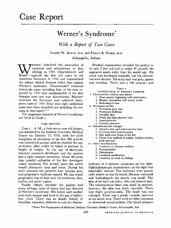
FIG. 1.
was 150/70 in both arms and the heart rate was regular and 80/minute. There was no axillary hair and the pubic hair was very sparse. The external genitalia were underdeveloped. There was atrophy of the skin, subcutaneous tissue and muscles of the lower extremities. The upper extremities were similarly involved but to a lesser degree. Areas of hyperkeratosis were present over the soles of the feet. The bones of the feet were prominent and the skin was stretched tightly over them. Large ulcers were present over both Achilles tendons and smaller ulcers were present over the internal malleoli and soles of the feet. The toes were only slightly movable because of the tight skin. The toe nails were absent except on the large toes. The terminal phalanges of the smaller toes were markedly atrophic. The dorsalis pedis and
AUGUST, 1953
posterior tibia1 arteries were palpable bilaterally. Oscillometric readings were essentially normal in the lower extremities except for the feet in which they were diminished. (Fig. 1.)
Laboratory studies included a normal uri- nalysis and a negative blood Wassermann test. The hemoglobin was 11 gm. per cent and the red blood count was 4.6 million. The white blood count was 7,500 with a normal differen- tial. Sedimentation rate was 5 and the hemato- crit 42. Total serum cholesterol was 263 mg. per cent and cholesterol esters determination was 158 mg. per cent. Serum protein-bound iodine was 4.1 gamma per 100 cc. Serum albumin was 4.5 gm. per cent and serum globulin was 2.5 gm. per cent. Non-protein nitrogen was 32 mg. per cent. Serum chloride was 98 mEq., serum sodium was 137 mEq. and serum potassium

Werner’s Syndrdme-hwin, Ward
was 5 mEq. Serum phosphorus was 4 mg. per cent, serum alkaline phosphatase was 2.4 King- Armstrong units and serum calcium was 10 mg. per cent. After being placed on a 150 mg. cal- cium diet for five days the total twenty-four-hour urinary excretion of calcium was 132 mg. for one day and 141 mg. on the following day. Two basal metabolic rates were plus 5 and minus 4. The thyroid uptake of 1131 at the end of forty-eight hours was 24 per cent. The 17- ketosteroid determination was low on two occasions and was 2.6 mg./24 hours and 5.8 mg./24 hours (method of Holtorff and Koch). The urinary reducing steroid determination was 3.0 mg./24 hours (method of Heard and Sobel). A four-hour Thorn test revealed an absolute eosinophil count of 200 before ACTH and a count of zero four hours after 25 mg. of ACTH was administered. A twenty-four-hour urinary estrogen determination was positive for 3.9 rat units and negative for 24 rat units. A twenty-four-hour urinary gonadotropin determi- nation was positive at 12 rat units and negative for 24 rat units. A glucose tolerance test re- vealed the following blood sugar values: fasting, 98 mg.; half an hour, 210 mg.; one hour, 262 mg.; two hours 152 mg.; and three hours, 136 mg. Sugar, 0.2 per cent, was present in the urine at two and three hours. Ultracentrifuge studies of the serum revealed the Sf 12-20 value to be 17 mg. per cent and the Sr 20-100 value to be 56 mg. per cent. Electrocardiogram was normal.
X-ray of the chest was normal except for moderate calcification of the aorta. There was marked calcific change in the tendons of the heel and foot. Moderate calcification of the arteries of the lower extremities was noted. There was generalized osteoporosis. X-rays of the skull were normal.
CASE II. E. H., a thirty-nine year old house- wife, was admitted to the Indiana University Medical Center on January 9, 1952. She stated that her growth had been normal until she reached the age of thirteen to fifteen,’ at which time she failed to increase in height or in weight. Her mother confirmed that she had been normal until that age. At the age of thirty-four bilateral cataracts developed which were extracted. About this time the patient noted that her scalp hair became progressively sparse and her hairline had receded. She also was aware of fast heart action and a blushing of the face and neck when she became excited. This patient had had eight pregnancies, with six living children
ranging in age from six to twenty-one years. During her last pregnancy she was told she had hypertension; she had much edema and con- vulsions occurred prior to delivery.
Family history revealed that she was the smallest person in her family. Two sisters were of normal development. The patient’s twelve year old daughter was over 5 feet tall and weighed more than 100 pounds. There was no family history of cataracts, diabetes, heart or vascular disease, or shortness of stature.
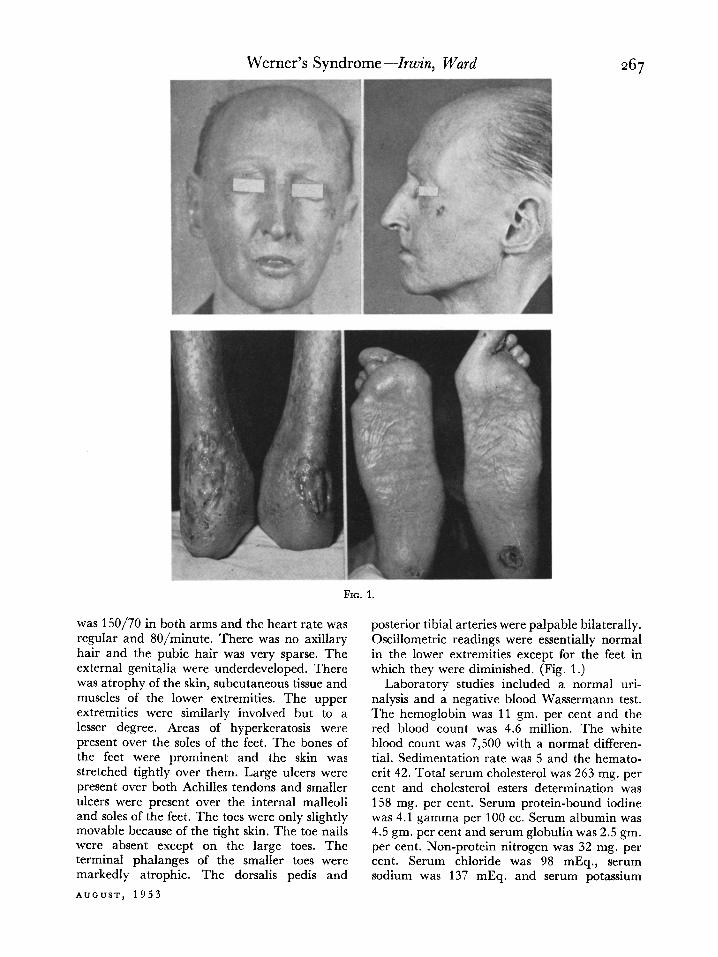
Physical examination revealed her height was only 4 feet 8 inches and her weight was 80 pounds. Her upper and lower extremities were small and spindly whereas her breasts were well developed and her abdomen obese and flabby. She appeared older than thirty-nine. Her hairline receded and her hair was sparse over the scalp and eyebrows. There was evi- dence of bilateral cataract extraction. Oph- thalmoscopic examination revealed minimal segmental constriction of the arterioles, slight increase in light reflex and minimal A-V com- pression. Vision was 20-30 bilaterally with cor- rection. The patient was edentulous. The skin over the face was thin and moved freely. Her voice was high pitched and laryngeal examina- tion revealed the mucous membrane over the vocal cords to be atrophic and thin. The lungs were normal and the heart was not enlarged. There was a grade II systolic murmur which was maximal at the aortic area. The aortic second sound was louder than the pulmonic second sound. Blood pressure was 220/70-O bilaterally in the upper extremities and 240/l 10 in the legs. The pulse was regular and varied from 76 to lbO/minute. The patient frequently demon- strated the flushing of face and neck, and other features characteristic of the diencephalic syn- drome. After amytal the blood pressure was 110/76. Histamine, benzodioxane and regitine caused no significant change in blood pressure. There was a rectocele and cystocele. The skin over the feet was thin and moved freely over the subcutaneous tissue. There were numerous callosities but no frank ulceration. The peripheral arterial pulsations were normal. (Fig. 2.)
Laboratory studies included normal urinalyses and a negative blood Wassermann test. The hemoglobin was 11.5 gm. per cent and the red blood count was 4.05 million. The white blood count was 6,200 with a normal differential. Sedimentation rate was 44 and the hematocrit 41. Total serum cholesterol was 218 mg. per
AMERICAN JOURNAL OF MEDICINE
Werner’s Syndrome--lrwin, Ward 269
FIG. 2.
cent. Serum protein-bound iodine was 5.2 gamma per 100 cc. Serum albumin was 4.7 gm. per cent and serum globulin was 1.9 gm. per cent. Non-protein nitrogen was 28 mg. per cent. Serum chloride was 104 mEq., serum sodium was 140 mEq., and serum potassium was 4.2 mEq. Serum phosphorus was 3.2 mg. per cent, serum alkaline phosphatase was 2.9 King- Armstrong units and serum calcium was 11 mg. per cent. After being placed on a 150 mg. cal- cium diet for four days the total twenty-four- hour urinary excretion of calcium was 139 mg. for one day and 126 mg. on the following day. Two basal metabolic rates were minus 6 per cent and minus 1 per cent. The uptake of 1131 over the thyroid at the end of forty-eight hours was 29 per cent. The 17-ketosteroid determina- tion was 14 mg./24 hours (method of Holtorff and Koch). The urinary reducing steroid determination was 2.1 mg./24 hours [method of Heard and Sobel). A four-hour Thorn test revealed an absolute eosinophil count of 154 before ACTH and a count of 21 four hours after 25 mg. of ACTH was administered. A twenty- four-hour urinary estrogen determination was
AUGUST, 1953
negative for 4.6 rat units. A twenty-four-hour urinary gonadotropin determination was posi- tive at 14 rat units and negative for 28 rat units. A glucose tolerance test revealed the following blood sugar values: fasting, 102 mg.; one-half hour, 198 mg.; one hour, 192 mg.; two hours, 150 mg.; and three hours, 146 mg. There was no glycosuria. Ultracentrifuge studies of the serum revealed the Si 12-20 value to be 4.5 mg. per cent and the Sf 20-100 value to be 219 mg. per cent. Electrocardiogram was normal.
X-ray and fluoroscopy of the chest revealed marked calcification of the entire thoracic aorta. The heart and lungs were otherwise normal. There was minimal calcification of the vessels of the lower extremities. A barium enema,
. . upper gastromtestmal series and intravenous pyelogram were normal except for a small esophageal diverticulum in the middle portion of the esophagus. Moderate osteoporosis of the spine was noted.
COMMENTS
The diagnosis of Werner’s syndrome in general is not difficult if the physician is aware of this

270 Werner’s Syndrome-I&n, Ward
FIG. 3.
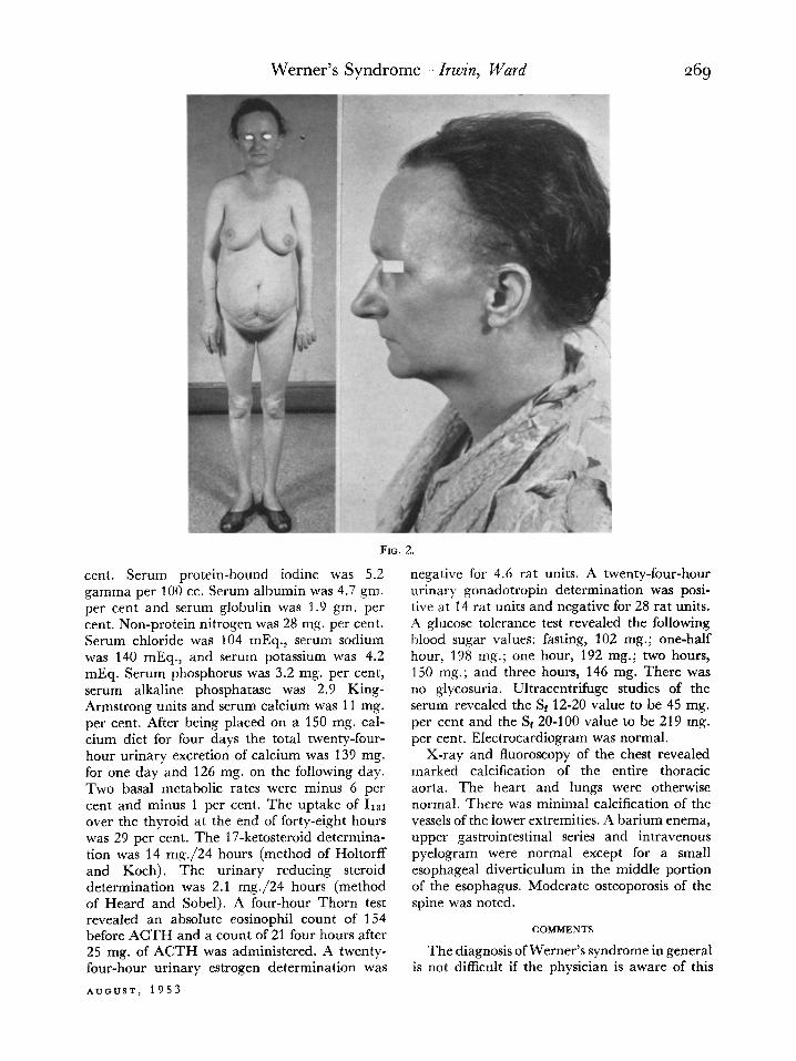
entity. Usually the general inspection of the patient’s facies, premature senility and body habitus reveals the characteristic features previ- ously outlined. Cases may be overlooked when one manifestation is predominant, and thus they may be considered cases of juvenile cataract, scleroderma, generalized arteriosclero- sis or a primary endocrine disorder. An incom- plete form may exist and our second case is an example in that frank skin ulceration and gray- ing of the hair have not developed to date. Clinically our cases support the opinion of Thannhauser3 that the skin changes are not true scleroderma. Their skin was thin but freely movable. The subcutaneous tissue especially of the face and feet was small in amount and the underlying muscle appears atrophic. Hyper- keratosis was prominent. Raynaud’s phenome- non was not present. A biopsy from our first patient revealed hyperkeratosis with a decrease in size of the rete pegs. The dermis was essen- tially normal in thickness and the collagen bundles were loosely and disorderly arranged. (Fig. 3.) This is distinct from scleroderma in which the dermis is increased in thickness and the collagen bundles are hypertrophic and densely packed causing obliterative changes of dermal vessels.
Although several endocrine abnormalities have been associated with Werner’s syndrome, there is no evidence that they are of primary etiologic significance. Diabetes mellitus is fre- quently associated with Werner’s syndrome. In
our review of fifty-five cases reported in the literature eleven patients were considered to have diabetes mellitus. Our patients did not have symptoms of diabetes or a fasting hyper- glycemia, although a glucose tolerance test did indicate a mild decrease in tolerance to glucose in both cases. A determination of urinary reduc- ing steroids (methods of Heard and Sobel) was normal for the second patient and was slightly elevated for the first patient who had the greater decrease in glucose tolerance. Clinically our cases were euthyroid and thyroid function was normal according to basal meta- bolic rates, serum cholesterol values, protein- bound iodine determinations and Ii31 uptake and excretion studies. The majority of the cases reported in the literature have had hypogonad- ism. There was clinical evidence of hypogonad- ism in our first case and the 17-ketosteroid determination was low. This man had mild gynecomastia, although the urinary excretion of estrogens was not high. The urinary gonado- tropin excretion in this case was considered normal. Our second patient was seen during the menopause and her urinary estrogen excre- tion was very low and urinary gonadotropin excretion was considered within normal limits. This patient was unusual in that she had had six normal pregnancies and the menopause did not occur until about the age of thirty-eight. Usually women with Werner’s syndrome men- struate for only a few years, although several cases in the literature have had multiple
AMERICAN JOURNAL OF MEDICINE

Werner’s Syndrome-bin, Ward 271 pregnancies. Clinically we wondered whether adrenal cortical insufficiency existed in our patients; however, this was not substantiated by normal serum electrolytes, a significant fall in eosinophils after ACTH, absence of low urinary reducing steroids, normal basal metabolic rates and absence of flat oral glucose tolerance tests. The first patient with the low 17-ketosteroid value and the normal gonadotropin value was thought most likely to have primary gonadal insufficiency rather than adrenal cortical in- sufficiency. Oppenheimer and Kuge12 believed that hyperparathyroidism might be a significant entity in this condition but this has never been substantiated. Our patients did not demon- strate a parathyroid disturbance in that re- peated serum calcium, phosphorus and alkaline phosphatase determinations were normal. The urinary excretion of calcium while the patients were on a 150 mg. calcium diet was also within normal limits. Osteoporosis has been reported frequently in Werner’s syndrome and was present in our two patients. Dr. John Gofman very kindly determined the values of S, 12-20 and St 20-100 concentration in the sera of our patients. It is interesting that these fractions were normal for the man but were quite high for the woman. Dr. Gofman informed us that there was some evidence to suggest that the aforementioned fractions have a tendency to become lower in men after the age of sixty-five whereas in women the values tend to increase after the age of sixty-five. Physiologically our man was perhaps comparable to a man near eighty years of age.
In our cases there was no familial history of this syndrome. However, in the majority of reported cases a hereditary factor was present with a tendency for occurrence in siblings.
Effective treatment of Werner’s syndrome is limited. Of course cataract extraction and management of diabetes mellitus when present are indicated. Treatment of the ulcers of the skin has not been very satisfactory. Sympa- thectomy has been reported as not being success- ful.’ Paravertebral sympathetic blocks also have been reported as having no benefit.b Thann- hauser believed that skin grafting was at least temporarily successful. We used testosterone in our case of hypogonadism; however, there has not been significant improvement after two years.
AUGUST, 1953
SUMMARY
Two cases of Werner’s syndrome are pre- sented, with a discussion of the important clinical features of this entity which include short stature, premature senility, juvenile cata- racts, scleroderma-like changes, skin ulcers over the feet and ankles, and certain endocrine abnormalities.
The diagnosis depends upon the physician’s awareness of this entity and can usually be made by general inspection of the patient.
The etiology of Werner’s syndrome is un- known. Hereditary factors seem to be important. Although endocrine abnormalities are usually present, they are variable in their occurrence and probably are of no etiologic significance.
An endocrine evaluation of our patients revealed that both had a decrease in glucose tolerance. One patient had clinical hypogonad- ism and both had osteoporosis. There was no evidence of primary thyroid, parathyroid or pituitary abnormality.
In both cases there was evidence of arterio- sclerosis. The Sr 12-20 and Sf 20-100 values of the sera were normal in one patient and elevated in another.
Treatment is not specific and is directed toward manifestations such as cataracts, diabetes mellitus, skin ulcers and hypogonadism.
REFERENCES
1. WERNER, C. W. 0. eber Katarakt in Verbindung mit Sclerodermie. Inaug. Dissert. Kiel, 1904.
2. OPPENHEIMJIR, B. S. and KUGEL, V. H. Werner’s syndrome, a heredofamilial disorder with sclero- derma, bilateral juvenile cataract, precocious gray- ing of the hair and endocrine stigmatization. Tr. A. Am. Physician, 49: 358, 1934.
3. THANNHAUSER, S. J. Werner’s syndrome and Roth- mund’s syndrome. Ann. Znt. Med., 23: 559, 1945.
4. MAEDER, G. Le syndrome de Rothmund et le syn- drome de Werner. Ann. d’ocul., 182: 809, 1949.
5. POMERANZ, M. M. Werner’s syndrome: case report. Radiolog, 51: 521, 1948.
6. SHEETS, R. F. Werner’s syndrome (progeria of the adult). Am. Pratt. @ Digest Treat., 1: 390, 1950.
7. BOATMIRIGHT, H., WHEELER, C. E. and CAWLEY, E. P. Werner’s syndrome. Arch. ht. Med., 90: 243, 1952.
8. FORCE, B. R. and POWELL, C. F. Progeria in the adult (Werner’s syndrome). U. S. Armed Forces M. J., 1: 578, 1950.
9. BRINK, A. J. and FINDLAY, C. H. Werner’s syndrome: ulcerating atrophodermia, cataract and premature senility; report of a case. South African M. J.. 24: 318. 1950.