weed could block h.i.v.’s spread. no, seriously. science news blog 20140217.docx · web viewthe...
TRANSCRIPT

1 5/12/23 Name Student number http://www.eurekalert.org/pub_releases/2014-02/uom-mns021014.php
Massive neutrinos solve a cosmological conundrumMass of ghostly sub-atomic particles called neutrinos deduced by combining data
Scientists have solved a major problem with the current standard model of cosmology identified by combining results from the Planck spacecraft and measurements of gravitational lensing in order to deduce the mass of ghostly sub-atomic particles called neutrinos.The team, from the universities of Manchester and Nottingham, used observations of the Big Bang and the curvature of space-time to accurately measure the mass of these elementary particles for the first time.The recent Planck spacecraft observations of the Cosmic Microwave Background (CMB) – the fading glow of the Big Bang – highlighted a discrepancy between these cosmological results and the predictions from other types of observations.The CMB is the oldest light in the Universe, and its study has allowed scientists to accurately measure cosmological parameters, such as the amount of matter in the Universe and its age. But an inconsistency arises when large-scale structures of the Universe, such as the distribution of galaxies, are observed.Professor Richard Battye, from The University of Manchester School of Physics and Astronomy, said: "We observe fewer galaxy clusters than we would expect from the Planck results and there is a weaker signal from gravitational lensing of galaxies than the CMB would suggest."A possible way of resolving this discrepancy is for neutrinos to have mass. The effect of these massive neutrinos would be to suppress the growth of dense structures that lead to the formation of clusters of galaxies."Neutrinos interact very weakly with matter and so are extremely hard to study. They were originally thought to be massless but particle physics experiments have shown that neutrinos do indeed have mass and that there are several types, known as flavours by particle physicists. The sum of the masses of these different types has previously been suggested to lie above 0.06 eV (much less than a billionth of the mass of a proton).In this paper, Professor Battye and co-author Dr Adam Moss, from the University of Nottingham, have combined the data from Planck with gravitational lensing observations in which images of galaxies are warped by the curvature of space-time. They conclude that the current discrepancies can be resolved if massive neutrinos are included in the standard cosmological model. They estimate that the sum of masses of neutrinos is 0.320 +/- 0.081 eV (assuming active neutrinos with three flavours).Dr Moss said: "If this result is borne out by further analysis, it not only adds significantly to our understanding of the sub-atomic world studied by particle physicists, but it would also be an important extension to the standard model of cosmology which has been developed over the last decade."The paper is published in Physical Review Letters and has been selected as an Editor's choice.A copy of the paper is available from http://arxiv.org/abs/1308.5870 or http://prl.aps.org/abstract/PRL/v112/i5/e051303
http://www.eurekalert.org/pub_releases/2014-02/miot-gme021014.phpGiant mass extinction may have been quicker than previously thought
MIT researchers find that the end-Permian extinction happened in 60,000 years -- much faster than earlier estimates
The largest mass extinction in the history of animal life occurred some 252 million years ago, wiping out more than 96 percent of marine species and 70 percent of life on land - including the largest insects known to have inhabited the Earth. Multiple theories have aimed to explain the cause of what's now known as the end-Permian extinction, including an asteroid impact, massive volcanic eruptions, or a cataclysmic cascade of environmental events. But pinpointing the cause of the extinction requires better measurements of how long the extinction period lasted.Now researchers at MIT have determined that the end-Permian extinction occurred over 60,000 years, give or take 48,000 years - practically instantaneous, from a geologic perspective. The new timescale is based on more precise dating techniques, and indicates that the most severe extinction in history may have happened more than 10 times faster than scientists had previously thought. "We've got the extinction nailed in absolute time and duration," says Sam Bowring, the Robert R. Shrock Professor of Earth and Planetary Sciences at MIT. "How do you kill 96 percent of everything that lived in the oceans in tens of thousands of years? It could be that an exceptional extinction requires an exceptional explanation."In addition to establishing the extinction's duration, Bowring, graduate student Seth Burgess, and a colleague from the Nanjing Institute of Geology and Paleontology also found that, 10,000 years before the die-off, the oceans experienced a pulse of light carbon, which likely reflects a massive addition of carbon dioxide to the atmosphere. This dramatic change may have led to widespread ocean acidification and increased sea temperatures by 10 degrees Celsius or more, killing the majority of sea life.

2 5/12/23 Name Student number But what originally triggered the spike in carbon dioxide? The leading theory among geologists and paleontologists has to do with widespread, long-lasting volcanic eruptions from the Siberian Traps, a region of Russia whose steplike hills are a result of repeated eruptions of magma. To determine whether eruptions from the Siberian Traps triggered a massive increase in oceanic carbon dioxide, Burgess and Bowring are using similar dating techniques to establish a timescale for the Permian period's volcanic eruptions that are estimated to have covered over five million cubic kilometers."It is clear that whatever triggered extinction must have acted very quickly," says Burgess, the lead author of a paper that reports the results in this week's Proceedings of the National Academy of Sciences, "fast enough to destabilize the biosphere before the majority of plant and animal life had time to adapt in an effort to survive."Pinning dates on an extinctionIn 2006, Bowring and his students made a trip to Meishan, China, a region whose rock formations bear evidence of the end-Permian extinction; geochronologists and paleontologists have flocked to the area to look for clues in its layers of sedimentary rock. In particular, scientists have focused on a section of rock that is thought to delineate the end of the Permian, and the beginning of the Triassic, based on evidence such as the number of fossils found in surrounding rock layers.Bowring sampled rocks from this area, as well as from nearby alternating layers of volcanic ash beds and fossil-bearing rocks. After analyzing the rocks in the lab, his team reported in 2011 that the end-Permian likely lasted less than 200,000 years. However, this timeframe still wasn't precise enough to draw any conclusions about what caused the extinction.Now, the team has revised its estimates using more accurate dating techniques based on a better understanding of uncertainties in timescale measurements.With this knowledge, Bowring and his colleagues reanalyzed rock samples collected from five volcanic ash beds at the Permian-Triassic boundary. The researchers pulverized rocks and separated out tiny zircon crystals containing a mix of uranium and lead. They then isolated uranium from lead, and measured the ratios of both isotopes to determine the age of each rock sample.From their measurements, the researchers determined a much more precise "age model" for the end-Permian extinction, which now appears to have lasted about 60,000 years — with an uncertainty of 48,000 years — and was immediately preceded by a sharp increase in carbon dioxide in the oceans.'Spiraling toward the truth'The new timeline adds weight to the theory that the extinction was triggered by massive volcanic eruptions from the Siberian Traps that released volatile chemicals, including carbon dioxide, into the atmosphere and oceans. With such a short extinction timeline, Bowring says it is possible that a single, catastrophic pulse of magmatic activity triggered an almost instantaneous collapse of all global ecosystems.To confirm whether the Siberian Traps are indeed the extinction's smoking gun, Burgess and Bowring plan to determine an equally precise timeline for the Siberian Traps eruptions, and will compare it to the new extinction timeline to see where the two events overlap. The researchers will investigate additional areas in China to see if the duration of the extinction can be even more precisely determined."We've refined our approach, and now we have higher accuracy and precision," Bowring says. "You can think of it as slowly spiraling in toward the truth."
http://www.eurekalert.org/pub_releases/2014-02/dumc-yua021014.phpYoung, unvaccinated adults account for severest flu cases
Patients who had not been vaccinated had severe cases and needed the most intensive treatmentDURHAM, N.C. – A snapshot of patients who required care at Duke University Hospital during this year's flu season shows that those who had not been vaccinated had severe cases and needed the most intensive treatment.In an analysis of the first 55 patients treated for flu at the academic medical center from November 2013 through Jan. 8, 2014, Duke Medicine researchers found that only two of the 22 patients who required intensive care had been vaccinated prior to getting sick. The findings were published online in Monday, Feb. 10, 2014, in the American Journal of Respiratory and Critical Care Medicine."Our observations are important because they reinforce a growing body of evidence that the influenza vaccine provides protection from severe illness requiring hospitalizations," said lead author Cameron Wolfe, M.D., assistant professor of medicine at Duke. "The public health implications are important, because not only could a potentially deadly infection be avoided with a $30 shot, but costly hospitalizations could also be reduced."Wolfe said this year's flu season was marked by hospitalizations of previously healthy young people, with a median age of 28.5 years. Among those who were hospitalized at Duke, 48 of the 55 were infected with the H1N1 virus that caused the 2009 pandemic. That outbreak also hit young adults particularly hard.

3 5/12/23 Name Student number "We observed a high percentage of hospitalized patients for influenza requiring ICU level care, which appears higher than observed in our hospital during the 2009 pandemic flu season," said co-author John W. Hollingsworth, M.D., associate professor of medicine at Duke. "It remains unclear whether the high rate of ICU admissions represents a diagnosis bias or whether the severity of illness being caused by the current H1N1 virus is higher."Of the 33 patients admitted to regular wards rather than the ICU at Duke University Hospital, only eleven had been vaccinated; most of those were immune compromised, chronically ill, or were on a medication that weakened the vaccine's protection.The study also echoes other studies that have highlighted problems with a rapid test for influenza. Wolfe said 22 of the patients treated at Duke University Hospital had been given a rapid influenza test that came up negative for flu, but they were actually positive when tested by other methods. As a result, they had not received anti-viral medications that might have eased flu symptoms had they been taken early."Together, our observations during this influenza season support a high prevalence of the H1N1 virus affecting young adults and requiring ICU care, high false negative rates of rapid flu tests, and delay in starting antiviral treatment," Wolfe said. "Added to the finding of very low vaccination rates among both hospitalized and ICU admissions, our observations support previous findings that vaccination reduces the severity of disease and vaccinations should be encouraged as recommended by the U.S. Centers for Disease Control and Prevention."In addition to Wolfe and Hollingsworth, study authors include J. Catania, L.G. Que and J.A. Govert.
http://www.eurekalert.org/pub_releases/2014-02/ehs-mcm021014.phpManga comics may help promote fruit consumption among youthAccording to a new study in the Journal of Nutrition Education and Behavior
AUDIOPHILADELPHIA, PA - A recent pilot study in Brooklyn, New York, with minority students found that exposure to Manga comics (Japanese comic art) promoting fruit intake significantly improved healthy snack selection. As snacking accounts for up to 27% of children's daily caloric intake, and childhood obesity has been linked to inadequate intake of fruits and vegetables, the results of this study could have wide-reaching implications."Manga comics could be used to promote healthier behaviors and beliefs related to fruit consumption in at-risk youth. The graphics and minimal text make it a promising format to engage younger populations," said lead author May May Leung, PhD, RD, City University of New York School of Public Health and Hunter College.The study was set in two after-school programs affiliated with Brooklyn Community Services, a New York City-based nonprofit community organization, in the summer and fall of 2011. It comprised 57 youth, approximately 11 years of age, nearly 90% of whom were either Black/African American or Hispanic and 54% were female. The school districts in the study had greater percentages of students eligible for free lunch (79 and 96%, respectively) compared to the citywide average of 66%.The researchers used an innovative intervention promoting positive dietary behaviors to capture the attention of youth living in a multimedia environment; specifically, Manga comics, which are Japanese comic art. Manga is a unique form of multimodal narrative media combining visual images and text. According to the Transportation-Imagery Model, persuasion of a story's messages occurs because an individual is ''transported'' or immersed into the narrative world, and images in a story are impactful in influencing behavior, which is why Manga was selected for this study.After reading either a Manga comic, titled "Fight for Your Right to Fruit," or a non-health-related newsletter, children were given the choice between a healthy snack (oranges, grapes, apples, strawberries) or an energy-dense snack (cookies, potato chips, nacho chips, and cheese-filled crackers). Sixty-one percent of children in the comic group chose a healthy snack after reading, opposed to just 35% of the control group.Approximately 30% to 45% of US children between the ages of 6 and 18 years do not meet recommended fruit consumption levels. Therefore, the results of this study could be useful in promoting healthy decision-making among youth as it relates to food consumption. However, because this was a pilot study, studies with a larger sample size are necessary, as are studies examining the effects of more traditional media.
http://nyti.ms/1lCyBOsDo Some Drugs Become Dangerous After Expiration?
Q. Are there drugs that turn into toxic substances as they age?By C. CLAIBORNE RAY
A. Though the main risks of using outdated medications lie elsewhere, a rare kidney ailment called renal tubular acidosis has been reported to result from taking the antibiotic tetracycline old enough to have become degraded into other chemicals. The initial case report, involving a form of the drug that is no longer used, was published in Annals of Internal Medicine in 1963.

4 5/12/23 Name Student number There were a handful of subsequent incidents, leading to a longstanding warning about taking that drug when it is outdated. A 2004 review of the literature on tetracycline and similar antimicrobials suggested that it might be difficult to determine when the kidney ailment is caused by such a drug.A notable problem with taking expired prescription drugs is that they may not deliver enough of their active ingredient to be effective. Some drugs are so unstable that they must be refrigerated or must be mixed by the pharmacist close to the time they are taken.On the other hand, a Defense Department program with the Food and Drug Administration has found that many drugs are effective long after their posted expiration date if they have been stored under ideal conditions.
http://slate.me/LQt5dLThe Most Dangerous Mushroom
The death cap is spreading. It looks, smells, and tastes delicious.By Cat Adams
The death cap mushroom likely kills and poisons more people every year than any other mushroom. Now there finally appears to be an effective treatment—but few doctors know about it.When someone eats Amanita phalloides, she typically won’t experience symptoms for at least six and sometimes as many as 24 hours. Eventually she’ll suffer from abdominal cramps, vomiting, and severely dehydrating diarrhea. This delay means her symptoms might not be associated with mushrooms, and she may be diagnosed with a more benign illness like stomach flu. To make matters worse, if the patient is somewhat hydrated, her symptoms may lessen and she will enter the so-called honeymoon phase.
The death cap, Amanita phalloides, from button stage to full sized fruiting body. The death cap, Amanita phalloides, from button stage to full-size fruiting body. Photo courtesy Justin Pierce via www.MushroomObserver.org
Meanwhile, the poison stealthily destroys her liver. It binds to and disables an enzyme responsible for making new proteins. Without this enzyme, cells can’t function, and liver failure results. Without proper, prompt treatment, the victim can experience rapid organ failure, coma, and death. A few mouthfuls of death cap mushroom can kill.Extremely adventurous mushroom connoisseurs have supposedly removed toxins from slightly poisonous mushrooms such as the fly agaric, Amanita muscaria—the archetypal red and white polka-dotted mushroom beloved by Nintendo video game enthusiasts and nature artists. A complicated boiling process is said to allow the nutty-tasting mushroom to be enjoyed with no harm.Despite folklore to the contrary, the death cap’s deadliest toxins, called amatoxins, cannot be removed this way. Amatoxins cannot be destroyed by any conventional cooking method, including boiling or baking. Freezing or drying the mushrooms also fails to remove any amount of amatoxin, instead preserving it to wreak havoc later.The death cap doesn’t taste remotely like death—many people who are poisoned claim the mushroom was the most delicious they’ve ever eaten.Its appearance doesn’t scream deadly, either: In its early “button” stage, it closely resembles immature edible white species, including the common field mushroom Agaricus campestris. Full-size death cap is reminiscent of other innocuous mushrooms. In California, a number of immigrants have confused it with the edible paddy straw mushroom Volvariella volvacea, which is harvested in Asia.Upon ingestion of death cap, about 60 percent of the absorbed amatoxins travel directly to the liver. Both poisoned and healthy liver cells spit out amatoxins into bile, which is then concentrated in the gall bladder. After each meal, the gall bladder releases bile into the gut, and the amatoxins travel with salts in the bile. At the end of the small intestine, most of the bile gets reabsorbed back into the liver. Amatoxins re-enter the liver via the same receptors as the bile salts, and the poisoning cycle repeats.The other 40 percent of absorbed amatoxins initially make a beeline to the kidneys, which serve as the blood-waste treatment center of the body. Healthy kidneys can extract amatoxins from the blood and send them to the bladder—an ability that is rare for liver poisons. Until the kidneys kick out every last bit of poison, amatoxins continue damaging the liver. The kidneys can continue to function only if the victim stays sufficiently hydrated. Without aggressive hydration, amatoxins poison the kidneys as well. After the kidneys fail, rapid organ failure is not far behind. But if the patient still has liver and kidney function, and enough fluid to urinate regularly, she can essentially pass the still-intact amatoxins out in urine, like the smallest, deadliest kidney stone.To keep the amatoxins from causing damage, a drug would have to protect the liver while the kidneys eliminated the poison. A nationwide clinical trial is testing a new treatment for amatoxin poisoning: silibinin, a

5 5/12/23 Name Student number drug derived from the plant milk thistle, Silybum marianum. When administered intravenously, the compound sits on and blocks the receptors that bring amatoxin into the liver, thus corralling the amatoxins into the blood stream so the kidneys can expel them faster.S. Todd Mitchell of Dominican Hospital in Santa Cruz, Calif., and his team have treated more than 60 patients suffering from amatoxin poisonings. Every patient who still had intact kidney function and was started on the drug within 96 hours of eating mushrooms has lived. Only a few patients sought treatment later and did not survive.The research hasn’t been published yet—60 patients aren’t enough to confirm that silibinin really is the liver savior it seems to be—but the researchers are confident. “When we present to FDA, it will be a slam dunk for approval,” Mitchell says. “The drug has virtually no side effects, it’s very well tolerated, and if used correctly it’s awesomely effective.” After ingesting amatoxins, “patients go into early renal failure for two reasons,” Mitchell explains. “One, they just present so late that their kidneys have already shut down. Or two, more commonly, they’re just not aggressively hydrated enough by the treating physicians.”Medical treatment often goes awry in the early stages of amatoxin poisoning. Poison control centers generally recommend three main treatments, none of which is effective.First, activated charcoal is recommended to prevent poisons from being absorbed by the gastrointestinal tract and causing liver damage. This works well for most poisonings, but by the time a patient usually seeks medical assistance for amatoxins, the poison has traveled well past the GI tract. Similarly, centers often recommend pumping the patient’s stomach, which is hard on the body and does nothing to remove the amatoxins damaging the liver. Third, acetylcysteine is often prescribed. It is very effective at preventing liver damage in acetaminophen poisoning. But in amatoxin poisonings, it is completely ineffective, thins the blood unnecessarily, and gives misleading liver-function test results. These recommendations make the patient sicker while diverting attention from the most effective weapon against amatoxins: aggressive hydration.Part of the challenge of recognizing the symptoms of amatoxin poisoning and properly treating it is that mushroom poisonings are relatively rare. The first time a physician treats a patient for amatoxin poisoning, Mitchell explains, is likely to be her last. Doctors may be encountering more cases in the near future, however.The death cap mushroom is an invasive species from Europe, now present on every continent except Antarctica. It became such a world traveler because humans spread the mushroom’s spores around like glitter at a kids’ glitter party.Fungi such as the death cap are ectomycorrhizal, meaning that they live symbiotically on the roots of trees. The fungus extends from the roots to form a network in the soil, called a mycelium, which is much finer than tree roots. The mycelium can more easily reach nutrients like nitrogen and phosphorous than the tree can, and it trades these nutrients with the tree in exchange for sugars, which the tree makes using photosynthesis.A mushroom is the lovechild of two sexually compatible mycelia. Mushrooms in turn make tiny spores that easily disperse and can grow into new mycelia.A shift from partnering with a deciduous oak to a coniferous pine tree is a very large step for a fungus.In the 19th century, people tried introducing their favorite trees to new continents. Seeds were planted but quickly died. Nothing seemed to help until someone had the bright idea to bring seedlings in pots with their native soil. The soil worked like a charm. The trees grew smashingly, but people didn’t know they had spread fungal spores and other soil microbes along with the trees.A few researchers in the mid-20th century did notice that some mushrooms seemed to have appeared in new areas, but because they lacked a historical baseline for fungal diversity, nothing could be proved. Most scientists simply assumed the death cap was native to both Europe and the United States.Anne Pringle became interested in the death cap as a postdoctoral fellow studying fungi at the University of California–Berkeley. (Disclosure: She later became my graduate adviser.) She was learning the local mushrooms by collecting them in the small canyon behind her house. She brought one sample to an adviser, Tom Bruns, who identified it as Amanita phalloides. He then hinted about an enticing rumor among the amateur mycological community that the death cap wasn’t actually native to California.Pringle admitted the idea was interesting but didn’t think too much about it until Bruns dropped some not-so-subtle hints that she should investigate, such as leaving drawings of a skull and crossbones on her desk.Pringle quickly learned that scientists in the early 20th century had been using descriptions to identify death cap that were so broad they encompassed several other species. By sequencing the DNA of old, dried specimens in collections across the country, she found that all specimens labeled before 1938 were actually different species of Amanita. While other North American mushrooms had long records in herbaria, the death cap made a sudden appearance in 1938 and became increasingly common after that year.

6 5/12/23 Name Student number Pringle also sequenced the DNA of wild A. phalloides mushrooms picked in the United States and Europe. She found much less genetic variation in U.S. mushrooms. That indicated that the species had started in Europe and that the U.S. mushrooms had undergone a “population bottleneck” in which a mere handful of individuals had colonized the continent.Why were most scientists wrong about the death cap? Prior to Pringle’s discovery, known invasive fungi fell exclusively into the category of plant or animal diseases, such as the one that wiped out the American chestnut. These fungi were ones we can usually see on the host, and they cause obvious symptoms.The death cap can’t live without its tree host. In order to become invasive, A. phalloides underwent something incredibly rare: a host shift. The fungus somehow switched from being able to grow only on European oak roots to growing on a completely different oak species, the California live oak. Not only was it able to colonize a new species of oak, but in the United States it has also been found to grow on native pines.A shift from partnering with a deciduous oak to canoodling with a coniferous pine tree is a very large step for a fungus. Pringle’s discovery shook up scientists’ ideas of what it means to be a symbiont.The death cap story intrigued me, and it is one of the reasons I joined the Pringle lab. I am currently conducting a literature review of research on Amanita phalloides and hope to eventually uncover the cellular mechanism by which the death cap was able to switch hosts.The death cap is now widely distributed in the United States. Based on the weather patterns within its native range, it appears to have spread as far as tolerable conditions allow on the East Coast. But there are still areas in the Pacific Northwest and Canada that it should be able to live in but where it hasn’t yet been recorded. The mushroom is spreading in Ohio, and marching south into Mexico.With this long history of confusion about whether or not the death cap is native, combined with the fact that it’s still spreading, it’s not surprising that people accidentally harvest and eat it. Similarly, it’s no wonder that people intentionally eat it: It’s large and meaty, it’s often plentiful, and it smells delicious. Even very experienced mushroom hunters aware of both the historical confusion and the death cap’s resemblance to edible fungi have been poisoned by Amanita phalloides. Because the mushroom is so deadly and can grow side by side with edible species, one wrong mushroom picked in the failing light can invite disaster.If you ever suspect you may be suffering from mushroom poisoning, ask your doctor to call Mitchell in Santa Cruz and request to be enrolled in the milk thistle treatment study. He will ship silibinin to anyone, anywhere in the world. And remember to stay hydrated if you want to live.
http://www.eurekalert.org/pub_releases/2014-02/asfm-bcd020614.phpBreast cancer drug fights fungal disease
Tamoxifen, a drug currently used to treat breast cancer, also kills a fungus that causes a deadly brain infection in immunocompromised patients.
The findings, which could lead to new treatments for a disease that kills more HIV/AIDS patients than tuberculosis, appear in mBio®, the online open-access journal of the American Society for Microbiology (ASM.) "This work sets the stage for additional animal studies to see if tamoxifen can be used as a drug in people and will allow us to design new drugs related to tamoxifen that are better antifungals," says Damian Krysan of the University of Rochester, an author on the study.Cryptococcosis is one of the most prevalent human fungal infections, responsible for approximately 1 million new infections and 620,000 deaths worldwide each year. The disease strikes primarily people living with HIV/AIDS and causes more deaths in this population than tuberculosis. It manifests as either pneumonia or a brain infection known as meningoencephalitis."The gold standard therapy for this infection is a combination of amphotericin B and 5-flucytosine. These drugs were first used in the late 1950s when penicillin was the antibiotic of choice. There have been no substantial improvements in the treatment of this disease in a half-century and the therapy is not available in many regions of the world that need it most," says Krysan.In areas of the world where the gold-standard therapy is not available, like sub-Saharan Africa, the drug of choice is fluconazole because it is widely available and inexpensive. Unfortunately, it is much less effective since it does not actually kill the fungus."Recently, interest in re-using old drugs to treat new diseases has increased as a way to develop new therapies more quickly. We screened a large collection of old drugs for drugs that kill Cryptococcus and rediscovered tamoxifen," says Krysan. "We used clinical microbiology tests to determine whether the molecules had promising activity against Cryptococcus both alone and in combination with other antifungal drugs such as fluconazole. The combination of tamoxifen and fluconazole was synergistic; this means that the combination is more than 4-times more active than either alone."

7 5/12/23 Name Student number Krysan and his colleagues also demonstrated that tamoxifen does not kill the fungus in the same way it works against breast cancer. Instead, it inhibits proteins related to calmodulin, an important calcium binding protein. They found that by making modifications to tamoxifen that improve its ability to interfere with calmodulin, they also improved its ability to kill Cryptococcus."An effective, widely available treatment for cryptococcal meningitis is an unmet clinical need of global importance," says Krysan. "These results indicate that tamoxifen is a pharmacologically attractive scaffold for the development of new anti-cryptococcal drugs and provide a mechanistic base for its further optimization."
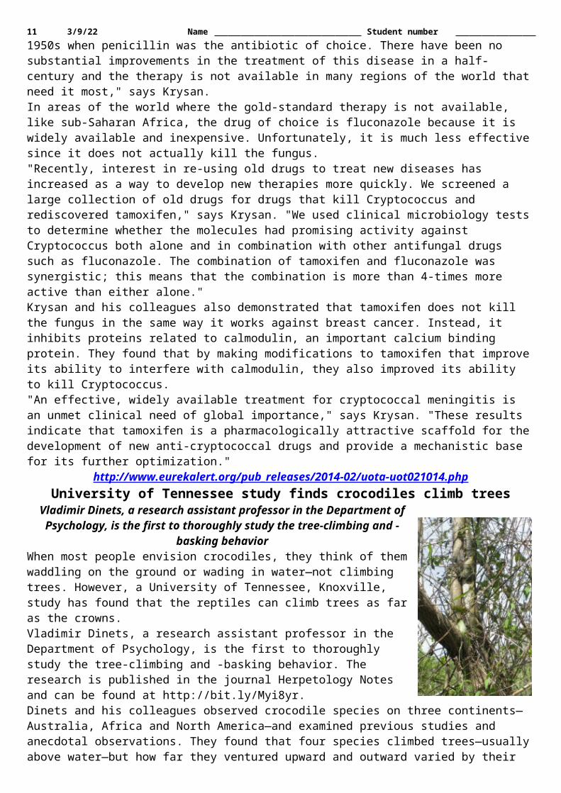
http://www.eurekalert.org/pub_releases/2014-02/uota-uot021014.phpUniversity of Tennessee study finds crocodiles climb trees
Vladimir Dinets, a research assistant professor in the Department of Psychology, is the first to thoroughly study the tree-climbing and -basking behavior
When most people envision crocodiles, they think of them waddling on the ground or wading in water—not climbing trees. However, a University of Tennessee, Knoxville, study has found that the reptiles can climb trees as far as the crowns. Vladimir Dinets, a research assistant professor in the Department of Psychology, is the first to thoroughly study the tree-climbing and -basking behavior. The research is published in the journal Herpetology Notes and can be found at http://bit.ly/Myi8yr.Dinets and his colleagues observed crocodile species on three continents—Australia, Africa and North America—and examined previous studies and anecdotal observations. They found that four species climbed trees—usually above water—but how far they ventured upward and outward varied by their sizes. The smaller crocodiles were able to climb higher and further than the larger ones. Some species were observed climbing as far as four meters high in a tree and five meters down a branch.
Crocodile climbing tree"Climbing a steep hill or steep branch is mechanically similar, assuming the branch is wide enough to walk on," the authors wrote. "Still, the ability to climb vertically is a measure of crocodiles' spectacular agility on land."The crocodiles seen climbing trees, whether at night or during the day, were skittish of being recognized, jumping or falling into the water when an approaching observer was as far as 10 meters away. This response led the researchers to believe that the tree climbing and basking are driven by two conditions: thermoregulation and surveillance of habitat."The most frequent observations of tree-basking were in areas where there were few places to bask on the ground, implying that the individuals needed alternatives for regulating their body temperature," the authors wrote. "Likewise, their wary nature suggests that climbing leads to improved site surveillance of potential threats and prey."The data suggests that at least some crocodile species are able to climb trees despite lacking any obvious morphological adaptations to do so. "These results should be taken into account by paleontologists who look at changes in fossils to shed light on behavior," said Dinets. "This is especially true for those studying extinct crocodiles or other Archosaurian taxa."Dinets collaborated with Adam Britton from Charles Darwin University in Australia and Matthew Shirley from the University of Florida.Research by Dinets published in 2013 found another surprising crocodile characteristic—the use of lures such as sticks to hunt prey. More of his crocodile research can be found in his book "Dragon Songs."
http://www.eurekalert.org/pub_releases/2014-02/w-cph021014.phpCould pizza herb prevent winter vomiting disease?
Scientists have found that carvacrol – the substance in oregano oil that gives the pizza herb its distinctive warm, aromatic smell and flavour – is effective against norovirus, causing the breakdown of the virus' tough
outer coat.The research is published today (12 February) in the Society for Applied Microbiology's Journal of Applied Microbiology. Norovirus, also known as the winter vomiting disease, is the leading cause of vomiting and diarrhoea around the world. It is particularly problematic in nursing homes, hospitals, cruise ships, and schools, and is a very common cause of foodborne-disease outbreaks. Although the disease is unpleasant, most people recover fully within a few days. But for people with an existing serious medical problem, this highly infectious virus can be dangerous.Dr Kelly Bright, who led the research at the University of Arizona said "Carvacrol could potentially be used as a food sanitizer and possibly as a surface sanitizer, particularly in conjunction with other antimicrobials. We

8 5/12/23 Name Student number have some work to do to assess its potential but carvacrol has a unique way of attacking the virus, which makes it an interesting prospect." Unfortunately the human form of norovirus is nearly impossible to work on in the laboratory so the research has been carried out using the mouse form of the virus, which is considered the most similar in its resistance to antimicrobials and disinfectants.In the experiments, carvacrol appeared to act directly on the virus capsid – a tough layer of proteins that surrounds the virus – causing it to break down. This would give another antimicrobial the opportunity to enter the internal part of the virus and kill it. So if carvacrol is used as a sanitizer in the future, it's likely to be in conjunction with another antimicrobial. And because it is slower acting than many disinfectants, such as bleach, it would be best used as part of a routine cleaning regimen to provide long-lasting antimicrobial residue on surfaces.The good news is that because carvacrol acts on the external proteins of the virus, it is unlikely that norovirus would ever develop resistance. It would also be safe, non-corrosive and it won't produce any noxious fumes or harmful by-products. This makes it particularly attractive for use in settings where people are likely to be vulnerable to traditional bleach or alcohol based cleaners, such as schools, hospitals, long-term care facilities, child day-care centres, and drug and alcohol rehabilitation facilities.The bad news: no amount of pizza could prevent norovirus, and quite apart from other negative health effects of a mainly pizza diet, concentrated carvacrol, although non-toxic, would be quite unpalatable, causing a burning sensation and then numbness of the tongue!Funding for this research came from the U.S. Department of Agriculture's Organic Research and Extension Initiative.
http://www.eurekalert.org/pub_releases/2014-02/tjnj-fdo020614.phpFewer doses of HPV vaccine still results in reduced risk of STD
Two doses of vaccine was associated with considerable reduction in riskAlthough maximum reduction in the risk of genital warts (condylomata) was seen after 3 doses of human papillomavirus (HPV) vaccine, receipt of 2 vaccine doses was associated with considerable reduction in risk, particularly among women who were younger than 17 years at first vaccination, according to a study in the February 12 issue of JAMA.HPV infection causes genital warts and cervical cancer, and HPV vaccine prevents both. The typical dose schedule requires 3 doses of vaccine, but small clinical trials have reported measures of vaccine efficacy with fewer than 3 doses. Although the primary goal of HPV vaccination programs is to prevent cervical cancer, genital warts related to HPV types 6 and 11 are prevented with a version of the vaccine and are the earliest measurable preventable disease outcome for the HPV vaccine, according to background information in the study.Eva Herweijer, M.Sc., of the Karolinska Institutet, Stockholm, Sweden, and colleagues assessed the association between the number of doses of HPV vaccination and genital warts among females 10 to 24 years of age living in Sweden (n = 1,045,165) who were followed up between 2006 and 2010, using the Swedish nationwide population-based health data registers.Among 20,383 new cases of genital warts, 322 occurred after receipt of at least 1 dose of the vaccine. The researchers found that maximum risk reductions were found after 3 doses, but 2 doses were also protective, although to a lesser extent; there was small difference in the number of cases prevented by 3 doses vs 2 doses.The authors caution that this study does not account for HPV disease outcomes other than genital warts, and that more studies with longer follow-up are needed to assess if these observed reductions apply for cervical cancer. (doi:10.1001/jama.2014.95; Available pre-embargo to the media at http://media.jamanetwork.com)Editor's Note: This study was supported by a grant from the Swedish Foundation for Strategic Research. Please see the article for additional information, including other authors, author contributions and affiliations, financial disclosures, etc.
http://www.eurekalert.org/pub_releases/2014-02/p-wmm020514.phpWhat makes memories last?
Stowers researchers identify protein that initiates the formation of stable, long-term memoriesPrions can be notoriously destructive, spurring proteins to misfold and interfere with cellular function as they spread without control. New research, publishingin the open access journal PLOS Biology on February 11 2014, from scientists at the Stowers Institute for Medical Research reveals that certain prion-like proteins, however, can be precisely controlled so that they are generated only in a specific time and place. These prion-like proteins are not involved in disease processes; rather, they are essential for creating and maintaining long-term memories."This protein is not toxic; it's important for memory to persist," says Stowers researcher Kausik Si, who led the study. To ensure that long-lasting memories are created only in the appropriate neural circuits, Si explains, the
9 5/12/23 Name Student number protein must be tightly regulated so that it adopts its prion-like form only in response to specific stimuli. He and his colleagues report on the biochemical changes that make that precision possible.Si's lab is focused on finding the molecular alterations that encode a memory in specific neurons as it endures for the days, months, or years—even as the cells' proteins are degraded and renewed. Increasingly, their research is pointing toward prion-like proteins as critical regulators of long-term memory.In 2012, Si's group demonstrated that prion formation in nerve cells is essential for the persistence of long-term memory in fruit flies. Prions are a fitting candidate for this job because their conversion is self-sustaining: once a prion-forming protein has shifted into its prion shape, additional proteins continue to convert without any additional stimulus.Si's team found that in fruit flies, the prion-forming protein Orb2 is necessary for memories to persist.Flies that produce a mutated version of Orb2 that is unable to form prions learn new behaviors, but their memories are short-lived. "Beyond a day, the memories become unstable. By three days, the memory has completely disappeared," Si explains.In the new study, Si wanted to find out how this process could be controlled so that memories form at the right time. "We know that all experiences do not form long-term memory—somehow the nervous system has a way to discriminate. So if prion-formation is the biochemical basis of memory, it must be regulated." Si says. "But prion formation is random for all the cases we know of so far."Si and his colleagues knew that Orb2 existed in two forms—Orb2A and Orb2B. Orb2B is widespread throughout the fruit fly's nervous system, but Orb2A appears only in a few neurons, at extremely low concentrations. What's more, once it is produced, Orb2A quickly falls apart; the protein has a half-life of only about an hour.When Orb2A binds to the more abundant form, it triggers conversion to the prion state, acting as a seed for the conversion. Once conversion begins, it is a self-sustaining process; additional Orb2 continues to convert to the prion state, with or without Orb2A. By altering the abundance of the Orb2A seed, Si says, cells might regulate where, when, and how the conversion process is engaged. But how do nerve cells control the abundance of the Orb2A seed?To look for leads, the scientists searched for proteins that physically interact with Orb2A. Because Orb2A is so scarce, finding it and identifying its molecular accomplices took perseverance and refinement of the standard protein-cataloging techniques. Post-doctoral researcher Erica White-Grindley led that effort, and after several years, turned up more than 60 suspected partners for Orb2A.One of these proteins, TOB, doubled the half-life of Orb2A, thereby temporarily increasing its abundance. The scientists were skeptical that this boost in stability would be enough to reliably stabilize long-term memories, but further biochemical analyses led them to a more satisfying explanation.Their experiments revealed that when TOB associates with Orb2A – which is known to occur in response to an incoming nerve signal—this triggers the addition of chemical tags known as phosphates to both of the proteins, altering both proteins' stability. Once phosphorylated, the TOB-Orb2A complex falls apart and Orb2A becomes much more stable, with a new half-life of 24 hours. This increases the prevalence of the prion-like state.This explained how Orb2's conversion could be specifically triggered following nerve cell stimulation. The next step was to determine how the process could be localized to the right neuronal connections. They found that the enzyme that places the phosphate tag on Orb2A is Lim kinase, a neuron-specific kinase that others had shown is activated at the synapse—the connection between neurons—when cells receive an impulse. Taken together, Si says, these experiments show how Orb2's conversion to the prion state can be confined in both time and space.The findings raise a host of new questions for Si, who now wants to understand what happens when Orb2 enters its prion-like state, as well as where in the brain the process occurs. While unraveling these mechanisms will likely be more accessible in the fruit fly than in more complex organisms, Si points out that proteins related to Orb2 and TOB have also been found in the brains of mice and humans. He's already shown that in the sea snail Aplysia, conversion to a prion-like state facilitates long-term change in synaptic strength. "This basic mechanism appears to be conserved across species," he notes.Researchers who also contributed to the work include Liying Li and Repon Mahammad Khan in the Department of Molecular and Integrative Physiology at the University of Kansas Medical Center, and Fengzhen Ren, Anita Saraf and Laurence Florens at the Stowers Institute for Medical Research.Please mention PLOS Biology as the source for this article and include the links below in your coverage to take readers to the online, open access articlesAll works published in PLOS Biology are open access, which means that everything is immediately and freely available. Use this URL in your coverage to provide readers access to the paper upon publication: http://www.plosbiology.org/article/info:doi/10.1371/journal.pbio.1001786
10 5/12/23 Name Student number Citation: White-Grindley E, Li L, Khan RM, Ren F, Saraf A, et al. (2014) Contribution of Orb2A Stability in Regulated Amyloid-Like Oligomerization of Drosophila Orb2. PLoS Biol 12(2): e1001786. doi:10.1371/journal.pbio.1001786Funding: The work is supported by the institutional fund from Stowers Institute for Medical Research and a fellowship to KSI from The McKnight Foundation. The funders had no role in study design, data collection and analysis, decision to publish, or preparation of the manuscript.
http://www.eurekalert.org/pub_releases/2014-02/uops-csb021114.phpCould statins be used to fight a deadly viral infection?
A way to use statins to fight the hantavirusTwo Perelman School of Medicine microbiologists may have found a way to use statins, the well-known blockbuster cholesterol-lowering drugs, to fight the hantavirus, a mysterious and lethal microorganism that appeared suddenly in the US southwest over 20 years ago. That first outbreak led to the deaths of more than a dozen people, most of them in their prime. The last reported outbreak happened in Yellowstone Park in 2012.Only about 30 known human cases of hantavirus are reported in the US each year. The respiratory syndrome caused by a hantavirus infection comes from breathing in small viral particles in the excrement of infected rodents. It starts out with flu-like symptoms that quickly deteriorate into a dangerous form of adult respiratory distress syndrome. It is among the most deadly known human viruses: 30 percent to 40 percent of people who are diagnosed die from hantavirus pulmonary fever.A PLOS Pathogens paper by Penn microbiologists Paul Bates, PhD, and Kenneth Briley, PhD, published this month reports that four proteins key to cholesterol synthesis and uptake are highjacked by the hantavirus to enter human host cells. To identify host-cell genes needed for viral replication, Bates and Briley first used a less dangerous virus that was engineered to exhibit some characteristics of a member of the hantavirus group found in South America called Andes virus (ANDV). Although the molecular details are still being deciphered, it appears that adequate cellular cholesterol levels are needed to transport the virus into the cell.This is not the first time that cholesterol-related proteins have been implicated in viral entry and infection but Bates says, "the hantaviruses seem to be exquisitely sensitive to the cellular cholesterol levels."The four proteins identified by the Penn researchers are part of a protein complex that regulates cholesterol production in the cells of mammals. They found that treating cells that originated from human airways with an experimental drug that targets one of the four proteins made the cells less susceptible to infection. The experimental drug also lowers cholesterol levels in cells, so Bates wondered whether statins could be used to fight a hantavirus infection.The researchers found that pre-treatment of human airway cells with a generic statin called mevastatin, which lowers cholesterol by a mechanism that does not involve the four proteins they identified, made the human airway cells less susceptible to ANDV infection. They tested both the experimental drug, PF-429242, and mevastatin, and both were effective against hantavirus, as measured by how many cells are infected with and without the drugs. Bates surmises that statins might be given after a known hantavirus infection, or even prophylactically to exposed individuals.Although the drug inhibition studies initially used the engineered virus, Bates used ANDV itself in high-containment Biosafety Level (BSL)-3 facilities in the Department of Microbiology at Penn to confirm that the results were true for ANDV as well. The next step is to test cholesterol-lowering drugs in an animal model for ANDV infection, working with collaborators to perform the animal studies in a BSL-4 at a national facility.
http://www.eurekalert.org/pub_releases/2014-02/uoc-maf021114.phpMales and females differ in specific brain structures
First meta-analysis of the evidence from research into sex differences in brain structureReviewing over 20 years of neuroscience research into sex differences in brain structure, a Cambridge University team has conducted the first meta-analysis of the evidence, published this week in the prestigious journal Neuroscience and Biobehavioral Reviews.The team, led by doctoral candidate Amber Ruigrok and Professors John Suckling and Simon Baron-Cohen in the Department of Psychiatry, performed a quantitative review of the brain imaging literature testing overall sex differences in total and regional brain volumes. They searched all articles published between 1990 and 2013. A total of 126 articles were included in the study, covering brains from individuals as young as birth to 80 years old.They found that males on average have larger total brain volumes than women (by 8-13%). On average, males had larger absolute volumes than females in the intracranial space (12%; >14,000 brains), total brain (11%; 2,523 brains), cerebrum (10%; 1,851 brains), grey matter (9%; 7,934 brains), white matter (13%; 7,515 brains), regions filled with cerebrospinal fluid (11.5%; 4,484 brains), and cerebellum (9%; 1,842 brains). Looking more
11 5/12/23 Name Student number closely, differences in volume between the sexes were located in several regions. These included parts of the limbic system, and the language system. Specifically, males on average had larger volumes and higher tissue densities in the left amygdala, hippocampus, insular cortex, putamen; higher densities in the right VI lobe of the cerebellum and in the left claustrum; and larger volumes in the bilateral anterior parahippocampal gyri, posterior cingulate gyri, precuneus, temporal poles, and cerebellum, areas in the left posterior and anterior cingulate gyri, and in the right amygdala, hippocampus, and putamen.
An overview of average regional sex differences in grey matter volume. Areas of larger volumes in women are in red and areas of larger volume in men are in blue. Neuroscience and Biobehavioral Reviews
By contrast, females on average had higher density in the left frontal pole, and larger volumes in the right frontal pole, inferior and middle frontal gyri, pars triangularis, planum temporale/parietal operculum, anterior cingulate gyrus, insular cortex, and Heschl's gyrus; bilateral thalami and precuneus; the left parahippocampal gyrus, and lateral occipital cortex.The results highlight an asymmetric effect of sex on the developing brain. Amber Ruigrok, who carried out the study as part of her PhD, said: "For the first time we can look across the vast literature and confirm that brain size and structure are different in males and females. We should no longer ignore sex in neuroscience research, especially when investigating psychiatric conditions that are more prevalent in either males or females."Professor Suckling added: "The sex differences in the limbic system include areas often implicated in psychiatric conditions with biased sex ratios such as autism, schizophrenia, and depression. This new study may therefore help us understand not just typical sex differences but also sex-linked psychiatric conditions. It is important to note that we only investigated sex differences in brain structure, so we cannot infer anything about how this relates to behaviour or brain function. Integrating across different levels will be an important goal for future research."Professor Baron-Cohen commented: "Although these very clear sex differences in brain structure may reflect an environmental or social factor, from other studies we know that biological influences are also important, including prenatal sex steroid hormones (such as foetal testosterone) as well as sex chromosome effects. Such influences need to be teased out, one by one."Dr Meng-Chuan Lai, another member of the team, noted: "The advantage of conducting a meta-analysis is that we can summarise the best knowledge from a vast, heterogeneous literature, with a very large sample size. However, we found a bias in the existing literature towards the use of volunteers over 18 years old, probably because this is the easiest age group to recruit and to brain scan. We need more research exploring brain development over the entire lifespan, especially in the early, formative years".
http://phys.org/news/2014-02-energy-consumption-paper-industry-percent.htmlNew process can reduce energy consumption of paper industry by 40 percent
New solvent may enable the paper industry to make big energy savings in productionEindhoven University of Technology (TU/e) signed an agreement last week with 14 European paper producers for the further development of a breakthrough new solvent. This new solvent, developed by TU/e professor Maaike Kroon, will potentially enable the paper industry to make big energy savings in production and to use raw materials more efficiently. The European paper industry has high expectations of the new solvent. "This is a game changer, and it means the paper industry will look very different 20 years from now", said Henk van Houtum, chairman of VNP, the Royal Netherlands' paper and board association.Kroon discovered that wood fibers easily dissolve in specific 'deep eutectic solvents' (DES). In the production of paper, the basic vegetable material (lignocellulose), such as wood chips or other biomass, has to be separated into lignine and cellulose. The cellulose is then used to make paper. The problem is that the two components are difficult to separate – the process still needs high pressures and temperatures, and it is costly to operate. Dissolving the wood chips has up to now not been an option because lignine is normally insoluble. But the new solvent, which Kroon has patented, makes this possible. As well as that the new solvent is entirely vegetable-

12 5/12/23 Name Student number based and biodegradable. Another advantage is that the new process produces very pure lignine, which the paper industry can use to develop new applications and markets such as making biodegradable plastics.Paper production is energy-intensive, which is why the Dutch paper industry took the initiative in 2004 for its 'Energy Transition Paper Chain 2004-2020' plan which aims to halve energy consumption. The Confederation of European Paper industries (CEPI) is looking even further ahead, and intends to reduce CO2 emissions by 80% before 2050. The industry has therefore focused strongly on innovation for a number of years, using natural raw materials in a high-tech process. In its search for breakthrough technologies, CEPI organized a competition last year to find the best new ideas. The winner was the 'deep eutectic solvents' which Kroon had already been working on for several years. Henk van Houtum of the VNP expects that the solvent developed by Kroon will make a substantial contribution to meeting the industry's energy targets. He hopes that the use of DES will lead to at least 40% lower energy costs and 20% less CO2 emissions.TU/e signed a letter of intent last week with 14 European paper producers, including seven in the Netherlands, to continue development of the solvent. Kroon will use the funding from these companies to recruit two PhD candidates for a further four years of research at TU/e to prepare the way for the building of a pilot plant in the Netherlands. Kroon emphasizes that this is a very special agreement because it has been reached directly with the industrial companies, and does not rely on government financial support. It underlines the potential that the companies see in this development by the TU/e chemistry professor, and the importance they place on quickly implementing it in practice. Large-scale applications are expected to be possible in around 15 years. The laboratory research will take another five to ten years, with a similar period being required for optimization in the pilot plant.Deep eutectic solvents were discovered in 2003 in the UK. They consist of a mixture of two compounds which, once they have been combined, have a much lower melting point than that of the individual components. Kroon believed that DES would make it possible to dissolve biomass, which formed the starting point for her present work. And it has indeed led to a process for dissolving lignine using different mixtures for specific types of wood.
http://www.eurekalert.org/pub_releases/2014-02/uos-nru021014.phpNew research uncovers debilitating effects of disease on toy dog breeds
New study has identified the specific effect Chiari malformation has on the shape of a dog's skullA new study from the University of Surrey, published today in the journal PLOS One, has identified the specific effect Chiari malformation has on the shape of a dog's skull and brain. This condition has become prevalent as a result of selective breeding and affects many toy dog breeds which have been bred to look more doll-like, including Griffon Bruxellois, Cavalier King Charles Spaniels, Chihuahuas and their crosses.Researchers took brain, skull and vertebrae measurements of 155 Griffon Bruxellois and compared dogs affected by the condition, with normal Griffons. They discovered that Griffons with the disease had taller foreheads and that it had also caused the shape of the brain to change, with severely affected animals having their cerebellum pushed underneath the main part of the brain.Although it can be asymptomatic, in many dogs Chiari malformation can cause headaches, problems with walking or even paralysis. The condition can also affect humans, when certain skull bones fuse too early, causing parts of the brain to push through an opening in the base of the skull. It currently affects 1 in 1280 humans. Indeed, researchers at the University of Surrey are working with human geneticists at the University of Montreal, in the hope that better understanding of the condition will lead to improved treatment for both dogs and humans.Lead author, Dr Clare Rusbridge from the new School of Veterinary Medicine at the University of Surrey, said: "Chiari malformation can be described as trying to fit a big foot into a small shoe. It can be very painful, causing headaches and pressure on the brain and can result in fluid filled cavities in the spinal cord. Our latest discoveries will be significant in driving this research forward and hopefully allow us to identify which genes may be associated with the condition. Our next steps will be to apply our technique to other breeds with Chiari malformation and investigate more sophisticated ways of screening, so that risk of disease can be detected more easily, at an earlier age and with a single MRI scan."We want to engage breeders and give them practical advice about the condition, but it is also important that the public recognises that breeding dogs in a certain way to influence how they look might not be in the animal's best interest. There are responsible breeders out there, who have invested in screening and who are breeding for health as well as producing attractive puppies, and it is vital that people only look to buy from them."The research was published in the journal PLOS ONE: http://dx.plos.org/10.1371/journal.pone.0088120The Guardian Good University Guide

13 5/12/23 Name Student number http://www.eurekalert.org/pub_releases/2014-02/aha-cil020414.php
Common infections linked to stroke in children; vaccines may reduce riskCommon infections are associated with a significantly higher chance of stroke in children, but routine
vaccinations may help decrease riskCommon infections are associated with a significantly higher chance of stroke in children, but routine vaccinations may help decrease risk, according to preliminary research (abstract 39) presented at the American Stroke Association's International Stroke Conference 2014."The protective association of routine vaccination against childhood stroke provides a widely available means of prevention, and this information can easily be dispersed by pediatric healthcare providers," said Nancy Hills, Ph.D., M.B.A., lead researcher and assistant professor of neurology at the University of California, San Francisco Medical Center.The international study, Vascular effects of Infection in Pediatric Stroke (VIPS) is a prospective study examining the link between infections and ischemic stroke, the most common type of stroke. (Ischemic stroke is caused by a clot that blocks blood flow in or leading to the brain.)Previous research by Hills and co-authors found that minor infections were related to an increased risk, but it was unclear whether infection actually could help predict future stroke.In the VIPS study, researchers found that common infections within the past week were linked to more than six times the risk of stroke, Hills said. Seventeen percent of the stroke patients vs. 3 percent of the non-stroke patients were reported to have had any minor infection in the prior week. The most frequent types of infection were colds and other upper respiratory infections (8 percent of the stroke and 2.4 percent of the non-stroke patients reported an occurrence of these kinds of infections in the prior week).However, routine vaccinations were associated with a lower stroke risk. Children who had "some, few or no" routine vaccinations were 6.7 times more likely to have an ischemic stroke than those receiving "all or most" vaccines, including those against polio, measles, mumps, rubella and pneumococcus.Researchers interviewed parents or guardians of 310 children who had a stroke to determine the presence and timing of any infectious illnesses prior to their stroke. They compared their findings with 289 children who hadn't experienced a stroke, but had visited the doctor for an annual checkup, routine follow-up for headaches or developmental delay, or trauma. The median age of the children who had a stroke was 7.5 years, and the median age among the comparison group was slightly more than 8."Because many childhood strokes appear to have no clear cause, and others likely have more than one cause, preventive measures have not been forthcoming," Hills said. "It is very promising that childhood vaccinations appear to have a protective effect."In other VIPS analyses (abstracts 36 & 38) researchers found that infections with parvovirus B19 (the cause of "slapped cheek syndrome") and different herpes viruses also were linked to a significantly greater stroke risk. Blood tests indicated that 41 percent of stroke patients had an active herpes infection, compared to 9 percent of non-stroke patients."VIPS is the largest-ever NIH-funded study of childhood stroke," said Heather J. Fullerton, M.D., M.A.S., principal investigator for the VIPS study and Professor of Neurology and Pediatrics at University of California San Francisco. "These three abstracts represent the first results of this important international effort."Other VIPS researchers are: Gabrielle A. DeVeber, M.D., M.Sc.; Mitchell S. Elkind, M.D., M.S.; Max Wintermark, M.D.; Carol A. Glaser, M.D.; Katherine Sear, M.P.H.; Jorge M. Luna, M.P.H; W. Ian Lipkin, M.D; Kawthar Muhammad, B.A.; and Rafal Tokarz, Ph.D. Author disclosures are on the abstracts.
http://www.eurekalert.org/pub_releases/2014-02/uop-dam021114.phpDoctors are missing chance to diagnose COPD in up to 85 percent of cases, study finds
Missed opportunities occur commonly in both primary and secondary careA retrospective, 20-year study led by researchers at Plymouth University Peninsula Schools of Medicine and Dentistry shows that in up to 85 per cent of patients with chronic obstructive pulmonary disease (COPD) the underlying disease was being overlooked. Missed opportunities occur commonly in both primary and secondary care. The paper demonstrates the pointers to help GP to come to a earlier diagnosis. The findings are published in The Lancet Respiratory Medicine today, Thursday 13th February 2014.The study encompassed almost 39,000 patients and showed that, in the UK, opportunities to diagnose COPD are frequently missed in both primary and secondary care settings.The study was led by Dr. Rupert Jones, Clinical Research Fellow at Plymouth University Peninsula Schools of Medicine and Dentistry and a working GP in Plymouth.He said: "This was a project which came from my work with the Department of Health, on the National COPD outcomes strategy - a stream of work which I have been involved in since 2005. We became acutely aware that
14 5/12/23 Name Student number many people were being diagnosed with COPD, a progressive and disabling lung disease, at a late stage when the damage done was severe and irreversible. Thus we wanted to examine the opportunities arising in primary care in order to diagnose COPD at an earlier stage and improve health outcomes, with potential to extend life expectancy and quality of life for patients."The research team used data from the General Practice and Optimum Patient Care Research databases. They assessed whether a diagnosis of COPD could have been made in an earlier visit to a doctor, whether in a primary or secondary care setting. From the databases 38,849 patients aged 40 or older and who had received a diagnosis for COPD were identified. The diagnoses had been made between 1990 and 2009 and for each data was available at least two years before and one year after diagnosis.Results showed that in the five years before diagnosis, 85 per cent of patients had visited their GP at least once with lower respiratory symptoms without the diagnosis of COPD being made. Opportunities for diagnosis were missed in 58 per cent of patients in the six to 10 years before diagnosis, and in 42 per cent in the 11 to 15 years before diagnosis.The study identified that, over the 20 year study period, there was a significant increase in the number of chest X-rays in the two years prior to diagnosis, but that only a third of those patients were given spirometry testing (a breathing test used to diagnose lung conditions and which measures how well the lungs work.It is estimated that around 2.2 million people in the UK remain undiagnosed for COPD. The UK Department of Health estimates that earlier diagnosis and treatment could save the NHS more than £1 billion over 10 years.Said Dr. Jones: "The numbers are large, both in terms of people affected and the cost to already stretched NHS provision of care. We believe that the results of our study provide clear support to the argument for improved identification and diagnosis of COPD in general practice, with greater awareness so that early opportunities to diagnose – such as presentation with lower respiratory tract symptoms or related conditions – are seized and acted upon."A link to the full article and comment for journalists is available at http://www.thelancet.com/journals/lanres/article/PIIS2213-2600%2814%2970008-6/fulltext
http://www.eurekalert.org/pub_releases/2014-02/bmj-qoa021014.phpRevision to rules for color in dinosaurs suggests connection between color and physiologyNew research that revises the rules allowing scientists to decipher color in dinosaurs may also provide a tool
for understanding the evolutionary emergence of flight and changes in dinosaur physiology prior to its origin.
In a survey comparing the hair, skin, fuzz and feathers of living terrestrial vertebrates and fossil specimens, a research team from The University of Texas at Austin, the University of Akron, the China University of Geosciences and four other Chinese institutions found evidence for evolutionary shifts in the rules that govern the relationship between color and the shape of pigment-containing organelles known as melanosomes, as reported in the Feb. 13 edition of Nature.At the same time, the team unexpectedly discovered that ancient maniraptoran dinosaurs, paravians, and living mammals and birds uniquely shared the evolutionary development of diverse melanosome shapes and sizes. (Diversity in the shape and size of melanosomes allows scientists to decipher color.) The evolution of diverse melanosomes in these organisms raises the possibility that melanosome shape and size could yield insights into dinosaur physiology.
The "rules" allowing color reconstruction from the shape of melanin-containing organelles originate with feathered dinosaurs, and are associated with an increase in melanosome diversity. However, fuzzy dinosaurs like T. rex and
Sinosauropteryx show a pattern found in other amniotes like lizards and crocodilians in which a limited diversity of shapes doesn't allow color reconstruction. An explosion in the distribution of the shapes of melanin-containing organelles preserved in living taxa and the fossil record may point to a key physiological shift within feathered
dinosaurs. Credit: Li et al. (authors).Melanosomes have been at the center of recent research that has led scientists to suggest the colors of ancient fossil specimens covered in fuzz or feathers. Melanosomes contain melanin, the most common light-absorbing pigment found in animals. Examining the shape of melanosomes from fossil specimens, scientists have recently

15 5/12/23 Name Student number suggested the color of several ancient species, including the fuzzy first-discovered feathered dinosaur Sinosauropteryx, and feathered species like Microraptor and Anchiornis.According to the new research, color-decoding works well for some species, but the color of others may be trickier than thought to reconstruct. Comparing melanosomes of 181 extant specimens, 13 fossil specimens and all previously published data on melanosome diversity, the researchers found that living turtles, lizards and crocodiles, which are ectothermic (commonly known as cold-blooded), show much less diversity in the shape of melanosomes than birds and mammals, which are endothermic (warm-blooded, with higher metabolic rates).The limited diversity in melanosome shape among living ectotherms shows little correlation to color. The same holds true for fossil archosaur specimens with fuzzy coverings scientists have described as "protofeathers" or "pycnofibers." In these specimens, melanosome shape is restricted to spherical forms like those in modern reptiles, throwing doubt on the ability to decipher the color of these specimens from fossil melanosomes.
These are two of the fossil specimens sampled from the Cretaceous and Jurassic of China. Fuzz-covered dinosaur Beipiaosaurus shows the rounder melanosomes seen in living lizards and crocodilians while the bird shows the unique
skinny melanosomes seen in living mammals, birds and many of the studied feathered dinosaurs to date. Changes in the diversity of these melanin-containing organelles may show a physiological shift occurred in feathered dinosaurs
closer to the origin of flight. Credit: Li et al. (authors).In contrast, in the dinosaur lineage leading to birds, the researchers found an explosion in the diversity of melanosome shape and size that appears to correlate to an explosion of color within these groups. The shift in diversity took place abruptly, near the origin of pinnate feathers in maniraptoran dinosaurs. "This points to a profound change at a pretty discrete point," says author Julia Clarke of The University of Texas at Austin's Jackson School of Geosciences. "We're seeing an explosion of melanosome diversity right before the origin of flight associated with the origin of feathers."What surprised the researchers was a similarity in the pattern of melanosome diversity among ancient maniraptoran dinosaurs, paravians, and living mammals and birds."Only in living, warm-blooded vertebrates that independently evolved higher metabolic rates do we see the melanosome diversity we also see in feathered dinosaurs," said co-author Matthew Shawkey of The University of Akron. Many of the genes involved in the melanin color system are also involved in other core processes such as food intake, the stress axis, and reproductive behaviors. Because of this, note the researchers, it is possible that the evolution of diverse melanosome shapes is linked to larger changes in energetics and physiology.Melanosome shape could end up offering a new tool for studying endothermy in fossil specimens, a notoriously challenging subject for paleontologists.Because the explosion of diversity in melanosomes appears to have taken place right at the origin of pinnate feathers, the change may indicate that a key shift in dinosaurian physiology occurred prior to the origin of flight."We are far from understanding the exact nature of the shift that may have occurred," says Clarke. "But if changes in genes involved in both coloration and other aspects of physiology explain the pattern we see, these precede flight and arise close to the origin of feathers." It is possible, notes Clarke, that a diversity in melanosome shape (and correlated color changes) resulted from an increased evolutionary role for signaling and sexual selection that had a carryover effect on physiology, or that a change in physiology closely preceded changes in color patterning. At this point, she stresses, both ideas are speculative."What is interesting is that trying to get at color in extinct animals may have just started to give us some insights into changes in the physiology of dinosaurs."
Analysis for the distribution of shapes of melanin-containing organelles (melanosomes) in fossil and living amniotes shows that fuzz-covered dinosaurs like Sinosauropteryx share similarities with living lizards, turtles and crocodilians.
In these living taxa color and the shape of the melanosomes are not linked in such a way that color can be reconstructed from melanosome shape alone. Melanosomes in Sinosauropteryx don't presently tell us if this animal

16 5/12/23 Name Student number was brown, blackish or grey. However, feathered dinosaurs are similar to birds, and we can estimate their color.
Credit: Li et al. (authors)Co-authors include Quanguo Li of the China University of Geosciences, Ke-Qin Gao of Peking University, Chang-Fu Zhou of Shenyang Normal University, Qingjin Meng of the Beijing Museum of Natural History, Daliang Li of the Museum of China University of Geosciences, and Liliana D'Alba also of the University of Akron.The research was supported by the U.S. National Science Foundation, Human Frontiers Science Program, Fundamental Research Funds for Central Universities (China), Beijing Municipal Bureau of Human Resources, and the Jurassic Foundation.
http://www.eurekalert.org/pub_releases/2014-02/bmj-qoa021014.phpQuality of acupuncture needles is less than perfect and must improve
Standards currently high, but surface irregularities and bent tips have not been completely eliminatedThe quality of acupuncture needles is high, but should still be universally improved to avoid potential problems, such as pain and skin reactions, finds research published online in Acupuncture in Medicine (AiM).Despite improvements to the manufacturing process, surface irregularities and bent tips have not been completely eliminated, say the researchers.In China, traditional Chinese medicine including acupuncture, accounts for 40% of all medical treatment, while in the West, acupuncture is one of the most frequently used complementary therapies. An estimated 1.4 billion acupuncture needles are used each year worldwide, with China, Japan, and Korea the main suppliers. China provides up to 90% of the world's needles.The growing popularity of acupuncture in recent decades has led to an increased focus on the safety and quality of this therapy, and adoption of single-use disposable needles has reduced the risk of infection. But a study of widely used acupuncture needles published a decade ago in AiM showed that several had surface irregularities or distorted points which could have led to allergic or painful reactions. Since then, there has been no further research in this area.A team of researchers in Australia therefore looked at the surface conditions and other physical properties of the two most commonly used stainless steel acupuncture needle brands. Scanning electron microscope images were taken of 10 randomly chosen needles from each brand, while further images were taken after each of these needles underwent a standard manipulation - the equivalent of using them on human skin - with an acupuncture needling practice gel. The researchers also compared forces and torques during the needling process.The images revealed significant surface irregularities and inconsistencies at the needle tips, especially for needles from one of the brands which had been manufactured in China. Metallic lumps and small, loosely attached pieces of material were observed on the surfaces of some needles. Some of this residue disappeared after the acupuncture manipulation.If these needles had been used on patients, the metallic residue could have been deposited in human tissues, potentially causing reactions, such as dermatitis, although these reactions are reported extremely rarely, say the authors. Malformed needle tips could also have caused other problems, including bleeding, bruising, or strong pain during needling, which are quite common, they suggest.Acupuncture, overall, is very safe, but it should be made even safer, say the researchers. "Acupuncture needle manufacturers, including the well established ones, should review and improve their quality control procedures for fabrication of needles," they conclude.In an accompanying podcast, Dr Mike Cummings, medical director of the British Medical Acupuncture Association and associate editor of the journal, comments that the pictures taken for the current study indicate that the needles "look as awful as they did 10 years ago."He adds: "We don't know if [this problem] is common to all needles, but it seems like it does happen with acupuncture needles." But he emphasises that acupuncture is safe, pointing out that "It's highly unlikely that [poor needle quality] will affect patient health." If people experience pain during acupuncture, they should ask their practitioner to check on the quality of the needles they use, he advises.Examination of surface conditions and other physical properties of commonly used stainless steel acupuncture needles Online First doi:10.1136/acupmed-2013-010472
http://www.eurekalert.org/pub_releases/2014-02/uoc-aoc021214.phpAmerica's only Clovis skeleton had its genome mapped
Clovis people were not the first humans in America, but they represent the first humans with a wide expansion on the North American continent
They lived in America about 13,000 years ago where they hunted mammoth, mastodons and giant bison with big spears. The Clovis people were not the first humans in America, but they represent the first humans with a wide expansion on the North American continent – until the culture mysteriously disappeared only a few hundred years after its origin. Who the Clovis people were and which present day humans they are related to
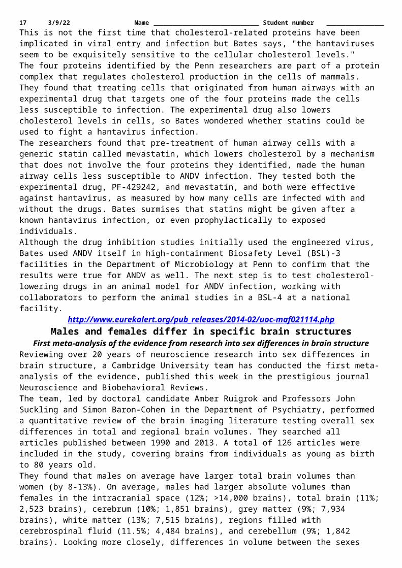
17 5/12/23 Name Student number has been discussed intensely and the issue has a key role in the discussion about how the Americas were peopled. Today there exists only one human skeleton found in association with Clovis tools and at the same time it is among the oldest human skeletons in the Americas. It is a small boy between 1 and 1.5 years of age – found in a 12,600 old burial site, called the Anzick Site, in Wilsall, Montana, USA. Now an international team headed by Danish researcher Eske Willerslev has mapped his genome thereby reviving the scientific debate about the colonization of the Americas.Roughly estimated some 80 % of all present-day Native American populations on the two American continents are direct descendants of the Clovis boy's family. The remaining 20 % are more closely related with the Clovis family than any other people on Earth, says Lundbeck Professor Eske Willerslev from the Centre for GeoGenetics at the Natural History Museum of Denmark, University of Copenhagen. This surprising result has now been published in the scientific journal Nature.The discovery is so decisive that Nature has chosen to send the article to the press at a later time than usual as they fear the media embargo may be broken. A comprehensive international telephone press conference has been arranged and will be held in the Crow tribe's reservation in Montana – close to where the boy was found. Behind the results are a group of international researchers led by Professor Eske Willerslev from Centre of Excellence in GeoGenetics, Natural History Museum at University of Copenhagen, Denmark.The missing linkIt is almost like finding the 'missing link' to the common ancestor of the Native Americans. The Clovis boy's family is the direct ancestor to roughly estimated 80 % of all present day Native Americans. Although the Clovis culture disappeared its people are living today.Put simply it is a sensation that we succeeded in finding an approximately 12,600 year old boy whose closest relatives can be regarded as the direct ancestor to so many people, Eske Willerslev says and adds:This also means that Clovis did not descend from Europeans, Asians or Melanesians, a theory that a number of scientists have advocated. They were Native Americans – and the Native American ancestors were the first people in America. This is now a fact.Shane Doyle, a historian from the Apsaalooke (Crow) tribe, who helped the team with consultations to the Montana tribes agrees:This discovery by Eske and his team proves something that tribal people have never doubted - we've been here since time immemorial and all the ancient artifacts located within our homelands are remnants from our direct ancestors.But the discovery is only part of the importance of this study. The other part being Eske and his team's respectful commitment to interacting face to face with tribal communities and listening to Native American leaders, which has lead directly to the reburial of this little boy."Also Sarah Anzick, a molecular biologist in the study and the steward of the remains that were found on private land is excited:After 46 years since the discovery on my family land, we are finally hearing this child's story through his genetic legacy. I find it remarkable that the descendants of the Clovis culture, which seemed to have vanished 12,600 years ago, are still alive and thriving today.Interestingly, the teams find that Native American ancestors coming in from Siberia split into two groups. One group is ancestors to the Native Americans presently living in Canada and the other one – which is represented by the Clovis boy – is the ancestor to virtually all Native Americans in South America and Mexico.The US is still a white spot on the map when it comes to genome-wide data from Native Americans. The team members hope to be able to accessing such data in the future to understand the full picture.The study validates the concept of continuity in the history of Native Americans, and suggests that modern Native Americans are direct descendants of the first people occupying this land, says Rasmus Nielsen, a co-author on the study and a Professor at UC Berkeley, who developed the method used for determining that many modern native Americans are direct descendants of the Clovis boy's family.An Asian homeland for the First AmericansThe first humans came from Siberia via the so-called Beringia Land Bridge, which during the latest ice age connected Siberia with North America and did not bring the Clovis culture with them. The Clovis culture arose after they arrived in America and the boy from Anzick was more a descendant of the first immigrants.Michael Waters, the key archaeologist connected to the study and who has worked on many Clovis and older sites in North America elaborates:The genetic findings mesh well with the archaeological evidence to confirm the Asian homeland of the First Americans, more clearly define their genetic heritage, and is consistent with occupation of the Americas a few thousand years before Clovis. The findings do not support a western European origin of the First Americans as

18 5/12/23 Name Student number suggested by the Solutrean hypothesis. The genetic information provided by the Anzick boy is part of the larger story of modern human dispersal across the Earth and is shedding new light on the last continent to be explored and settled by our species.Then who were the first immigrants? We don't know. Yet. Maybe a Native American, maybe an ancestor related to the Mal'ta boy from Siberia and another one who was East Asian. We don't know. But our results eliminate all other theories about the origins of the first people in America. The first people in America were the direct ancestors of Native Americans, says Professor Willerslev and continues:We can see that the Clovis boy shares about 1/3 of his genes with the 24,000 year old child from Mal'ta at the Siberian Lake Baikal who we have analyzed previously. The same goes for all present day Native Americans. Therefore the encounter between East Asians and the Mal'ta group happened before Clovis.The human remains from the Anzick site will be reburied sometime this year in cooperation with Native American tribes in Montana. Eske Willerslev has in connection with the genome work on the Clovis boy visited several of Montana's Native American tribes with Crow historian Shane Doyle to discuss the findings. All the members of the research team hope that the Anzick study leads to more and enhanced cooperation between Native peoples and scientists. Therefore he has also chosen the Crow reservation in Montana to hold the press conference.
http://bit.ly/1jGPlG9Drug References Found on Walls of Ancient Egyptian School
Archaeologists working in the western desert of Egypt have discovered a school dating back about 1,700 years
Feb 12, 2014 09:50 AM ET // by Owen Jarus, LiveScienceArchaeologists working in the western desert of Egypt have discovered a school dating back about 1,700 years that contains ancient Greek writings on its walls, including a text about ancient drug use that references Homer's "The Odyssey." The school - which contains benches that students could sit on to read, or stand on and write on the walls - dates back to a time when the Roman Empire controlled Egypt, and Greek was widely spoken.In use for less than 20 years, the school structure eventually became part of a large house that contained colorful art, including images of the Olympian gods, the researchers said. We look at some of the most amazing pyramids from South America to Egypt.
The teacher's text (shown here) was written very carefully and was apparently a model for composition. At the time it was written Egypt was part of the Roman Empire and Greek was widely spoken.
Paola Davoli CC Attibution 2.5 Generic The house and school are located in the ancient town of Trimithis (modern-day Amheida), which is in the Dakhla Oasis, about 200 miles (322 kilometers) west of the Nile River. The house, and some of the art, was first discovered in 1979.In 2001, a new exploration project at Amheida, now sponsored primarily by New York University, led to the discovery of the school, its Greek writings and more art scenes from the house.A unique discoveryIn the ancient world, schools were often part of other places - like private residences, city halls or temples - and, as such, are very difficult for archaeologists to identify, Raffaella Cribiore, a professor at New York University, wrote in the journal Zeitschrift für Papyrologie und Epigraphik (a journal that publishes ancient texts).Although archaeologists know of another ancient school in Egypt - a university in Alexandria -0 the school at Amheida is unique because it was found with texts on its walls, Cribiore said.The texts are "further proof that teaching and learning took place there, and confirm that they belong to the only building so far discovered from antiquity that was certainly a school and showed educational activities," Cribiore wrote.For instance, The text referring to "The Odyssey" tells a legendary story of ancient drug use: Helen of Troy, for whom the Trojan War had been fought, gives her guests a drug (possibly opium) that "takes away grief and anger, and brings forgetfulness of every ill," the text reads. "Whoever should drink this down when it is mixed in the bowl would not let fall a tear down his cheek in the course of that day at least. Imitate." The word "imitate" appears to indicate the students should copy the passage in some way. Ancient records say that some people believed this passage had a magical quality to it that could calm young people.

19 5/12/23 Name Student number
https://news.wustl.edu/news/Pages/26496.aspxSpecial glasses help surgeons ‘see’ cancer
High-tech glasses developed at Washington University School of Medicine in St. Louis may help surgeons visualize cancer cells, which glow blue when viewed through the eyewear.
The wearable technology, so new it’s yet unnamed, was used during surgery for the first time today at Alvin J. Siteman Cancer Center at Barnes-Jewish Hospital and Washington University School of Medicine.Cancer cells are notoriously difficult to see, even under high-powered magnification. The glasses are designed to make it easier for surgeons to distinguish cancer cells from healthy cells, helping to ensure that no stray tumor cells are left behind during surgery.“We’re in the early stages of this technology, and more development and testing will be done, but we’re certainly encouraged by the potential benefits to patients,” said breast surgeon Julie Margenthaler, MD, an associate professor of surgery at Washington University, who performed today’s operation. “Imagine what it would mean if these glasses eliminated the need for follow-up surgery and the associated pain, inconvenience and anxiety.”Current standard of care requires surgeons to remove the tumor and some neighboring tissue that may or may not include cancer cells. The samples are sent to a pathology lab and viewed under a microscope. If cancer cells are found in neighboring tissue, a second surgery often is recommended to remove additional tissue that also is checked for the presence of cancer. The glasses could reduce the need for additional surgical procedures and subsequent stress on patients, as well as time and expense.Margenthaler said about 20 to 25 percent of breast cancer patients who have lumps removed require a second surgery because current technology doesn’t adequately show the extent of the disease during the first operation.“Our hope is that this new technology will reduce or ideally eliminate the need for a second surgery,” she said.The technology, developed by a team led by Samuel Achilefu, PhD, professor of radiology and biomedical engineering at Washington University, incorporates custom video technology, a head-mounted display and a targeted molecular agent that attaches to cancer cells, making them glow when viewed with the glasses.In a study published in the Journal of Biomedical Optics, researchers noted that tumors as small as 1 mm in diameter (the thickness of about 10 sheets of paper) could be detected.Ryan Fields, MD, a Washington University assistant professor of surgery and Siteman surgeon, plans to wear the glasses later this month when he operates to remove a melanoma from a patient. He said he welcomes the new technology, which theoretically could be used to visualize any type of cancer.“A limitation of surgery is that it’s not always clear to the naked eye the distinction between normal tissue and cancerous tissue,” Fields said. “With the glasses developed by Dr. Achilefu, we can better identify the tissue that must be removed.”In pilot studies conducted on lab mice, the researchers utilized indocyanine green, a commonly used contrast agent approved by the Food and Drug Administration. When the agent is injected into the tumor, the cancerous cells glow when viewed with the glasses and a special light.Achilefu, who also is co-leader of the Oncologic Imaging Program at Siteman Cancer Center and professor of biochemistry and molecular biophysics, is seeking FDA approval for a different molecular agent he’s helping to develop for use with the glasses. This agent specifically targets and stays longer in cancer cells. “This technology has great potential for patients and health-care professionals,” Achilefu said. “Our goal is to make sure no cancer is left behind.”Viktor Gruev, PhD, assistant professor of engineering at Washington University, and Ron Liang, PhD, of the University of Arizona, assisted with development of the glasses. Washington University graduate students Suman Mondal, Shengkui Gao and Yang Liu and postdoctoral fellow Nan Zhu also played key roles.
High-tech glasses developed at the School of Medicine help breast surgeon Julie Margenthaler, MD, visualize cancer cells in a patient on Feb. 10. Here is real-time video of the lymph node
removal, as seen by Margenthaler as she wore the eyewear. A florescent marker injected into the patient and special lighting made cancer cells glow blue when viewed with the technology. The lighter the shade of blue, the more
concentrated the cancer cells are. Dr. Achilefu has worked with Washington University’s Office of Technology Management and has a patent pending for the technology.The research is funded by the National Cancer Institute (R01CA171651) at the National Institutes of Health (NIH).

20 5/12/23 Name Student number
http://www.wired.com/wiredscience/2014/02/fusion-power-not-yet/We’re One Step Closer to Nuclear Fusion Energy
Scientists with the National Ignition Facility (NIF) at Lawrence Livermore National Laboratory announced today that they have achieved a critical step in fusion research: For the first time, their hydrogen fuel has
given off more energy than it took in.By Adam Mann
Though an important milestone, the result does not mean that your Delorean is soon going to sport a Mr. Fusion reactor. NIF would need to sustain temperatures and pressures much greater than they are currently capable of before they can harness fusion energy.Nuclear fusion is the energy source of the stars. Deep in our sun’s belly, hydrogen atoms slam into one another at high speed, getting mashed together to form helium atoms and releasing copious amounts of energy. Creating viable fusion energy here on Earth has been a dream since the dawn of the Atomic Age. With true fusion power, the amount of water you use in a single shower could provide all your energy needs for a year. But for six decades, fusion has remained a far-off dream.To create fusion reactions at NIF, scientists shoot 192 lasers simultaneously with a peak power of 500 trillion Watts, roughly 1,000 times the power output of the U.S. grid. This heats up a 1 centimeter gold cylinder to millions of degrees, producing X-rays that get focused at a plastic shell the size of a BB pellet. The X-rays blast the shell, creating an implosion that shrinks the gas inside pellet to 1/35th of its size, compressing isotopes of hydrogen known as deuterium and tritium to incredible densities. At the center of this hydrogen plasma, in an area smaller than the width of a human hair, the atoms fuse. This gives off energy, which should in theory set off a chain reaction that ignites the rest of the hydrogen and creates a self-sustaining ball of fusion.Because of this convoluted process, only 1/200th of the energy that the lasers generate is imparted to the hydrogen fuel, compressing it enough to produce a small amount of fusion. Until now, the energy given off by the fusing hydrogen hasn’t been enough to set off a chain reaction. The hydrogen fuel also always consumed more energy than it put out. But during experiments late last year, NIF researchers were finally able to get the hydrogen to give off as much as 1.7 times more energy than it had taken in, a result that appears today in Nature. In subsequent experiments last month, the team was able to produce as much as 2.6 times more energy than was put into the hydrogen fuel.“The physics is a breakthrough,” said physicist Riccardo Betti of the University of Rochester, who was not involved in the work. “If fusion will ever become a viable source of energy, we may look back and say that in 2013, for the first time, a plasma produced more energy out than it took in.” But the dream of fusion energy isn’t yet a reality. “In terms of making energy to power the grid, it’s still light-years away,” Betti said.NIF is a $3.5-billion facility that was built to study the dynamics of nuclear explosions for the National Nuclear Security Administration and to test the integrity of the country’s nuclear stockpile without exploding any bombs. After the 1963 Partial Test Ban Treaty, the U.S., Russia, and many other countries agreed to only test atomic bombs underground, and since 1992 the U.S. has placed a moratorium on any nuclear testing. But not being able to physically test the bombs “is like having a car that you’re studying but not allowed to start,” said Livermore Lab physicist Paul Springer, a co-author of the recent fusion results. NIF was the answer to this problem.When NIF was first being built, researchers were confident that it would produce fusion reactions fairly quickly. The point when fusion becomes self-sustaining is known as ignition. The fusing hydrogen atoms at the fuel center send out helium nuclei, which knock into other hydrogen atoms, setting off a cascading chain-reaction of expansion fusion that should produce more energy than the entire experiment consumes. While ignition requires extremely high temperatures and pressures, computer simulations in 2009 predicted that NIF would achieve the energies to generate it by 2012. Of course, reality doesn’t work as well as a digital model, and the deadline passed without achieving ignition.Troubles came when scientists found it was extremely difficult to get their hydrogen fuel to compress in the right way. In order to generate the intense pressure and temperatures inside the hydrogen gas needed for fusion, the tiny pellet had to collapse perfectly symmetrically. But small instabilities appearing in the pellet meant that the plasma imploded unevenly, sending fingers of cold gas into the center that doused the fusion reactions.Over the years, NIF scientists learned from their experiments. They studied the way that the pellet collapsed, and tweaked their designs. They also learned how to time their laser pulses to give the hydrogen the perfect kick. With this knowledge, researchers have been able to go back and improve their simulations, which are now in better agreement with what is physically seen in experiments.

21 5/12/23 Name Student number But the latest achievements are still a long way from creating self-sustaining fusion reactions, which would require the hydrogen to reach temperatures of hundreds of millions of degrees and pressures a thousand times more than what is currently possible. The implosion continues to be more of an amorphous blob than a perfect spherical cave-in. To go forward, researchers will have to “make the collapse rounder and more stable against things that cause distortions,” said Springer.Still, a future with fusion power is starting to look more possible. A European team is also attempting to generate fusion energy at the $20 billion International Thermonuclear Experimental Reactor (ITER) currently under construction in France. That facility will trap superheated hydrogen plasma in a donut-shaped magnetic chamber, an entirely different technique than what has been achieved at NIF, meaning that the lessons from the Livermore Lab won’t be entirely applicable. Rather than set unrealistic deadlines, ITER is moving forward at a very slow and steady pace.In the meantime, the NIF team is happy with their achievements and cautiously optimistic of their future prospects. “We’ve all been extremely excited about the results that we’ve been getting,” said physicist Denise Hinkel of Livermore Lab, another co-author. “Many people have been waiting for something like this to happen.”Update 2/13/14: The comparison of the peak power of the NIF lasers has been changed to a correct statement. The lasers’ peak power occurs for roughly four nanoseconds, and the U.S. power grid output is about 0.47 TW.
http://nyti.ms/1kJjogITracing Ancestry, Researchers Produce a Genetic Atlas of Human Mixing Events
Geneticists applying new statistical approaches have taken a first shot at both identifying and dating the major population mixture events of the last 4,000 years
By NICHOLAS WADE FEB. 13, 2014The rise and fall of empires, the march of armies, the flow of trade routes, the practice of slavery — all these events have led to a mixing of populations around the world. Such episodes have left a record in the human genome, but one that has so far been too complex to decipher on a global scale.Now, geneticists applying new statistical approaches have taken a first shot at both identifying and dating the major population mixture events of the last 4,000 years, with the goal of providing a new source of information for historians.Some of the hundred or so major mixing events they describe have plausible historical explanations, while many others remain to be accounted for. For instance, many populations of the southern Mediterranean and Middle East have segments of African origin in their genomes that were inserted at times between A.D. 650 and 1900, according to the geneticists’ calculations. This could reflect the activity of the Arab slave trade, which originated in the seventh century, and the absorption of slaves into their host populations.
By The New York Times Source: ScienceThe lowest amount of African admixture occurs in the Druse, a religious group of the Middle East that prohibited slavery and has been closed to converts since A.D. 1043.Another mixing event is the injection of European-type DNA into the Kalash, a people of Pakistan, at some time between 990 and 210 B.C. This could reflect the invasion of India by Alexander the Great in 326 B.C. The Kalash claim to be descended from Alexander’s soldiers, as do several other groups in the region.

22 5/12/23 Name Student number The genetic atlas of human mixing events was published on Thursday in the journal Science by a team led by Simon Myers of Oxford University, Garrett Hellenthal of University College London and Daniel Falush of the Max Planck Institute for Evolutionary Anthropology in Leipzig, Germany. Having sampled genomes from around the world, they found they could detect about 95 distinguishable populations.Though all humans have the same set of genes, their genomes are studded with mutations, which are differences in the sequence of DNA units in the genome. These mutations occur in patterns because whole sets of mutations are passed down from parent to child and hence will be common in a particular population. Based on these patterns, geneticists can scan a person’s genome and assign the ancestry of each segment to a particular race or population.The team led by Dr. Myers has developed a statistical technique for identifying the chromosomal segments with particular precision. This enables them to perform a second feat, that of assigning a date to the one or more mixing events that have affected a population.The dating system is based on measuring the length of chromosome segments of a particular ancestry that occur in a population. When people of two different populations intermarry, their children’s genomes carry large chunks of DNA of one parent’s ancestry interspersed with large chunks from the other’s.In each successive generation, the average size of the chunks becomes smaller because when DNA is swapped between the parents’ genomes in making the eggs or sperm, the cuts needed to generate the swapped sections are made in different places. Therefore, from the average size of the chunks in a person’s genome, the geneticists can calculate the number of generations since the mixing event.“We are among the first to try to date ancestry events, and we have more ability to determine the source populations,” Dr. Myers said.One of the most widespread events his group has detected is the injection of Mongol ancestry into populations within the Mongol empire, such as the Hazara of Afghanistan and the Uighur Turks of Central Asia. The event occurred 22 generations ago, according to genetic dating, which corresponds to the beginning of the 14th century, fitting well with the period of the Mongol empire. In another example, the European colonization of America is recorded in the genomes of the Maya and Pima Indians. And Cambodian genomes mark the fall of the Khmer empire in the form of ancestral DNA from the invading Tai people.Dr. Myers and his colleagues have detected European ancestry that entered the Tu people of central China between the 11th and 14th centuries; this, they surmise, could be from traders traveling the Silk Road. They find among Northern Italians an insertion of Middle Eastern DNA that occurred between 776 B.C. and A.D. 550, and may represent the Etruscans, a mysterious people said by the ancient Greek historian Herodotus to have emigrated from Lydia in Turkey.The Myers group has posted its results on a web page that records the degree of admixture in each population. The English, however, known to be a rich medley of Celts with invaders such as the Angles, Saxons, Jutes, Danes and Norwegians, carry the notation “No strong evidence of admixture.” Dr. Myers said his method cannot yet detect genetic mixing between very similar populations, as was the case with the English and their invaders from Scandinavia and Northern Germany. He said he hoped to distinguish all these groups in a separate project on British ancestry.Dr. Hellenthal said, “We’re fairly confident that increasing our sample size will help us follow local migrations.”John Novembre, a geneticist at the University of Chicago, described the new genetic atlas as a “landmark study” because of its scale and the fact that the authors had been able to extract complex signatures from the data. “The detailed historical interpretations may need further questioning and testing,” he said.Dr. Myers and Dr. Hellenthal said that they hoped historians would find their work useful, but that they had not collaborated with historians.“In some sense we don’t want to talk to historians,” Dr. Falush said. “There’s a great virtue in being objective: You put the data in and get the history out. We do think this is a way of reconstructing history by just using DNA.”
http://www.eurekalert.org/pub_releases/2014-02/aha-amt020514.phpAmbulance magnesium treatment fails to improve stroke outcome
Intravenous magnesium to stroke patients soon after the start of symptoms failed to improve stroke-related disability 3 months later
Giving intravenous magnesium to stroke patients soon after the start of symptoms, in an attempt to protect brain cells deprived of oxygen, failed to improve stroke-related disability 3 months later, according to research presented at the American Stroke Association's International Stroke Conference 2014. Investigators showed

23 5/12/23 Name Student number that paramedics can successfully deliver intravenous medications to most stroke patients within an hour after symptoms begin. This is the "golden hour" the time in which patients have the best chance to survive and avoid long-term neurological damage."We hoped magnesium would be beneficial, but in any case the study was a success in demonstrating we can get a drug to patients in this early time frame when there is the greatest amount of threatened brain tissue that might still be saved. There are lots of other promising agents in the pipeline that could be helpful and we now have a system for testing and using them," said Jeffrey L. Saver, M.D., principal investigator and professor of neurology and director of the stroke center at UCLA.Currently, the only immediate treatment for clot-caused strokes is the clot-dissolving medication tissue plasminogen activator (tPA). However, this drug can't be given until the patient arrives at the hospital and a clot is confirmed by CT scan or other imaging."Giving tPA in the ambulance before brain imaging is not an option – it could harm patients having a bleeding type of stroke. Neuroprotective drugs can be delivered in the field as they are safe for both types of stroke," Saver said.The Field Administration of Stroke Therapy – Magnesium Phase 3 Clinical Trial (FAST-MAG) tested whether IV magnesium, a potential neuroprotective agent, could be delivered in a timely manner and was effective in improving the neurological outcome of patients. Magnesium was chosen because, in animal studies, it dilates blood vessels in the brain, increasing blood flow, and counters the dangerous calcium overload that occurs in cells that are deprived of oxygen. In smaller human trials, magnesium given up to 12 hours after a stroke showed neither harm nor benefit overall, but there were indications that it was helpful in the small number of patients who received it within a few hours of stroke onset.In the current study, IV magnesium proved to be safe, resulting in no more serious adverse reactions than placebo infusions. However, there was no benefit in outcome. Ninety days after the stroke, the average level of disability in both magnesium and placebo patients was 2.7 on the modified Rankin scale, indicating between a slight and moderate level of disability in which patients are unable to carry out their previous activities without assistance.FAST-MAG involved collaboration between 315 ambulances, 40 emergency medical service agencies, 60 receiving hospitals, and 2,988 paramedics in Los Angeles and Orange Counties in California. Between 2005 and 2012, paramedics evaluated and began infusions of study medications to 1,700 patients (42.7 percent female, average age 69) within 2 hours of symptom onset.In the study, the median time for receiving treatment was 45 minutes after symptoms began, and 74 percent of patients were started on treatment within an hour."The FAST-MAG investigators are enormously indebted to the paramedics who implemented the trial and showed incredible skill and dedication as first responders for this devastating brain condition," Saver said."Similarly the National Institute of Neurological Disorders and Stroke (NINDS) is extremely grateful to the investigators, emergency medical technicians, and patients who participated in this landmark study demonstrating how to test therapies in stroke patients before they arrive at the hospital," said Walter Koroshetz, M.D., Deputy Director of the NIH NINDS, the sponsor of the FAST-MAG study. "NINDS has just set up a new national stroke trials network which can incorporate the lessons learned in FAST-MAG, but also increase the likelihood that the treatments we test will improve patients' lives."Co-authors are Marc Eckstein, M.D.; Samuel Stratton, M.D., M.P.H.; Frank Pratt, M.D.; Scott Hamilton, Ph.D.; Robin Conwit, M.D.; David Liebeskind, M.D.; Patrick Lyden, M.D.; Nerses Sanossian, M.D.; Gene Sung, M.D.; Ian Kramer, M.D.; Gary Moreau, M.D.; Robert Goldweber, M.D.; and Sidney Starkman, M.D.; writing for the FAST-MAG investigators and coordinators.Author disclosures are on the abstract.Downloadable video/audio interviews, B-roll, animation and images related to this news release are located on the right column of the release link located at http://newsroom.heart.org/news/ambulance-magnesium-treatment-fails-to-improve-stroke-outcome?preview=61dca0f34d1dd42e05b1507a422f4323.
http://www.eurekalert.org/pub_releases/2014-02/su-sps021314.phpStanford psychologist shows why talking to kids really matters
Fifty years of research has revealed the sad truth that the children of lower-income, less-educated parents typically enter school with poorer language skills than their more privileged counterparts.
By some measures, 5-year-old children of lower socioeconomic status (SES) score two years behind on standardized language development tests by the time they enter school.In recent years, Anne Fernald, a psychology professor at Stanford University, has conducted experiments revealing that the language gap between rich and poor children emerges during infancy. Her work has shown
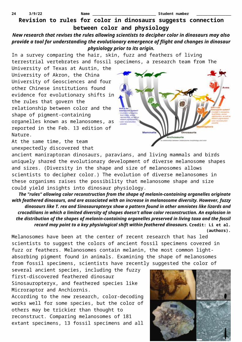
24 5/12/23 Name Student number that significant differences in both vocabulary and real-time language processing efficiency were already evident at age 18 months in English-learning infants from higher- and lower-SES families. By age 24 months, there was a six-month gap between SES groups in processing skills critical to language development.Fernald's work has also identified one likely cause for this gap. Using special technology to make all-day recordings of low-SES Spanish-learning children in their home environments, Fernald and her colleagues found striking variability in how much parents talked to their children. Infants who heard more child-directed speech developed greater efficiency in language processing and learned new words more quickly. The results indicate that exposure to child-directed speech – as opposed to overheard speech – sharpens infants' language processing skills, with cascading benefits for vocabulary learning.Fernald and colleagues are now running a parent-education intervention study with low-income Spanish-speaking mothers in East San Jose, California, funded by the W. K. Kellogg Foundation. This new program, called ¡Habla conmigo! (Talk with Me!), teaches Latina mothers how they can support their infants' early brain development and helps them learn new strategies for engaging verbally with their children. Although they only have data from 32 families so far, the preliminary results are promising. Mothers in the ¡Habla conmigo! program are communicating more and using higher quality language with their 18-month-olds compared to mothers in a control group."What's most exciting," said Fernald, "is that by 24 months the children of more engaged moms are developing bigger vocabularies and processing spoken language more efficiently. Our goal is to help parents understand that by starting in infancy, they can play a role in changing their children's life trajectories."
http://www.medscape.com/viewarticle/820618?src=rssGreen Tea, Blueberry Supplement May Improve Cognition
Nutraceutical supplement composed of, among other ingredients, green tea and blueberries led to modest improvements in cognitive processing speed in older, cognitively healthy adults
Megan BrooksA commercially available nutraceutical supplement composed of, among other ingredients, green tea and blueberries led to modest improvements in cognitive processing speed in older, cognitively healthy adults, according to results of a randomized, placebo-controlled trial.This finding is "noteworthy" given that processing speed is "most often affected early on in the course of cognitive aging, and successful performance on these tasks often underlies more complex cognitive outcomes, such as memory and verbal ability," the researchers write.The study was published in Rejuvenation Research.Clinically Meaningful?The nutraceutical NT-020 contains a proprietary formulation of blueberry, carnosine, green tea, vitamin D3, and Biovin. It was patented by the University of South Florida (USF) in partnership with the James A. Haley Veterans' Hospital and licensed to Natura Therapeutics, Inc. The supplement is commercially available as NutraStem.The study team randomly assigned 113 healthy adults aged 65 to 85 years to NT-020 (2 pills daily) or matching placebo for 2 months. Eight people dropped out (6 in the NT-020 group and 2 in the placebo group), most indicating that they no longer had time to participate, leaving 105 to complete the study.At baseline and after 2 months, participants underwent a battery of cognitive performance tests of episodic memory, processing speed, verbal ability, working memory, executive functioning, and complex speed.The researchers note that adults taking NT-020 "improved significantly" from baseline on 2 measures of processing speed, namely, the identical pictures test ( P = .021) and the number comparison task ( P = .012). The placebo group had no change on the identical pictures test and a slight decline on the number comparison task.None of the other cognitive ability measures were related to intervention group. NT-020 was well tolerated.Co–principal investigator Brent Small, PhD, of the School of Aging Studies at USF, in Tampa, told Medscape Medical News that it is "somewhat unclear" whether the improvement in processing speed is clinically meaningful, mainly due to the study population."Gains of the magnitude that we saw here are positive, but it is hard to say if they are clinically meaningful because we are dealing with a population that is not clinically impaired," said Dr. Small. "Currently, we are seeking funding for additional studies that will focus on persons with mild cognitive impairment," he added.In prior preclinical studies, the researchers gave aging laboratory rats NT-020 and found that it promoted the growth of stem cells in the brain, produced an overall rejuvenating effect, benefited animals with simulated stroke, and led to better cognitive performance.

25 5/12/23 Name Student number "NT-020 is 95% polyphenols," study co–principal investigator Paula C. Bickford, PhD, of the Department of Neurosurgery and Brain Repair, USF Health Morsani College of Medicine, and senior research career scientist at the James A. Haley Veterans' Hospital in Tampa, said in a statement."The basis for the use of polyphenol-rich nutritional supplements as a moderator of age-related cognitive decline is the age-related increase in oxidative stress and inflammation. Nonvitamin polyphenols are the most abundant modulators of oxidative stress and inflammation in our diet," she explained.Blueberry EffectReached for comment, Robert Krikorian, PhD, of the Department of Psychiatry and Behavioral Neuroscience, University of Cincinnati Academic Health Center in Ohio, said the current study "appears to have been designed well, and the discussion concerning the modest positive findings and the limitations of the study seem thorough.""In particular," said Dr. Krikorian, "as the authors noted, the relatively high preintervention level of functioning may have been a factor mitigating ability to measure benefit from the supplementation. That is, there may have been a ceiling effect." "Also, the time frame of the intervention was briefer than many of our natural products studies. On the other hand, showing improvement in processing speed is impressive, as diminished speed of processing with aging is a well-established observation," Dr. Krikorian noted.Although the team's observations "need to be corroborated, it provides a basis for further investigation of this product, perhaps with a sample of individuals with mild cognitive decline," he said.In his own research, Dr. Krikorian has found that blueberry juice may boost memory in older adults with early memory decline, as previously reported by Medscape Medical News.The current study was supported by a grant from the USF Neuroscience Collaborative. Dr. Small has disclosed no relevant financial relationships. Two of the authors are cofounders of Natura Therapeutics, which developed and markets NT-020. Dr. Krikorian has disclosed no relevant financial relationships.Rejuvenation Res. Published online October 17, 2013. Abstract
http://www.eurekalert.org/pub_releases/2014-02/uops-pst021314.phpPenn study: Topiramate reduces heavy drinking in patients seeking to cut down on
alcohol consumptionFindings have important implications for the personalized treatment of alcohol abuse
PHILADELPHIA – Heavy drinking is common in the United States and takes a personal and societal toll, with an annual estimated cost of $223.5 billion due to losses in workplace productivity, health care and criminal justice expenses. Data shows that 23 percent of individuals age 12 or older reported drinking five or more drinks on one occasion in the previous month, and almost seven percent reported doing so on at least five days per month. Despite this, few heavy drinkers seek out treatment—especially those who do not meet the clinical criteria for an alcohol use disorder, but whose drinking causes substantial damage to individuals, their families and the community.Researchers at Penn Medicine have shown that the anticonvulsant medication, topiramate, previously shown to reduce drinking in patients committed to abstinence from alcohol, can also be helpful in treating problem drinkers whose aim is to curb their alcohol consumption – particularly among a specific group of patients whose genetic makeup appears to be linked to the efficacy of the therapy. Their findings are published in the current issue of the American Journal of Psychiatry."This study represents an important next step in understanding and treating problem drinking," says Henry R. Kranzler, MD, professor of Psychiatry, director of Penn's Center for Studies of Addiction and lead author on the study. "Our study is the first we are aware of in which topiramate was evaluated as a treatment option for patients who want to limit their drinking to safe levels, rather than stop drinking altogether."The randomized double-blind trial included a total of 138 heavy drinkers, approximately half of whom received 12 weeks of treatment with topiramate at a maximal dosage of 200 mg/day and half of whom received a placebo. Both groups underwent brief counseling to reduce drinking and increase abstinent days. The study was initiated at the University of Connecticut Health Center and completed at the Center for Studies of Addiction at the University of Pennsylvania.The study had three phases: a one-week pre-treatment assessment period, a 12-week treatment period and a nine-day medication taper period.Patients were seen weekly during the first six weeks of treatment, followed by three biweekly visits in which their breath alcohol concentration, weight and vital signs were measured and concurrent medications, the occurrence of adverse events and protocol adherence monitored. Patients were also interviewed at each visit on their drinking and medication use since the last visit.
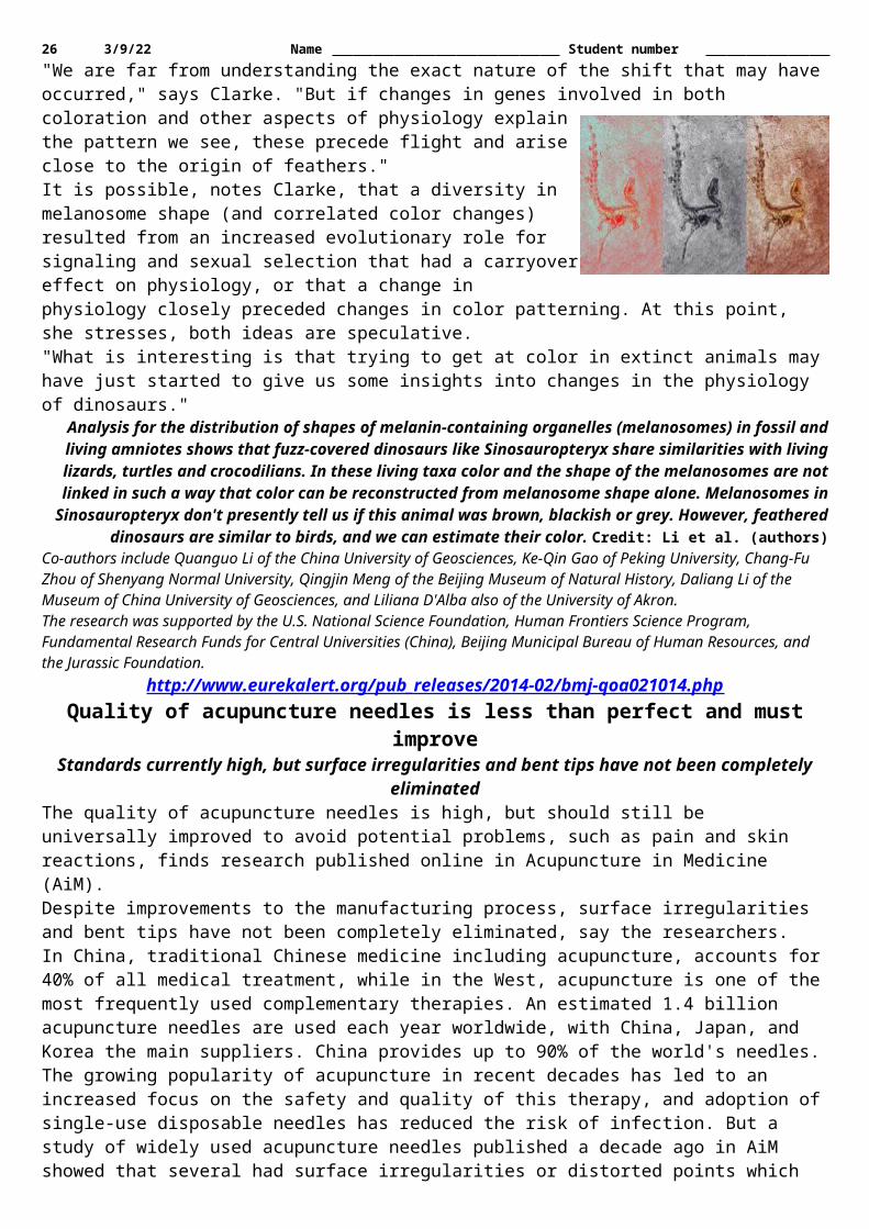
26 5/12/23 Name Student number The results showed that the patients who received topiramate had fewer heavy drinking days than those in the placebo group. By the end of treatment, the odds of experiencing a heavy drinking day in the placebo group was five times more than that of the topiramate group; and the number of patients who experienced no heavy drinking days on the last four weeks of treatment in the topirmate group was more than double that of the placebo group. In addition, topiramate patients reported more abstinent days than placebo patients.The study has important implications for the personalized treatment of heavy drinking. Analysis showed that only individuals with a specific genotype found in 40 percent of European-Americans benefitted from treatment with topiramate. The genotype involves two copies of a variant in the gene encoding a subunit of the receptor for an excitatory amino acid neurotransmitter, glutamate. This study, by virtue of showing that only individuals with a certain form of the kainate (glutamate) receptor reduced drinking with topiramate treatment, indicates that this receptor plays a key role in topiramate's effects on drinking. Because topiramate interacts with multiple neurotransmitter and enzyme systems, this provides a specific target for the development of medications to reduce heavy drinking. Targeting this receptor could yield the greatest therapeutic effect in heavy drinkers, while reducing topiramate's common side effects, which include fatigue, dizziness, and memory problems.Kranzler is optimistic about the potential for the personalized treatment of heavy drinking. "Our hope is that the study will result in additional research on how best to help patients who have struggled with heavy drinking and the problems it causes, but who are unable or unwilling to abstain from alcohol altogether. These findings may allow us to predict, in advance, who may benefit from topiramate treatment, thereby avoiding the unnecessary use of the medication."This study was supported by grants P60 AA03510 and K24 AA13736 from the National Institute on Alcohol Abuse and Alcoholism, and the VISN 4 Mental Illness Research, Education and Clinical Center of the US Department of Veterans Affairs.
http://www.eurekalert.org/pub_releases/2014-02/uoc-rfb021314.phpResearchers find brain's 'sweet spot' for love in neurological patient
A region deep inside the brain controls how quickly people make decisions about love, according to new research at the University of Chicago.
The finding, made in an examination of a 48-year-old man who suffered a stroke, provides the first causal clinical evidence that an area of the brain called the anterior insula "plays an instrumental role in love," said UChicago neuroscientist Stephanie Cacioppo, lead author of the study. In an earlier paper that analyzed research on the topic, Cacioppo and colleagues defined love as "an intentional state for intense [and long-term] longing for union with another" while lust, or sexual desire, is characterized by an intentional state for a short-term, pleasurable goal.
These fMRI scans show brain regions activated by sexual desire (in blue) compared to love (in pink) in healthy patients. The overlap in red illustrates how a patient's brain lesion affected the area in the brain associated with
decision-making in love. Courtesy of Chris Frum and James Lewis/West Virginia University; Robin Weiss/University of Chicago
In this study, the patient made decisions normally about lust but showed slower reaction times when making decisions about love, in contrast to neurologically typical participants matched on age, gender and ethnicity. The findings are presented in a paper, "Selective Decision-Making Deficit in Love Following Damage to the Anterior Insula," published in the journal Current Trends in Neurology."This distinction has been interpreted to mean that desire is a relatively concrete representation of sensory experiences, while love is a more abstract representation of those experiences," said Cacioppo, a research associate and assistant professor in psychology. The new data suggest that the posterior insula, which affects sensation and motor control, is implicated in feelings of lust or desire, while the anterior insula has a role in the more abstract representations involved in love.In the earlier paper, "The Common Neural Bases Between Sexual Desire and Love: A Multilevel Kernel Density fMRI Analysis," Cacioppo and colleagues examined a number of studies of brain scans that looked at differences between love and lust.The studies showed consistently that the anterior insula was associated with love, and the posterior insula was associated with lust. However, as in all fMRI studies, the findings were correlational."We reasoned that if the anterior insula was the origin of the love response, we would find evidence for that in brain scans of someone whose anterior insula was damaged," she said.

27 5/12/23 Name Student number In the study, researchers examined a 48-year-old heterosexual male in Argentina, who had suffered a stroke that damaged the function of his anterior insula. He was matched with a control group of seven Argentinian heterosexual men of the same age who had healthy anterior insula.The patient and the control group were shown 40 photographs at random of attractive, young women dressed in appealing, short and long dresses and asked whether these women were objects of sexual desire or love. The patient with the damaged anterior insula showed a much slower response when asked if the women in the photos could be objects of love. "The current work makes it possible to disentangle love from other biological drives," the authors wrote. Such studies also could help researchers examine feelings of love by studying neurological activity rather than subjective questionnaires.Co-authors of the paper were John Cacioppo, the Tiffany & Margaret Blake Distinguished Service Professor in Psychology at the University of Chicago; Blas Couto, Lucas Sedeno, Facundo Manes, Augustin Ibanez, all of Favaloro University, Buenos Aires; Mylene Bolmont of the University of Geneva; and Chris Frum and James Lewis of West Virginia University.
http://www.eurekalert.org/pub_releases/2014-02/ki-irf021414.phpImpaired recovery from inflammation linked to Alzheimer's
New research from Karolinska Institutet in Sweden shows that the final stage of the normal inflammatory process may be disrupted in patients with Alzheimer's disease.
A study published in the journal Alzheimer's & Dementia shows that levels in the brain and cerebrospinal fluid of the molecules necessary for tissue recovery through the clearance of harmful inflammatory substances are lower than normal in patients with Alzheimer's disease. The study also showed association between the lower levels of these molecules with impaired memory function.Alzheimer's disease is the most common form of dementia, eventually leading to neuronal death associated with an increasing degree of memory impairment. As with other neurodegenerative diseases, Alzheimer's is characterised by an inflammatory process in the brain. Prolonged inflammation with the release of inflammatory and toxic substances can cause further damage and neuronal death.The inflammatory process normally ends in what is known as resolution. This is an active process regulated by certain molecules, so called specialized pro-resolving mediators, where the tissue is cleared from microorganisms, debris from dead cells via an uptake mechanism (phagocytosis), and where the release of growth factors stimulates tissue repair.Together with colleagues in the United States, researchers at Karolinska Institutet have now shown that the levels of resolution-regulating molecules in the brain and in the cerebrospinal fluid are lower in Alzheimer's disease than normal. The researchers have also shown that the lower levels of these molecules correlate with a lower degree of cognitive function, that is, memory capacity. The results are based on analyses of cerebrospinal fluid from 15 patients with Alzheimer's disease, 20 patients with mild cognitive impairment and 21 control subjects. The researchers also analysed brain tissue from 10 Alzheimer's patients and 10 control subjects."Our hypothesis is that stimulation of resolution of inflammation in Alzheimer's disease may result in reduced neuronal death in the brain, and in turn have a beneficial effect in disease progression and cognition. This is an entirely new approach and provides the opportunity to develop new treatment principles for Alzheimer's disease," says Professor Marianne Schultzberg, who led the study at the Department of Neurobiology, Care Sciences and Society.In ongoing studies, the researchers are now investigating how the pro-resolving molecules affect neuronal death in cell cultures, and whether treatment in animal experiments with these substances can prevent neurodegeneration and improve memory functions. The pro-resolving molecules identified so far are derivatives of omega-3 fatty acids, which constitute a popular food supple-ment that has been ascribed several health benefits, and have received attention for beneficial effects also on factors related to Alzheimer's disease, in line with the new results described above. In previous studies, the researchers behind these new findings have shown that omega-3 also stimulates cells to take up amyloid-beta, a protein that kills neurons and occurs in the brain in the form of plaques in Alzheimer's disease.The research was funded with support, among others, from the Swedish Research Council, Swedish Brain Power, Stockholm Community Council, the Chinese Scholarship Council, the Knut and Alice Wallenberg Foundation, Stiftelsen för Gamla Tjänarinnor, Alzheimerfonden, the Gun and Bertil Stohne Foundation, the Petrus and Augusta Hedlund Foundation.Publication: 'Resolution of inflammation is altered in Alzheimer's disease', Wang X, Zhu M, Hjorth E, Cortés Toro V, Eyjolfsdottir H, Graff C, Nennesmo I, Palmblad J, Eriksdotter M, Sambamurti K, Fitzgerald JM, Serhan CN, Granholm A-C & Schultzberg M, Alzheimer's & Dementia, online 14 February 2014.
http://www.eurekalert.org/pub_releases/2014-02/hsop-gno021214.phpGrowing number of chemicals linked with brain disorders in children

28 5/12/23 Name Student number Toxic chemicals may be triggering the recent increases in neurodevelopmental disabilities among children—
such as autism, attention-deficit hyperactivity disorder, and dyslexiaBoston, MA – Toxic chemicals may be triggering the recent increases in neurodevelopmental disabilities among children—such as autism, attention-deficit hyperactivity disorder, and dyslexia—according to a new study from Harvard School of Public Health (HSPH) and Icahn School of Medicine at Mount Sinai. The researchers say a new global prevention strategy to control the use of these substances is urgently needed.The report will be published online February 15, 2014 in Lancet Neurology."The greatest concern is the large numbers of children who are affected by toxic damage to brain development in the absence of a formal diagnosis. They suffer reduced attention span, delayed development, and poor school performance. Industrial chemicals are now emerging as likely causes," said Philippe Grandjean, adjunct professor of environmental health at HSPH.The report follows up on a similar review conducted by the authors in 2006 that identified five industrial chemicals as "developmental neurotoxicants," or chemicals that can cause brain deficits. The new study offers updated findings about those chemicals and adds information on six newly recognized ones, including manganese, fluoride, chlorpyrifos and DDT (pesticides), tetrachloroethylene (a solvent), and the polybrominated diphenyl ethers (flame retardants).The study outlines possible links between these newly recognized neurotoxicants and negative health effects on children, including: Manganese is associated with diminished intellectual function and impaired motor skills Solvents are linked to hyperactivity and aggressive behavior Certain types of pesticides may cause cognitive delaysGrandjean and co-author Philip Landrigan, Dean for Global Health at Mount Sinai, also forecast that many more chemicals than the known dozen or so identified as neurotoxicants contribute to a "silent pandemic" of neurobehavioral deficits that is eroding intelligence, disrupting behaviors, and damaging societies. But controlling this pandemic is difficult because of a scarcity of data to guide prevention and the huge amount of proof needed for government regulation. "Very few chemicals have been regulated as a result of developmental neurotoxicity," they write.The authors say it's crucial to control the use of these chemicals to protect children's brain development worldwide. They propose mandatory testing of industrial chemicals and the formation of a new international clearinghouse to evaluate industrial chemicals for potential developmental neurotoxicity."The problem is international in scope, and the solution must therefore also be international," said Grandjean. "We have the methods in place to test industrial chemicals for harmful effects on children's brain development—now is the time to make that testing mandatory."Funding for the study came from the National Institutes of Health, National Institute for Environmental Health Sciences (ES09584, ES09797, and ES11687)."Neurobehavioural effects of developmental toxicity," Philippe Grandjean, Philip J. Landrigan, Lancet Neurology, online February 15, 2014. http://www.thelancet.com/journals/laneur/article/PIIS1474-4422(13)70278-3/abstract
http://www.medscape.com/viewarticle/820665?src=rssTo Stop Lyme, Vaccinate...the Mice?
Field trials of a new oral vaccine against Borrelia burgdorferi in mice significantly reduced the level of infected ticksJanis C. Kelly
Field trials of a new oral vaccine against Borrelia burgdorferi in mice significantly reduced the level of infected ticks in the treated areas and might help lower the risk for human infection, researchers report in an article published online February 11 in the Journal of Infectious Diseases.The oral bait vaccine elicits antibody responses against the outer surface protein A (OspA) of B burgdorferi. According to the authors, injectable vaccines based on OspA have been shown to protect humans, dogs, and mice against infection. This study provides proof of concept that an oral anti-OspA can provide similar protection.Maria Gomes-Solecki, DVM, and colleagues tested a new oral anti-OspA formulation that is being commercialized by US Biologic. Dr. Gomes-Solecki is assistant professor at the University of Tennessee Health Sciences Center, Memphis, and owner of Biopeptides, Inc.The field trial targeted white-footed mice ( Peromyscus leucopus) because they are the major reservoir for B burgdorferi, which is then transmitted to the Ixodes scapularis tick, the vector for transmission to humans. "A high prevalence of infection with Borrelia burgdorferi in ixodid ticks is correlated with a high incidence of Lyme disease," the authors write.

29 5/12/23 Name Student number Gary M. Green, MD, infectious disease specialist at Kaiser Permanente, Santa Rose, California, told Medscape Medical News, "This is a novel approach aimed at environmental control of Lyme disease by breaking the cycle of transmission between the tick vector and the reservoir host, which is the white-footed mouse. It seems clear now that [for] Lyme disease, which occurs in complex peridomestic areas where forest regrowth has increased contact between humans and Lyme vectors, a multipronged strategy for disease control is needed. This type of vaccine aimed at the reservoir host might be one of those prongs." Dr. Green was not involved in the study.The researchers conducted a prospective 5-year field trial in Lyme-endemic Dutchess County, New York, which involved seven 1.1-hectare plots of similar oak-dominated forest and understory vegetation, as well as soil type, slope, and drainage. Four of the plots were treated with an oatmeal-based OspA/RTV bait including 200 mg Escherichia coli expressing OspA; 3 control plots were treated with oatmeal only. Each plot had an 8 by 8 array of live traps and was baited in the late afternoon for 5 consecutive nights per week from mid-May until mid-September. Vaccine consumption was recorded the next morning, and captured mice were tagged with numbered ear tags.During the peak time for ticks, 10 mice per grid were brought into the lab, and their blood was tested for anti-OspA antibody levels. Host-seeking nymphal ticks were collected in May and June from each plot, B burgdorferi DNA was extracted from 16 to 150 ticks per site per year, and real-time polymerase chain reaction was used to assess tick infection rate and vaccine efficacy.The authors report that oral vaccination of the mice resulted in OspA-specific seropositivity followed by reductions in the number of infected ticks by 23% at year 2 and 76% at year 5 in the treated areas."Although eliminating B. burgdorferi from its natural enzootic cycle seems unrealistic, diminishing its threat to humans via reduction of the tick infection prevalence is an achievable goal with remarkable public health ramifications.... Our results suggest that prevention of Lyme disease can be shifted from the current standard of direct vaccination of humans to an indirect strategy of containment of transmission, as the pathogen moves through its natural enzootic cycle, before it spins out into a zoonotic human disease," the authors write.The study was supported by the Centers for Disease Control and Prevention, the National Institute of Allergy and Infectious Diseases, and the National Science Foundation. Dr. Gomes-Solecki has relevant patents that might pose a conflict of interest and is chair of the scientific advisory of and a shareholder in US Biologic. The other authors and Dr. Green have disclosed no relevant financial relationships.J Infect Dis. Published online February 11, 2014. Abstract
http://nyti.ms/1g8h1yjMedicines Made in India Set Off Safety Worries
India, the second-largest exporter of over-the-counter and prescription drugs to the United States, is coming under increased scrutiny by American regulators for safety lapses, falsified drug test results and selling fake
medicines.By GARDINER HARRISFEB. 14, 2014
NEW DELHI - Dr. Margaret A. Hamburg, the commissioner of the United States Food and Drug Administration, arrived in India this week to express her growing unease with the safety of Indian medicines because of “recent lapses in quality at a handful of pharmaceutical firms.” India’s pharmaceutical industry supplies 40 percent of over-the-counter and generic prescription drugs consumed in the United States, so the increased scrutiny could have profound implications for American consumers.F.D.A. investigators are blitzing Indian drug plants, financing the inspections with some of the roughly $300 million in annual fees from generic drug makers collected as part of a 2012 law requiring increased scrutiny of overseas plants. The agency inspected 160 Indian drug plants last year, three times as many as in 2009. The increased scrutiny has led to a flood of new penalties, including half of the warning letters the agency issued last year to drug makers.Dr. Hamburg was met by Indian officials and executives who, shocked by recent F.D.A. export bans of generic versions of popular medicines - like the acne drug Accutane, the pain drug Neurontin and the antibiotic Cipro - that the F.D.A. determined were adulterated, suspect that she is just protecting a domestic industry from cheaper imports. “There are some people who take a very sinister view of the F.D.A. inspections,” Keshav Desiraju, India’s health secretary until this week, said in a recent interview.The F.D.A.'s increased enforcement has already cost Indian companies dearly — Ranbaxy, one of India’s biggest drug manufacturers, pleaded guilty to felony charges and paid a $500 million fine last year, the largest ever levied against a generic company. And many worry that worse is in store. “If I have to follow U.S. standards in inspecting facilities supplying to the Indian market,” G. N. Singh, India’s top drug regulator, said in a recent interview with an Indian newspaper, “we will have to shut almost all of those.”

30 5/12/23 Name Student number The unease culminated Tuesday when a top executive at Ranbaxy — which has repeatedly been caught lying to the F.D.A. and found to have conditions such as flies “too numerous to count” in critical plant areas — pleaded with Dr. Hamburg at a private meeting with other drug executives to allow his products into the United States so that the company could more easily pay for fixes. She politely declined.India’s drug industry is one of the country’s most important economic engines, exporting $15 billion in products annually, and some of its factories are world-class, virtually undistinguishable from their counterparts in the West. But others suffer from serious quality control problems. The World Health Organization estimated that one in five drugs made in India are fakes. A 2010 survey of New Delhi pharmacies found that 12 percent of sampled drugs were spurious. In one recent example, counterfeit medicines at a pediatric hospital in Kashmir are now suspected of playing a role in hundreds of infant deaths there in recent years.One widely used antibiotic was found to contain no active ingredient after being randomly tested in a government lab. The test was kept secret for nearly a year while 100,000 useless pills continued to be dispensed.More tests of hospital medicines found dozens more that were substandard, including a crucial intravenous antibiotic used in sick infants.“Some of the fake tablets were used by pregnant women in the post-surgical prevention of infections,” said Dr. M. Ishaq Geer, senior assistant professor of pharmacology at the University of Kashmir. “That’s very serious.”Investigations of the deaths are continuing, but convictions of drug counterfeiters in India are extremely rare.Satish Reddy, president of the Indian Pharmaceutical Alliance, said Indian drug manufacturers were better than the F.D.A. now contends. “More rigorous enforcement is needed, for sure, but this impression that India is overrun with counterfeits is unjustified,” Mr. Reddy said.But Heather Bresch, chief executive of Mylan, which has plants in the United States and India, said regulatory scrutiny outside the United States was long overdue. “If there were no cops around, would everyone drive the speed limit?” Ms. Bresch asked. “You get careless, start taking risks. Our government has enabled this.”For Dr. Hamburg, the trip is part of a long-running effort to create a global network of drug and food regulators to help scrutinize the growing flood of products coming into the United States, including 80 percent of the seafood consumed in the United States, 50 percent of the fresh fruit, 20 percent of the vegetables and the vast majority of drugs. She has gone to conclaves of regulators from Europe and elsewhere to coordinate policing, but Indian officials have so far not attended such meetings.Many of India’s drug manufacturing facilities are of top quality. Cipla, one of the industry’s giants, has 40 plants across the country that together can produce more than 21 billion tablets and capsules annually, and one of its plants in Goa appeared just as sterile, automated and high tech on a recent tour as those in the United States.Cipla follows F.D.A. guidelines at every plant and on every manufacturing line, and the company exports more than 55 percent of its production, said Yusuf Hamied, the company chairman.But Benjamin Mwesige, a pharmacist at the Uganda Cancer Institute in Kampala, said in an interview in July that the institute had stopped buying cancer drugs from India in 2011 because it had received shipments of drugs that turned out to be counterfeit and inactive, with Cipla labels that Mr. Mwesige believed were forged.He became suspicious when doctors began seeing chemotherapy patients whose cancer showed none of the expected responses to the drugs — and who also had none of the usual side effects. The drugs that had been prescribed were among the mainstays of cancer treatment — methotrexate, docetaxel and vincristine. Laboratory tests confirmed that the drugs were bogus, and Mr. Mwesige estimated that in 2011 20 percent of the drugs that the institute bought were counterfeit.Enforcement of regulations over all is very weak, analysts say, and India’s government does a poor job policing many of its industries. Last month, the United States Federal Aviation Administration downgraded India’s aviation safety ranking because the country’s air safety regulator was understaffed, and a global safety group found that many of India’s best-selling small cars were unsafe.India’s Central Drugs Standard Control Organization, the country’s drug regulator, has a staff of 323, about 2 percent the size of the F.D.A.'s, and its authority is limited to new drugs. The making of medicines that have been on the market at least four years is overseen by state health departments, many of which are corrupt or lack the expertise to oversee a sophisticated industry. Despite the flood of counterfeit drugs, Mr. Singh, India’s top drug regulator, warned in meetings with the F.D.A. of the risk of overregulation.This absence of oversight, however, is a central reason India’s pharmaceutical industry has been so profitable. Drug manufacturers estimate that routine F.D.A. inspections add 25 percent to overall costs. In the wake of the 2012 law that requires the F.D.A. for the first time to equalize oversight of domestic and foreign plants, India’s cost advantage could shrink significantly.
31 5/12/23 Name Student number Some top manufacturers are already warning that they may leave, tough medicine for an already slowing economy. “I’m a great nationalist, an Indian first and last,” Dr. Hamied said. “But companies like Cipla are looking to expand their businesses abroad and not in India.”American businesses and F.D.A. officials are just as concerned about the quality of drugs coming out of China, but the F.D.A.'s efforts to increase inspections there have so far been frustrated by the Chinese government.“China is the source of some of the largest counterfeit manufacturing operations that we find globally,” said John P. Clark, Pfizer’s chief security officer, who added that Chinese authorities were cooperative.Using its new revenues, the F.D.A. tried to bolster its staff in China in February 2012. But the Chinese government has so far failed to provide the necessary visas despite an announced agreement in December 2013 during a visit by Vice President Joseph R. Biden Jr., said Erica Jefferson, an F.D.A. spokeswoman.The United States has become so dependent on Chinese imports, however, that the F.D.A. may not be able to do much about the Chinese refusal. The crucial ingredients for nearly all antibiotics, steroids and many other lifesaving drugs are now made exclusively in China.Denise Grady contributed reporting from Kampala, Uganda, and Hari Kumar from Srinagar, Kashmir.
http://www.bbc.co.uk/news/science-environment-26023166Wavier jet stream 'may drive weather shift'
New research suggests that the main system that helps determine the weather over Northern Europe and North America may be changing.
By Pallab Ghosh Science correspondent, BBC News, ChicagoThe study shows that the so-called jet stream has increasingly taken a longer, meandering path. This has resulted in weather remaining the same for more prolonged periods. The work was presented at the annual meeting of the American Association for the Advancement of Science (AAAS) in Chicago. The observation could be as a result of the recent warming of the Arctic. Temperatures there have been rising two to three times faster than the rest of the globe. According to Prof Jennifer Francis of Rutgers University in New Jersey: "This does seem to suggest that weather patterns are changing and people are noticing that the weather in their area is not what it used to be." The meandering jet stream has accounted for the recent stormy weather over the UK and the bitter winter weather in the US Mid-West remaining longer than it otherwise would have."We can expect more of the same and we can expect it to happen more frequently," says Prof FrancisThe jet stream, as its name suggests, is a high-speed air current in the atmosphere that brings with it the weather.It is fuelled partly by the temperature differential between the Arctic and the mid-latitudes. If the differential is large then the jet stream speeds up, and like a river flowing down a steep hill, it ploughs through any obstacles - such as areas of high pressure that might be in its way. If the temperature differential reduces because of a warming Arctic then the jet stream weakens and, again, like a river on a flat bed, it will meander every time it comes across an obstacle. This results in weather patterns tending to becoming stuck over areas for weeks on end. It also drives cold weather further south and warm weather further north. Examples of the latter are Alaska and parts of Scandinavia, which have had exceptionally warm conditions this winter.With the UK, the US and Australia experiencing prolonged, extreme weather, the question has been raised as to whether recent patterns are due to simple natural variations or the result of manmade climate change? According to Prof Francis, it is too soon to tell. "The Arctic has been warming rapidly only for the past 15 years," she says. "Our data to look at this effect is very short and so it is hard to get a very clear signal. "But as we have more data I do think we will start to see the influence of climate change."Prof Francis was taking part in a session on Arctic change involving Mark Serreze, the director of the US National Snow and Ice Data Center in Colorado. He said the idea that changes in the polar north could influence the weather in middle latitudes - so-called "Santa's revenge" - was a new and lively area of research and somewhat controversial, with arguments for and against."Fundamentally, the strong warming that might drive this is tied in with the loss of sea-ice cover that we're seeing, because the sea-ice cover acts as this lid that separates the ocean from a colder atmosphere," Dr Serreze explained. "If we remove that lid, we pump all this heat up into the atmosphere. That is a good part of the signal of warming that we're now seeing, and that could be driving some of these changes."
http://thebea.st/1gDfnWZ

32 5/12/23 Name Student number
Weed Could Block H.I.V.’s Spread. No, Seriously.But the U.S. government won’t let scientists try out this promising treatment on humans.
On a warm summer day in Chicago at the International Cannabinoid Research Conference, hundreds of marijuana researchers were giggling.It wasn’t the groundbreaking research they’d just heard—proving the ability of THC, one of the active ingredients in marijuana, to stave off HIV (or RIV in monkeys)—that did it. Nor was it the author of the study, Dr. Patricia E. Molina, who had them laughing. It was the rogue researcher daring enough to taint the victory with a harsh dose of reality: “What’s next, testing this on humans?”As the laughter subsided and the gravity of Dr. Molina’s results sank in, reality did too. THC is one of 500 active ingredients in marijuana. And marijuana, despite many studies proving its medical value, is sill classified by the government as a Schedule I Substance. In the face of mounting evidence that it is beneficial in treating diseases ranging from Alzheimer’s and Multiple Sclerosis, it remains a controlled substance. The joke wasn’t funny so much as painfully true: proving that an illegal drug can stop a deadly disease in humans—without testing it on them—is impossible.This bleak truth renders Dr. Molina’s discovery—at this point—futile. She’s found a key to a door that hasn’t been built.When the journal Aids Research and Human Retroviruses published Dr. Molina’s story this week—more than three years after the study was completed—it was followed by a small amount of buzz. But it was largely overlooked by the mainstream media—perhaps because THC is already well known for treating HIV’s “wasting” symptoms, like nausea and loss of appetite.For those well-versed in the medical marijuana community, however, the results are too powerful to ignore. Amanda Reiman, California policy manager for the Drug Policy Alliance was at the conference in 2011 when Dr. Molina presented her results. “It was groundbreaking. Everyone was in awe,” she tells The Daily Beast.The study itself was fairly simple. For 17 months, Dr. Molina and her team at Louisiana State University administered a high concentration of THC to 4-to-6-year-old male rhesus monkeys who were RIV-positive (a virus in chimps similar to HIV), twice daily. An examination of the tissue in their intestines before and after the chronic THC exposure revealed dramatic decreases in immune tissue damage in the stomach and a significant increase in the numbers of normal cells.Mirroring other studies that link marijuana to HIV, the study illustrates how THC works by targeting so-called “CB2” receptors in the brain. One of two known cannabinoid receptors activated by cannabinoids (terpenophenolic compounds present in Cannabis), the CB2 receptors manifest in cells connected with the immune system, such as the gastrointestinal tract and the spleen. Unlike CB1 receptors, which respond to the psychoactive qualities of THC (producing a feeling of “high”), CB2 receptors react to the therapeutic aspects of THC—for example, reducing swelling and relieving pain.“There is no place in our scientific protocol to investigate the benefits of illicit substances—including cannabis.”The changes that THC produces in the gut a process formally known as “microbial translocation,” isn’t as complicated as it sounds. During HIV infection, one of the earliest effects is that the virus spreads rapidly throughout the body and kills a significant part of cells in the gut and intestine. This activity damages the gut in a way that allows the HIV to leak through the cell wall of the intestines and into the bloodstream.When THC is introduced into this environment, it activates the CB2 receptors in the intestines to build new, healthy bacterial cells that block the virus from leaking through the cell walls. In other words, the body works hard to keep bad stuff in the intestines and the good stuff out.Put another way: HIV kills the cells that protect the walls— THC brings them back. Reducing the amount of the virus in the lower intestines could then help keep uninfected people uninfected.The results of Molina’s study were bigger than even she imagined. “When we started the study, we thought [THC] was going to increase viral load [the amount of the HIV virus that is present in the gut],” Dr. Molina told Leaf Science. It did the opposite. “It adds to the picture and it builds a little bit more information around the potential mechanisms that might be playing a role in the modulation of the infection,” Molina said.While some are praising Dr. Molina’s work, others take issue with classifying it as a potential way to decrease the spread of HIV. Dr. Leslie Walker, chief of the Division of Adolescent Medicine at Seattle Children’s Hospital, disagrees with the study for a variety of reasons. “One would need to actually read the study and then help them see animal model as a beginning; one cannot make the leap to preventing HIV from this type of study,” she wrote in an email to The Daily Beast, adding: “Many things can fight infection in the stomach lining that may have no impact on an overall infection.”

33 5/12/23 Name Student number Dr. Kevin Sabet, Director of ProjectSAM an anti-marijuana group co-founded with Patrick Kennedy, feels equally as strong. “This study looked at THC—not marijuana—and they should not confuse the two issues,” he told The Daily Beast. “This is not about marijuana—and any characterization as such is flawed…It would be like saying people should smoke opium because Morphine might help with X condition.”Government researchers are only a bit more enthusiastic. Dr. Carl Dieffenbach, Director of the Division of AIDS (DAIDS) of the National Institute of Allergy and Infectious Diseases (NIAID), had just read the study when we talked. “In my mind, I don’t see its relevance to the human condition,” he said. “I’m speaking as a taxpayer and a scientist.”Part of the issue, now that a study such as this one could technically be performed in Colorado or Washington, is the stopgap on government funding. Still, Dr. Dieffenbach, who has dedicated his life to researching ways to prevent, diagnose, and treat HIV doesn’t feel strongly about it being tested on humans. But what if it did? Should he be allowed to test it at the point, despite its illicit nature? Our colorful conversation is interrupted by an awkward response: silence. “I don’t have an opinion on that,” he says moments later.It’s views like these that infuriate those in the drug policy world, like Amanda Reiman. “Look at the amount of research that started with animals and moved to humans. That’s the normal progression,” she says in response to critiques on the results stemming from monkeys alone. “What’s the alternative? We just give up? We’ll just never study it in humans, so we’ll never know?” she adds with audible frustration. “I’m sure the hundreds of thousands of people living with HIV would disagree with that plan. I think they’d say that ‘hey, if it worked on a monkey, let’s try it.”The main issue Reiman highlights isn’t a lack of research—but a lack of government funding. The National Institute on Drug Abuse (NIDA) handles all of the grants to perform research with cannabis. “There is no place in our scientific protocol to investigate the benefits of illicit substances—including cannabis,” she says.The process of obtaining research-grade marijuana is no walk in the park. Any independent U.S. group wanting to do research must first get approval from a special Department of Health and Human Services committee who reviews the project. A spokesperson for NIDA told The Daily Beast that independently-funded requests are “extremely rare.” Since 2001, only 18 requests have been submitted to the HHS review committee—15 of which have been approved. Additional approval from the Drug Enforcement Administration is required before the request makes it to NIDA’s drug supply program. Beyond being illegal, the spokesperson raises other issues with testing THC on human subjects. “It is difficult to do human research on any kind of potentially addictive drug,” the spokesperson said. “It is not ethical to give these drugs to naïve subjects, so we are often limited in the kind of clinical research we can do.”Mason Tvert, Director of Communications for the Marijuana Policy Project (MPP), says Dr. Molina’s research is a small step forward in a race that’s already being run. “There is a growing body of evidence demonstrating the efficacy of marijuana in the treatment of a variety of medical conditions,” he tells The Daily Beast. “This is not the first time researchers have identified the benefits marijuana can provide to people with HIV.” He’s right. The findings stand on the shoulders of a great deal of research linking marijuana and HIV. In a 1999 study (PDF) by The Institute of Medicine, researchers called cannabis a “promising treatment” for “nausea, appetite loss, pain, and anxiety.”According to data from the Centers for Disease Control and Prevention, approximately 1.1 million Americans are living with HIV—200,000 of whom are unaware of it. Each year an estimated 50,000 are diagnosed and while treatment is available, the fight for a cure rages on. In 2011 alone, 636,048 people died from AIDS.In the wake of such studies, marijuana has become a fixture in the world of HIV treatment, something that Tvert hopes will continue. “Marijuana’s ability to stimulate appetite has proven to be a life-saver for patients suffering from HIV wasting syndrome, which can result in dangerous levels of weight loss,” Tvert says. “Research like this will continue to come out, and it is only a matter of time before more states and our federal government stop blocking HIV patients from accessing this valuable medicine. Unfortunately, there are still many people who are very sick and do not have time to wait.”
http://arstechnica.com/science/2014/02/nasa-solves-mystery-of-jelly-doughnut-shaped-rock/NASA solves mystery of jelly doughnut-shaped rock on Mars
Opportunity drove over it.by Megan Geuss - Feb 16 2014, 5:30am TST
NASA announced this week that it has solved the mystery of the “jelly doughnut-shaped” rock that suddenly appeared in front of the Mars rover Opportunity earlier this year. The small white-ish rock with a deep red center was dubbed “Pinnacle Island” by scientists at

34 5/12/23 Name Student number NASA, who say the rock wasn't there one day, and then 12 Martian days later, it showed up in images that Opportunity sent back. How a slow drive on Mars has changed what we expect from space exploration.This image from the panoramic camera (Pancam) on NASA's Mars Exploration Rover Opportunity shows the location
of "Pinnacle Island" rock before it appeared in front of the rover in early January 2014.NASA/JPL-Caltech/Cornell Univ./Arizona State Univ.
While NASA took the discovery in stride, the information instigated some outlandish speculation from people outside of the space agency, and one “astrobiologist” even sued NASA for failing to expedite an investigation of the rock. (He demanded 100 high-resolution photographs of the anomaly, which he was sure was a “mushroom-like fungus, a composite organism consisting of colonies of lichen and cyanobacteria, and which on Earth is known as Apothecium.”) The space agency, for its part, maintained that the rock was either a close call with a meteor or the result of something Opportunity did.It turns out that NASA's latter speculation was correct: Pinnacle Island is just a small piece of a larger rock that broke off when Opportunity drove over it.According to the Jet Propulsion Laboratory, "Once we moved Opportunity a short distance, after inspecting Pinnacle Island, we could see directly uphill an overturned rock that has the same unusual appearance.We drove over it. We can see the track. That's where Pinnacle Island came from."Although the air of mystery has been diffused, the rock is still quite interesting to NASA scientists.According to the Los Angeles Times “Opportunity's instruments have revealed that [Pinnacle Island] has high levels of sulfur and manganese, water soluble ingredients that may have been concentrated in the rock thanks to the action of water."It seems that Opportunity is living up to its name for NASA scientists, even in the rocks it drives over.
http://www.eurekalert.org/pub_releases/2014-02/uoc-lia021214.phpLoneliness is a major health risk for older adults
Feeling extreme loneliness can increase an older person's chances of premature death by 14 percent, according to research by John Cacioppo, professor of psychology at the University of Chicago.
Cacioppo and his colleagues' work shows that the impact of loneliness on premature death is nearly as strong as the impact of disadvantaged socioeconomic status, which they found increases the chances of dying early by 19 percent. A 2010 meta-analysis showed that loneliness has twice the impact on early death as does obesity, he said.Cacioppo, the Tiffany & Margaret Blake Distinguished Service Professor in Psychology at the University, joined other scholars at a seminar on "The Science of Resilient Aging" Feb. 16 at the American Association for the Advancement of Science Annual meeting in Chicago.The researchers looked at dramatic differences in the rate of decline in physical and mental health as people age. Cacioppo and colleagues have examined the role of satisfying relationships on older people to develop their resilience, the ability to bounce back after adversity and grow from stresses in life.The consequences to health are dramatic, as feeling isolated from others can disrupt sleep, elevate blood pressure, increase morning rises in the stress hormone cortisol, alter gene expression in immune cells, and increase depression and lower overall subjective well-being, Cacioppo pointed out in a talk, "Rewarding Social Connections Promote Successful Aging."Cacioppo, one of the nation's leading experts on loneliness, said older people can avoid the consequences of loneliness by staying in touch with former co-workers, taking part in family traditions, and sharing good times with family and friends – all of which gives older adults a chance to connect others about whom they care and who care about them."Retiring to Florida to live in a warmer climate among strangers isn't necessarily a good idea if it means you are disconnected from the people who mean the most to you," said Cacioppo.Population changes make understanding the role of loneliness and health all the more important, he explained."We are experiencing a silver tsunami demographically. The baby boomers are reaching retirement age. Each day between 2011 and 2030, an average of 10,000 people will turn 65," he said. "People have to think about how to protect themselves from depression, low subjective well-being and early mortality."Although some people are happy to be alone, most people thrive from social situations in which they provide mutual support and develop strong rapport. Evolution encouraged people to work together to survive and accordingly most people enjoy companionship over being alone.Research by Cacioppo and his colleagues has identified three core dimensions to healthy relationships —intimate connectedness, which comes from having someone in your life you feel affirms who you are; relational
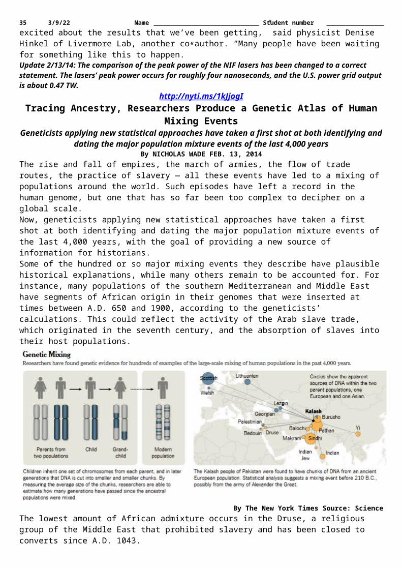
35 5/12/23 Name Student number connectedness, which comes from having face-to-face contacts that are mutually rewarding; and collective connectedness, which comes from feeling that you're part of a group or collective beyond individual existence.It is not solitude or physical isolation itself, but rather the subjective sense of isolation that Cacioppo's work shows to be so profoundly disruptive.Older people living alone are not necessary lonely if they remain socially engaged and enjoy the company of those around them. Some aspects of aging, such as blindness and loss of hearing, however, place people at a special risk for becoming isolated and lonely, he said. —William Harms
http://www.eurekalert.org/pub_releases/2014-02/uoa-sof021414.phpStudy on flu evolution may change textbooks, history books
A new study reconstructing the evolutionary tree of flu viruses challenges conventional wisdom and solves some of the mysteries surrounding flu outbreaks of historical significance
A new study reconstructing the evolutionary tree of flu viruses challenges conventional wisdom and solves some of the mysteries surrounding flu outbreaks of historical significance.The study, published in the journal Nature, provides the most comprehensive analysis to date of the evolutionary relationships of influenza virus across different host species over time. In addition to dissecting how the virus evolves at different rates in different host species, the study challenges several tenets of conventional wisdom, for example the notion that the virus moves largely unidirectionally from wild birds to domestic birds rather than with spillover in the other direction. It also helps resolve the origin of the virus that caused the unprecedentedly severe influenza pandemic of 1918.The new research is likely to change how scientists and health experts look at the history of influenza virus, how it has changed genetically over time and how it has jumped between different host species. The findings may have implications ranging from the assessment of health risks for populations to developing vaccines."We now have a really clear family tree of theses viruses in all those hosts – including birds, humans, horses, pigs – and once you have that, it changes the picture of how this virus evolved," said Michael Worobey, a professor of ecology and evolutionary biology at the University of Arizona, who co-led the study with Andrew Rambaut, a professor at the Institute of Evolutionary Biology at the University of Edinburgh. "The approach we developed works much better at resolving the true evolution and history than anything that has previously been used."Worobey explained that "if you don't account for the fact that the virus evolves at a different rates in each host species, you can get nonsense – nonsensical results about when and from where pandemic viruses emerged.""Once you resolve the evolutionary trees for these viruses correctly, everything snaps into place and makes much more sense," Worobey said, adding that the study originated at his kitchen table."I had a bunch of those evolutionary trees printed out on paper in front of me and started measuring the lengths of the branches with my daughter's plastic ruler that happened to be on the table. Just like branches on a real tree, you can see that the branches on the evolutionary tree grow at different rates in humans versus horses versus birds. And I had a glimmer of an idea that this would be important for our public health inferences about where these viruses come from and how they evolve.""My longtime collaborator Andrew Rambaut implemented in the computer what I had been doing with a plastic ruler. We developed software that allows the clock to tick at different rates in different host species. Once we had that, it produces these very clear and clean results."The team analyzed a dataset with more than 80,000 gene sequences representing the global diversity of the influenza A virus and analyzed them with their newly developed approach. The influenza A virus is subdivided into 17 so-called HA subtypes – H1 through H17 – and 10 subtypes of NA, N1-N10. These mix and match, for example H1N1, H7N9, with the greatest diversity seen in birds.Using the new family tree of the flu virus as a map showed which species moved to which host species and when. It revealed that for several of its 8 genomic segments avian influenza virus is not nearly as ancient as often assumed."What we're finding is that the avian virus has an extremely shallow history in most genes, not much older than the invention of the telephone," Worobey explained.The research team, which included UA graduate student Guan-Zhu Han and Andrew Rambaut, a professor from the University of Edinburgh who is also affiliated with the U.S. National Institutes of Health, found a strong signature in the data suggesting that something revolutionary happened to avian influenza virus, with the

36 5/12/23 Name Student number majority of its genetic diversity being replaced by some new variant in a selective sweep in an extremely synchronous event.Worobey said the timing is provocative because of the correlation of that sudden shift in the flu virus' evolution with historical events in the late nineteenth century."In the 1870s, an immense horse flu outbreak swept across North America," Worobey said, "City by city and town by town, horses got sick and perhaps five percent of them died. Half of Boston burned down during the outbreak, because there were no horses to pull the pump wagons. Out here in the West, the U.S. Cavalry was fighting the Apaches on foot because all the horses were sick. This happened at a time when horsepower was actual horse power. The horse flu outbreak pulled the rug out from under the economy."According to Worobey, the newly generated evolutionary trees show a global replacement of the genes in the avian flu virus coinciding closely with the horse flu outbreak, which the analyses also reveal to be the closest relative to the avian virus."Interestingly, a previous research paper analyzing old newspaper records reported that in the days following the horse flu outbreak, there were repeated outbreaks described at the time as influenza killing chickens and other domestic birds," Worobey said. "That's another unexpected link in the history, and the there is a possibility that the two might be connected, given what we see in our trees."He added that the evolutionary results didn't allow for a definitive determination of whether the virus jumped from horses to birds or vice versa, but a close relationship between the two virus species is clearly there.With regard to humans, the research sheds light on a longstanding mystery. Ever since the influenza pandemic of 1918, it has not been possible to narrow down even to a hemisphere the geographic origins of any of the genes of the pandemic virus."Our study changes that," Worobey said. "It is now clear that most of its genome jumped from birds very close to 1918 in the Western Hemisphere, and there is a suggestion that it was North America in particular."The results also challenge the accepted wisdom of wild birds as the major reservoir harboring the flu virus, from where it jumps to domestic birds and other species, including humans. Instead, the genetic diversity across the whole avian virus gene pool in domestic and wild birds often appears to trace back to earlier outbreaks of the virus in domestic birds, Worobey explained."People tend to think of wild birds as the source of everything, but we see a very strong indication of spillover from domestic birds to wild birds," he said. "It turns out the animals we keep for food and eggs may be substantially shaping the diversity of these viruses in the wild over time spans of decades. That is a surprise."
http://www.bbc.co.uk/news/science-environment-26189722Genetically modified potatoes 'resist late blight'
Scientists have developed a variety of Desiree potatoes that are resistant to late blightMatt McGrath By Matt McGrath Environment correspondent, BBC News
British scientists have developed genetically modified potatoes that are resistant to the vegetable's biggest threat.A three-year trial has shown that these potatoes can thrive despite being exposed to late onset blight. That disease has plagued farmers for generations and triggered the Irish potato famine in the 1840s. EU approval is needed before commercial cultivation of this GM crop can take place. The research is published in the journal, Philosophical Transactions of the Royal Society B. Potatoes are particularly vulnerable to late blight, a fungus-like organism that loves the damp and humid conditions that often occur during the growing season in Europe.Curbing the spraysThe speed with which this infection takes hold and the devastating impacts on the crop, makes it the number one threat to six million tonnes of potatoes produced in the UK each year. Farmers have to be continuously on their guard and need to spray up to 15 times a season to protect against the disease.As part of an EU-wide investigation into the potential for biotechnology to protect crops, scientists at the John Innes Centre and the Sainsbury Laboratory began a trial with blight-resistant potatoes in 2010. The researchers added a gene to Desiree potatoes from a wild South American relative, which helps the plant turn on its natural defences to fight off blight. The scientists involved say that the use of techniques to add in extra genes was crucial in developing a plant that was resistant to the blight."Breeding from wild relatives is laborious and slow and by the time a gene is successfully introduced into a cultivated variety, the late blight pathogen may already have evolved the ability to overcome it," said Professor Jonathan Jones from the Sainsbury Laboratory, the lead author of the research paper. "And I think it is better to control disease with genetics than with chemistry."

37 5/12/23 Name Student number In 2012, the third year of the trial, all the non-GM potatoes in the trial became infected with late blight by August while the modified vegetables remained fully resistant to the end of the experiment. There was also a difference in yield with the GM variety producing double the amount of tubers. The scientists say that since the potatoes are grown from tubers rather than seeds, they are sterile and the issue of GM pollen escaping into the wild does not arise.One area the scientists cannot comment on is the taste, as they were barred from eating the GM variety. However they do not believe there is any mechanism by which the new genes can impact the flavour.As late blight is a highly adaptive organism, the scientists at the Sainsbury Laboratory are eager to find more resistance genes and add them into the plant in a "stack". This would make the chances of late blight overwhelming these potatoes very low. However it might make the GM variety more expensive to plant."The balance will be in favour of the farmer," said Prof Jones. "Yes they may pay more for the seed but they will spend an awful lot less on fungicide."The scientists believe the big challenge will be in getting regulatory approval for the new variety in Europe. The researchers have licensed the technology to an American firm, Simplot, who want to grow them in the US."I think it is unfortunate that American famers are going to benefit from the fruits of European taxpayers funded work way before Europeans," said Prof Jones. "This kind of product will likely be on the US market within a couple of years and if we are lucky within 8-10 years in Europe."Critics of GM crops said that no matter how big the scale of the environmental benefits, they believe that consumers wouldn't be interested. "Is anyone really going to grow, sell or buy genetically modified potatoes?" said Liz O'Neil, director of GM Freeze. "The law says that they will have to be labelled GM, experience shows that the UK doesn't want GM in its shopping basket and British farmers are far too smart to grow something they can't sell."Regulatory hurdlesOther researchers in the field have welcomed the development but were equally negative about the chances of these new potatoes being grown in the UK. "Late blight of potatoes is a difficult disease to control, and using genes from distant relatives is a valuable tool," said Professor Chris Pollock, from Aberystwyth University. "Unfortunately, the problems in the current European regulatory process, which is expensive and extremely slow, means that this advance by UK scientists is far more likely to help farmers in other countries."Only 600 of the GM potato plants have been grown, but the scientists have had to spend £40,000 to protect them over the three years of the trial.
http://www.eurekalert.org/pub_releases/2014-02/uon-nel021314.phpNew eye layer has possible link to glaucoma
A new layer in the human cornea plays a vital role in the structure of the tissue that controls the flow of fluid from the eye
A new layer in the human cornea - discovered by researchers at The University of Nottingham last year - plays a vital role in the structure of the tissue that controls the flow of fluid from the eye, research has shown.The findings, published in a paper in the British Journal of Ophthalmology, could shed new light on glaucoma, a devastating disease caused by defective drainage of fluid from the eye and the world's second leading cause of blindness.The latest research shows that the new layer, dubbed Dua's Layer after the academic Professor Harminder Dua who discovered it, makes an important contribution to the sieve-like meshwork, the trabecular meshwork (TM), in the periphery of the cornea.The TM is a wedge-shaped band of tissue that extends along the circumference of the angle of the anterior chamber of the eye. It is made of beams of collagen wrapped in a basement membrane to which trabecular cells and endothelial cells attach. The beams branch out randomly to form a 'meshwork'. Pressure within the eye is maintained by the balance of aqueous fluid production by eye tissue called the ciliary body and drainage principally through the TM to the canal of Schlemm, a circular channel in the angle of the eye.Defective drainage through the TM is an important cause of glaucoma, a condition that leads to raised pressure in the eye that can permanently affect sight. Around 1 to 2% of the world's population yearly have chronic
GM spuds squashed GM giant Monsanto attained approval for a NewLeaf GM potato in 1995. This variety was designed to protect against the Colorado potato beetle. Initially welcomed by growers, the high costs and development of more effective pesticides against the beetle saw the NewLeaf decline and it was discontinued in 2001. In 2010 BASF were given approval by the EU to grow a GM potato called Amflora. This was designed to produce large amounts of starch for industrial use. In 2012 BASF decided to stop growing these potatoes in Europe due to widespread resistance to the technology.

38 5/12/23 Name Student number glaucoma and globally around 45 million people have open angle glaucoma which can permanently damage the optic nerve — 10% of whom are blind.The latest research by Professor Dua and colleagues in Academic Ophthalmology at The University of Nottingham sheds new light on the basic anatomy of Dua's Layer, which is just 15 microns thick but incredibly tough. Comprised of thin plates of collagen, it sits at the back of the cornea between the corneal stroma and Descemet's membrane.By examining human donor eyes using electron microscopy, the researchers were able to look at Dua's Layer beyond the central part of the cornea to shed more light on its features at the extreme periphery of the cornea. They discovered that the collagen fibres of Dua's Layer also branch out to form a meshwork and that the core of TM is in fact an extension of Dua's Layer.It is hoped the discovery will offer new clues on why the drainage system malfunctions in the eyes of some people, leading to high pressure.Professor Dua said: "Many surgeons who perform lamellar corneal transplant recognise this layer as an important part of the surgical anatomy of the cornea. This new finding resulting from a study of the microanatomy of the periphery of the layer could have significance beyond corneal surgery."The breakthrough discovery of Dua's Layer was first revealed in a paper in the academic journal Ophthalmology in June last year and was widely covered by the world's scientific and lay media.The paper became the number one downloaded ophthalmology paper from the website ScienceDirect between July and September 2013 and was ranked the 11th most downloaded from the website for the whole of medicine and dentistry.The latest research paper, The Collagen Matrix of the Human Trabecular Meshwork is an Extension of the Novel Pre-Descemet's layer (Dua's layer), can be viewed online (after the embargo lifts) at http://dx.doi.org/10.1136/bjophthalmol -2013-304593
http://www.bbc.co.uk/news/health-26189827Cancer: 'Tumour monorail' can lead cancers to their doom
Cancer "monorails" can be used to kill tumours by luring them into toxic pits or areas of the body that are safer to operate on, say US researchers.
By James Gallagher Health and science reporter, BBC NewsA team at the Georgia Institute of Technology designed nanofibres thinner than a human hair which cancers "choose" to travel down.Animal studies showed brain tumours could be shrunk by tricking cancer cells into migrating down the fibres.Cancer Research UK said it was a fascinating idea, but early days.The team were working with difficult-to-treat brain cancers - glioblastomas, which have a tendency to spread inside the brain.The cancerous cells travel down nerves and blood vessels as they invade the brain.The nanofibre technology, reported in Nature Materials, mimics the channels cancerous cells use to move.One of the researchers Prof Ravi Bellamkonda said: "The cancer cells normally latch on to these natural structures and ride them like a monorail to other parts of the brain."By providing an attractive alternative fibre, we can efficiently move the tumours along a different path to a destination that we choose."Deadly brain tumoursA variety of different types of cancer were able to ride the monorail in tests in a Petri dish.Animal studies showed that tumours could be drawn out of the brain and into an implanted toxic gel.The size of the tumour was 93% smaller in animals fitted with the cancer monorail than in rats in which the tumour was untreated.Prof Bellamkonda told the BBC: "It's a way of bringing the tumour to the drug, not the drug to the tumour."You can move a tumour along a path you specify and then kill it, it's not creating extra tumour and the primary tumour actually shrinks. "He suggested that controlling the growth of a tumour might be able to make cancer something people live with, like diabetes, if it cannot be cured.Another idea is to make cancer surgery easier.Normally the tumour and the surrounding tissue are removed, but this is a challenge in the brain where removing any unnecessary tissue could have dire consequences.Prof Bellamkonda suggested doctors might be able to move a tumour to an area more easily operated on.However, the research is still at a very early stage and there will be far more animal studies before it is considered in people.

39 5/12/23 Name Student number Dr Emma Smith, senior science information officer at Cancer Research UK, said: "This fascinating, cutting-edge approach could lead to new ways of stopping tumours growing without damaging healthy tissue, which is particularly important for people with brain tumours."But it's still in its infancy and so far has only been tested in rats, so there is a long way to go before we know if it will be safe and effective as a cancer treatment."